Hailey-Hailey Disease with Nail Changes Preceding the Skin Changes- A Case Report - Medwin Publishers
←
→
Page content transcription
If your browser does not render page correctly, please read the page content below
Clinical Dermatology Open Access Journal
                                                                                                                   ISSN: 2574-7800

   Hailey-Hailey Disease with Nail Changes Preceding the Skin
                                             Changes- A Case Report

Das P1, Bachaspatimayum R 2* and Bhattacharjee N1
                                                                                                             Case Report
1Junior   Resident, Department of Dermatology, Venereology & Leprology, Regional
                                                                                                             Volume 4 Issue 1
Institute of Medical Sciences, Manipur University, India
                                                                                                     Received Date: December 31, 2018
2Assistant   Professor, Department of Dermatology, Venereology & Leprology, Regional                 Published Date: January 23, 2019
Institute of Medical Sciences, Manipur University, India                                              DOI: 10.23880/cdoaj-16000170

*Corresponding author: Romita Bachaspatimayum, Nongmeibung SeramIeirak, Imphal– East, Manipur, India – 795005,
Tel: 09862897399; Email: dr.romita.bachaspatimayum@gmail.com

 Abstract
Hailey-Hailey disease is an uncommon genodermatoses characterised by recurrent vesicular and erosive lesions that
favour the intertriginous areas, especially axilla and groin. Longitudinal leukonychia in multiple finger nails may appear
concurrently, prior to or may follow the skin lesions in this disease (two-third patients). Here we are presenting a case of
Hailey-Hailey disease in a 40 years old male with nail changes preceding the skin manifestations.

Keywords: Hailey-hailey disease; Vesiculobullous lesions; Intertriginous areas; Longitudinal leukonychia

 Introduction                                                              formation of erosions. Recurrent exacerbation was
                                                                           reported during summer. There was no period of
    Hailey-Hailey disease is a rare genodermatoses                         remission throughout the entire course of the disease.
characterised by recurrent vesicular and erosive lesions                   There was history of using local indigenous ointments
that favour the intertriginous areas, especially axilla and                over the lesions on and off with healing of few lesions
groin. The disease was first described by Hailey brothers                  leaving hyperpigmented spots. There was no history of
in 1939 as “familial benign chronic pemphigus” [1]. Here                   similar complaints in any other family member.
we report a case of Hailey-Hailey disease in a 40 years old
male with nail changes preceding the skin manifestations.                     On cutaneous examination, few intact vesicles
                                                                           containing clear fluid on erythematous base, few erosions
 Case Report                                                               and multiple hyperpigmented macules and patches were
                                                                           present over bilateral groins and axilla (Figure 1a).
    A 40 years old male presented with itchy                               Asymptomatic longitudinal white bands (longitudinal
vesiculobullous eruptions over both axilla and groins for                  leukonychia) were observed in multiple finger nails
last 4 years and asymptomatic longitudinal white bands in                  (Figure 1b). There was no involvement of neck folds,
multiple finger nails for last 5 years which was of gradual                antecubital fossae, popliteal fossae, palms, soles and
onset. The vesicles ruptured spontaneously leading to                      mucous membranes.

 Hailey-Hailey Disease with Nail Changes Preceding the Skin Changes- A Case Report                                      Clin Dermatol J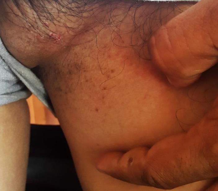
2
Clinical Dermatology Open Access Journal
Hailey disease was made. He was prescribed oral and
topical antibiotics. The patient was lost to follow up.
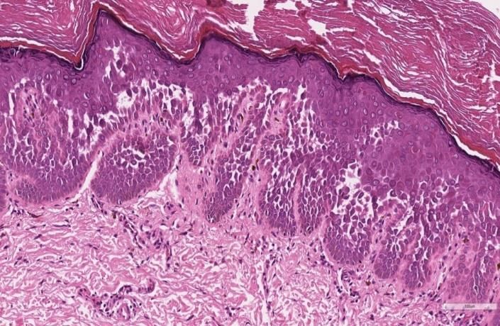
Figure 2: H.P.E. (H&E, 10x) Showing intraepidermal
bullae containing many acantholytic cells imparting a
Figure 1(a): Showing few intact vesicles containing dilapidated brick wall appearance.
clear fluid on erythematous base, few erosions and
multiple hyperpigmented macules and patches over
left groin. Discussion
Hailey-Hailey disease is a rare autosomal dominant
disorder with incomplete penetrance. Family history is
obtained in about two-third of the patients [2]. However
in our case, no positive family history was reported. The
lesions usually develop during second and third decade of
life and affect both sexes equally [2]. Our patient was a 45
years old male. The disease is caused by mutation of the
ATP2C1 gene on chromosome 3q21-q24 which encodes a
calcium channel that is localized to the Golgi apparatus of
keratinocytes. It sequesters calcium within Golgi
apparatus. There is a major role of cytosolic calcium
concentration in keratinocyte differentiation [3].
Increased intracellular calcium concentration leads to
expression of involucrin, a protein which envelops
keratinocytes and responsible for keratinocyte adhesion.
Figure 1(b): Showing longitudinal white bands in Alberg, et al. found decreased level of involucrin in Hailey-
bilateral thumbnails. Hailey disease [4]. In affected individuals, there is a loss of
cohesion between keratinocytes (acantholysis) and
development of vesicles.
All routine haematological and biochemical
investigations were normal. A potassium hydroxide wet Recurrent, fragile vesicles and erosions over the
mount preparation from the axillary eruption did not intertriginous areas (axilla, groin, lateral aspect of neck,
show any fungal hyphae. Tzanck smear showed a few inframammary area and perianal region) is the
acantholytic cells. Histopathological examination (H.P.E) characteristic clinical finding [2]. Scalp, antecubital fossae,
of a punch biopsy specimen from groin demonstrated popliteal fossaeare less frequently involved and
intraepidermal bullae containing many acantholytic cells involvement of oral, vulvar and conjunctival mucosa is
imparting a dilapidated brick wall appearance (Figure 2). rare [5]. However, in our patient, bilateral groin and
Direct immunofluorescence was negative. On the basis of axillary lesions were seen. The cutaneous lesions can be
clinical and histological findings, a diagnosis of Hailey-
Bachaspatimayum R, et al. Hailey-Hailey Disease with Nail Changes Preceding Copyright© Bachaspatimayum R, et al.
the Skin Changes- A Case Report. Clin Dermatol J 2019, 4(1): 000170.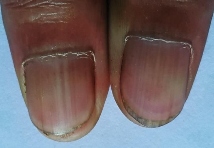
3
                                                                          Clinical Dermatology Open Access Journal

pruritic, painful and malodorous. Healing occurs without                    2.    Burge SM (1992) Hailey-Hailey disease: the clinical
scarring leaving post inflammatory hyperpigmentation                              features, response to treatment prognosis. Br J
[2]. Painful intertriginous erosions, malodorous discharge                        Dermatol 126(3): 275-282.
and pruritus may interfere with physical and professional
activities and give rise to psychosocial distress of the                    3.    Hakuno M, Shimizu H, Akiyama M, Amagai M, Wahl JK,
patients. Itching was the major complaint in our patient                          et al. (2000) Dissociation of intra- and extracellular
and few lesions healed with hyperpigmented macules and                            domains of desmosomalcadherins and E-cadherin in
patches following application of indigenous ointments.                            Hailey-Hailey disease and Darier's disease. Br J
Longitudinal white bands (longitudinal leukonychia) in                            Dermatol 142(4): 702-711.
the fingernails may serve as a diagnostic clue which was
seen in 71% cases in a study by Burge SM, et al. [2]. In our                4.    Aberg KM, Racz E, Behne MJ, Mauro TM (2007)
case, longitudinal white bands in multiple finger nails                           Involucrin expression is decreased in Hailey-Hailey
appeared prior to the onset of cutaneous lesions. The                             keratinocytes owing to increased involucrin mRNA
disease has a chronic course with periods of exacerbation                         degradation. J Invest Dermatol 127(8): 1973-1979.
and remission [2]. In our patient, the lesions were
                                                                            5.    Vaclavinkova V, Neumann E (1982) Vaginal
continuously present throughout the course of the disease,
                                                                                  involvement in familial benign chronic pemphigus
without any period of remission. The disease can be
                                                                                  (Morbus Hailey-Hailey). Acta Derm Venereol 62(1):
triggered by friction, heat, sweating, infection and stress.
                                                                                  80-81.
    Differential diagnosis includes intertrigo, eczema,                     6.    Bianchi L, Chimenti MS, Giunta A (2004) Treatment of
Darier disease and pemphigus vegetans. The treatment of                           Hailey-Hailey disease with topical calcitriol. J Am
Hailey-Hailey disease is challenging. Avoidance of                                Acad Dermatol 51(3): 475-476.
potential is aggravating factors, such as friction and
sweating, maintaining weight at appropriate levels,                         7.    Rabeni EJ, Cunningham NM (2002) Effective
comfortable loose clothing to minimize frictionare the                            treatment of Hailey-Hailey disease with topical
conservative mode of treatments. Various treatment                                tacrolimus. J Am Acad Dermatol 47(5): 797-798.
modalities include topical corticosteroids, antimicrobials,
calcipotriol, tacrolimus and retinoids, systemic                            8.    Berger EM, Galadari HI, Gottlieb AB (2007) Successful
antimicrobials, retinoids, dapsone, immunosuppressants                            treatment of Hailey-Hailey disease with acitretin. J
like methotrexate and cyclosporine, botulinum toxin,                              Drugs Dermatol 6(7): 734-736.
dermabrasion, carbon dioxide laser, photodynamic
therapy with variable efficacies [6-10]. Wide excision of                   9.    Berth-Jones J, Smith SG, Graham-Brown RA (1995)
the involved area with replacement by split graft is                              Benign familial chronic pemphigus (Hailey-Hailey
indicated in recalcitrant cases [11].                                             disease) responds to cyclosporin. Clin Exp Dermatol
                                                                                  20(1): 70-72.
    Conclusion
                                                                            10. Don PC, Carney PS, Lynch WS, Zaim MT, Hassan MO
   Asymptomatic white bands in finger nails                                     (1987) Carbon dioxide laser abrasion: a new
(longitudinal leukonychia) may appear concurrently,                             approach to management of familial benign chronic
prior to or may follow the skin lesions in Hailey- Hailey                       pemphigus (Hailey-Hailey disease). J Derm Surg Oncol
disease (two-third patients). In our case, nail changes                         13(11): 1187-1194.
preceded the appearance of skin lesions by 1 year. Hence,
nail changes may serve as a diagnostic window for the                       11. Menz P, Jackson IT, Connolly S (1987) Surgical control
early diagnosis of this rare genodermatoses.                                    of Hailey -Hailey disease. Br J Plast Surg 40: 557-561.

    References
1.     Hailey H, Hailey H (1939) Familial benign chronic
       pemphigus. Arch Dermatol 39: 679-685.

    Bachaspatimayum R, et al. Hailey-Hailey Disease with Nail Changes Preceding                        Copyright© Bachaspatimayum R, et al.
    the Skin Changes- A Case Report. Clin Dermatol J 2019, 4(1): 000170.
You can also read