Headaches Due to Low and High Intracranial Pressure
←
→
Page content transcription
If your browser does not render page correctly, please read the page content below
REVIEW ARTICLE
Headaches Due to Low
C O N T I N UU M A UD I O
I NT E R V I E W A V AI L A B L E
ONLINE
and High Intracranial
Pressure
By Deborah I. Friedman, MD, MPH, FAAN
Downloaded from https://journals.lww.com/continuum by BhDMf5ePHKav1zEoum1tQfN4a+kJLhEZgbsIHo4XMi0hCywCX1AWnYQp/IlQrHD33D9/FQ5Fz8lLeSB0H4GzbuKu7eSMk/dy05HjsKdWzmIT/T6yvczBGQ== on 08/25/2018
ABSTRACT
PURPOSE OF REVIEW: Headache disorders attributed to low and high
intracranial pressure are commonly encountered in specialty headache
practices and may occur more frequently than realized. While the
headaches resulting from intracranial pressure disorders have what are
conventionally thought of as defining characteristics, a substantial minority
CITE AS: of patients do not manifest the “typical” features. Moreover, patients with
CONTINUUM (MINNEAP MINN) intracranial pressure disorders may also have a preexisting primary
2018;24(4, HEADACHE):1066–1091.
headache disorder. Heightening the complexity of the presentation, the
Address correspondence to
headaches of intracranial pressure disorders can resemble the phenotype
Dr Deborah I. Friedman, University of a primary disorder. Lastly, patients with so-called intracranial
of Texas Southwestern Medical “hypotension” often have normal CSF pressure and neuroimaging studies.
Center, 5323 Harry Hines Blvd,
MC 9322, Dallas, TX 75390, Thus, a high index of suspicion is needed. The published literature has
Deborah.Friedman@ inherent bias as many types of specialists evaluate and treat these
UTSouthwestern.edu. conditions. This article reviews the key points to emphasize the history,
RELATIONSHIP DISCLOSURE: examination, and laboratory evaluation of patients with intracranial
Dr Friedman has received pressure disorders from a neurologist’s perspective.
personal compensation for
serving on the advisory boards of
Alder BioPharmaceuticals, RECENT FINDINGS:Lumbar puncture opening pressure in patients with
Inc; Amgen Inc; Avanir spontaneous intracranial hypotension was low enough to meet diagnostic
Pharmaceuticals, Inc; Biohaven
Pharmaceutical; ElectroCore, criteria (≤60 mm CSF) in only 34% of patients in one study. Most patients
LLC; Supernus Pharmaceuticals, had an opening pressure in the low normal to normal range, and 5% had
Inc; Teva Pharmaceutical
Industries Ltd; and Zosano
an opening pressure of 200 mm CSF or more. Diskogenic microspurs are a
Pharma Corporation. common cause of this syndrome. The Idiopathic Intracranial Hypertension
Continued on page 1091 Treatment Trial found that most participants had a headache phenotype
UNLABELED USE OF
resembling migraine or tension-type headache. No “typical” or
PRODUCTS/INVESTIGATIONAL characteristic headache phenotype was found, and headache-related
USE DISCLOSURE: disability was severe at baseline. Headache disability did not correlate
Dr Friedman discusses the
unlabeled/investigational with the lumbar puncture opening pressure at baseline or at the 6-month
use of acetazolamide for the primary outcome period. Although participants who were randomly
treatment of pseudotumor
assigned to acetazolamide had a lower mean CSF opening pressure at
cerebri syndrome, the use of
gadolinium for MRI myelography 6 months, headache disability in that group was similar to the group who
for the diagnosis of spontaneous received placebo.
intracranial hypotension, and the
use of zonisamide for the
treatment of associated SUMMARY: Significant overlap is seen in the symptoms of high and low CSF
headache. pressure disorders and in those of primary headache disorders.
Neurologists are frequently challenged by patients with headaches who
© 2018 American Academy
of Neurology. lack the typical clinical signs or imaging features of the pseudotumor
1066 AUGUST 2018
Copyright © American Academy of Neurology. Unauthorized reproduction of this article is prohibited.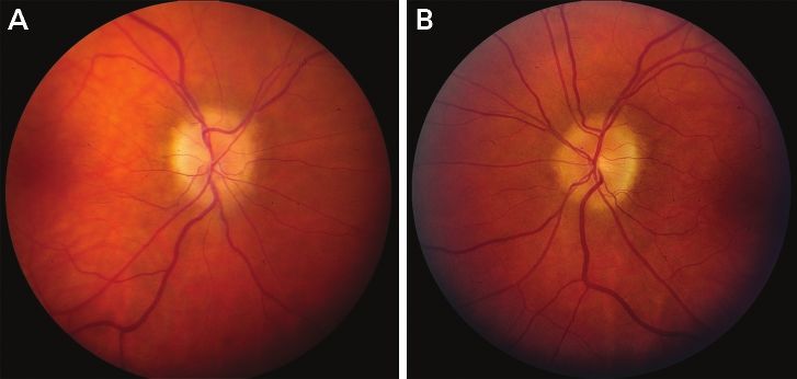
cerebri syndrome or spontaneous intracranial hypotension. Even when
characteristic symptoms and signs are initially present, the typical features
of both syndromes tend to lessen or resolve over time; consider these
diagnoses in patients with long-standing “chronic migraine” who do not
improve with conventional headache treatment. While the diagnostic
criteria for pseudotumor cerebri syndrome accurately identify most
patients with the disorder, at least 25% of patients with spontaneous
intracranial hypotension have normal imaging and over half have a normal
lumbar puncture opening pressure. Detailed history taking will often give
clues that suggest a CSF pressure disorder. That said, misdiagnosis can
lead to significant patient morbidity and inappropriate therapy.

INTRODUCTION

A
              lthough they represent opposite extremes on the intracranial
              pressure spectrum, many similarities occur between the clinical
              features of high- and low-pressure disorders. Additionally, both
              conditions share properties seen with primary headache disorders
              (TABLE 6-1 and TABLE 6-2). Both syndromes can produce new daily
persistent headaches, although the headaches are not always daily in either
disorder. No typical headache phenotype exists for either condition; nocturnal
awakening and worsening with bending over or Valsalva maneuvers are helpful
clues for both conditions but are not specific. The longer the duration of
symptoms, the more difficult it is to confirm either diagnosis by brain imaging
techniques, and even the lumbar puncture opening pressure can be misleading.
Although neurologists usually evaluate patients with these conditions because of
headaches and other neurologic symptoms, either disorder can be asymptomatic
or acephalgic. Finally, there is a fine line between being hypervigilant when
considering the two diagnoses in clinical practice and overdiagnosing these
conditions, which can lead to inappropriate investigations and treatments,
potentially causing harm.2

SPONTANEOUS INTRACRANIAL HYPOTENSION
The term spontaneous intracranial hypotension is a misnomer. The syndrome is not
always spontaneous; a precipitating event is often identified. Additionally, the
majority of patients with spinal CSF leaks do not have low CSF pressure, defined
as a lumbar puncture opening pressure below 60 mm CSF.3 It is more accurately
conceptualized as low CSF volume, low CSF pressure, and possibly low
compliance of the caudal spinal dura. “Intracranial” implies that the problem is
within the skull. While most of the clinical manifestations are intracranial, the
underlying defect is spinal.4
   TABLE 6-3 lists the diagnostic criteria for spontaneous intracranial hypotension.
                                                                                    5

The estimated prevalence of spontaneous intracranial hypotension is 1 per 50,000,
with an annual incidence of 5 per 100,000.6 This is likely an underestimation, as
patients without typical brain imaging findings are less likely to be diagnosed and
are therefore excluded from population-based studies. Additionally, such studies
rely on a specific diagnosis code, which does not currently exist for this disorder.
Spontaneous intracranial hypotension is more common in women than men and
typically presents in the fourth or fifth decades, although it may occur at any
age, and patients may have symptoms for years or even decades before being

CONTINUUMJOURNAL.COM                                                                                                1067

              Copyright © American Academy of Neurology. Unauthorized reproduction of this article is prohibited.
HEADACHES DUE TO LOW AND HIGH INTRACRANIAL PRESSURE
TABLE 6-1 Overlap of Clinical Features of High and Low Cerebrospinal Fluid Pressure
Disorders and Primary Headache Disorders
Pseudotumor Cerebri Intracranial
Feature Syndrome Hypotension Primary Headache Disorder
Location of pain Often frontal or retro-orbital Often posterior but Varies
but varies varies
Timing Worse in the morning or no Worse as the day Patterns vary by headache type;
fluctuation progresses migraine is often present upon
awakening
Nocturnal awakening Yes Yes Yes; frequent in cluster headache,
infrequent in migraine, defining of
hypnic headache
Worse with Valsalva Yes Yes Yes; migraine, primary exertional
maneuver, exercise, headaches, secondary causes (eg,
bending over reversible cerebral
vasoconstriction syndrome,
aneurysm, Chiari malformation)
Effect of caffeine None or worsens Improvement Either; caffeine may provoke
migraine or relieve it
Orthostatic/positional Sometimes worse lying flat Usually better lying Varies; patients with migraine often
component flat prefer to lie down, which may be
related to avoiding movement
Effect of high altitude Usually worsens Usually improves Either; migraine is a risk factor for
headache at high altitude1
Effect of Trendelenburg None (may theoretically Often improves None
position worsen)
Pulsatile tinnitus Common Rare (but may have Not associated
nonpulsatile tinnitus)
Transient obscurations Common No No; transient visual loss in migraine
of vision lasts longer than 1 to 2 minutes and
is not postural
Joint hypermobility Not associated Common Not associated
Neck or back pain Common Common Common
Radicular pain Common Rare No
Papilledema Usually present No No
Spontaneous venous Absent Usually present Usually present
pulsations
Associated with cerebral Yes Yes No
venous sinus thrombosis
Sex Marked female preponderance More common in Male or female; depends on
after puberty females primary headache diagnosis
Body habitus Usually obese Often slim or normal No association
1068 AUGUST 2018
Copyright © American Academy of Neurology. Unauthorized reproduction of this article is prohibited.Comparison of Diagnostic Tests Between High and Low Cerebrospinal TABLE 6-2
Fluid Pressure Disorders and Primary Headaches
Pseudotumor Cerebri Primary Headache
Feature Syndrome Intracranial Hypotension Disorder
Sella/pituitarya Usually empty sella Usually enlarged and hyperemic No pattern
pituitary
Ventricular size Normal Normal Normal
Tonsillar descent Possible Common, may resemble Chiari No relationship
malformation
Flattening of anterior pons No Common No
Optic nerve sheath complex Distended and/or tortuous Narrowed, straight No
Lumbar puncture opening High Low or normal, occasionally high Usually normal
pressureb
Post–lumbar puncture headache Yes Yes Yes
possible
Headache improves after lumbar Often No, symptoms may worsen Possibly
puncture
a
Pituitary gland may enlarge during pregnancy.
b
May be elevated with Valsalva maneuver, extreme pain.
Diagnostic Criteria for Spontaneous Cerebrospinal Fluid Leak and TABLE 6-3
Intracranial Hypotensiona
A Demonstration of a spinal CSF leak (ie, presence of extrathecal CSF)
Or, if criterion A not met,
B Cranial MRI changes of intracranial hypotension (ie, presence of subdural fluid
collections, enhancement of the pachymeninges, or sagging of the brain) and the
presence of at least one of the following:
1 Low opening pressure (≤60 mm H2O)
2 Spinal meningeal diverticulum
3 Improvement of symptoms after epidural blood patching
Or, if criteria A and B not met:
C The presence of all of the following or at least two of the following if typical orthostatic
headaches are present:
1 Low opening pressure (≤60 mm H2O)
2 Spinal meningeal symptoms
3 Improvement of symptoms after epidural blood patching
Note: Patients with onset of symptoms after dural puncture or other penetrating spinal trauma
are excluded.
CSF = cerebrospinal fluid; MRI = magnetic resonance imaging.
a
Reprinted with permission from Schievink WI, et al, AJNR Am J Neuroradiol.5
© 2008 American Society of Neuroradiology.
CONTINUUMJOURNAL.COM 1069
Copyright © American Academy of Neurology. Unauthorized reproduction of this article is prohibited.HEADACHES DUE TO LOW AND HIGH INTRACRANIAL PRESSURE
KEY POINTS correctly diagnosed. Predisposing factors include hypermobility disorders,
including Ehlers-Danlos and Marfan syndromes, and degenerative disk
● Although they represent
opposite ends of a
disease. It is postulated that individuals with connective tissue disorders and
spectrum, spontaneous hypermobility have dural weakness leading to tears and diverticula that allow
intracranial hypotension and CSF to egress into the epidural space.6 TABLE 6-4 lists other causes of the
pseudotumor cerebri syndrome, such as trauma, intentional or accidental dural puncture, and various
syndrome share many
physical activities.8 The physical trauma may be trivial or related to usual
clinical similarities.
activities, such as exercise.
● Neurologists are often The most common symptom of spontaneous intracranial hypotension is
faced with the dilemma of headache. Spontaneous intracranial hypotension is a secondary cause of new
evaluating patients who may daily persistent headache, and the headache may start abruptly. The classic
have either spontaneous
intracranial hypotension or headache is orthostatic, worsening in the upright posture and improving with
pseudotumor cerebri recumbence. This feature suggests that downward traction on pain-sensitive
syndrome, but are “atypical.” upper cervical and cranial structures (nerve roots, meninges, ligaments, veins) is
responsible for the headache. However, nocturnal awakening is not uncommon,
● When evaluating a patient
with possible spontaneous
and paradoxical headaches (worse in recumbence and improved in the upright
intracranial hypotension or posture) may rarely occur.9 Patients will often relate that their headache is absent
pseudotumor cerebri or least intense when they first awaken. It may worsen almost immediately after
syndrome, there is a fine line sitting or arising or progressively worsen throughout the day. Others will
between being hypervigilant
experience headache that begins later in the day, sometimes starting at a very
when considering the two
diagnoses in clinical practice specific time. The location and quality of the headache are extremely variable,
and overdiagnosing the with the latter ranging from annoying to completely incapacitating. Bilateral
conditions, which can lead to posterior head pain is commonly present, but the pain may be unilateral and in
inappropriate investigations
any cranial location. Associated neck pain commonly occurs, and trigeminal
and treatments, potentially
causing harm. neuralgic pain has been reported.10 The orthostatic component may dissipate
over time.
● Spontaneous intracranial Exacerbating factors, which may also occur with intracranial hypertension
hypotension is not and other headache disorders, include coughing, sneezing, laughing, lifting,
necessarily spontaneous, is
not of intracranial origin, and
bending, straining (Valsalva maneuver), sexual activity, and exercise.
may not arise solely from (Worsening with Valsalva maneuvers seems paradoxical, but such maneuvers
low CSF pressure. CSF may exacerbate the CSF leakage through spinal dural defects.) The headache
volume and compliance of may improve at high altitude, with caffeine, with greater occipital nerve
the caudal dura may also be
contributing factors.
blockade, and possibly with onabotulinumtoxinA injections.11,12
Other manifestations of spontaneous intracranial hypotension include chest
● Typical orthostatic and or back pain (“coat hanger” headache), photophobia, diplopia, blurred vision,
“end of the day” headaches facial pain, imbalance, hearing abnormalities, tinnitus, cognitive and mental
may be less prominent in
status changes, hyperkinetic and hypokinetic movement disorders, galactorrhea,
spontaneous intracranial
hypotension over time. subdural fluid collections, and intracranial hemorrhage (TABLE 6-2).
● A marked variability occurs Diagnosis
in the location and character The key to diagnosis is a high level of clinical suspicion and a careful history. As
of spontaneous intracranial
hypotension–related
the manifestations vary and neuroimaging may be normal, the diagnosis may be
headaches. delayed for many years. The neurologic examination is usually normal or may
reveal abnormalities referable to the nonheadache symptoms. Spontaneous
venous pulsations on fundoscopy are supportive although not universally
present. Patients may be slim with an elongated, slender neck. Improvement of
symptoms in the Trendelenburg position (10- to 20-degree head-down tilt for 5
to 10 minutes) is highly suggestive of spontaneous intracranial hypotension.13
Patients should be queried and examined for joint hypermobility; the Beighton
scale is a helpful assessment tool.13,14 These points are demonstrated in CASE 6-1.
1070 AUGUST 2018
Copyright © American Academy of Neurology. Unauthorized reproduction of this article is prohibited.Laboratory Studies
Lumbar puncture is only helpful for diagnosing spontaneous intracranial
hypotension if the CSF pressure is low (≤60 mm CSF). In a study of 106 patients
meeting the diagnostic criteria of headache due to spontaneous intracranial
hypotension developed by Schievink and colleagues,15 34% of patients had a CSF
pressure of 60 mm CSF or less, 45% had opening pressures between 60 mm CSF
and 120 mm CSF, 16% had opening pressure between 120 mm CSF and 200 mm
CSF, and 5% had pressures greater than 200 mm CSF.3,15 Thus, most patients
with spontaneous intracranial hypotension have normal CSF pressure. A
24-gauge needle is recommended to avoid a post–dural puncture leak that may
Causes and Predisposing Factors for Spontaneous Intracranial TABLE 6-4
Hypotension Syndromea
Connective Tissue Matrix Disorders
◆ Marfan syndrome
◆ Ehlers-Danlos syndrome type II
◆ Autosomal dominant polycystic kidney disease
◆ Joint hypermobility: hyperflexibility, “party tricks,” naturally good at gymnastics, dance,
or yoga (inquire about flexibility in childhood)
◆ Retinal detachment at a young age
◆ Personal or family history of arterial dissection, aneurysms, nonrheumatic valvular
heart disease
◆ Secondary to unrecognized intracranial hypertension
Trauma
◆ Previous spine surgery
◆ History of lumbar puncture
◆ Nerve root sleeve tears or avulsions
◆ Previous spinal or epidural anesthesia
◆ Trivial trauma
◇ Valsalva related: heavy lifting, coughing, straining
◇ Repetitive truncal torsion: tennis, golf, yoga, kayaking, canoeing
◆ Impact: motor vehicle accident, whiplash, sports injury
Spine Disorders
◆ Calcified herniated disks
◆ Osteophytes and spondylotic spurs
◆ CSF venous fistula
Bariatric Surgery7
Unknown
CSF = cerebrospinal fluid.
a
Modified with permission from Mokri B, Continuum (Minneap Minn).8
© 2015 American Academy of Neurology.
CONTINUUMJOURNAL.COM 1071
Copyright © American Academy of Neurology. Unauthorized reproduction of this article is prohibited.HEADACHES DUE TO LOW AND HIGH INTRACRANIAL PRESSURE
worsen the condition. Rarely, spontaneous intracranial hypotension mimics
aseptic meningitis with a lymphocytic pleocytosis or elevated CSF protein.16
Contrast-enhanced MRI of the brain is generally the first imaging study
obtained and is abnormal about 75% of the time. The typical findings are brain
sag with cerebellar tonsillar descent, pituitary enlargement and hyperemia,
flattening of the anterior pons, straightening of the optic chiasm, distention of
the cerebral venous sinuses, ventricular collapse, venous sinus dilatation, and
descent and distortion of the midbrain (FIGURE 6-2). Most findings are best
viewed on the midline sagittal T1-weighted images. Coronal images reveal
thickening and enhancement of the pachymeninges resulting from venous
distention (FIGURE 6-3).17 Subdural fluid collections, subdural hematoma,
cerebral venous sinus thrombosis, evidence of reversible cerebral
vasoconstriction syndrome, or subarachnoid hemorrhage may be seen.
Spontaneous intracranial hypotension occurring in children and young adults
may alter the skull morphology with calvarial thickening, expansion of the
CASE 6-1 A 31-year-old woman developed diplopia and intermittent headaches
that had become constant within 2 weeks of onset. One month prior to
the onset of symptoms, she had begun taking vigorous aerobic exercise
classes, and she rode a roller coaster a few days prior to symptom onset.
An optometrist had found a right abducens palsy, and a CT scan of the
orbits was normal. Two weeks later, the diplopia persisted, and the
headache became constant.
At her neurologic evaluation 2 months later, the headache had evolved
into a right temple pain with intermittent burning of the right cheek and
right ear and sharp retro-orbital pain. The initial dull pain spread to
the right neck and occipital regions with intermittent sharp pain just to
the right of the vertex. She had severe phonophobia, mild photophobia,
nausea, confusion, tinnitus, and dry heaves. The headache was rated
4 out of 10 intensity upon awakening and worsened over hours to 8 out of
10 by the end of the day. It awakened her from sleep at times. Coughing,
sneezing, and bearing down increased the pressure sensation in her neck.
Lying completely flat improved the headache, and standing worsened it.
Caffeine helped the headache. She reported being “double jointed” with
a strong family history of joint hypermobility and heart murmurs.
MRI of the brain with contrast was normal (FIGURE 6-1). Neurologic
examination showed 50% of normal right eye abduction and normal fundi
with spontaneous venous pulsations. Joint hypermobility was present in
the fingers, wrists, and hips. Cardiac auscultation was normal. Headache
severity prior to the Trendelenburg test was rated 3 out of 10; the
patient’s headache resolved, and the right abducens palsy improved
after being in the Trendelenburg position for 10 minutes.
She underwent two sequential high-volume lumbar epidural blood
patches with brief rebound intracranial hypertension that was treated
with acetazolamide. Genetic testing for Ehlers-Danlos syndrome was
negative. Her symptoms ultimately resolved.
1072 AUGUST 2018
Copyright © American Academy of Neurology. Unauthorized reproduction of this article is prohibited.paranasal sinuses, aeration of bones at the skull base, and reduction in the size of
the sella turcica.18
In one series, patients without dural enhancement had a longer duration of
symptoms than those with enhancement; brain sag and venous distention did not
correlate with symptom duration.19 Misinterpretation of the findings may have
devastating consequences for the patient. Brain sag may be erroneously
diagnosed as a Chiari malformation type I, leading to unnecessary surgery that
may make the patient worse. Draining resultant subdural hematomas without
addressing the intracranial hypotension may cause rebleeding that can be fatal.
Spinal imaging modalities are incorporated to identify the site, nature, and
cause of a leak in order to plan therapy. Collaboration with a neuroradiologist
having expertise in the techniques used to diagnose spinal CSF leaks is critical for
optimal patient management. Areas of dural thinning and dehiscence allow the
herniation of the arachnoid layer through the dural defect, leading to meningeal
diverticula that are prone to tear.19 These diverticula tend to be located along the
FIGURE 6-1
Imaging of the patient in CASE 6-1. A, Sagittal noncontrast T1-weighted MRI shows no
evidence of brain sag, pituitary enlargement, or chiasmal flattening. B, Coronal postcontrast
T1-weighted image reveals normal meningeal contrast enhancement.
Despite normal brain imaging, the patient’s symptoms were highly COMMENT
suggestive of spontaneous intracranial hypotension. She had orthostatic
head, face, and neck pain that worsened during the day and was associated
with tinnitus. Predisposing factors included joint hypermobility, vigorous
exercise, and riding a rollercoaster. The headache and sixth nerve palsy
improved in the Trendelenburg position. Intracranial hypertension may occur
after epidural blood patches, which can usually be managed medically.
CONTINUUMJOURNAL.COM 1073
Copyright © American Academy of Neurology. Unauthorized reproduction of this article is prohibited.HEADACHES DUE TO LOW AND HIGH INTRACRANIAL PRESSURE
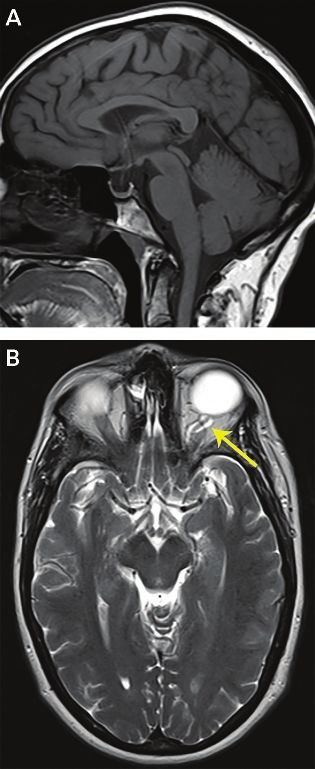
FIGURE 6-2
Imaging findings of spontaneous intracranial hypotension. A 61-year-old man, who worked as
a high-level financial executive, underwent neuroimaging for a 3-year history of cognitive
decline, which had progressed to the point that he missed paying his bills and could not
recall the day of the week. A, T1-weighted sagittal MRI showed pronounced brain sag with
tonsillar descent, flattening of the anterior pons, midbrain collapse, downward
displacement of the posterior corpus callosum, obliteration of the third ventricle, and
straightening of the optic chiasm. B, Axial fluid-attenuated inversion recovery (FLAIR) image
showed marked distention of the midbrain with abnormal high signal in the right hippocampal
formation indicating partial herniation. No abnormal pachymeningeal enhancement was
seen on the postcontrast images (not shown). The patient had minimal improvement with
blood patches and ultimately required surgical repair of his spinal CSF leak, resulting in
significant improvement in his cognition. This case illustrates that symptoms other than
headache may be predominant in spontaneous intracranial hypotension.
FIGURE 6-3
Spontaneous intracranial hypotension. A 30-year-old man developed a severe orthostatic
headache, intense vomiting, vertigo, blurred vision, and neck pain. Imaging 2 weeks later
revealed mild flattening of the anterior pons and pituitary enlargement on T1-weighted
sagittal MRI (A). Despite the relatively normal precontrast images, meningeal thickening and
enhancement is seen on the postcontrast T1-weighted coronal MRI (B).
1074 AUGUST 2018
Copyright © American Academy of Neurology. Unauthorized reproduction of this article is prohibited.nerve root sleeves and are often large and irregular in contour. Degenerative KEY POINTS
changes, such as osteophytes and calcified disk protrusions, can directly tear
● Patients with spontaneous
the dura and are most commonly located ventrally in the lower cervical or thoracic intracranial hypotension
spine.20 CSF venous fistulas are identified in a small percentage of patients. The may be asymptomatic or
anatomy of a leak may be complex, and localizing the exact leak site or sites may experience visual,
be elusive. In 46% to 55% of cases, including those with slow or intermittent vestibulocochlear, and
cognitive problems, as well
leaks and those caused by CSF venous fistulas, the leak site cannot be found.21,22
as an altered level of
In fast (high-flow) leaks, a pool of contrast material extravasates into the consciousness, movement
epidural space surrounding the thecal sac. It may be extensive in some cases, disorders, and intracranial
arising at multiple levels and tracking into the paraspinal soft tissues.3 An extensive hemorrhage.
leak may impede exact localization of the leak site, which requires high resolution
● Patients who have a
and rapid imaging after the administration of contrast. A large pool of epidural typical headache pattern or
contrast is usually absent with slow (low-flow) leaks. Slow leaks generally occur abnormal brain imaging are
around nerve root sleeves and are easier to treat than fast ventral leaks. However, generally identified early.
if the leak is quite slow or intermittent, identification of the leak site is difficult. The lack or subtle nature of
orthostatic symptoms
Delayed imaging may be helpful. One strategy is to inject the contrast material for coupled with normal brain
the CT and MRI simultaneously after injecting a bolus (approximately 15 mL) imaging leads to
of intrathecal saline or artificial CSF to increase the CSF volume. The patient is considerable delay in
immediately imaged with CT, followed by delayed imaging using MRI. Both diagnosis, sometimes for
decades. Spontaneous
techniques are invasive, and the use of gadolinium for intrathecal injection is intracranial hypotension
off-label. Radiation dose must always be considered with CT. should be considered in
Digital subtraction myelography is performed under fluoroscopy and patients with headaches of
allows the real-time visualization of the contrast agent traversing along the any phenotype that are
refractory to conventional
spine to identify the site of the leak.3 The leak appears as a split in the column
headache therapies.
of contrast material creating a parallel track in the epidural space. The
resolution is excellent, but there is a limit to the area of coverage possible, so ● Brain sag may be
the lower cervical and thoracic spine are generally scanned unless a leak is erroneously diagnosed as a
suspected elsewhere. The patient must be absolutely immobile, which may Chiari malformation type I,
leading to unnecessary
require general anesthesia. This technique is performed in the angiography surgery that may make the
suite and is not widely available. patient worse.
Spinal MRI with heavily T2-weighted images and fat suppression is a
noninvasive technique that best identifies the presence (but not necessarily ● In cases of spontaneous
intracranial hypotension, the
the location) of high-flow leaks. A retrospinal fluid collection at C1-C2 seen leak site cannot be
with this technique does not indicate the site of the leak and is a false localizing identified in about half of
sign.23 Disadvantages of this technique are the lower spatial resolution, cases, and intermittent
higher rate of artifacts, decreased likelihood of localizing the leak, and the leaking may occur, which
can make identification of
need for very homogeneous fat suppression.19 This imaging technique is
the leak site challenging.
not generally included in standard MRI software and may require
additional programming. ● Nerve sheath diverticula
Radionuclide cisternography incorporates an iodinated tracer (indium 111 and osseous changes are
diethylenetriamine pentaacetic acid [111InDTPA]), which is injected indirect signs of a potential
leak site.
intrathecally with image acquisition immediately and at 1, 2, 4, 24, and
sometimes 48 hours after injection. It may show direct or indirect evidence ● In cases of spontaneous
of a leak. Unilateral or bilateral focal areas of increased activity within intracranial hypotension, a
paraspinal tissues indicate direct evidence.13 Indirect signs include early large pool of epidural
contrast suggests a
uptake in the kidneys and bladder within 4 hours, absence of activity along high-flow leak.
cerebral convexities at 24 hours (similar to normal pressure hydrocephalus),
and rapid loss of spinal activity. Cisternography has only modest sensitivity and
specificity, and the cost of the tracer is sometimes prohibitive. False-positive and
false-negative results may occur.
CONTINUUMJOURNAL.COM 1075
Copyright © American Academy of Neurology. Unauthorized reproduction of this article is prohibited.HEADACHES DUE TO LOW AND HIGH INTRACRANIAL PRESSURE
Treatment
Conservative treatments may be attempted but are generally unsuccessful
(TABLE 6-5); the refractory nature of spontaneous intracranial hypotension
headaches to typical headache medications is a clue that the patient may have a
secondary disorder. Occasionally, leaks will resolve spontaneously.
A nontargeted, autologous, high-volume epidural blood patch is often the first
step in management, and each attempt is successful approximately 30% of the
time.21 A disagreement exists regarding whether this procedure should be tried
empirically or whether patients should be evaluated to determine the leak site
first. Even transient relief of symptoms supports the diagnosis of spontaneous
intracranial hypotension, so the procedure also has diagnostic value in suspected
cases with normal brain imaging. Symptoms may subsequently recur,
requiring additional blood patches, and at least 5 days are recommended
between procedures.26
“Nontargeted” midline epidural blood patches are generally performed at one
or more spinal levels from T12-L1 through L4-L5, and multilevel patches may be
done in one session. The success rate is potentially enhanced by pretreating the
patient with acetazolamide (250 mg orally given at 18 and 6 hours prior to the
procedure) to decrease CSF volume and allow a higher volume of blood to be
injected in the epidural space and placing the patient in the Trendelenburg
position during and immediately after the procedure.27 A volume of 10 mL to 20
mL of autologous blood is initially injected, which may be increased as tolerated in
subsequent attempts. The amount of blood injected is generally determined by
the patient’s level of procedure-related pain or anatomic constraints limiting the
volume of injection.
Another technique employs a single puncture in the lower thoracic or lumbar
space, passing a guidewire into the epidural space, and advancing in a 4-French
vertebral catheter superiorly into the dorsal epidural space. After confirming
epidural placement with contrast administration, the guidewire is removed,
and autologous blood is injected into the epidural space while slowly withdrawing
the catheter to the access site.28
The mechanism of epidural blood patches leading to improvement is
uncertain and may be related to tamponade and sealing of the leak, restriction of
TABLE 6-5 Strategies for Conservative Management of Spontaneous Intracranial
Hypotension
◆ Bedrest (patients can be bedbound because of their symptoms)
◆ Elevate the foot of the bed (home Trendelenburg position)
◆ Caffeine, theophylline (often helpful but may produce anxiety and insomnia)
◆ Abdominal binder
◆ Analgesics
◆ Corticosteroids (2- to 4-week gradual prednisone taper starting with 50 mg/d)24
◆ Bilateral greater occipital nerve blocks25
◆ Overhydration
◆ Time
1076 AUGUST 2018
Copyright © American Academy of Neurology. Unauthorized reproduction of this article is prohibited.CSF egress into the epidural space, mild compression of the thecal sac by the
epidural blood and secondary increased CSF pressure rostral to the injection, or
decreasing the elasticity of the thecal sac. Aspirin, anticoagulant therapy, and
nonsteroidal anti-inflammatory drugs all interfere with blood clotting and should
be temporarily limited in the periprocedural period, if possible. Detailed
postprocedure instructions (TABLE 6-6) are recommended to prolong the
duration of relief, although they have not undergone rigorous study.
If leak site(s) or potential leak site(s) are identified, targeted epidural blood
patches with percutaneous placement of fibrin sealant (“glue”) have the best
chance of alleviating the patient’s symptoms.26 This procedure is generally
performed with CT guidance and conscious sedation.
Surgery may be needed in cases of a calcified disk or osteophyte causing a
dural defect. Leaking meningeal diverticula can be ligated or clipped. Larger
dural defects are closed with a muscle or fat pledget, with gelatin sponge and
fibrin sealant, or sutured.26 Suturing may be less successful in patients with thin
and friable dural composition. Lumbar dural reduction surgery is occasionally
employed in otherwise refractory cases.29 Transient rebound intracranial
hypertension is possible after successful closure of a spinal leak and can generally
be managed medically. Intrathecal saline or artificial CSF infusion is a
temporizing measure that is incorporated in patients with coma or a decreased
level of consciousness, indicative of brain herniation.30
Prognosis
Once a leak has been successfully treated, the prognosis is generally good.
However, leaks can recur, and new leaks may develop, particularly in patients
with underlying connective tissue disorders.
Postprocedure Instructions Following Epidural Blood Patches TABLE 6-6
Bedrest and light activity only for 24–48 hours after the procedure
Unless medically necessary, avoid nonsteroidal anti-inflammatory drugs for at least 48 hours
Patients requiring anticoagulation who were transitioned to enoxaparin sodium may resume
enoxaparin sodium 12 hours after the procedure
For the first 4 weeks:
◆ No lifting more than 4.5 kg (10 lb)
◆ Avoid straining to have a bowel movement; patients who are prone to constipation should
take a stool softener (sennosides, docusate sodium, fiber supplementation)
◆ Do not bend over to lift objects, tie your shoes, or pick something up; either ask someone
else to help you or, if you must bend over, do so from the knees rather than the waist
◆ Avoid activities that cause twisting of the trunk, such as golf, tennis, canoeing, kayaking,
yoga, martial arts, mopping, vacuuming, or contact sports
◆ If you engage in sexual activity, you should be on the bottom
After 4 weeks, you may gradually increase your activity level as tolerated; however, activities
that require heavy lifting, straining, or trunk rotation (see above) may increase your risk of
developing another leak and should be kept to a minimum if possible
Chiropractic manipulation or adjustment and similar procedures should be avoided
indefinitely as they can tear the dura; massage is acceptable after the first week
CONTINUUMJOURNAL.COM 1077
Copyright © American Academy of Neurology. Unauthorized reproduction of this article is prohibited.HEADACHES DUE TO LOW AND HIGH INTRACRANIAL PRESSURE
KEY POINTS
PSEUDOTUMOR CEREBRI SYNDROME
The pseudotumor cerebri syndrome primarily affects children and adults younger
● CT myelography than age 50. Boys and girls are equally affected until puberty, when the female
performed immediately preponderance manifests.31 For the most part, the clinical presentation in teenage
after the instillation of
girls with pseudotumor cerebri syndrome is similar to adult women with the
intrathecal contrast is the
preferred technique for disorder. The etiology is unknown.32 Many secondary causes have been associated
detecting fast leaks in cases with pseudotumor cerebri syndrome such as exogenous agents, obstruction to
of spontaneous intracranial cerebral venous outflow, endocrine disorders, obstructive sleep apnea, and head
hypotension; delayed MRI trauma (TABLE 6-7).34 When no secondary cause is identified, the syndrome is
myelography is more
sensitive for detecting termed idiopathic intracranial hypertension (IIH), which most commonly affects
slow leaks. women of childbearing age who are obese. As pseudotumor cerebri syndrome
potentially causes blindness, early recognition and treatment are essential.
● Close collaboration Headache is the most common presenting symptom of pseudotumor cerebri
with a neuroradiologist
who is experienced in
syndrome and often persists for years after the other symptoms resolve and the
the diagnostic imaging CSF pressure normalizes.35–37
modalities, interpretation of
findings, and interventional Clinical Features
treatments of spontaneous
The diagnostic criteria of pseudotumor cerebri syndrome are listed in TABLE 6-8.
intracranial hypotension
is imperative. A definite diagnosis requires the presence of papilledema, normal level of
consciousness, normal MRI (including ventricular size) except for signs referable
● A retrospinal fluid to intracranial hypertension, and a lumbar puncture with an opening pressure
collection at C1-C2 is a false measurement and normal CSF composition confirming the diagnosis. In the
localizing sign of a spinal
CSF leak. absence of papilledema or an abducens palsy, the diagnosis can only be suggested
if neuroimaging criteria are met.
● A nontargeted lower Headache is the most common symptom, present in approximately 80%
thoracic or lumbar to 90% of patients at diagnosis, and is frequently the initial symptom. No
high-volume epidural blood
patch is successful in
distinguishing characteristics of the headache occur, although it often represents
alleviating symptoms in a new or different headache and may be quite severe. In the Idiopathic
about 30% of patients with Intracranial Hypertension Treatment Trial (IIHTT) that prospectively studied
spontaneous intracranial 165 patients newly diagnosed with papilledema and mild visual field loss from IIH
hypotension. However, the
(perimetric mean deviation from −2 dB to −7 dB on Humphrey automated
symptoms may recur
over time. perimetry), headache was present in 84% at the baseline visit.38 Headache
phenotypes, characterized using the International Classification of Headache
● Of the neurointerventional Disorders, Third Edition (ICHD-3) criteria,39 were migraine (52%), probable
procedures for spontaneous migraine (16%), tension-type headache (22%), probable tension-type headache
intracranial hypotension,
targeted epidural blood (4%), and unclassifiable (7%). Of the participants, 68% described frontal pain
patches with fibrin sealant that was either pressurelike or throbbing, but many also experienced posterior
have the best chance of (39%), ocular (47%), and neck pain (47%); 36% reported global pain, and
alleviating the patient’s 30% had unilateral pain. Migraine-associated symptoms were common and
symptoms.
included photophobia (70%), phonophobia (52%), nausea (47%), vomiting
● Headache is the most (15%), and worsening with routine physical activity (50%). Participants indicated
common symptom of substantial to severe headache disability as measured by the six-item Headache
pseudotumor cerebri Impact Test (HIT-6) questionnaire, and concurrent photophobia significantly
syndrome and may persist
worsened the HIT-6 score. The mean headache frequency was 12 days in the
after other symptoms
resolve and the CSF month prior to study entry, with 23% having constant, daily head pain.40
pressure normalizes. Most enrollees in the IIHTT with headache also had other symptoms
suggesting a secondary cause, such as constant visual loss (34%), transient
visual obscurations (68%), diplopia (22%), and dizziness (53%). However, 14%
of those with headache had none of those symptoms despite having papilledema.
Of all enrollees, intermittent or daily pulse-synchronous tinnitus occurred in
1078 AUGUST 2018
Copyright © American Academy of Neurology. Unauthorized reproduction of this article is prohibited.Causes of Pseudotumor Cerebri Syndrome and Commonly Associated TABLE 6-7
Conditionsa
Primary Pseudotumor Cerebri Syndrome
◆ Idiopathic intracranial hypertension
Secondary Pseudotumor Cerebri Syndrome
◆ Cerebral venous abnormalities
◇ Cerebral venous sinus thrombosis
◇ Jugular vein obstruction
◆ Decreased CSF absorption from previous intracranial infection or subarachnoid hemorrhage
◇ Increased right heart pressure
◇ Superior vena cava syndrome
◆ Associated with systemic venous hypertension
◆ Medications and exposures
◇ Antibiotics (tetracycline family, fluoroquinolones, nalidixic acid)33
◇ Vitamin A and retinoids (including isotretinoin, all-transretinoic acid, hypervitaminosis A)
◇ Hormones
→ Human growth hormone
→ Thyroxine (in children)
→ Leuprorelin acetate
◇ Anabolic steroids
◇ Withdrawal from chronic corticosteroids
◇ Lithium
◇ Chlordecone
◆ Medical conditions
◇ Endocrine disorders (Addison disease, hypoparathyroidism)
◇ Hypercapnia (sleep apnea, pickwickian syndrome)
◇ Anemia
◇ Renal failure
◇ Turner syndrome
◇ Down syndrome
CSF = cerebrospinal fluid.
a
Modified with permission from Friedman DI, et al, Neurology.34 © 2013 American Academy of Neurology.
CONTINUUMJOURNAL.COM 1079
Copyright © American Academy of Neurology. Unauthorized reproduction of this article is prohibited.HEADACHES DUE TO LOW AND HIGH INTRACRANIAL PRESSURE
52% of patients and was most frequently bilateral (66%).37 Daily nonpulsatile
tinnitus occurred in 23% of patients. Back pain, including radicular pain, was
experienced by 53% of the study cohort. Occasionally, IIH is diagnosed in
asymptomatic patients (often children) when papilledema is discovered
during a routine ophthalmic examination.41
Headaches arising from pseudotumor cerebri syndrome may cause nocturnal
awakening and may worsen in high altitude or in recumbence. Young children
with pseudotumor cerebri syndrome may not be able to articulate their
symptoms, which must be inferred by manifestations such as behavior change,
decline in school performance, malaise, withdrawal, light avoidance, apparent
inability to see well, decreased appetite, or vomiting.
Papilledema from pseudotumor cerebri syndrome may be asymmetric and is
sometimes difficult to discern with direct ophthalmoscopy, so ophthalmologic
consultation is recommended. However, misdiagnosis is possible even using
more sophisticated techniques of evaluating the optic nerve.2 The diagnosis of
pseudotumor cerebri syndrome in patients without papilledema is challenging
TABLE 6-8 Diagnostic Criteria for the Pseudotumor Cerebri Syndromea
1 Required for Diagnosis of Pseudotumor Cerebri Syndromeb
A Papilledema
B Normal neurologic examination except for cranial nerve abnormalities
C Neuroimaging: normal brain parenchyma without evidence of hydrocephalus, mass, or
structural lesion and no abnormal meningeal enhancement on MRI, with and without
gadolinium, for typical patients (female and obese), and MRI, with and without gadolinium,
and magnetic resonance venography for others; if MRI is unavailable or contraindicated,
contrast-enhanced CT may be used
D Normal CSF composition
E Elevated lumbar puncture opening pressure (≥250 mm CSF in adults and ≥280 mm CSF in
children [≥250 mm CSF if the child is not sedated and not obese]) in a properly performed
lumbar puncture
2 Diagnosis of Pseudotumor Cerebri Syndrome Without Papilledema
A In the absence of papilledema, a diagnosis of pseudotumor cerebri syndrome can be
made if B–E from above are satisfied and, in addition, the patient has a unilateral or
bilateral abducens nerve palsy
B In the absence of papilledema or sixth nerve palsy, a diagnosis of pseudotumor cerebri
syndrome can be suggested but not made if B–E from above are satisfied and, in addition,
at least three of the following neuroimaging criteria are satisfied:
i Empty sella
ii Flattening of the posterior aspect of the globe
iii Distention of the perioptic subarachnoid space with or without a tortuous optic nerve
iv Transverse venous sinus stenosis
CSF = cerebrospinal fluid; CT = computed tomography; MRI = magnetic resonance imaging.
a
Reprinted with permission from Friedman DI, et al, Neurology.34 © 2013 American Academy of Neurology.
b
A diagnosis of pseudotumor cerebri syndrome is definite if the patient fulfills criteria A–E. The diagnosis is
considered probable if criteria A–D are met but the measured CSF pressure is lower than specified for a
definite diagnosis.
1080 AUGUST 2018
Copyright © American Academy of Neurology. Unauthorized reproduction of this article is prohibited.and potentially problematic. Papilledema may not be present in patients with KEY POINTS
preexisting optic atrophy, in cases of recurrence, or in patients with mild disease
● When present, the
who were not evaluated for disc edema at symptom onset (which may have been headache of pseudotumor
years prior). cerebri syndrome is
Of the 165 participants in the IIHTT, 67 had a self-reported history of heterogeneous in
migraine, which is more than twice the expected prevalence in the general phenotype, severe, and
disabling.
population.37,38 This may ultimately lead to uncertainty about the etiology of
headaches in patients with pseudotumor cerebri syndrome in the long-term. ● The presence of
However, the visual disturbances of pseudotumor cerebri syndrome are different pulse-synchronous tinnitus
from those that occur in migraine. and transient obscurations
Transient obscurations of vision are brief episodes of visual loss in one of vision supports a
diagnosis of pseudotumor
or both eyes that are often precipitated by arising after bending over. They cerebri syndrome.
may also be provoked by eye movement or occur spontaneously. The visual
loss may be complete (“black out” or “white out”) or partial, often described ● A history of migraine was
as cloudy or foggy. The episodes last seconds to a few minutes (shorter over twice as common in
participants in the Idiopathic
than typical migraine aura) and may occur many times during the day. Intracranial Hypertension
These episodes were one of the most disabling symptoms reported by Treatment Trial as in the
participants in the IIHTT, likely because of their unpredictability.42 Diplopia general population.
in pseudotumor cerebri syndrome is binocular, meaning that the patient sees
● Elevated CSF pressure in
double only when viewing from both eyes simultaneously. Reflecting
patients with pseudotumor
abducens palsy, most patients with double vision experience horizontal cerebri syndrome may lead
diplopia that is worse at a distance and is usually constant. It is extraordinarily to skull base CSF leaks or
uncommon for diplopia to occur in the absence of papilledema in the initial intracranial hypotension.
presentation of this condition. Other visual symptoms include subjective
visual loss (eg, blurred vision, scotomas, dimness, decreased peripheral
vision) and visual distortion (if associated macular edema is present).
Flashes of light (photopsia) lasting seconds to hours may be similar to those
experienced by patients with migraine and are generally provoked by headache,
postural change, darkness, fatigue, eye closure, or watching television.43 The
daily occurrence of photopsia was more common in patients with pseudotumor
cerebri syndrome than in controls.43 However, patterned positive visual
phenomena, such as fortification spectra or scintillating scotomata, do not occur
in pseudotumor cerebri syndrome. Eye pain does not reliably distinguish
pseudotumor cerebri syndrome from migraine or controls.43 Functional
(psychogenic) visual loss may occur and, in one study, was more common in
patients with IIH who did not have papilledema.44,45
Other symptoms of pseudotumor cerebri syndrome may be quite similar to
those of spontaneous intracranial hypotension. Tinnitus can occur with either
condition, although pulse-synchronous tinnitus suggests pseudotumor cerebri
syndrome. Hearing loss or a “high altitude” sensation, neck pain, dizziness, and
back pain are common to both disorders.
Prolonged elevated intracranial pressure from pseudotumor cerebri syndrome
may cause bony erosion at the skull base with a subsequent empty sella and
CSF rhinorrhea or otorrhea. Because patients with skull base CSF leaks from
pseudotumor cerebri syndrome have largely “self-decompressed,” their
CSF pressures tend to be only minimally elevated prior to intervention, and
papilledema is absent.46,47 Similarly, rupture of spinal diverticula from
increased intracranial pressure may lead to spontaneous intracranial
hypotension, which reverts to a high-pressure syndrome when the spinal
leak is repaired (CASE 6-2).
CONTINUUMJOURNAL.COM 1081
Copyright © American Academy of Neurology. Unauthorized reproduction of this article is prohibited.HEADACHES DUE TO LOW AND HIGH INTRACRANIAL PRESSURE
CASE 6-2 A 46-year-old woman presented with a history of orthostatic headaches
that had begun at least 10 years prior to her initial evaluation. The
headaches occurred after being upright for 6 to 7 hours and gradually
increased to a 7 out of 10 in severity. The pain was located at the top of
her head, was sharp in quality, and was
associated with nuchal aching; it was
daily and constant, relieved only with
sleep and traveling to high altitude.
Associated symptoms included
phonophobia, constant tinnitus, and
pulsatile tinnitus (whooshing) when
arising in the morning. She also
experienced occipital headaches with
intrascapular tension and a burning neck
pain. The previous year, she had woken
up 2 days in a row with a “wet ear” with a
halo of blood and clear liquid on the
pillowcase. Her headaches worsened
after this, although no CSF leak was
found on imaging. She took topiramate
100 mg/d for headache prevention.
Lumbar puncture 5 years prior for
suspected intracranial hypotension
showed an opening pressure of 17 cm
CSF. A CT myelogram showed multiple
perineural cysts but no CSF leak; her
headaches resolved for a month after a
nontargeted lumbar epidural blood
patch. MRI 2 years prior to her current
evaluation had shown an expanded and
partially empty sella, flattening of the
posterior sclerae, and distension of the
perioptic subarachnoid space with
normal ventricles and brain parenchyma
(FIGURE 6-4). She had Ehlers-Danlos
FIGURE 6-4
MRI of the patient in CASE 6-2. A, Sagittal syndrome type A.
T1-weighted image shows an expanded Examination revealed a body mass
and partially empty sella. B, Axial index of 28 kg/m2 and normal fundi with
T2-weighted image reveals flattening
spontaneous venous pulsations. The
of the posterior sclerae and a tortuous
optic nerve (arrow) with distention of Trendelenburg test reduced the
the perioptic subarachnoid space (left headache severity from a 7 out of 10 to a
eye shown). 5 out of 10 in 10 minutes.
1082 AUGUST 2018
Copyright © American Academy of Neurology. Unauthorized reproduction of this article is prohibited.An epidural blood patch was performed to target a large perineural cyst
at T10-T11 (FIGURE 6-5) with short-lived relief. Topiramate was discontinued
for potentially exacerbating intracranial hypotension. She had subsequent
blood patches with relief for 5 to 9 weeks. However, 10 days after her last
blood patch, she developed a different headache that worsened when
lying flat. She awakened with a headache that resolved within 10 to
15 minutes of being upright, then the previous orthostatic headache
began 4 hours later. She had gained weight (13.6 kg [30 lb]) after stopping
topiramate and related a “lifelong” history of transient visual
obscurations upon standing.
Her examination showed mild bilateral papilledema (FIGURE 6-6) with
absent spontaneous venous pulsations. A trial of acetazolamide was
somewhat helpful although poorly
tolerated with nausea and cognitive
dysfunction, and therapeutic lumbar
punctures were required. She was not
a candidate for optic nerve sheath
fenestration because of good vision,
and shunting was undesirable because
of its general limitation for headache
treatment and potential for increased
complications related to
Ehlers-Danlos syndrome. Magnetic
resonance venography (MRV)
revealed a dominant right transverse
sinus with a focal narrowing or
arachnoid granulation. The left
transverse sinus was small with a focal
stenosis. Venous manometry showed
a gradient of more than 20 mm across
the stenotic area in the right
transverse sinus, which was
successfully stented, albeit with
difficulty due to a congenital
fenestration in the sinus. She
improved considerably after the FIGURE 6-5
CT myelography of the patient in
procedure, but she continued to have CASE 6-2 showing a large, irregular nerve
mild symptoms of both high and low sheath diverticulum at T10-T11 on the
CSF pressure during the day. left side (arrow).
CONTINUED ON
PAGE 1084
CONTINUUMJOURNAL.COM 1083
Copyright © American Academy of Neurology. Unauthorized reproduction of this article is prohibited.HEADACHES DUE TO LOW AND HIGH INTRACRANIAL PRESSURE
Diagnosis
The diagnosis of pseudotumor cerebri syndrome is based on the presence of
papilledema or abducens nerve palsy, neuroimaging findings, lumbar puncture
opening pressure, and CSF analysis. The status of the patient’s vision helps
determine the appropriate therapy.
VISION EVALUATION. The importance of visual assessment in pseudotumor
cerebri syndrome cannot be overemphasized. Any patient with headache and
visual symptoms needs, at a minimum, a measurement of visual acuity in each
eye, assessment of visual fields, pupil examination to look for an afferent
pupillary defect or poor pupillary reaction, and a fundus examination.
Patients with suspected pseudotumor cerebri syndrome should have a
complete ophthalmologic evaluation with a stereoscopic viewing of the optic
discs and perimetry. Fundus photography, optical coherence tomography,
and fluorescein angiography or orbital ultrasound (if the diagnosis of
papilledema is uncertain) are helpful to document the disc appearance for
subsequent comparison.
CONTINUED FROM
PAGE 1083
FIGURE 6-6
Fundus image of the patient in CASE 6-2 shows mild optic disc edema in both eyes
(A, right eye; B, left eye).
COMMENT This complex case has features of both intracranial hypotension and
intracranial hypertension. Considering her brain MRI findings, most likely,
the patient’s initial problem was intracranial hypertension, with a secondary
self-limited skull base leak (otorrhea) and spinal manifestations of
intracranial hypotension. Ehlers-Danlos syndrome predisposed her to
developing spinal diverticula and a CSF leak. She initially had a good
response to epidural blood patches, but her weight gain after discontinuing
topiramate caused the intracranial hypertension to recur, producing a
“mixed” CSF pressure headache syndrome.
1084 AUGUST 2018
Copyright © American Academy of Neurology. Unauthorized reproduction of this article is prohibited.You can also read