Hereditary eye disease in dogs - British Veterinary Association
←
→
Page content transcription
If your browser does not render page correctly, please read the page content below
Hereditary
eye disease
in dogs
Revised by Sheila Crispin, August 2018

With acknowledgements to past and present members of the
Eye Panel and Eye Panel Working Party and with grateful thanks
to Dr Cathryn Mellersh and the Animal Health Trust

The British Veterinary Association and the Kennel Club
— working together for excellence in canine health

Hereditary eye
disease in dogs

PART I
Clinical Examination for inherited eye disease
By Sheila Crispin

The main purpose of the British Veterinary Association/Kennel Club/International Sheep Dog Society (BVA/KC/ISDS)
Eye Scheme is to ensure that there is no clinical evidence of hereditary eye disease in dogs that are to be used for
breeding. A secondary purpose is to identify breed-related problems which may be inherited, especially if they have
welfare implications for the dog. Following examination of the eye an Eye Examination Certificate is issued which records
the inherited eye disease status (Schedule A) relevant to the breed being examined as either ‘clinically unaffected’ or
‘clinically affected’ together with any additional comments about other clinical findings. In breeds in which primary
glaucoma is recognised clinical examination is supplemented by examination of the drainage angle — gonioscopy
(primary closed angle glaucoma) or measurement of intraocular pressure — tonometry (primary open angle glaucoma).
In addition, clinical examination of puppies when they are still part of a litter (litter screening) can be used to identify
signs of congenital/early onset inherited eye disease in affected puppies. When new potentially inherited conditions
are considered for listing under Schedule A, their inclusion is based on the scientific evidence including a clinical
prevalence of at least 1% over a minimum three-year period and/or the peer-reviewed scientific literature.

Over the years, since its inception in the 1960s, the Eye Scheme       ●● Ensuring that the dog remains free of the inherited eye
has been expanded to include assessment not just of the eye,              diseases listed for the breed being examined under
but also of adjacent (adnexal) structures such as the eyelids. The        Schedule A of the Eye Scheme. A number of inherited eye
result of this expansion is that certification under the Eye Scheme       problems may only be detected later in life (for example,
has important subsidiary benefits; notably, recording anomalies           various types of hereditary cataract and some forms of
(findings of no clinical significance) and abnormalities (findings        progressive retinal atrophy);
of potential or actual clinical significance) whatever their origin.
Examples are provided at the end of this section (Part I).             ●● Indicating whether late onset, potentially inherited, conditions
                                                                          are emerging in older animals;
It is clearly sensible for all dogs (pure bred and cross bred)
which are to be used for breeding to be examined under the             ●● Identifying age-related ocular and generalised diseases with
Eye Scheme prior to being bred from, as this is the simplest way          ocular manifestations, some of which may need treatment.
of identifying breed-related and potentially inherited problems.
Advice on the frequency of re-testing is provided each time            Further information is provided in Why should we check the eyes
the dog is examined under the Eye Scheme. Examination and              of older dogs? (available from www.bva.co.uk/chs).
certification of older dogs, usually those no longer used for
breeding, should be regarded as essential, because longitudinal
                                                                        Healthy eyes enhance dogs’ quality of life
information collected over time is a crucial means of providing
owners and breeders with the information that they need in
order to make informed breeding decisions and, in addition, a
reduced fee provides a financial incentive for certification of
dogs aged eight years and older. In summary, examination of
the older dog is recommended for a number of reasons:

●● Enabling longitudinal information to be collected. A
   longitudinal study is an observational research method in
   which data is gathered for the same subjects repeatedly
   over a period of time;

2   Hereditary eye disease in dogs
The normal eye
                     1                1: Normal adult eye of           2
                                      a Border Collie with a
                                      pigmented iris. 2: Ocular
                                      fundus of that eye, showing
                                      the tapetal fundus (yellow)
                                      dorsally and heavily
                                      pigmented non-tapetal
                                      fundus ventrally.

                     3                3: Normal adult eye of           4
                                      a Crossbred dog with
                                      variations of pigmentation
                                      (heterochromia) in different
                                      sectors of the iris. 4: Ocular
                                      fundus of that eye. Note
                                      that there is less pigment
                                      ventrally, corresponding
                                      with the area of reduced
                                      pigmentation in the iris.

                     5                5: Subalbinotic eye of a         6
                                      normal adult Border Collie.
                                      6: Ocular fundus of that eye.
                                      Both retinal and choroidal
                                      vessels are visible and there
                                      is no tapetum.
Picture, Sue Jones

                     7                7: Normal ocular fundus of       8
                                      the eye of the Border Collie
                                      puppy pictured on the left.
                                      8: The eyes of newborn
                                      puppies are not fully
                                      developed at birth and the
                                      tapetum has not yet formed
                                      in this five-week-old puppy.

                                                                           Hereditary eye disease in dogs   3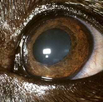
The inherited eye conditions currently certified under the          1. Litters of puppies in breeds with congenital/neonatal
Eye Scheme are reviewed in Part I of this article, together            inherited ocular disease (Litter Screening Eye Examination
with examples of some other conditions of the eye and                  Certificate);
adnexa which may be inherited, as well as other examples
of non-inherited conditions. The potentially inherited and          2. All dogs, whether purebred or cross bred, before they are
non-inherited conditions are currently recorded in the middle          used for breeding and when they are subsequently bred
section of the certificate.                                            from (Eye Examination Certificate). For dogs that are bred
                                                                       from year on year (for example, popular sires), annual
Inherited eye disease status (Schedule A) is recorded in the           re-examination (Eye Examination Certificate) is required
bottom section of the certificate, as congenital (present from         throughout the dog’s breeding life;
birth) and non-congenital (acquired later in life) in type. This
simple classification is not entirely satisfactory as the eyes of   3. Breeds in which primary glaucoma is recognised require
puppies cannot be examined until the puppy is at least 4 weeks         additional assessment as part of certification (Eye
of age, so there is a presumption that abnormalities viewed at         Examination Certificate) under the Eye Scheme. Clinical
this stage are of congenital origin, whereas it is possible that       examination should be supplemented by gonioscopy to
some of the conditions identified may actually be neonatal             assess the drainage angle in breeds susceptible to primary
rather than congenital. In addition, because the eye is immature       closed angle/angle closure glaucoma and tonometry to
at birth, it is possible that some developmental conditions may        measure the intraocular pressure in breeds susceptible to
not be apparent at litter screening (puppies of up to 12 weeks         primary open angle glaucoma. Both these procedures may
of age) and others may become less obvious (for example,               need to be repeated in later life;
Collie eye anomaly) because of postnatal maturation. It is more
rational to describe inherited congenital conditions as those       4. Dogs of eight years of age or older should be examined, at
identifiable during the neonatal stage (congenital/neonatal).          a reduced fee, in order to check ocular and general health,
                                                                       to provide valuable longitudinal data and to ensure that
In summary, clinical examination for certification (Schedule A         later onset inherited conditions are recorded accurately
conditions) under the BVA/KC/IDS Eye Scheme includes:                  (Eye Examination Certificate).

Inherited eye diseases certified under
the Eye Scheme Schedule A
Glaucoma                                                            the anterior chamber of the eye. Aqueous outflow is through
Glaucoma is the term used to describe the effects of a              the iridocorneal (drainage) angle and in the dog the angle is
sustained pathological elevation of intraocular pressure.           extended posteriorly into the ciliary body as the ciliary cleft. It
In normal eyes the rate of aqueous humour formation and             is within the ciliary cleft that the trabecular meshwork is found
the rate of aqueous outflow are in balanced equilibrium             and the canine equivalent of the primate canal of Schlemm, the
and the normal canine intraocular pressure (IOP) measured           aqueous plexus, is situated in the scleral tissues which form the
with an applanation or rebound electronic tonometer is              outer wall of the cleft. In cases of primary glaucoma, a defect
approximately 10–25mmHg. The clinical features shown are            of the iridocorneal angle and the structures associated with the
those which are the result of structural ocular damage and          ciliary cleft is responsible for inadequate drainage, leading to an
the consequent visual impairment or blindness. In particular,       increase of intraocular pressure. The secondary glaucomas are
it is the damage to the retinal ganglion cells and axons of the     associated with antecedent eye disease such as uveitis, primary
optic nerve, particularly the prelaminar portion, which is the      lens luxation, trauma and neoplasia.
most significant feature in sight loss. Once the process of
retinal ganglion cell and optic nerve degeneration has begun,       Classification
the most that appropriate therapy can achieve is slowing            Currently, two types of primary glaucoma may be
down the loss of sight.                                             distinguished, primary closed angle glaucoma (or primary
                                                                    angle closure glaucoma) and primary open-angle glaucoma
Glaucoma is not a single disease entity, but rather a               (see Primary Glaucoma — available from www.bva.co.uk/chs).
degenerative process with a number of possible causes and           The nomenclature has been ‘borrowed’ from human medicine
a final common pathway. Two broad categories of glaucoma            and, although acceptable, does not describe the situation
are recognised — primary and secondary. In primary glaucoma         completely. When these terms are used to describe primary
there is no antecedent intraocular disease and, although the        glaucoma in the dog they denote the appearance of the
aetiology is complex, all the canine primary glaucomas are          entrance to the ciliary cleft. Thus, in closed angle glaucoma the
due to impairment or cessation of aqueous outflow from              cleft is closed and in open-angle glaucoma the cleft is open.

4   Hereditary eye disease in dogs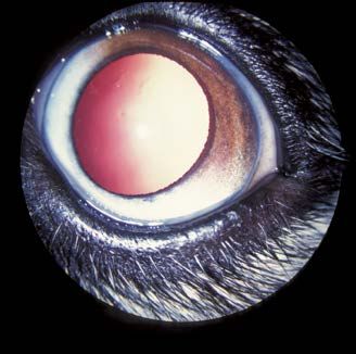
9                                 10                                11                                12                                 13

14                                 15                                16                                17

9: Anterior segment – gross globe – the iris root processes that comprise the pectinate ligament span the entrance to the ciliary cleft. The iris, from which the
processes arise, is on the left and the posterior cornea, where the processes insert, is on the right. 10: Acute closed angle glaucoma. 11: Welsh Springer Spaniel –
chronic closed angle glaucoma with globe enlargement (left eye). 12: Gonioscopy, Barkan goniolens in situ, a column of saline within the silicone tubing creates a
negative pressure to keep the lens in place. 13: Gonioscopy – Koeppe goniolens in situ, coupling gel keeps the lens in place. 14: Normal drainage angle of a Siberian
Husky. The drainage angle is of normal width and is spanned by the pectinate ligament. In this poorly pigmented eye, the white band of the scleral shelf is clearly
distinguished. There is great variation in the number, width, pigmentation and distribution of the fibres that comprise the pectinate ligament in different breeds,
but the width of the normal drainage angle is not subject to such variation. 15: Normal drainage angle of a Flat Coated Retriever. The width of the drainage angle
is normal and the fibres of the pectinate ligament are clearly defined. This eye is more heavily pigmented than the one pictured on the left and the scleral shelf is
obscured by pigment. 16: Goniodysgenesis in a Flat Coated Retriever. There is extensive pectinate ligament dysplasia and sheets of mesenchymal tissue occlude
the majority of the drainage angle. Aqueous drainage is via a limited number of ‘flow holes’. The drainage angle is slightly narrowed and normal pigment obscures
the scleral shelf. The eye was normotensive at the time of examination (intraocular pressure of 18 mmHg measured with a Mackay-Marg tonometer), despite the
compromised drainage angle. 17: Welsh Springer Spaniel – goniodysgenesis.

●● Primary closed angle glaucoma (PCAG)/primary angle                                    can be used as a method        Primary closed angle/angle
   closure glaucoma (PACG):                                                              of screening to determine      closure glaucoma breeds
   In the normal dog, the ciliary cleft entrance is between 1.5                          those animals that are         under Schedule A
   and 2 mm in width and spanned by a number of iris root                                predisposed before             Basset Hound
   processes or fibres, collectively referred to as the pectinate                        the disease makes its          Dandie Dinmont Terrier
   ligament. Dogs which develop primary closed angle glaucoma                            appearance and affected        Japanese Shiba Inu
                                                                                                                        Leonberger
   demonstrate a congenital predisposition, in that the entrance                         animals should not be
                                                                                                                        Retriever (Flat Coated)
   to the ciliary cleft is abnormal and both eyes are affected.                          bred from. Routine             Siberian Husky
   The pectinate ligament is dysplastic, or abnormal, and may be                         gonioscopy can be              Spaniel (American Cocker)
   seen as sheets of undifferentiated mesenchymal tissue rather                          performed at five to six       Spaniel (Cocker)
   than the normal fibrocellular processes. Pectinate ligament                           months of age in most          Spaniel (English Springer)
                                                                                                                        Spaniel (Welsh Springer)
   abnormality with, or without narrowing of the iridocorneal                            breeds and should be
                                                                                                                        Spanish Water Dog
   angle, is referred to as goniodysgenesis. To assist owners and                        repeated, as progression
   breeders and as part of data collection for goniodysgenesis                           of pectinate ligament
   a simple grading system is being piloted to help in the initial                       abnormality over time has been reported in a number of
   assessment of the drainage angle and any changes to the                               breeds, including the Flat Coated Retriever, Basset Hound,
   drainage angle over time. A grading scheme will almost                                Dandie Dinmont Terrier and Welsh Springer Spaniel. The
   certainly replace the ‘Clinically Unaffected’ or Clinically                           Eye Panel Working Party has suggested that gonioscopy
   Affected’ classification that was used previously to record                           is performed at approximately 1, 4 and 7–8 years of age in
   the results of examination (see Primary Glaucoma — available                          breeds at risk.
   from www.bva.co.uk/chs).
                                                                                         The precise modes of inheritance have not been
     The age at which glaucoma develops tends to vary with                               determined for affected breeds, but clear breed and line
     breed but, in most, the disease is one of middle age                                predisposition indicate a genetically determined cause,
     and the presentation can be acute. In addition to raised                            albeit with a likely complex mode of inheritance.
     intraocular pressure the common clinical signs of acute
     closed angle glaucoma include pain, episcleral congestion,                      ●● Primary open angle glaucoma (POAG):
     corneal oedema and a dilated non-responsive pupil. The                             Primary open angle glaucoma presents both dog breeder
     canine drainage angle cannot be viewed directly and a                              and clinician alike with real problems in its early diagnosis,
     goniolens is needed to assess the pectinate ligament                               for the usual clinical features of glaucoma are not present.
     and drainage angle width. The Barkan and Koeppe direct                             It is silent in onset, with either vision impairment or globe
     goniolenses are used most commonly for assessment of                               enlargement usually being the first abnormalities reported
     the iridocorneal angle under the Eye Scheme. Gonioscopy                            by the owner. Goniodysgenesis is not a feature of this

                                                                                                                                      Hereditary eye disease in dogs    5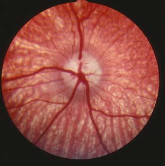
18: Tonopen in use. 19: Tonovet in use.
                                                                                                     20: Primary open angle glaucoma
                                                                                                     — the globe is enlarged and secondary
                                                                                                     lens luxation has occurred.

18                                    19                            20

     type of glaucoma and there are no other predisposing           Persistent hyperplastic primary vitreous
     features that can be used to forecast the advent of the        The embryonic lens is supplied with nutrients by the hyaloid
     disease. It is inherited as a simple recessive trait and has   artery (which grows forward from the optic stalk to reach
     been described extensively in the Beagle. In this breed the    the posterior lens surface at about day 25 of gestation) and
     causal mutation has been identified in the ADAMSTS 10          the tunica vasculosa lentis (which is formed by day 30 of
     gene and a DNA based test has been developed. POAG             gestation). Regression of the vascular supply starts at about
     has been reported in other breeds, notably the Norwegian       day 45 of gestation and is complete some two to four weeks
     Elkhound and, most recently in the UK, the Petit Basset        after birth. Persistence of these vessels and proliferation of
     Griffon Vendeen (PBGV), Basset Hound and Basset Fauve          associated mesodermal elements of the tunica vasculosa
     de Bretagne and DNA tests are available.                       lentis posterioris produce the main lesion of fibrovascular
                                                                    plaque formation on the posterior lens capsule. This plaque
In the PBGV the earliest clinical presentation is either lens       appears as a dense yellow/white opacity with multiple
instability (phacodonesis), lens subluxation in a normotensive      pigment foci attached to the posterior lens capsule. Blood
eye, or actual globe enlargement with possible lens                 vessels may be visible within the plaque and at its periphery.
subluxation/luxation and vision impairment. The disease             Other features of persistent hyperplastic primary vitreous
affects most dogs from 3 to 6 years of age, but a later onset       include persistent capsulopupillary vessels (iridohyaloid
is possible. When the intraocular pressure (IOP) increases,         vessels), coloboma of the lens, posterior lenticonus,
variable degrees of episcleral congestion and mild corneal          intralenticular and retrolental haemorrhage, secondary
oedema may be seen. The pupil may be partially dilated, but         cataract, persistence of the hyaloid artery and Bergmeister’s
with the passage of time the pupillary light reflex (PLR) is        papilla (remnant of the glial sheath around hyaloid vessels on
eventually impaired or lost. Signs of ocular pain are subtle and    the optic disc), and retinal dysplasia.
affected dogs are usually lethargic and sleep more; systemic
non-steroidal anti-inflammatory drugs benefit such patients.        Currently, the Dobermann and Staffordshire Bull Terrier are
With progression globe enlargement occurs, but it is only in        certified for persistent hyperplastic primary vitreous under
advanced disease that cupping of the optic disc and retinal         the Eye Scheme. The mode of inheritance is complex and to
degeneration characterised by increased tapetal reflectivity        date no mutations have been identified that play a role in
and blood vessel attenuation are seen. Lens subluxation may         the development of this disease, although the genetic data
occur even before there is a rise in IOP, but total lens luxation   available suggest an autosomal dominant gene with variable
would appear to be unusual unless there is gross globe              or incomplete penetrance.
enlargement. The elevation of IOP is moderate, with pressures
between 30 and 40 mmHg being routinely recorded.                    Significance
                                                                    Severe lesions cause marked visual loss or blindness. Yellow/
In terms of the BVA/KC/ISDS          Primary open angle glaucoma    brown focal dots on the posterior lens capsule, the mildest
Eye Scheme as applied to POAG, breeds under Schedule A              form of PHPV, have no discernible effect on vision. Small
gonioscopy is not necessary,         Basset Hound                   areas of retrolental plaque formation may spare the lens
for closure of the iridocorneal      Petit Basset Griffon Vendeen   periphery and allow adequate vision. More extensive plaques
angle occurs only in late disease Shar Pei                          and cataract, or other lens abnormalities, usually cause severe
where there is marked globe                                         visual impairment or blindness. The Staffordshire Bull Terrier
enlargement, but tonometry can prove helpful where early            suffers less from the posterior lens capsule deformities, but
disease is present. Annual examination in dogs of 3 to 9 years of   has more widespread retinal folds and rosettes than the
age is advisable. Lens subluxation is common before and after       Dobermann. The condition is not common in either breed in
the rise in IOP, but POAG should not be confused with primary       the UK, but represents a serious congenital inherited problem
lens luxation (see below).                                          in some affected dogs. Surgical treatment of those cases with
                                                                    visual problems is fraught      Persistent hyperplastic primary
                                                                    with difficulty and there is    vitreous breeds under Schedule A
                                                                    a high risk of postoperative Dobermann
                                                                    complications.                  Staffordshire Bull Terrier

6    Hereditary eye disease in dogs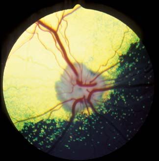
21                                        22                                         23                                        24
21: Persistent hyperplastic primary vitreous in a Dobermann. The white opacity visible through the pupil involves the vitreous and posterior lens capsule.
22: Persistent hyperplastic primary vitreous with intralenticular haemorrhage ventrolaterally. The intralenticular haemorrhage progressed to involve the
whole lens. 23: Persistent hyperplastic primary vitreous. In this dog, the hyaloid vessel has remained patent and there is haemorrhage into the lens. Note the
numerous vacuoles within the lens cortex, which are indicative of progressive cataract formation. 24: The same eye as that pictured in 23 some months later
after cataract formation.

Retinal dysplasia                                                                         multifocal retinal dysplasia are the Cavalier King Charles
The term retinal dysplasia embraces a number of congenital/                               Spaniel, Hungarian Puli, Rottweiler, Golden Retriever and
neonatal conditions resulting from disorderly proliferation                               American Cocker Spaniel.
and atypical differentiation of the retina during embryonic
life. In addition to genetically determined hereditary retinal                      Litter screening is useful, although subtle changes are not
dysplasia, a wide variety of extraneous insults (for example,                       always clearly defined. In older animals remodelling of some or
infectious agents such as canine herpes virus and irradiation)                      all multifocal lesions may result in them becoming less obvious,
to the developing retina may cause acquired, non-inherited,                         even disappearing, over time. This does not appear to be the
retinal dysplasia. Defective retinal development results                            case with the geographic form.
in extremely varied clinical and microscopic appearances
so that, for example, folds, ridges, rosettes, geographic                           ●● Total retinal dysplasia:
abnormalities and localised detachments are all possible                               Somewhat more complex, this form of retinal dysplasia is
manifestations of multifocal retinal dysplasia, whereas total                          associated with non-attachment or complete detachment of
retinal dysplasia is most commonly associated with non-                                the retina. Non-attachment may result from apparent failure
attachment or complete detachment of the retina.                                       of contact of the inner (retinal) and outer (retinal pigment
                                                                                       epithelial) layers of the optic cup during embryogenesis; other
Classification                                                                         ocular abnormalities, such as microphthalmos and nystagmus,
●● Multifocal retinal dysplasia:                                                       are often present in these cases. The Bedlington Terrier,
   Linear folding of the sensory retina and the formation of                           Labrador Retriever and Sealyham Terrier are certified for
   rosettes composed of variable numbers of neuronal retinal                           total retinal dysplasia under the Eye Scheme, although total
   cells are the histological characteristics of multifocal retinal                    retinal dysplasia has been recorded in other breeds, including
   dysplasia. Typically, the lesions range from vermiform grey                         the Yorkshire Terrier and Samoyed. In the Bedlington Terrier
   streaks, dots and circles to multiple focal sites of tapetal                        most affected dogs have an infundibular retinal detachment.
   hyperreflectivity, which may or may not be associated with                          Puppies are blind from birth and may present with leukocoria,
   hypertrophy of the retinal pigment epithelium. Irregularly                          a white pupil, because the retina is immediately behind
   shaped (geographic) areas of retinal dysplasia may also be                          the posterior lens capsule. Retinal neovascularisation may
   encountered. In most cases, the lesions are most obvious                            result in intraocular haemorrhage. In the Sealyham Terrier
   in the tapetal fundus dorsal to the optic disc. In the English                      a total detachment of the retina is similarly present and
   Springer Spaniel, dysplastic changes occur in the developing                        microphthalmos and nystagmus are common. Two forms
   sensory retina at 45 to 50 days of gestation. The other                             of total retinal dysplasia are recognised as inherited in the
   breeds at present certified under the Eye Scheme for                                Labrador Retriever. In one form associated with complete

25                                26                               27                                28                               29
25: Golden Retriever — multifocal retinal dysplasia. 26: Multifocal retinal dysplasia in an English Springer Spaniel. Some of the larger focal lesions with
pigmented centres to the right of the dorsal primary retinal vessels resemble inactive chorioretinopathy, but there are also classical rosettes and vermiform
lesions to the left of the vessels. 27: Cavalier King Charles Spaniel puppy — geographic retinal dysplasia. 28: Cavalier King Charles Spaniel — geographic retinal
dysplasia. 29: Golden Retriever — geographic retinal dysplasia and haemorrhage.

                                                                                                                                    Hereditary eye disease in dogs   7
detachment, the defect               Multifocal retinal dysplasia/               Collie eye anomaly (CEA)
                                          Total retinal dysplasia breeds
     seems to result from an                                                          The prevalence of Collie eye anomaly in the UK is high, in
                                          under Schedule A
     inability of the developing                                                      excess of 60% in the Rough Collie, Smooth Collie and Shetland
                                          Certified – Schedule A (TRD)
     retina to match the rapid                                                        Sheepdog, with the Lancashire Heeler and Border Collie much
                                          Bedlington Terrier
     growth of the choroid                Sealyham Terrier                            less affected. The condition has a worldwide distribution and
     and sclera. The resulting                                                        ocular lesions of identical ophthalmoscopic appearance have
     detachment leads to                  Certified – Schedule A (MRD)                been described in a number of other collie and non-collie
     degeneration of the                  Cavalier King Charles Spaniel               breeds, such as the Bearded Collie and Australian Shepherd.
                                          Hungarian Puli
     neurosensory retina                                                              CEA is a complex disorder affecting retinal, choroidal and
                                          Retriever (Golden)
     because of ischaemic                 Rottweiler                                  scleral development. The classical lesion is of choroidal
     anoxia and such animals              Spaniel (American Cocker)                   hypoplasia in the lateral or dorsolateral region of the fundus
     are blind.                           Spaniel (English Springer)                  near the optic disc. In some animals the hypoplasia may be
                                                                                      more extensive and it is not uncommon for the two eyes to be
                                     Certified – Schedule A
     Other ocular defects,                                                            dissimilar. The lesion is apparent as a ‘pale patch’ and is due to
                                     (TRD and MRD)
     such as microphthalmos,         Retriever (Labrador)                             a localised lack of some, or all, retinal and choroidal pigment
     nystagmus and cataract,                                                          and tapetum. The choroidal vessels in the affected region are
     may be present. The             TRD Total retinal dysplasia, MRD                 also abnormal, usually in size, number and disposition. In merle
     second form of total            Multifocal retinal dysplasia                     dogs, with little pigmentation in the fundus and no tapetum,
     retinal dysplasia, which                                                         choroidal hypoplasia will be less obvious and the appearance
     has not been reported in the UK, is ocular-skeletal dysplasia                    of the choroidal vessels then becomes the important
     associated with severe ocular defects and short-limbed                           diagnostic feature. In addition to choroidal hypoplasia,
     dwarfism. In addition to the Labrador Retriever (dwarfism                        colobomas and staphylomas of the optic nerve head and/or
     with retinal dysplasia type 1 — DRD1) it has also been                           adjacent tissues may be part of the extended phenotype and
     reported in the Samoyed (dwarfism with retinal dysplasia                         can sometimes be the only visible abnormality. It has been
     type 2 — DRD2). This phenotype is inherited as an autosomal                      suggested that adult dogs in this category may be examples of
     recessive in both breeds and mutations have been                                 so-called ‘go normals’. The term ‘go normal’ has been applied
     identified; a 1-base insertional mutation in exon 1 of COL9A3                    to cases where post natal development (pigmentation and
     in the Labrador Retriever and a 1,267-bp deletion mutation                       tapetal development) obscures the choroidal hypoplasia which
     in the 5’ end of COL9A2 in the Samoyed.                                          is the key diagnostic feature, so that adult dogs have a fundus
                                                                                      of ‘normal’ appearance; however, this description might be
Significance                                                                          regarded as inappropriate because such dogs are genotypically
A simple autosomal recessive gene is responsible for                                  affected. The phenomenon is common enough to call into
retinal dysplasia in most of the breeds studied. Diagnosis                            question the relevance of examining dogs as adults rather than
is complicated by the fact that retinal dysplasia may be                              as puppies. Data on Collie eye anomaly in the Rough Collie in
the result of both genetic and non-genetic influences, the                            Norway, for example, has indicated that the diagnosis of the
ophthalmoscopic changes may be more difficult to detect in                            condition in a group of dogs of more than three months of
the developing eye (ie, in puppies of less than six months of                         age was almost half that for
                                                                                                                            Collie eye anomaly breeds
age) and there is not always a clear distinction between the                          a group of puppies of seven
                                                                                                                            under Schedule A
various ocular manifestations of the multifocal and total types.                      weeks to three months of age.
                                                                                                                            Border Collie
To add yet further complexity, remodelling of dysplastic lesions                      Furthermore, when puppies             Collie (Rough)
may occur over time.                                                                  which had been diagnosed              Collie (Smooth)
                                                                                      as having Collie eye anomaly          Lancashire Heeler
While many dogs with multifocal retinal dysplasia will have                           with mild choroidal hypoplasia Shetland Sheepdog
no obvious visual defect, some are severely visually impaired,                        at between seven weeks and
as are all dogs affected with total retinal dysplasia. There is,                      three months of age were re-examined at about one year of
therefore, no question of not examining ‘at risk’ breeds under                        age, 68 per cent had a fundus of normal appearance.
the Eye Scheme.

30                                          31                                         32
30: Total retinal dysplasia in a Labrador Retriever puppy. 31: Close-up of the eye of this puppy. The retina can be
visualised behind the lens. 32: Gross globe — total retinal dysplasia with infundibular retinal detachment.

8    Hereditary eye disease in dogs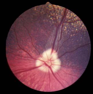
33                                         34                                         35                                         36

37                                         38                                         39

33: Collie eye anomaly in a five-week-old Border Collie puppy. A region of chorioretinal hypoplasia (pale patch) is obvious lateral and slightly dorsal to the optic
nerve head. 34: Border Collie — Collie eye anomaly right eye. 35: Border Collie — Collie eye anomaly left eye. 36: Collie eye anomaly in an adult Rough Collie. The
most striking feature is the peripapillary coloboma ventral to the optic nerve head. 37: Collie eye anomaly in an adult Border Collie. This image is dominated by a
large colobomatous defect to the right of the picture and there is also extensive chorioretinal hypoplasia lateral to the coloboma. 38: Rough Collie — Collie eye
anomaly with retinal detachment. 39: Shetland Sheepdog — Collie eye anomaly with intraocular haemorrhage.

Complications such as retinal detachment and intraocular                             cell layer that produces new                Hereditary cataract breeds
haemorrhage are fortunately rare; thus, the majority of dogs                         lens fibres throughout life.                under Schedule A
with Collie eye anomaly show no apparent visual defect.                              The epithelial cells migrate                Alaskan Malamute
                                                                                     peripherally and elongate at                Australian Shepherd
Tortuosity of the retinal vessels and retinal folds, the latter                      the equator (circumference)                 Belgian Shepherd Dog
usually in the form of vermiform streaks, are not now regarded as                    of the lens. Each fibre extends             Bichon Frise
                                                                                                                                 Boston Terrier*
part of the syndrome, but may relate to the smallness of the eye.                    anteriorly and posteriorly to
                                                                                                                                 Cavalier King Charles Spaniel
                                                                                     meet fibres to the front and rear           German Shepherd Dog
Significance                                                                         to form the suture lines. The               Giant Schnauzer
A variety of fundamental issues combine to make this                                 suture lines form an upright ‘Y’            Irish Red and White Setter
a difficult clinical diagnosis on occasions, however,                                anteriorly and an inverted ‘Y’              Large Munsterlander
                                                                                                                                 Leonberger
it is worth emphasising that Collie eye anomaly is a                                 posteriorly.
                                                                                                                                 Miniature Schnauzer*
congenital condition which can be diagnosed as soon as                                                                           Norwegian Buhund
eye examination is possible (ie at five to six weeks of age),                        Cataract is defined as                      Old English Sheepdog
and that it is diagnosed clinically with greatest accuracy in                        any opacity of the lens                     Poodle (Standard)
such young dogs. In aiming to eliminate CEA from a breed,                            or its capsule. There are                   Retriever (Chesapeake Bay)
                                                                                                                                 Retriever (Golden)
litter screening, combined with DNA testing is the best                              many reasons for cataract
                                                                                                                                 Retriever (Labrador)
approach. Advances in canine genetics have shown that the                            formation — cataracts may be                Siberian Husky
primary CEA mutation has arisen as a single disease allele in                        congenital, due to in utero                 Spaniel (American Cocker)
a common ancestor of herding breeds and that all affected                            insult; traumatic, as a result              Spaniel (Welsh Springer)
dogs share a homozygous deletion of 7.8 kb in the NHEJ1                              of blunt or penetrating injury              Staffordshire Bull Terrier*
gene. The availability of a DNA test has proved of great value                       to the eye; metabolic, as a
                                                                                                                      *More than one type of
in devising comprehensive breeding strategies.                                       consequence of, for example,     hereditary cataract can occur
                                                                                     diabetes mellitus; toxic, caused within an individual breed
                                                                                     by some drugs; nutritional,
Hereditary cataract (HC)                                                             produced by inappropriate diets; or a complication of
The canine lens is an asymmetrical, transparent, biconvex sphere,                    other primary ocular diseases such as uveitis and neoplasia.
with the more convex aspect posteriorly. The adult lens consists                     Cataracts may also form in dogs with generalised progressive
of a central nucleus surrounded by cortical lens fibres and the                      retinal atrophy.
nucleus itself is divisible into various regions according to age:
the oldest, central, portion of the lens is the embryonic nucleus;                   Additionally, primary inherited cataracts have been reported in
surrounding that is the foetal nucleus; and the outermost portion                    a number of breeds and it is these with which the Eye Scheme
is the adult nucleus. The whole lens is contained within an                          is concerned. Fortunately, the age of onset, appearance and
acellular capsule (the anterior capsule is thicker than the posterior                evolution of the cataracts which are certified under Schedule A
capsule). A single layer of epithelial cells lies immediately beneath                of the Eye Scheme are usually quite specific within the affected
the anterior capsule and it is these cells which form the germinal                   breeds, enabling inherited cataracts to be distinguished from

                                                                                                                                      Hereditary eye disease in dogs   9
40                                          41                                         42                                         43
40: Congenital hereditary cataract in a Miniature Schnauzer. The nuclear portion of the lens is affected and there is a pyramid-shaped extension medially. Picture:
Dr Keith Barnett. 41: Hereditary cataract in a Norwegian Buhund. There is an obvious opacity, located posteriorly, involving the posterior pole and posterior
suture lines. In this breed pulverulent nuclear cataracts have also been reported as inherited (see Figure 85). 42: Hereditary cataract in a Golden Retriever. The
characteristic Y-shaped cataract is located in a posterior polar subcapsular position. 43: Total hereditary cataract in a Labrador Retriever.

other non-inherited types of cataract. At present, congenital                         the German Hunt Terrier), the Lancashire Heeler, the Miniature
cataract in the Miniature Schnauzer is the only congenital                            Bull Terrier, the Jack Russell Terrier, the Parson Russell Terrier,
inherited cataract included in the Eye Scheme; the remainder                          the Patterdale Terrier, the Rat Terrier, the Sealyham Terrier,
are all non-congenital types and, as some of them have a                              the Tenterfield Terrier, the
variable age of onset, it is important to examine dogs of over                        Tibetan Terrier, the Toy Fox            Primary lens luxation breeds
eight years of age to ensure that animals that have been used                         Terrier, the Volpino Italiano, the under Schedule A
for breeding remain free of inherited cataract.                                       Welsh Terrier, the Wire-haired          Border Collie
                                                                                                                              Bull Terrier (Miniature)
                                                                                      Fox Terrier and the Yorkshire
                                                                                                                              Fox Terrier (Smooth)
Significance                                                                          Terrier). This mutation has been Fox Terrier (Wire)
Quite apart from the undesirable perpetuation of abnormality                          excluded from involvement in            Lancashire Heeler
within breeding lines, a proportion of inherited cataracts progress                   the Border Collie and the Shar          Parson Russell Terrier
to produce visual impairment and blindness. The only treatment                        Pei, indicating other mutations         Sealyham Terrier
                                                                                                                              Tibetan Terrier
for cataract is surgical and, although modern techniques give                         are the cause in these breeds.
good results, the procedure is expensive.
                                                                                      The condition is essentially bilateral, but almost invariably
                                                                                      presents as a uniocular condition, as one eye may be affected
Primary lens luxation (PLL)                                                           weeks or months in advance of the other. Observant owners
Primary lens luxation is a condition in which an inherent defect                      may notice a change in the appearance of the affected eye
in the zonule (the suspensory ligament of the lens) leads to                          which correlates with the lens moving out of its normal
partial or complete dislocation of the lens at approximately                          position. When the lens moves anteriorly, secondary glaucoma
four to five years of age; clinical signs are not usually observed                    develops rapidly and pain, blepharospasm, photophobia and
before three years of age or later than seven years of age. It                        lacrimation, an increase in intraocular pressure, together with a
is a common cause of secondary glaucoma and, as such, an                              widely dilated non-responsive pupil, visual loss and episcleral
important disease to recognise because of the potential for pain                      and conjunctival congestion, are the most obvious clinical
and visual loss. Primary lens luxation is recognised as a familial                    features. With posterior lens luxation, secondary glaucoma is
problem in certain of the terrier breeds (Miniature Bull Terrier,                     less likely, although most lenses will move forward at some
Smooth Fox Terrier, Wire Fox Terrier, Parson Jack Russell Terrier                     stage. Careful observation will reveal the displaced lens
and Sealyham Terrier), the Tibetan Terrier (which is not a true                       (usually the lens equator is highlighted by the penlight used for
terrier breed), the Lancashire Heeler and the Border Collie. A                        examination), in addition vitreal prolapse and instability of the
single nucleotide substitution in the ADAMTS17 gene has been                          lens may also be apparent (phacodenesis). In addition, the iris
shown to be the cause of PLL in 17 breeds (the Australian Cattle                      trembles slightly with head and eye movement (iridodonesis)
Dog, the Chinese Crested Dog, the Jagdterrier (also known as                          because it has lost the support of the lens.

44                                         45                                          46

44: Primary lens luxation in a Miniature Bull Terrier. The changes were acute, and the eye painful and red (episcleral congestion), indicative of glaucoma (the
intraocular pressure measured with a Mackay-Marg tonometer was 60 mmHg). The other eye was normotensive (intraocular pressure 22 mmHg). The lens has
luxated anteriorly and an area of corneal oedema is apparent as a result of endothelial damage from contact with the lens. The lens equator is highlighted by
illumination from a penlight. 45: Tibetan Terrier – primary lens luxation (anterior). 46: Tibetan Terrier – primary lens luxation, the anteriorly dislocated lens has
developed cataract.

10   Hereditary eye disease in dogs
47 48 49
47: Progressive retinal atrophy in a Cocker Spaniel. Attenuation of the retinal vessels and pallor of the optic nerve head are the most obvious features as tapetal islets (a
normal variant) do not produce the striking hyperreflectivity seen with a more extensive tapetum. Vision was seriously compromised in this dog. 48: Progressive retinal
atrophy in a Miniature Poodle. Tapetal hyperreflectivity is obvious, the optic nerve head is pale and the retinal vessels scarcely visible. The animal was almost totally
blind. 49: Progressive retinal atrophy in a Cocker Spaniel. An abnormal zone is apparent slightly dorsal to the optic nerve head. The zone appears dull or hyperreflective
depending on the direction of the light source. The retinal vessels are marginally narrower than usual and the animal’s vision was apparently unaffected.
Significance Cataracts, which form late Progressive retinal atrophy breeds
Primary lens luxation is an inherited problem which can on in the condition, may under Schedule A
cause persistent pain and blindness without prompt surgical manifest as opacities in the Australian Cattle Dog
intervention. Affected dogs should not be bred from and the ‘at posterior cortex, or as radial Collie (Rough)
risk’ breeds that are to be used for breeding should be examined opacities, before progressing Dachshund (Miniature Long-Haired)
Finnish Lapphund
under the Eye Scheme. to total cataract. Glen of Imaal Terrier
Gordon Setter
In those disorders in which Irish Setter
Progressive retinal atrophy (PRA) the cones or rods are Irish Wolfhound
Progressive retinal atrophy (PRA), is a generic term for a range of preferentially affected (for Lhasa Apso
Miniature Schnauzer
genetically heterogeneous inherited retinal diseases affecting example, cone degeneration Norwegian Elkhound
many breeds of dog. PRA involves the retinal photoreceptors in the Alaskan Malamute, Poodle (Miniature)
and two major types are recognised — developmental or rod dysplasia in the Poodle (Toy)
(dystrophies) and degenerative. The developmental disorders Norwegian Elkhound) the Retriever (Chesapeake Bay)
are of early onset and involve the rod or cone photoreceptors, visual defect will reflect Retriever (Golden)
Retriever (Labrador)
or both, the loss of photoreceptors and rate of progression the type of photoreceptor Retriever (Nova Scotia Duck Tolling)
is usually rapid as the affected photoreceptors fail to involved — tending to Spaniel (American Cocker)
differentiate normally. The degenerative disorders by contrast day blindness when cone Spaniel (Cocker)
usually involve photoreceptors that have differentiated photoreceptors are abnormal Spaniel (English Springer)
normally and the age of onset is later and progression slower. and night blindness when Swedish Vallhund
Tibetan Terrier
For example, in a breed such as the Irish Setter, with rod/cone rod photoreceptors are Welsh Corgi (Cardigan)
dysplasia, the photoreceptors are abnormally formed and begin abnormal.
to degenerate before they are mature. The disease, therefore,
affects these dogs at a relatively young age. The age of onset Significance
is later in, for example, the rod/cone degeneration of the Most types of PRA are autosomal recessive traits but, less
Miniature and Toy Poodle, as the photoreceptors degenerate commonly, autosomal dominant and X-linked types of PRA have
after reaching maturity. also been reported.
The clinical findings in PRA are strikingly similar whatever There is currently no effective treatment for these conditions, so
the underlying pathogenesis. Owners usually notice a loss it is DNA testing that underpins efforts to eliminate PRA in many
of night vision, especially when the dog is in unfamiliar breeds, including a form of PRA, breed specific retinopathy, in the
surroundings. The condition progresses to produce a loss Swedish Vallhund.
of vision under all lighting conditions and there is a poor
pupillary light reflex with dilated pupils. In time, secondary
cataract formation is common. Ophthalmoscopic examination Retinal pigment epithelial dystrophy (RPED) /
indicates a generalised, bilaterally symmetrical increase in central progressive retinal atrophy (CPRA)
tapetal reflectivity (a consequence of retinal atrophy). There Retinal pigment epithelial dystrophy, for many years referred
is attenuation (narrowing) of the retinal vessels, especially to as Central Progressive Retinal Atrophy (CPRA), is a disease
the small peripapillary arterioles, which may become barely of the retinal pigment epithelium cells. The breeds at present
visible (‘ghost vessels’) or invisible on ophthalmoscopy. In dogs certified under the Eye Scheme are the Border Collie, Briard,
with a poorly developed tapetum or an atapetal fundus, the Rough Collie, Smooth Collie, Golden Retriever, Labrador
attenuation of the retinal vessels may be the only obvious Retriever, Shetland Sheepdog, Cocker Spaniel, English Springer
ophthalmoscopic sign of early progressive retinal atrophy, Spaniel and Cardigan Welsh Corgi. Ophthalmoscopic signs
necessitating careful observation. Later in the course of the may be detected on occasion in dogs of just over 12 months of
disease the optic disc becomes paler due to atrophy of its age, but it is more usual to make the diagnosis from about 18
capillaries and nerve fibres. The non-tapetal fundus also shows months of age onwards. Electroretinography is not of value in
extensive areas of depigmentation as the condition progresses. early diagnosis.
Hereditary eye disease in dogs 1150 51 52
50: Retinal pigment epithelial dystrophy in a Cocker Spaniel. At this relatively early stage, multiple focal accumulations
of lipopigment are the most obvious feature, together with some vascular attenuation and a slightly pale optic nerve
head. 51: Cocker Spaniel – retinal pigment epithelial dystrophy, other eye of the same dog (as 50) photographed in blue
light. 52: Retinal pigment epithelial dystrophy in another Cocker Spaniel at a later stage of disease. The lipopigment has
migrated to produce a more cobweb-like appearance.
The disease is caused by the inability of the retinal pigment disease develops in a working dog, the effects are predictably
epithelial cells to degrade spent photoreceptor metabolites, serious. The inheritance of the disease appears complex and
with the resultant accumulation of lipopigment within the environmental factors (for example, poor quality diet) may
retinal pigment epithelium. There are focal concentrations of influence the phenotypic expression. It is prudent to advise
lipopigment-laden cells which migrate into the true retinal layers. against breeding from affected dogs and their relatives,
Degeneration of the photoreceptors (rods and cones) and retinal although the mode of inheritance has not been confirmed.
atrophy are secondary to the lipopigment accumulation and
retinal pigment epithelial cell malfunction.
In dogs of working breeds, the owner may notice an inability to
work in bright light, while vision in dim light may be adequate until
the disease is advanced. In pet dogs, suspect vision may not be
noticed as early. Affected dogs may exhibit a central visual defect,
Examples of
but the pupillary light response is often reasonable and complete
blindness is unusual.
conditions recorded
Ophthalmoscopic examination of early cases indicates light
in comments section
brown foci in the tapetal fundus. These become more numerous
Written descriptive comments can be made on both the Eye
and eventually coalesce into
Retinal pigment epithelial Examination Certificate and Litter Screening Eye Examination
larger areas of lipopigment dystrophy (RPED/CPRA) breeds Certificate providing further information on any Schedule A
with hyperreflective areas under Schedule A
inherited eye diseases identified and as a means of recording
between. In advanced cases the Border Collie
other conditions identified in the course of the clinical
pigment becomes less obvious Briard
Collie (Rough)
examination. Examples include:
as hyperreflectivity increases.
The retinal blood vessels may Collie (Smooth)
Retriever (Golden) ●● Breed-related eye diseases which may be inherited, currently
become attenuated late in the Retriever (Labrador) designated as ‘Under Investigation’ (Schedule B);
disease, but the appearance Shetland Sheepdog
of the non-tapetal fundus and Spaniel (Cocker) ●● Other breed-related anomalies and abnormalities of the eye
optic disc alters little. Both eyes Spaniel (English Springer) and adnexa which may be inherited;
are affected. Welsh Corgi (Cardigan)
●● Acquired ocular abnormalities (for example, post-traumatic
damage, neoplasia, active/inactive inflammation);
The Briard is of interest in that a defect in retinal polyunsaturated
fatty acid metabolism may underlie a form of congenital night ●● Ocular changes indicative of systemic disease.
blindness in which the appearance of the ocular fundus is initially
normal, but by two to three years of age subtle hyper-reflectivity Such observations are important if the abnormalities identified
of the tapetal fundus is apparent, together with sparse greyish have welfare implications, particularly so if they might be passed
spots, which increase in number as the disease progresses, on to subsequent generations. Examples of relevant breed-
accompanied by moderate attenuation of the retinal vessels. related abnormalities with a genetic component include ocular
problems associated with excessive amounts of loose skin,
Significance imperfect eyelid anatomy (for example, entropion, ectropion
Unlike generalised progressive retinal atrophy, retinal pigment and combinations of entropion and ectropion) and ocular-
epithelial dystrophy rarely causes blindness and secondary related disease associated with brachycephaly (for example,
cataract formation is also unusual. However, when the lagophthalmos and corneal damage).
12 Hereditary eye disease in dogsBreed-related ocular conditions that may be inherited, currently listed under Schedule B
53 54 55 56
57 58 59 60
53: Multiocular defects in a Cocker Spaniel. The eye is microphthalmic and a congenital cataract is present. Retinal
dysplasia was an additional finding. 54: Multiocular defects in an Old English Sheepdog. The eye is microphthalmic
and both a congenital cataract and persistent papillary membrane remnants are present. 55: Persistent pupillary
membrane. Most of the remnants arise from the iris collarette and extend anteriorly to the cornea where a discrete
opacity is present at the point of contact. 56: Persistent pupillary membrane. Most of the remnants arise from the iris
collarette and extend posteriorly to the lens. Note the associated pigment deposition on the anterior lens capsule. 57:
Congenital hereditary cataract and uveitis in a Golden Retriever. Note the darkly pigmented iris cyst in the pupillary
aperture medially. 58: Ocular melanosis (abnormal pigment deposition) in a Labrador Retriever. 59: Ocular melanosis
(abnormal pigment deposition) in a Cairn Terrier – secondary glaucoma has resulted in an enlarged globe. 60: Optic nerve
hypoplasia in a Miniature Poodle. 61: Papillary coloboma.
61
Other breed-related ocular conditions of known and potential inheritance
62 63 64 65
66 67 68 69
62: Pug – brachycephaly is associated with respiratory and ocular problems. The
common anatomical-related problems that render the eye more susceptible to
mechanical insult can be summarised as a prominent eye, macropalpebral fissure
and lagophthalmos. Chronic pigmentary keratitis is also present in this dog.
63: Shar Pei – multiple skin problems (including poor eyelid conformation and
entropion) associated with hereditary cutaneous hyaluronosis. 64: Neapolitan
mastiff puppy – excessive skin folds and poor eyelid conformation. 65: Bulldog –
prolapsed nictitans gland, sparse distichia, subtle medial lower lid entropion.
66: Golden Retriever – lower eyelid entropion. 67: Boerbel puppy – severe
bilateral entropion (right). 68: Boerbel puppy – severe bilateral entropion (left).
69: Great Dane – lower eyelid ectropion. 70: St Bernard – ‘diamond eye’ –
70 71 combined entropion and ectropion (right). 71: St Bernard – ‘diamond eye’ –
combined entropion and ectropion (left).
Hereditary eye disease in dogs 1372 73 74 75
76 77 78 79
80 81 82 83
84 85 86 86
87
72: Basset Hound – combined entropion and ectropion and trichiasis.
73: American Cocker Spaniel – distichiasis and cataract. 74: Shetland Sheepdog
– ectopic cilia and corneal ulcer. 75: Cocker Spaniel – epiphora (right) associated
with lower lacrimal punctal aplasia (imperforate punctum). 76: West Highland
White Terrier – keratoconjunctivitis sicca. 77: Boxer – epithelial basement
membrane dystrophy. 78: Cavalier King Charles Spaniel – crystalline corneal
dystrophy (Schnyder-like corneal dystrophy). 79: Labrador Retriever – macular
corneal dystrophy. Picture: Animal Health Trust. 80: English Springer Spaniel –
endothelial dystrophy. 81: German Shepherd Dog – corneal arcus (arcus lipoides
corneae) secondary to hypothyroidism (right eye). 82: German Shepherd Dog
– corneal arcus (arcus lipoides corneae) secondary to hypothyroidism (left eye).
88 89 83: Golden Retriever – lipid keratopathy. 84: Labrador Retriever – iris melanoma.
85: Multiple iris cysts and early, non-inherited, cataract in an aged Labrador
Retriever. 86: Senile nuclear sclerosis in an aged Border Collie. 87: Alaskan
Malamute – pulverulent nuclear cataract. Opacities involving the lens or its
capsule are frequently observed as incidental findings under the Eye Scheme
and there are many causes, of which possible inheritance is just one.
88: Akita – uveodermatological syndrome. 89: Cocker Spaniel – immune-
mediated thrombocytopenia.
14 Hereditary eye disease in dogsExamples of other conditions observed as part of examination under the Eye Scheme
90 91 92 93
90: Corneal foreign body (thorn). 91: Briard – nevus identified at examination
under the Eye Scheme and then followed over several years. 92: Briard –
nevus some two years later. 93: Focal granuloma (ocular larva migrans).
94:Border Collie – inactive focal chorioretinopathy lesions (probably ocular
larva migrans originally). 95: Systemic hypertensive disease associated with
hyperadrenocorticism.
94 95
BVA/ KC/ ISDS Eye Scheme Schedule A 2018
Alphabetical list of breeds and eye conditions for certification under the Inherited Eye Disease Status section of the Certificate of Examination (ie those specified in
Schedule A of the current Procedure Notes and for which “Clinically Unaffected” or “ Clinically Affected” boxes should be ticked):
●● Alaskan Malamute – HC ●● Leonberger – G, HC
●● Australian Cattle Dog – PRA ●● Lhasa Apso – PRA Inherited eye disease status (NB: For a
●● Australian Shepherd – HC ●● Miniature Schnauzer – CHC, PRA, HC number of breeds a DNA test is available
●● Basset Hound – G, POAG ●● Norwegian Buhund – HC for certain eye conditions – please refer to
●● Bedlington Terrier – TRD ●● Norwegian Elkhound – PRA current list)
●● Belgian Shepherd Dog (all varieties) – HC ●● Old English Sheepdog – HC CEA = Collie Eye Anomaly
●● Bichon Frise – HC ●● Parson Russell Terrier – PLL CHC = Congenital Hereditary Cataract
●● Border Collie – CEA, RPED, PLL ●● Petit Basset Griffon Vendeen – POAG G = Goniodysgenesis/Primary
●● Boston Terrier – HC (two forms) ●● Poodle (Miniature) – PRA Glaucoma
●● Briard – RPED ●● Poodle (Standard) – HC HC = Hereditary Cataract
●● Bull Terrier (Miniature) – PLL ●● Poodle (Toy) – PRA MRD = Multifocal Retinal Dysplasia
●● Cavalier King Charles Spaniel – MRD, HC ●● Retriever (Chesapeake Bay) – PRA, HC PLL = Primary Lens Luxation
●● Collie (Rough) – CEA, PRA, RPED ●● Retriever (Flat Coated) – G PHPV = Persistent Hyperplastic Primary
●● Collie (Smooth) – CEA, RPED ●● Retriever (Golden) – MRD, PRA, RPED, HC Vitreous
●● Dachshund (Miniature Long–Haired) – PRA ●● Retriever (Labrador) – MRD, TRD, PRA, RPED, HC POAG = Primary Open Angle Glaucoma
●● Dandie Dinmont Terrier – G ●● Retriever (Nova Scotia Duck Tolling) – PRA PRA = Progressive Retinal Atrophy
●● Dobermann – PHPV ●● Rottweiler – MRD RPED = Retinal Pigment Epithelial
●● Finnish Lapphund – PRA ●● Sealyham Terrier – TRD, PLL Dystrophy (formerly Central
●● Fox Terrier (Smooth) – PLL ●● Shar Pei – POAG Progressive Retinal Atrophy
●● Fox Terrier (Wire) – PLL ●● Shetland Sheepdog – CEA, RPED = CPRA)
●● German Shepherd Dog – HC ●● Siberian Husky – G, HC TRD = Total Retinal Dysplasia
●● Giant Schnauzer – HC ●● Spaniel (American Cocker) – MRD, G, PRA, HC,
●● Glen of Imaal Terrier – PRA ●● Spaniel (Cocker) – G, PRA, RPED
●● Gordon Setter – PRA ●● Spaniel (English Springer) – MRD, G, PRA, RPED,
●● Hungarian Puli – MRD ●● Spaniel (Welsh Springer) – G, HC
●● Irish Red and White Setter – HC ●● Spanish water Dog – G
●● Irish Setter – PRA ●● Staffordshire Bull Terrier – PHPV, HC
●● Irish Wolfhound – PRA ●● Swedish Vallhund – PRA
●● Japanese Shiba Inu – G ●● Tibetan Spaniel – PRA
●● Lancashire Heeler – CEA, PLL ●● Tibetan Terrier – PRA, PLL
●● Large Munsterlander – HC ●● Welsh Corgi (Cardigan) – PRA, RPED
Hereditary eye disease in dogs 15BVA/KC/ISDS Eye Scheme Schedule B 2018
Conditions under investigation (i.e those that are not yet included in Schedule A of the Procedure notes, but for which information is being actively sought by
examination of the breeds for the conditions specified). These conditions should be commented upon only in the middle section of the eye certificate.
●● Akita – PRA ●● Irish Setter – PRA (late onset)
●● Australian Shepherd – C ●● Lancashire Heeler – HC (early developing), PPM APD = Abnormal Pigment Deposition
●● Beagle – MRD ●● Norwegian Elkhound – MRD C = Coloboma
●● Bloodhound – MOD ●● Old English Sheepdog – MOD, CHC CEA = Collie Eye Anomaly
●● Border Collie – HC (early developing), G ●● Papillon – PRA CHC = Congenital Hereditary Cataract
●● Border Terrier – HC (late onset) ●● Petit Basset Griffon Vendeen – PPM G = Goniodysgenesis/Primary
●● Bullmastiff – PPM ●● Polish Lowland Sheepdog – RPED Glaucoma
●● Cairn Terrier – APD ●● Poodle (Miniature) – ONH HC = Hereditary Cataract
●● Cavalier King Charles Spaniel – MOD ●● Poodle (Standard) – MOD MOD = Multi-ocular defects
●● Collie (Rough) – MOD, MRD ●● Poodle (Toy) – ONH MRD = Multifocal Retinal Dysplasia
●● Dachshund (Miniature Long–Haired) – ONH ●● Retriever (Flat Coated) – PRA ONH = Optic Nerve Hypoplasia
●● Dachshund (Miniature Smooth–Haired) – PRA ●● Retriever (Golden) – MOD, CHC, G PHPV = Persistent Hyperplastic Primary
●● Dachshund (Miniature Wire–Haired) – PPM ●● Retriever (Labrador) – APD Vitreous
●● Dandie Dinmont Terrier – G ●● Rottweiler – MOD, PPM PLL = Primary Lens Luxation
●● Dobermann – MOD ●● Siberian Husky – PPM PPM = Persistent Pupillary Membrane
●● Finnish Lapphund – MRD, PHPV, HC, PPM ●● Spaniel (Cocker) – MOD, PPM PRA = Progressive Retinal Atrophy
●● French Bulldog – HC (early developing) ●● Spaniel (Field) – HC (early developing), MRD RPED = Retinal Pigment Epithelial
●● German Shepherd Dog – MRD ●● Spaniel (Sussex) – MRD Dystrophy (formerly Central
●● German Spitz – MRD ●● Staffordshire Bull Terrier – HC Progressive Retinal Atrophy
●● Giant Schnauzer – MRD (variable age of onset) (CPRA)
●● Great Dane – G ●● Tibetan Terrier – HC (early developing)
●● Greenland Dog – HC (early developing) ●● Welsh Terrier – G
●● Griffon Bruxellois – HC (early developing) ●● West Highland White Terrier – MOD, CHC, PPM
●● Hungarian Vizsla – G ●● Yorkshire Terrier – HC (late onset), PRA
Litter screening checklist BVA/KC/ISDS Eye Scheme synopsis sheet 2018
Alphabetical list of breeds and congenital inherited ocular diseases (those ophthalmoscopically identifiable during the neonatal stage) for those specified in Schedule A
of the current Procedure Notes:
●● Bedlington Terrier – TRD ●● Retriever (Golden) – MRD
●● Border Collie – CEA ●● Retriever (Labrador) – TRD, MRD Congenital inherited ocular diseases
●● Cavalier King Charles Spaniel – MRD ●● Rottweiler – MRD CEA = Collie Eye Anomaly
●● Collie (Rough) – CEA ●● Sealyham Terrier – TRD CHC = Congenital Hereditary Cataract
●● Collie (Smooth) – CEA ●● Shetland Sheepdog – CEA PHPV = Persistent Hyperplastic Primary
●● Dobermann – PHPV ●● Spaniel (American Cocker) – MRD Vitreous
●● Hungarian Puli – MRD ●● Spaniel (English Springer) – MRD TRD = Total Retinal Dysplasia
●● Lancashire Heeler – CEA ●● Staffordshire Bull Terrier – PHPV MRD = Multifocal Retinal Dysplasia
●● Miniature Schnauzer – CHC
Summary
●● The BVA/KC/ISDS Eye Scheme offers a means of identifying ●● Checking puppies’ eyes when they are seen for the first time;
the presence or absence of inherited eye disease in a variety
of breeds of dog. There is little doubt that conscientious ●● Informing all pet owners, not just breeders, about the Eye
breeders of all types of dog, both purebred and crossbred, Scheme; and
wish to use sound stock with known freedom from inherited
eye disease and breed-related ocular disorders as part of ●● Ensuring that owners recognise the need for eye
their breeding programme. However, in the context of a examination in any dog which is to be used for breeding and
comprehensive breeding programme, it is important to are aware of the importance of annual examination for dogs
recognise that inherited problems without any impact on used regularly for breeding.
the dog’s quality of life may well rank below maintaining
genetic diversity and ensuring that breeding pairs are of good ●● In addition, older dogs (those over eight years of age)
temperament and fit for function, an aspect of particular should be examined, in order to ascertain the dog’s status in
importance in working dogs. Understanding the welfare relation to possible later onset inherited ocular conditions,
implications of inherited disease and breed-related ocular any changes that may have occurred with pre-existing
disorders is crucial and those conditions that may be a inherited ocular conditions and as a way of assessing ocular
cause of pain or blindness, require surgical correction, or and general health.
lifelong medical therapy should be regarded as priorities for
elimination, as they have substantial effects on the individual’s Up to date information on the Eye Scheme, which includes
quality of life. All veterinary surgeons involved in clinical the conditions certified in individual breeds and those under
practice can help to achieve this ideal by: investigation (see tables) can be obtained from the BVA website.
16 Hereditary eye disease in dogsYou can also read