HHS Public Access Author manuscript Ann N Y Acad Sci. Author manuscript; available in PMC 2021 November 01 - CDC stacks
←
→
Page content transcription
If your browser does not render page correctly, please read the page content below
HHS Public Access
                                Author manuscript
                                Ann N Y Acad Sci. Author manuscript; available in PMC 2021 November 01.
Author Manuscript

                    Published in final edited form as:
                     Ann N Y Acad Sci. 2020 November ; 1479(1): 196–209. doi:10.1111/nyas.14347.

                    Methylene blue and monosodium glutamate improve neurologic
                    signs after fluoroacetate poisoning
                    Vanessa E. DeLey Cox1, Matthew A. Hartog1, Erin Pueblo1, Michelle Racine1, Laura
                    Jennings1, Justin Tressler1, Wing Y. Tuet1, Samuel Stone1, Samuel A. Pierce1, Lily
                    Thompson1, Aliyah Dukes1, Heidi Hoard-Fruchey1, Benjamin Wong1, Bryan J. McCranor1
                    1PharmaceuticalSciences Department, U.S. Army Medical Research Institute of Chemical
Author Manuscript

                    Defense, Aberdeen Proving Ground, MD

                    Abstract
                         Fluoroacetate (FA) is a tasteless, odorless, water-soluble metabolic poison with severe
                         toxicological effects. Characterized in the mid-1900s, it has been used as a rodenticide but is
                         comparably lethal to all mammals. Many countries have restricted its use, and modern-day
                         accidental human exposures are rare, but recently, concerns have been raised about its application
                         as a chemical weapon with no known antidote. A combined treatment of methylene blue (MB), an
                         antioxidant, and monosodium glutamate (MSG), a precursor of the citric acid cycle substrate
                         alpha-ketoglutarate, has been recommended as an effective countermeasure; however, no peer-
                         reviewed articles documenting the efficacy of this therapy have been published. Using a rodent
                         model, we assessed the effects of MB and MSG on the neurologic, cardiac, and pulmonary
Author Manuscript

                         systems. Transcriptomic analysis was used to elucidate inflammatory pathway activation and
                         guide bioassays, which revealed the advantages and disadvantages of these candidate
                         countermeasures. Results show MB and MSG can reduce neurologic signs observed in rats
                         exposed to sodium FA and improve some effects of intoxication. However, while this strategy
                         resolved some signs of intoxication, ultimately it was unable to significantly reduce lethality.

                    Graphical Abstract
                         Using a rodent model, we assessed the effects of methylene blue (MB) and monosodium
                         glutamate (MSG) on the neurologic, cardiac, and pulmonary systems. Results show MB and MSG
                         can reduce neurologic signs observed in rats exposed to sodium fluoroacetate and improve some
                         effects of intoxication. However, while this strategy resolved some signs of intoxication, ultimately
                         it was unable to significantly reduce lethality.
Author Manuscript

                    Address for correspondence: Bryan J. McCranor, Pharmaceutical Sciences Department, U.S. Army Medical Research Institute of
                    Chemical Defense, 8350 Ricketts Point Rd., Aberdeen Proving Ground, MD 21010. bryan.j.mccranor.civ@mail.mil.
                    Author contributions
                    V.E.D.C. wrote the manuscript, designed experiments, and analyzed data. L.J., E.P., and M.R. performed and analyzed the biochemical
                    assays. M.A.H. wrote the transcriptomics sections. M.A.H. and L.T. performed and analyzed the transcriptomics analysis. E.P., M.R.,
                    S.S., A.D., S.A.P., J.T., W.Y.T., and L.J. performed and analyzed the animal experiments. H.H.F., B.W., and B.J.M. conceptualized this
                    work, edited the manuscript, and performed analysis. B.J.M. accepts responsibility for the integrity of data presented.
                    Disclosure
                    The views expressed are solely those of the authors and do not necessarily represent the official views of the CCRP, NIAID, NIH,
                    HHS, USAMRICD, or DoD.
                    Competing interests
                    The authors declare no competing interests.
DeLey Cox et al. Page 2
Keywords
Author Manuscript
metabolic toxin; 1080; fluorocitrate; rat models; antioxidant; TCA cycle
Introduction
Fluoroacetate (FA) was first identified as a botanically derived metabolic poison.1 In the
1940s, a synthetic version, sodium FA (1080), was characterized and mass-produced as a
rodenticide.2, 3 Exposure to 1080 can be fatal to any species, but mammals are especially
sensitive. In humans, the median lethal dose (LD50) of 1080 is estimated to be 2–10 mg/kg.4
Since no antidote or therapeutic treatment currently exists, many countries, including the
United States, heavily restrict its use; however, some countries, including Mexico, South
Korea, Australia, and New Zealand, have more liberal regulations on the use of 1080,
Author Manuscript
leading to debates about both public safety and the ethics of using 1080 to control invasive
and nuisance species.5–10
Signs and symptoms of 1080 intoxication are nonspecific. Across species, neurologic effects
are the most common with secondary symptoms including respiratory distress and cardiac
dysfunction.11 In humans, symptoms include nausea, muscle weakness, nerve pain,
tachypnea, cyanosis, and decreased body temperature.12–14 The most common clinical
symptom of 1080 poisoning, metabolic acidosis, is observed in many disease states
including cancer and alcohol intoxication, which confounds proper clinical identification.
Additionally, the onset of symptoms is often delayed postexposure in warm-blooded
animals. The lack of specific signs and symptoms combined with the delayed onset
complicates efforts to diagnose poisoning. In the past 10 years, the identification of 1080
intoxication in livestock and domesticated animals has continued to be a focus.15–18 These
Author Manuscript
studies have been complemented by novel strategies to identify 1080 contamination in food
and water sources.19, 20 However, despite ongoing research, there is still no reliable
treatment for intoxication.
Owing to the severity of intoxication following exposure, 1080 has recently become
recognized as a potential chemical weapon that may be used by assassins or terrorists.21–23
In 2007, the CIA released a statement identifying it as a weapon of interest for the Iraqi
Security Service under Saddam Hussain.24 In 2011, a dozen people were arrested for
smuggling 1080 into New York City along with other poisons.25 The route of administration
does not influence lethality; while ingestion is the most common route, 1080 can be inhaled,
injected, or absorbed through dermal abrasions.
Author Manuscript
Despite limited progress identifying medical countermeasures, the mechanism of toxicity
has been understood for decades. FA itself is nontoxic. As a small molecule, it readily passes
through the cellular membrane, and because it mimics acetic acid, FA is converted into F-
acetyl-SCoA and can enter the citric acid cycle. Citrate synthase then catalyzes the reaction
between F-acetyl-SCoA and oxaloacetate to form fluorocitrate (FC), a highly toxic molecule
that inhibits aconitase, thereby blocking cellular respiration.26–28 The conversion of 2-
carbon FA to 6-carbon FC is known as lethal synthesis.29
Ann N Y Acad Sci. Author manuscript; available in PMC 2021 November 01.DeLey Cox et al. Page 3
FC is a competitive inhibitor of aconitase; however, it binds very strongly, mimicking a
suicide substrate.30 Only 3% of FA is estimated to convert to FC.31, 32 The remaining FA is
Author Manuscript
defluorinated via a glutathione-dependent mechanism and excreted in the urine.33 The
binding affinity of FC for aconitase is significantly greater than that of the native substrate,
citrate, so that even a small amount of FC can be fatal. A number of structurally similar
molecules are also converted in vivo to FC with comparable cellular effects and
physiological signs and symptoms.34 Countermeasures against 1080 intoxication would
apply to any chemicals in this group.
Currently, treatment of 1080 poisoning is limited to supportive care. Animal studies have
evaluated the efficacy of various acetate donors (i.e., ethanol, glycerol, and acetate), but the
therapeutic potential is limited, and these countermeasures must be administered close to the
time of exposure.34–36 Investigations into naturally occurring 1080 tolerance in mammals
and microorganism have yet to establish clear routes to developing an antidote, though
Author Manuscript
amino acid metabolism has been suggested to play a role in 1080-tolerant bacteria.37–39
Expectations of providing prompt treatment are unrealistic as symptoms present several
hours postexposure. Some in vitro studies have evaluated the therapeutic potential of
antioxidants, but further research in animal models would be required.40
In 2010, a U.S. patent application was published outlining a therapeutic treatment to counter
1080 intoxication.41 The patent named methylene blue (MB) and monosodium glutamate
(MSG) as a combined treatment to decrease the lethality of 1080 in rats. Data were
presented in the patent, and preliminary data have been included in textbook chapters;
however, no comprehensive peer-reviewed article has been published.14, 42
As an antioxidant, MB can mitigate damage from reactive oxygen species originating from
mitochondrial dysfunction.43 As a medical therapeutic, MB is FDA approved for the
Author Manuscript
treatment of methemoglobinemia and has also shown efficacy in treating hypoxia,
hypotension, and cyanide intoxication.44–47 MSG is metabolized to alpha-ketoglutarate, a
substrate of the citric acid cycle that would bypass the aconitase blockade following 1080
poisoning. By supporting glutamine metabolism, MSG may circumvent the bottleneck in the
citric acid cycle that prevents pyruvate oxidation. Administration of MSG may allow cells to
produce ATP despite the damaging cellular effects of 1080.
Through analysis of biosamples and evaluation of respiratory parameters, the therapeutic
benefit of MB and MSG as countermeasures against 1080 was assessed in an ingestion rat
model. MB and MSG were administered in combination at 0.5 and 2 h after exposure.
Though we found MB and MSG had some therapeutic benefits, in our hands, these
compounds did not reduce lethality in any of the therapeutic models evaluated. These data
Author Manuscript
suggest important directions for future research.
Materials and methods
Animals
Male Sprague–Dawley rats (300–500 g) were housed individually under standard conditions
with a 12 h light/dark cycle, and LabDiet® Rodent Feed 5001 (Lab Supply, Fort Worth, TX)
Ann N Y Acad Sci. Author manuscript; available in PMC 2021 November 01.DeLey Cox et al. Page 4
and water available ad libitum. All research complied with relevant federal statutes and
Author Manuscript
regulations regarding animal care and research including the Animal Welfare Act of 1966
(and amendments) and the National Research Council’s Guide for the Care and Use of
Laboratory Animals (National Academy Press, 2011). The study protocol was reviewed and
approved by the Institutional Animal Care and Use Committee, the United States Army
Medical Research Institute of Chemical Defense (USAMRICD), Aberdeen Proving Ground,
MD.
In vivo exposure—Before exposure, animals were placed in individual whole-body
plethysmography (WBP) chambers for 10 min to acclimate. Baseline measurements of
respiratory parameters were then recorded for 10 minutes. After baseline, animals were
removed and administered either 1080 or water via a non-invasive assisted-drinking method.
48, 49 To mimic real-world conditions, animals were not fasted prior to experiments. Control
animals not exposed to 1080 were used to establish a baseline for healthy animals. Exposure
Author Manuscript
concentrations of 1080 were calculated based on an LD50 of 2.08 mg/kg.48 Doses of 1080
and therapeutics were scaled to preexposure animal weight, and liquid volumes administered
were normalized. Animals were then returned to WBP chambers where respiratory function
was tracked for 24 hours. Physiological and behavioral signs were observed continuously for
at least 2 h postexposure. Therapeutics were administered after exposure via either a
subcutaneous (SC) or an intraperitoneal (IP) injection. A sham treatment (injectable sterile
water) was used as vehicle control. Treatment cohorts for 1080-exposed and control animals
were as follows: (1) exposure to 1 × LD50 and administration at 0.5 h postexposure of a
single injection of both MB (5 mg/kg, SC) and MSG (250 mg/kg, IP) or sham injections
(water, SC and IP) (n = 39); (2) exposure to 1 × LD50 and two administrations at 0.5 and 2 h
postexposure of MB (5 mg/kg SC) and MSG (250 mg/kg IP) or sham injections (SC and IP)
(n = 40); (3) exposure to 1 × LD50 and no treatments, and animals euthanized for genomic
Author Manuscript
analysis at 1 h postexposure (n = 8), 2 h postexposure (n = 8), and 3 h postexposure (n = 8);
and (4) exposure to 0.5 × LD50 and administration at 0.5 h postexposure of a single injection
of both MB (5 mg/kg, SC) and MSG (250 mg/kg, IP) or sham injections (water, SC and IP)
(n = 24). The total number of animals used was 127.
Prior to euthanasia, animals were deeply anesthetized via an intramuscular injection of
ketamine (90 mg/kg) and xylazine (10 mg/kg). Animals were then euthanized via
exsanguination. Blood was collected from the descending aorta and placed in an untreated
collection tube. The left lung was tied off, and bronchoalveolar lavage was performed on the
right lung using 3 mL of phosphate-buffered saline. Tissues were collected, processed, and
flash-frozen in liquid nitrogen. For biochemical analysis, animals were euthanized, and
tissue and biosamples were collected 24 h postexposure.
Author Manuscript
Comprehensive metabolic panel and complete blood count—For the
comprehensive metabolic panel, blood was collected from the descending aorta and placed
in an untreated collection tube. Samples were run on an automated hematology analyzer to
determine concentrations of blood urea nitrogen (BUN), aspartate transaminase (AST), and
alanine aminotransferase (ALT). For the complete blood count, blood was collected from the
Ann N Y Acad Sci. Author manuscript; available in PMC 2021 November 01.DeLey Cox et al. Page 5
descending aorta and placed in a blood collection tube treated with EDTA, and samples were
Author Manuscript
run on an automated hematology analyzer.
BALF analysis—Bronchoalveolar lavage fluid (BALF) was centrifuged at 2254 × g for 10
min at 10 °C. The supernatant was collected, aliquoted, and stored at −8 0 °C. Total protein
content was assessed via the Pierce 660 protein assay (Cat. No. 22662, Thermo-Fisher
Scientific) using the company’s recommended protocol with the following modifications: 10
μL of BALF and 150 μL of Pierce® 660 nm protein assay reagent were added directly to a
96-well plate. The plate was shaken for 1 min, then incubated for 4 min at room
temperature. Results were read on a SpectraMax M5 microplate reader. Samples were
prepared in triplicate. Prediluted protein assay standards (bovine serum albumin, Cat. No.
23208, Thermo Scientific, Rockford, IL) were used to generate a protein concentration
curve. Hemoglobin (Hb) concentrations were based on the absorption of BALF at 540
nanometers. BALF samples were aliquoted into a 96-well plate and read on a SpectraMax
Author Manuscript
M5 microplate reader. Samples were prepared in triplicate.
Inflammatory cytokine analysis—Cytokine-specific assays were run to quantify serum
concentrations of interleukin (IL)-10. A serum separator tube was used to fractionate blood
by centrifugation. Isolated serum samples were aliquoted and frozen at −80 °C. Thawed
samples were run on a Bio-Plex® 200 system (Bio-Rad Laboratories, Hercules, CA) using
the ProcartaPlex Rat IL-10 Simplex Kit (Cat. No. EPX10A-36049–901, Thermo-Fisher
Scientific) following the manufacturer’s instructions with a 21-h incubation at 4 °C.
Undiluted samples were assayed in triplicate and analyzed using the Bio-Plex manager
software (v6.1, Bio-Rad Laboratories). The analyte concentration range used to generate the
standard curve for IL-10 was 9.89–40500 pg/mL.
Author Manuscript
Data and statistical analysis—Unless otherwise noted, data are presented as the mean ±
one standard deviation. When appropriate, two groups were compared using a Student’s t-
test or >2 groups were analyzed using an ANOVA followed by Dunnett’s test. Significant
differences were defined as P < 0.05. Specialized software was used to collect respiratory
dynamics (FinePointe Software v2.3.1.16, DSI). All raw data were exported and analyzed
using custom-designed programs (Microsoft Visual Basic for Applications v.7.0.1639,
Microsoft Corporation, Redmond, WA), spreadsheet software (Microsoft Excel
v.14.1.7166.5000 [32-bit], Microsoft Corporation, Redmond, WA), and statistical and
graphing software (GraphPad Prism v5.04, v.7.04, GraphPad Software, Inc., La Jolla, CA).
Genomics—At 1, 2, or 3 h postexposure the heart, lungs, liver, kidneys, and brain were
collected from 1080-exposed and vehicle control animals. The tissues were immediately
Author Manuscript
snap-frozen in liquid nitrogen and stored at −80 °C. Total RNA was isolated using RNeasy®
Plus Mini Kits (QIAGEN, Germantown, MD). RNA concentration, quality, and integrity
were determined using a NanoDrop® ND-8000 UV–Vis spectrophotometer (Thermo-Fisher
Scientific; Waltham, MA) and an Agilent 2200 TapeStation (Agilent Technologies, Santa
Clara, CA). RNA was processed for hybridization to GeneChip® HT RG 230 PM array
plates using the GeneChip 3’ IVT PLUS Reagent Kit (Affymetrix, Santa Clara, CA)
according to the manufacturer’s instructions. The array plates were processed and scanned
Ann N Y Acad Sci. Author manuscript; available in PMC 2021 November 01.DeLey Cox et al. Page 6
using an Affymetrix GeneTitan system. The raw signal intensities were imported into
Author Manuscript
Partek® Genomics Suite® version 7.0 (Partek Inc., St. Louis, MO) and normalized using
robust multi-array averaging (RMA). Principal component analysis (PCA) was used to
identify patterns and major sources of variability in the data. An analysis of variance
(ANOVA) was used to identify significant changes in gene expression induced by 1080
exposure (≤−1.5 fold change ≥1.5; false discovery rate adjusted P ≤ 0.05). The probeset ID,
fold change, and P value of genes with significant changes in expression level were imported
into the Ingenuity® Pathway Analysis (IPA; Qiagen Inc., Valencia, CA) and mapped to
biological and toxicological pathways.
Chemicals—Sterile water for injection (SWFI; Cat. No. A1287301, Thermo-Fisher
Scientific, Waltham, MA) was used as a vehicle. Control groups received only SWFI
without dissolved chemicals added. Sodium FA (Cat. No. N-13216–250MG, Chem Service
Inc., West Chester, PA), MB (Cat. No. M9140, Sigma Aldrich, St. Louis, MO), and L-
Author Manuscript
glutamic acid monosodium salt (Cat. No. RES5063G-A701X, Sigma Aldrich, St. Louis,
MO) were used for animal studies. All chemicals were dissolved in SWFI.
Results
Based on the patent application from Goncharov et al., we pursued similar treatment
strategies using a combination of MB and MSG as countermeasures to 1080 intoxication.41
Additionally, upstream regulator analysis of the transcriptomic data predicted that MSG may
be a regulator of response to 1080 exposure in cardiac tissue at 2 h postexposure (Z-score:
2.31) and in hepatic tissue at 3 h postexposure (Z-score: 3.06) (Fig. S1, online only).
Previously we identified convulsions as a common neurologic sign in rats following 1080
exposure. One cohort of rats was treated with a single administration of 5 mg/kg MB and
Author Manuscript
250 mg/kg MSG at 0.5 h, which significantly reduced the number of rats experiencing
convulsions within 2 h following exposure. In the exposed group treated 0.5 h postexposure,
10% of animals experienced convulsions compared with 70% of sham-treated animals. A
second cohort was treated with the same concentrations of MB and MSG, which were
administered at 0.5 h and again at 2 hours. Of these animals, 50% of treated animals
experienced convulsions as compared with 90% of sham-treated animals, supporting the
argument that MB and MSG reduce neurologic signs in rats exposed to 1080. However,
despite these promising data, treatment with MB and MSG did not improve survival as
compared with sham-treated animals. In fact, more sham animals survived 24 h
postexposure than treated animals (treatment at 0.5 h led to 70% sham and 50% treated
survival; and treatment at 0.5 and 2 h resulted in 90% sham and 70% treated survival.) In
subsequent studies, the effects of MB and MSG on rats exposed to 1080 were evaluated for
Author Manuscript
metabolic changes, respiratory function, and inflammatory response.
Prior research has established multiorgan failure as a characteristic of 1080 exposure.11
Metabolic panels run on blood collected 24 h postexposure have been consistent with
hepatic, renal, and cardiac damage.48 In this study, treatment with a combination of 5 mg/kg
MB and 250 mg/kg MSG, at 0.5 and 2 h postexposure seemed to reduce markers of organ
damage. Concentrations of enzymes indicative of multiorgan damage were similar in treated
animals and control animals but elevated in sham-treated animals (Fig. 1A and B). Elevated
Ann N Y Acad Sci. Author manuscript; available in PMC 2021 November 01.DeLey Cox et al. Page 7
ALT and AST levels are indicators of liver damage. Levels of both enzymes were elevated in
Author Manuscript
serum collected from sham-treated animals, but concentrations were normal in animals
treated at 0.5 and 2 h with MB and MSG. Results from the cohort treated at 0.5 h showed a
similar trend in ALT, but owing to low numbers of blood samples collected from treated
survivors (n = 4), definitive statements cannot be made from this data set (Table S1, online
only). Since hepatocytes are highly metabolically active and can be especially susceptible to
mitochondrial dysfunction, increases in serum markers of liver damage in 1080-exposed
animals are not surprising.50 The decrease in apparent liver injury, based on ALT and AST
levels, in MB- and MSG-treated animals is encouraging.
BUN concentrations, an indicator of renal dysfunction, were also elevated in exposed
animals (Fig. 1C). Elevated BUN concentrations can be directly caused by renal failure or
indirectly caused by cardiac dysfunction.51 In exposed animals, overall BUN concentrations
were significantly elevated. It is not clear how effective treatment was in returning BUN
Author Manuscript
concentrations to normal because data is not consistent between the two cohorts. Treatment
at 0.5 h returned BUN concentrations to normal, suggesting animals treated with MB and
MSG have normal renal function despite exposure to 1080 (Table S1, online only). However,
treatment at 0.5 and 2 h did not appear to reduce circulating BUN (Fig. 1C).
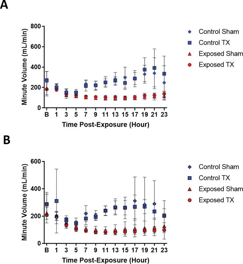
Prior data has shown 1080 diminishes respiratory function in exposed animals; however, as
administered, MB and MSG did not improve respiratory parameters.48 Minute volume, the
product of tidal volume multiplied by the respiratory rate, was tracked for 24 h to evaluate
the effects of therapeutics on respiration. Consistent with previously published data, animals
exposed to 1080 showed signs of respiratory distress. MB and MSG treatment had no
appreciable effect on respiratory parameters in 1080-exposed animals (Fig. 2). Following
1080 exposure, respiratory function decreased around 7 h postexposure and remained
Author Manuscript
depressed throughout the remainder of the study.
Previously published a histologic analysis of lung tissue following exposure to 1080 has
revealed evidence of pulmonary hemorrhage and inflammation.48 To further evaluate the
potential of MB and MSG to mitigate 1080-induced pulmonary toxicity, BALF was
analyzed for evidence of heme (Fig. 3) and protein in the lungs (Fig. 4). An increase in
BALF absorbance at 540 nm was observed in the sham-treated 1080-exposed animals of
both cohorts (Fig. 3A and B). This is consistent with the presence of Hb, indicating
hemorrhage in the lungs.52 We observed that animals treated with MB and MSG
postexposure appeared to have no increase in the amount of Hb in BALF over that, which
was observed in sham-treated exposed animals. Higher BALF protein content was also
observed in exposed animals (Fig. 4), consistent with pulmonary edema.53 Treatment with
Author Manuscript
MB and MSG had no significant effect on the elevated levels of protein when administered
at 0.5 h only (Fig. 4A). In animals that were exposed to 1080 and received MB and MSG at
0.5 and 2 h, the protein content in BALF was elevated, but not statistically significant (Fig.
4B). These data suggest that while MB and MSG are able to reduce the hemorrhagic
pulmonary edema, they are not able to improve overall respiratory function, possibly due to
pulmonary edema.
Ann N Y Acad Sci. Author manuscript; available in PMC 2021 November 01.DeLey Cox et al. Page 8
Inflammation in response to acute pulmonary damage is often marked by increased counts
Author Manuscript
of neutrophils and elevated concentrations of IL-17 and IL-6, both proinflammatory
cytokines, and IL-10, an anti-inflammatory cytokine. Previous publications quantifying
concentrations of cytokines in blood serum have shown elevated concentrations of the
proinflammatory cytokine IL-17 in animals exposed to 1080.48
Transcriptomic analysis revealed changes in gene expression associated with the generation
of an inflammatory response in multiple organs following 1080 exposure. The expression of
genes regulating the acute phase response pathway was found to be altered in cardiac tissue
at 2 h and 3 h postexposure (Fig. 5). This pathway was predicted to be activated (Z-score ≥
2.0) in cardiac tissue at 2 h postexposure, as well as in hepatic and renal tissues 3 h
postexposure (Table S2, online only). The IL-8 signaling pathway was predicted to be
activated in cardiac tissue 2 h and 3 h postexposure, in hepatic tissue at 3 h postexposure,
and in pulmonary tissue at 2 h postexposure (Table S3, online only). IL-8 is a potent
Author Manuscript
chemotactic and neutrophil-activating factor that is an important contributor to the
propagation of an inflammatory response. Additionally, the proinflammatory IL-6 signaling
pathway was predicted to be activated in cardiac and hepatic tissue at 2 h and 3 h
postexposure, as well as in pulmonary and renal tissue 3 h postexposure (Table S4, online
only). Since IL-6 is one of the cytokines activated by IL-17, predicted data are consistent
with the inflammatory response demonstrated in the literature.
A proinflammatory response by IL-6 may be complemented by elevated levels of IL-10, a
protective anti-inflammatory cytokine. The severity of 1080 poisoning means inflammatory
pathways may not be able to be activated before organ systems shut down. Thus, in an effort
to elicit an inflammatory response before multiorgan failure, the third cohort of animals was
exposed to 0.5 × LD50 and treated with MB and MSG 0.5 h postexposure. Concentrations of
Author Manuscript
IL-10 in serum collected from 1080-exposed animals were elevated compared with control
animals but not statistically significant (Fig. S2, online only). Treatment with MB and MSG
did not seem to affect concentrations of IL-10 in animals exposed to 1080. The treatment
did, however, affect healthy animals: IL-10 concentrations were low in control animals
dosed with MB and MSG. The antioxidant properties of MB may explain these data. The
lack of protective anti-inflammatory cytokines suggests MB and MSG are not affecting this
pathway; however, blood would need to be taken at multiple time points to make a firm
determination.
White blood cell (WBC) counts further support an acute inflammatory response in the lungs.
Decreases in total WBCs were driven by a reduction in lymphocytes. Elevated numbers of
neutrophils, consistent with acute pulmonary injury, were observed in exposed animals and
Author Manuscript
treated controls relative to sham controls (Fig. 6). Since neutrophils secrete both IL-17 and
IL-6, these findings are consistent with both the transcriptomic analysis and previously
published data on acute pulmonary inflammation.54 These data are also consistent with
transcriptomic analysis predicting activation of IL-8 that recruits neutrophils. The leucopenia
observed in exposed animals is consistent with other models of 1080 intoxication.18
Ann N Y Acad Sci. Author manuscript; available in PMC 2021 November 01.DeLey Cox et al. Page 9
Discussion
Author Manuscript
Collectively, these data suggest MB and MSG are able to mitigate some signs of 1080
intoxication, though they are unable to prevent mortality. Overall, data from sham-treated
animals and control animals were consistent with previously published results that list
neurologic signs, multiorgan failure, respiratory distress, and an acute inflammatory
response as signs of 1080 exposure. Data trends from biosample analysis suggest MB and
MSG support neurologic, muscular, and organ function but cannot rescue respiratory
function.
The metabolic panel suggests impaired organ function in sham-treated animals, consistent
with previous results. Increased concentrations of AST and ALT in blood reflect impaired
liver function, while elevated BUN concentrations are associated with renal damage and
possible cardiac dysfunction. Overall, treatment with MB and MSG was able to shift
Author Manuscript
averages toward normal levels, suggesting this course of treatment supports organ function
either by directly mitigating toxic effects of 1080 or by indirectly rescuing organ function.
As highly metabolically active cells, hepatocytes may be especially susceptible to 1080-
induced toxicity due to the mechanism of action. Mitochondrial dysfunction in hepatocytes
would be expected to dramatically impair liver function. Blood panel results suggest
treatment with MB and MSG may support hepatic mitochondrial function, potentially
leading to improved liver function, protection against toxic effects of 1080, and reduced
occurrence or severity of multiorgan failure, which is characteristic of 1080 poisoning.
Cardiomyocytes may also be particularly sensitive to mitochondrial dysfunction.55 Cardiac
cells have a high number of mitochondria to fulfill the energy demands of the heart muscle.
The mechanism of toxicity for 1080 affects the citric acid cycle, directly leading to
Author Manuscript
mitochondrial dysfunction and downstream to cardiac dysfunction.48 Cardiac dysfunction is
one possible explanation for elevated concentrations of BUN and pulmonary hemorrhagic
edema. It is possible cardiac dysfunction may be one of the primary signs of 1080 poisoning,
with renal damage and pulmonary edema being cardiogenic secondary effects. Treatment
with MB and MSG reduced average BUN concentrations, suggesting improved cardiac
function may be placing less stress on the kidneys. BALF from treated animals suggests
treatment with MB and MSG reduced pulmonary hemorrhage, though pulmonary distress
was still evident.
Improved cardiac function could also explain the decrease in pulmonary hemorrhage in
treated animals. Analysis of BALF from treated animals showed less evidence of heme,
suggesting MB and MSG reduced pulmonary hemorrhage. However, decreases in total
Author Manuscript
protein concentrations in BALF showed minimal improvement, suggesting that although
treatment can mitigate the more severe hemorrhage, treatment with MB and MSG fails to
entirely prevent pulmonary edema. Minute volume was also depressed in exposed animals
and unaffected by treatment strategies. Collectively, these data suggest MB and MSG can
support pulmonary function but only to a limited extent. One possible explanation is that
MB and MSG may indirectly support respiration through improving cardiac function,
leading to a reduction in pulmonary hemorrhage. Collectively, these data suggest by
Ann N Y Acad Sci. Author manuscript; available in PMC 2021 November 01.DeLey Cox et al. Page 10
supporting the citric acid cycle, MB and MSG may support cardiac function sufficiently to
Author Manuscript
reduce pulmonary hemorrhage, though not enough to restore respiratory function.
To understand the molecular mechanism of toxicity, transcriptomic analysis was used to
further investigate the pulmonary inflammatory response. Increased neutrophils, IL-8, IL-17,
and IL-6 are among the physiological markers consistent with acute pulmonary injury.54
Previous publications have shown elevated IL-17 concentrations.48 Transcriptomic analysis
of animal tissues predicted activation of the IL-8 and IL-6 inflammatory pathways within
pulmonary tissue. Consistent with unresolved acute pulmonary injury, increased circulating
neutrophils suggest an inflammatory response. Expression of IL-10, an anti-inflammatory
cytokine, would also be expected to be upregulated to protect pulmonary tissue from
possible damage from proinflammatory cytokines; however, the transcriptomic data are
inconclusive on whether the IL-10 pathway is activated following exposure at the time
points tested, and serum concentrations of IL-10 were not elevated 24 h postexposure. This
Author Manuscript
acute inflammatory response may reflect the inability of MB and MSG to resolve pulmonary
toxicity. These data suggest that while MB and MSG support organ function, they do not
resolve the pulmonary inflammatory response. Future studies may include candidate
countermeasures to improve cardiac function and rescue pulmonary function. In these
studies, the evaluation of inflammatory markers in BALF may provide more detailed
information about the localized pulmonary effects of 1080.
Collectively, these data suggest that although treatment with MB and MSG is not sufficient
to reduce lethality, these countermeasures do resolve some signs of 1080 poisoning. The
results are consistent with the expected mechanisms of action of these therapeutics, neither
of which is a true antidote. As an antioxidant, MB would be expected to support
mitochondrial and overall cell function by neutralizing reactive oxygen species. MSG
Author Manuscript
bypasses the citric acid cycle blockage and supports ATP production via the glutamine
metabolic pathway. These results show that these therapeutics can indirectly support
neurologic, muscular, and organ function, and suggest that these classes of therapeutics may
be critical to countering the signs and symptoms caused by exposure to 1080. Future studies
investigating alternative therapeutics from these classes may yield improved results.
Critically, these data suggest a path forward. 1080 poisoning seems to require a multi-
pronged therapeutic approach. Although MB and MSG were unable to improve survival
outcomes as applied, these candidate countermeasures have some important therapeutic
benefits improving signs and symptoms and supporting organ function. However, respiratory
parameters did not show significant improvement, and pairing MB and MSG with another
therapeutic targeted at specifically addressing respiratory distress may resolve a broader
Author Manuscript
range of signs and symptoms and shift lethality. Rather than seeking a single-strategy
antidote, these results suggest 1080 poisoning may best respond to a pharmaceutical cocktail
that counters signs and symptoms while supporting organ function.
Supplementary Material
Refer to Web version on PubMed Central for supplementary material.
Ann N Y Acad Sci. Author manuscript; available in PMC 2021 November 01.DeLey Cox et al. Page 11
Acknowledgements
Author Manuscript
The research described was supported by an interagency agreement (AOD 18015–001-00000) between the NIH
Office of the Director (OD) and the U.S. Army Medical Research Institute of Chemical Defense under the oversight
of the Chemical Countermeasures Research Program (CCRP) within the Office of Biodefense Research (OBRS) at
the National Institute of Allergy and Infectious Diseases (NIAID/NIH). Some project performers were supported in
part by an appointment to the Postgraduate Research Participation Program at the U.S. Army Medical Research
Institute of Chemical Defense administered by the Oak Ridge Institute for Science and Education through an
interagency agreement between the U.S. Department of Energy and USAMRDC.
References
1. Marais JSC 1944 Monofluoroacetic acid, the toxic principle of “gifblaar” Dichapetalum cymosum.
Onderstepoort J. Vet. Sci. Anim. Ind 20: 67.
2. Gratz NG 1973 “A criticle review of currently used single-dose rodenticides”. In Bull. W. H. O, Vol.
48: 469–477. World Health Organization. [PubMed: 4543551]
3. Kalmbach ER 1945 “Ten-eighty,” a war-produced rodenticide Science (New York, N.Y.). 102: 232–
Author Manuscript
233. [PubMed: 17778513]
4. Egekeze JO & Oehme FW. 1979 Inorganic and organic fluoride concentrations in tissues after the
oral administration of sodium monofluoroacetate (Compound 1080) to rats. Toxicology. 15: 43–53.
[PubMed: 542959]
5. 2010 “Factsheet: The livestock protection collar”. In. W. Services, Ed.: 1–3. Washington, DC:
Animal and Plant Health Inspection Service.
6. Sherley M 2007 Is sodium fluoroacetate (1080) a humane poison? Anim. Welfare. 16: 449–458.
7. Eason C 2002 Sodium monofluoroacetate (1080) risk assessment and risk communication.
Toxicology. 181-182: 523–530.
8. Eason C, Miller A, Ogilvie S, et al. 2011 An updated review of the toxicology and ecotoxicology of
sodium fluoroacetate (1080) in relation to its use as a pest control tool in New Zealand. N. Z. J.
Ecol. 35: 1–20.
9. Twigg LE & Parker RW. 2010 Is sodium fluoroacetate (1080) a humane poison? The influence of
mode of action, physiological effects, and target specificity. Anim. Welfare. 19: 249–263.
Author Manuscript
10. Eason CT, Shapiro L, Adams P, et al. 2010 Advancing a humane alternative to sodium
fluoroacetate (1080) for wildlife management - welfare and wallaby control. Wildl. Res. 37: 497–
503.
11. Sherley M 2004 The traditional categories of fluoroacetate poisoning signs and symptoms belie
substantial underlying similarities. Toxicol. Lett. 151: 399–406. [PubMed: 15261984]
12. Brockmann JL, McDowell AV & Leeds WG. 1955 Fatal poisoning with sodium fluoroacetate;
report of a case. J. Am. Med. Assoc. 159: 1529–1532. [PubMed: 13271109]
13. Taitelman U, Roy A & Hoffer E. 1983 Fluoroacetamide poisoning in man: the role of ionized
calcium. Arch. Toxicol. Suppl. 6: 228–231. [PubMed: 6578726]
14. Goncharov N, Glashkina L, Savelieva E, et al. 2009 “Fluoroacetate”. In Handbook of Toxicology
of Chemical Warfare Agents. R.C. Gupta, Ed.: 177–198. New York: Academic Press.
15. Shokry E, dos Santos FC, da Cunha PHJ, et al. 2017 Earwax: A clue to discover fluoroacetate
intoxication in cattle. Toxicon. 137: 54–57. [PubMed: 28716647]
16. Giannitti F, Anderson M, Caspe SG, et al. 2013 An outbreak of sodium fluoroacetate (1080)
Author Manuscript
intoxication in selenium- and copper-deficient sheep in California. Vet. Pathol. 50: 1022–1027.
[PubMed: 23613492]
17. Brower A, Struthers J & Schmidt J. 2017 Sodium fluoroacetate toxicity: a case report of malicious
poisoning in dogs across a Phoenix, Arizona neighborhood. Forensic Sci., Med., Pathol. 13: 450–
453.
18. Collicchio-Zuanaze R, Sakate M, Langrafe L, et al. 2010 Hematological and biochemical profiles
and histopathological evaluation of experimental intoxication by sodium fluoroacetate in cats.
Hum. and Exp. Toxicol. 29: 903–913.
Ann N Y Acad Sci. Author manuscript; available in PMC 2021 November 01.DeLey Cox et al. Page 12
19. Parry E & Willison SA. 2018 Direct aqueous injection of the fluoroacetate anion in potable water
for analysis by liquid chromatography tandem mass-spectrometry. Anal. Methods. 10: 5524–5531.
Author Manuscript
20. Wong YT, Law WK, Lai SSL, et al. 2018 Ultra-trace determination of sodium fluoroacetate (1080)
as monofluoroacetate in milk and milk powder by GC-MS/MS and LC-MS/MS. Anal. Methods.
10: 3514–3524.
21. Holstege CP, Bechtel LK, Reilly TH, et al. 2007 Unusual but potential agents of terrorists. Emerg.
Med. Clin. North Am. 25: 549–566; abstract xi. [PubMed: 17482032]
22. Hume T 2015 “‘Eco-terrorism’ threat to poison infant formula in New Zealand”. In. https://
www.cnn.com/2015/03/10/asia/new-zealand-dairy-threat/index.html: CNN.
23. Cooney TP, Varelis P & Bendall JG. 2016 High-Throughput Quantification of Monofluoroacetate
(1080) in Milk as a Response to an Extortion Threat. J. Food Prot. 79: 273–281. [PubMed:
26818988]
24. CIA. (CIA). IIS undeclared research on poisons and toxins for assassination: Iraq’s chemical
warfare program. https://www.cia.gov/library/reports/general-reports-1/iraq_wmd_2004/
chap5_annxA.html.
25. Rashbaum WK 2011 “12 held in sale of pest poisons, one 60 times as potent as the legal limit”. In
Author Manuscript
The New York Times. https://www.nytimes.com/2011/09/20/nyregion/12-arrested-in-sales-of-
illegal-pesticides-in-chinatown.html.
26. Buffa P & Peters RA. 1949 The in vivo formation of citrate induced by fluoroacetate and its
significance. J. Physiol. 110: 488–500. [PubMed: 15406444]
27. Buffa P & Pasquali-Ronchetti I. 1977 Biochemical lesions of respiratory enzymes and
configurational changes of mitochondria in vivo. II. Early ultrastructural modifications correlated
to the biochemical lesion induced by fluoroacetate. Cell Tissue Res. 183: 1–23. [PubMed: 922823]
28. Carrell HL, Glusker JP, Villafranca JJ, et al. 1970 Fluorocitrate inhibition of aconitase: relative
configuration of inhibitory isomer by x-ray crystallography. Science (New York, N.Y.). 170: 1412–
1414. [PubMed: 5481856]
29. Peters RA 1952 Lethal synthesis. Proc. R. Soc. London, Ser. B: Biol. Sci. 139: 143–170. [PubMed:
14911820]
30. Lauble H, Kennedy MC, Emptage MH, et al. 1996 The reaction of fluorocitrate with aconitase and
the crystal structure of the enzyme-inhibitor complex. Proc. Natl. Acad. Sci. U. S. A. 93: 13699–
Author Manuscript
13703. [PubMed: 8942997]
31. Gal EM, Drewes PA & Taylor NF. 1961 Metabolism of fluoroacetic acid-2-C-14 in the intact rat.
Arch. Biochem. Biophys. 93: 1–14. [PubMed: 13702988]
32. Schaefer H & Machleidt H. 1971 Conversion of fluoroacetic acid to amino acids in the mammal.
Biochim. Biophys. Acta. 252: 83–91. [PubMed: 5141830]
33. Soiefer AI & Kostyniak PJ. 1983 The enzymatic defluorination of fluoroacetate in mouse liver
cytosol: the separation of defluorination activity from several glutathione S-transferases of mouse
liver. Arch. Biochem. Biophys. 225: 928–935. [PubMed: 6625615]
34. Goncharov NV 2009 “Fluoroacetate”. In Handbook of Toxicology of Chemical Warfare Agents.
R.C. Gupta, Ed.: 177–198. New York: Academic Press.
35. Goncharov NV, Jenkins RO & Radilov AS. 2006 Toxicology of fluoroacetate: a review, with
possible directions for therapy research. J. Appl. Toxicol. 26: 148–161. [PubMed: 16252258]
36. Hoyos CLA, Galvis MAC, Estrada DO, et al. 2018 Intravenous lipid emulsion and ethanol for
sodium fluoroacetate poisoning. Am. J. Ther. 25: E756–E758. [PubMed: 29668489]
37. Pimentel MFA, Paula DAJ, Riet-Correa F, et al. 2019 Detection and characterization of bovine
Author Manuscript
rumen microorganisms resistant to sodium fluoroacetate. Acta Scientiae Veterinariae. 47.
38. Leong LEX, Denman SE, Hugenholtz P, et al. 2016 Amino acid and peptide utilization profiles of
the fluoroacetate-degrading bacterium synergistetes strain MFA1 under varying conditions.
Microb. Ecol. 71: 494–504. [PubMed: 26111963]
39. Deakin JE, Cooper DW, Sinclair JJ, et al. 2013 Towards an understanding of the genetic basis
behind 1080 (sodium fluoroacetate) tolerance and an investigation of the candidate gene ACO2.
Aust. J. Zool. 61: 69–77.
Ann N Y Acad Sci. Author manuscript; available in PMC 2021 November 01.DeLey Cox et al. Page 13
40. Mead RJ, Moulden DL & Twigg LE. 1985 Significance of sulfhydryl compounds in the
manifestation of fluoroacetate toxicity to the rat, brush-tailed possum, woylie and western grey
Author Manuscript
kangaroo. Aust. J. Biol. Sci. 38: 139–149. [PubMed: 4051904]
41. Goncharov NV, Kuznetsov AV, Glashkina LM, et al., inventors. 2010 US patent Date of
application: 2007.
42. Goncharov N, Savelieva E, Zinchenko V, et al. 2015 “Fluoroacetate”. In Handbook of Toxicology
of Chemical Warfare Agents. Gupta RC, Ed.: 193–214. Elsevier.
43. Ginimuge PR & Jyothi SD. 2010 Methylene blue: revisited. J. Anaesthesiol. Clin. Pharmacol. 26:
517–520. [PubMed: 21547182]
44. F.D.A. 2016 PROVAYBLUE label. https://www.accessdata.fda.gov/drugsatfda_docs/label/
2016/204630s000lbl.pdf.
45. Ryou M-G, Choudhury GR, Li W, et al. 2015 Methylene blue-induced neuronal protective
mechanism against hypoxiareoxygenation stress. Neuroscience. 301: 193–203. [PubMed:
26047733]
46. Weissgerber LAJ 2008 Methylene blue for refactory hypotension: A case report. AANA Journal
76: 271–274. [PubMed: 18777811]
Author Manuscript
47. Haouzi P, McCann M, Tubbs N, et al. 2019 Antidotal effects of the phenothiazine chromophore
methylene blue following cyanide intoxication. Toxicological Sciences. 107: 82–94.
48. McCranor BJ, Young TD, Tressler J, et al. 2019 The cardiopulmonary effects of sodium
fluoroacetate (1080) in sprague-dawley rats. Cogent Biol. 5.
49. Rice NC, Rauscher NA, Langston JL, et al. 2018 Behavioral toxicity of sodium cyanide following
oral ingestion in rats: Dose-dependent onset, severity, survival, and recovery. Food Chem. Toxicol.
114: 145–154. [PubMed: 29454866]
50. Mukai M, Bischoff K & Ramaiah SK. 2012 “Liver toxicity”. In Toxicology Veterinary. Gupta RC,
Ed.: 246–263. Waltham: Elsevier.
51. Gwaltney-Brant SM 2012 “Renal toxicity”. In Veterinary Toxicology. Gupta RC, Ed.: 264–277.
Waltham: Elsavier.
52. Beck-Shimmer B, Rosenberger DS, Neff SB, et al. 2005 Pumonary Aspiration Anesthesiology.
103: 556–566.
53. Aggarwal S, Jilling T, Doran S, et al. 2019 Phosgene inhalation causes hemolysis and acute lung
Author Manuscript
injury Toxicol. Lett. 312: 204–213.
54. Ward PA & Lentsch AB. 1999 The acute inflammatory response and its regulation. AMA Arch.
Surg. 134: 666–669. [PubMed: 10367878]
55. Zhou B & Tian R. 2018 Mitochondrial dysfunction in pathophysiology of heart failure. J. Clin.
Invest. 128: 3716–3726. [PubMed: 30124471]
Author Manuscript
Ann N Y Acad Sci. Author manuscript; available in PMC 2021 November 01.DeLey Cox et al. Page 14
Author Manuscript
Figure 1.
Metabolic blood panel. The concentration of ALT, AST, and BUN 24 h after exposure to 1 ×
Author Manuscript
LD50 of 1080. Male rats were exposed to 1 × LD50 of 1080 and then treated with MB and
MSG or sham injection. Treated animals were given MB (5 mg/kg, SC) and MSG (250
mg/kg, IP) twice at 0.5 and 2 h after exposure. Significant increases in all three makers were
observed in the double sham-treated animals, and treatment with MB and MSG appears to
restore alanine aminotransferase (A) and aspartate transaminase (B) concentrations to
control levels. BUN levels were also elevated in exposed animals, but treatment did not
appear to be of any benefit (C). Dash = average, n = 6–10, *P < 0.05 with a one-way
ANOVA followed by Dunnett’s multiple comparison test versus sham control.
Author Manuscript
Author Manuscript
Ann N Y Acad Sci. Author manuscript; available in PMC 2021 November 01.DeLey Cox et al. Page 15
Author Manuscript
Author Manuscript
Author Manuscript
Figure 2.
Author Manuscript
Respiratory parameters following 1080 exposure. Minute volume was measured for animals
exposed to 1 × LD50 of 1080 (red) and control animals (blue). Baseline respiration (B) and
the first 23 h are shown. As relevant, MB (5 mg/kg, SC) and MSG (250 mg/kg, IP) were
administered at either 0.5 (A) or 0.5 and 2 h (B) postexposure. Sham animals were treated
with sterile water. All exposed animals showed a marked reduction in minute volume
compared with the control animals starting around 7 h postexposure. Treatment with MB
and MSG showed no effect on respiration. Error bars indicate standard deviation, n = 9–10.
Ann N Y Acad Sci. Author manuscript; available in PMC 2021 November 01.DeLey Cox et al. Page 16
Author Manuscript
Author Manuscript
Author Manuscript
Figure 3.
Author Manuscript
Determination of heme in BALF. Animals were exposed to 1 × LD50 of 1080, and treated
animals were given MB (5 mg/kg, SC) and MSG (250 mg/kg, IP) either once at 0.5 h (A) or
at both 0.5 and 2 h (B). BALF was collected 24 h after exposure. In both cases, treating with
MB and MSG was able to reduce the amount of absorbance at 540 nm, indicating a
reduction in Hb in BALF. Error bars indicate standard deviation with average marked, n = 5–
8, * P < 0.05 with a one-way ANOVA followed by Dunnett’s multiple comparison test
versus sham control.
Ann N Y Acad Sci. Author manuscript; available in PMC 2021 November 01.DeLey Cox et al. Page 17
Author Manuscript
Author Manuscript
Author Manuscript
Figure 4.
Author Manuscript
Protein content in BALF. Animals were exposed to 1 × LD50 of 1080. Treated animals were
given MB (5 mg/kg, SC) and MSG (250 mg/kg, IP) either at 0.5 h (A) or at 0.5 and 2 h (B).
BALF was collected 24 h after exposure and total protein content was analyzed. Exposure to
1080 caused an increase in protein in the lungs, consistent with the development of
pulmonary edema. A single administration of MB and MSG was unable to reduce the
amount of protein in BALF (A), but 24 h after two administrations, the elevated protein
concentration was no longer significant (B). Error bars indicate standard deviation with
Ann N Y Acad Sci. Author manuscript; available in PMC 2021 November 01.DeLey Cox et al. Page 18
average marked, n = 5–10, **P < 0.01 with a one-way ANOVA followed by Dunnett’s
Author Manuscript
multiple comparison test versus sham control.
Author Manuscript
Author Manuscript
Author Manuscript
Ann N Y Acad Sci. Author manuscript; available in PMC 2021 November 01.DeLey Cox et al. Page 19
Author Manuscript
Author Manuscript
Figure 5.
Transcriptomic pathway analysis. Analysis of the acute phase response pathway in cardiac
tissue at 2 (A) and 3 (B) h after 1080 exposure. Downregulated genes are shown in green
and upregulated genes are in red. The P value and fold change, respectively, of significantly
altered genes are shown adjacent to the affected genes. The acute phase response pathway
was predicted to be activated at 2 h postexposure (Z-score: 2.31) but not at 3 h (Z-score:
1.79).
Author Manuscript
Author Manuscript
Ann N Y Acad Sci. Author manuscript; available in PMC 2021 November 01.DeLey Cox et al. Page 20
Author Manuscript
Author Manuscript
Author Manuscript
Figure 6.
White blood cell (WBC) count. Treated animals were given MB (5 mg/kg, SC) and MSG
(250 mg/kg, IP). Injections were administered at 0.5 and 2 h after exposure. From animals
that survived to 24 h, blood was collected from the descending aorta, and a complete blood
count was performed with an automated hematology analyzer. Overall, the WBC counts
were decreased in exposed animals (A), which was driven largely from a reduction in
lymphocytes (C). Neutrophils were elevated in both exposed and treated control animals (B),
while monocytes remained largely unchanged (D). Dash = average, n = 6–110, *P < 0.05
Author Manuscript
with a one-way ANOVA followed by Dunnett’s multiple comparison test versus sham
control.
Ann N Y Acad Sci. Author manuscript; available in PMC 2021 November 01.You can also read