Hibernation factors directly block ribonucleases from entering the ribosome in response to starvation
←
→
Page content transcription
If your browser does not render page correctly, please read the page content below
Nucleic Acids Research, 2021 1
                                                                                                                           doi: 10.1093/nar/gkab017

Hibernation factors directly block ribonucleases from
entering the ribosome in response to starvation
Thomas Prossliner* , Michael Askvad Sørensen and Kristoffer Skovbo Winther*

Department of Biology, University of Copenhagen, Ole Maaløes Vej 5, DK-2200 Copenhagen, Denmark

                                                                                                                                                                  Downloaded from https://academic.oup.com/nar/advance-article/doi/10.1093/nar/gkab017/6121461 by guest on 19 February 2021
Received November 20, 2020; Revised January 03, 2021; Editorial Decision January 04, 2021; Accepted January 11, 2021

ABSTRACT                                                                          bosomes (7,8) and rapid modulation or inhibition of exist-
                                                                                  ing ribosomes by a variety of factors (4).
Ribosome hibernation is a universal translation                                      One of the most distinctive and striking examples of ri-
stress response found in bacteria as well as plant                                bosome modulation is the dimerization of 70S ribosomes
plastids. The term was coined almost two decades                                  into inactive 100S complexes (5). Variations of this phe-
ago and despite recent insights including detailed                                nomenon, termed ribosome hibernation, are almost ubiqui-
cryo-EM structures, the physiological role and un-                                tously distributed in prokaryotes as well as in plant plastids
derlying molecular mechanism of ribosome hiber-                                   (5,9). In most bacteria, including gram-positives, cyanobac-
nation has remained unclear. Here, we demonstrate                                 teria and some gram-negatives, ribosome hibernation is in-
that Escherichia coli hibernation factors RMF, HPF                                duced by long hibernation promoting factor (lHPF), which
and RaiA (HFs) concurrently confer ribosome hiber-                                binds 70S ribosomes and induces the formation of a 100S
nation. In response to carbon starvation and result-                              ribosome dimer (10–16). In some organisms, lHPFs sta-
                                                                                  bilize ribosomes in a 70S assembled form as reported
ing growth arrest, we observe that HFs protect ri-
                                                                                  for mycobacteria and plant plastids (17–19). In most ␥ -
bosomes at the initial stage of starvation. Consis-                               proteobacteria, such as Escherichia coli, two distinct hiber-
tently, a deletion mutant lacking all three factors                               nation mechanisms exist: the formation of 100S dimers by
(HF) is severely inhibited in regrowth from star-                                the combined action of ribosome modulation factor (RMF)
vation. HF cells increasingly accumulate 70S ribo-                               and hibernation promoting factor (HPF) (9,20,21), and the
somes harbouring fragmented rRNA, while rRNA in                                   stabilization of 70S ribosomes by ribosome associated in-
wild-type 100S dimers is intact. RNA fragmentation                                hibitor A (RaiA) (22,23). Importantly, these two forms of
is observed to specifically occur at HF-associated                                hibernating ribosomes are thought to be simultaneously
sites in 16S rRNA of assembled 70S ribosomes. Sur-                                present in the cell and we will refer to these protein factors
prisingly, degradation of the 16S rRNA 3 -end is de-                             as hibernation factors (or HFs) (20). The formation of 100S
creased in cells lacking conserved endoribonuclease                               dimers occurs in two steps: binding of RMF at the mRNA
                                                                                  exit channel induces dimerization of 70S ribosomes to a
YbeY and exoribonuclease RNase R suggesting that
                                                                                  90S intermediate followed by joining of HPF to form a ma-
HFs directly block these ribonucleases from access-                               ture 100S complex (20,24). RaiA is a homolog of HPF, but
ing target sites in the ribosome.                                                 contains a short C-terminal extension of 18 residues (20).
                                                                                  Consistent with the highly similar sequence and structure of
INTRODUCTION                                                                      HPF and RaiA, both factors are thought to bind in the same
                                                                                  position at the codon-anticodon interaction region of the
Translation is one of the most important cellular processes
                                                                                  ribosomal A- and P-site. The extended C-terminus present
and consumes a considerable fraction of available resources
                                                                                  in RaiA is thought to protrude towards the mRNA chan-
(1,2). During translation in bacteria, free 30S and 50S ri-
                                                                                  nel and into the binding site of RMF, likely interfering with
bosomal subunits are assembled into translating 70S ribo-
                                                                                  RMF binding and 100S formation (25,26).
somes, which are mostly present in polysomic chains of
                                                                                     HFs in E. coli are transcriptionally induced in a growth-
ribosomes. Canonical translation has been studied exten-
                                                                                  rate dependent manner by major stress response pathways:
sively and is well understood (3). Recently, a more com-
                                                                                  rmf, hpf and raiA are all induced by starvation alarmone
plex picture has emerged where translating ribosomes rep-
                                                                                  (p)ppGpp upon transition to stationary phase (27–32). In
resent only a part of a heterogeneous ribosome population
                                                                                  addition, rmf and raiA are also induced by cAMP in re-
(4,5). When protein synthesis is reduced during stress condi-
                                                                                  sponse to glucose starvation (33). Consistently, HFs are pri-
tions, ribosome capacity is generally adjusted by inhibition
                                                                                  marily present in stationary phase, while RaiA has also been
of de novo ribosome synthesis (6), degradation of excess ri-

* To
   whom correspondence should be addressed. Tel: +45 35336986; Email: kristoffer.winther@bio.ku.dk
Correspondence may also be addressed to Thomas Prossliner. Email: thomas.prossliner@bio.ku.dk

C The Author(s) 2021. Published by Oxford University Press on behalf of Nucleic Acids Research.
This is an Open Access article distributed under the terms of the Creative Commons Attribution-NonCommercial License
(http://creativecommons.org/licenses/by-nc/4.0/), which permits non-commercial re-use, distribution, and reproduction in any medium, provided the original work
is properly cited. For commercial re-use, please contact journals.permissions@oup.com
2 Nucleic Acids Research, 2021
detected at low levels during exponential growth (20,34,35). (MOPS MM) supplemented with 0.2% glucose, 1.32 mM
Interestingly, 100S dimers are exclusively formed in station- K2 HPO4 and 0.01 g/l of each nucleobase was used (45,46).
ary phase or during stress and can rapidly dissociate when Bacterial growth in liquid culture was determined by mea-
nutrients are replenished (27,34,36). This observation has suring optical density at 600 nm (OD600 ) in a DU730 spec-
led to the hibernation model in which 100S dimers serve as trophotometer (Beckman Coulter).
a translationally inactive, RNase-protected ribosome reser-
voir (21). The physiological significance of ribosome hiber- Bacterial strains and plasmids
nation appears to differ from organism to organism (5).
The strains used in this study are listed in Supplementary
In E. coli, loss of hibernation factors has been implicated
Table S1. Escherichia coli K-12 MG1655 rmf, hpf and
Downloaded from https://academic.oup.com/nar/advance-article/doi/10.1093/nar/gkab017/6121461 by guest on 19 February 2021
in reduced stationary phase survival and fitness, tolerance
raiA were generated by P1 transduction (47) using E. coli
against heat, acid and osmotic stress (37–39), as well as
K-12 derivative BW25113 rmf::kanR, hpf::kanR and
increased susceptibility to aminoglycoside antibiotics gen-
raiA::kanR as donor strains (48). The kanamycin resis-
tamicin and netilmicin (40,41).
tance cassette (kanR) was removed by FLP-FRT recombi-
Recent high resolution structures of hibernation factors
nation (49). Double and triple deletion mutants were gener-
in complex with ribosomes have shed light on the struc-
ated by sequential P1 transduction of single and double mu-
tural basis of ribosome hibernation (9,18,25,42–44). How-
tants, respectively. Correct insertion and removal of the re-
ever, the physiological role of ribosome hibernation on a
sistance cassette was confirmed by PCR with primers flank-
mechanistic level is not clear. Here, we present a compre-
ing the gene of interest and sequencing of the PCR product
hensive analysis of the collective role of hibernation factors
(for primer sequences see Supplementary Table S2 in sup-
RMF, HPF and RaiA in E. coli. We show that ribosome hi-
plementary).
bernation confers a fitness advantage by ensuring efficient
resuscitation from growth arrest in response to glucose star-
Viability measurements
vation. Analysis of ribosome integrity, subunit distribution
and rRNA fragment accumulation in a strain lacking HFs Viability in selected media was quantified as colony-
(HF) shows increased release of free ribonucleotides dur- forming units per ml of culture (CFU/ml) by spot-plating
ing starvation, accompanied by a stark alteration of the ri- serial dilutions of cultures at indicated time points. To de-
bosome profile. Intriguingly, hibernation-deficient cells ex- termine viability in defined media a single colony was inoc-
hibit increased accumulation of fragmented rRNA in free ulated into MOPS MM and incubated at 37◦ C and shaking
subunits and assembled 70S particles. Furthermore, map- at 160 rpm overnight. Overnight cultures were diluted 100-
ping of 16S rRNA fragments of 70S particles in the absence fold into 15 ml pre-warmed MOPS MM as pre-culture and
of HFs revealed degradation taking place at the 30S-50S grown to an OD600 of ∼0.3 before dilution into 30 ml pre-
subunit interface and the 3 -terminus including the anti- warmed medium to a final OD600 of 0.01. Cultures were in-
Shine-Dalgarno sequence. Strikingly, the degraded rRNA cubated for up to 10 days at 37◦ C and shaking at 160 rpm.
domains all map to positions coinciding with binding sites To determine viability in phosphate-buffered saline (PBS)
of hibernation factors HPF/RaiA and RMF. Surprisingly, an exponential phase culture prepared as described above
the degradation of the 16S rRNA 3 -end is dependent on the was harvested by centrifugation at 4◦ C and 2700 x g, washed
conserved ribonuclease YbeY and exonuclease RNase R, twice in cold PBS (Oxoid) and resuspended in pre-warmed
which have been reported to be involved in ribosome qual- PBS before continuing incubation for up to 10 days at 37◦ C
ity control and 16S rRNA maturation. and shaking at 160 rpm. At indicated time points OD600 of
In conclusion, our data is consistent with HFs directly cultures was measured and serial dilutions of cells in PBS
protecting ribosomes by preventing ribonuclease access to were spotted onto NA plates. Colonies were counted after
cleavage target sites within the ribosome. Furthermore, overnight incubation at 37◦ C and after an additional 24 h of
our study uncovers a new functional role of YbeY and incubation at 37◦ C to include late-appearing colonies after
RNase R in degradation of ribosomes disengaged from prolonged starvation.
translation.
Regrowth assays
MATERIALS AND METHODS
Cultures in defined media were prepared as described above:
Media and growth conditions overnight cultures were diluted 100-fold in fresh medium,
grown to exponential phase and back-diluted to an OD600
For growth on solid medium cells were routinely streaked
of 0.01 before incubation at 37◦ C and 160 rpm shak-
out from frozen stocks on nutrient agar (NA; Oxoid) and
ing for up to 5 days. At the indicated time points cul-
incubated overnight (ON; ∼16 h) at 37◦ C. For cloning and
tures were diluted 1:100 into 30 ml of pre-warmed MOPS
recombination purposes, cells were cultured in lysogeny
MM and regrowth followed at 37◦ C and shaking at 160
broth (LB; Oxoid) at 37◦ C and orbital shaking at 160
rpm. Lag time was calculated as the intercept of the initial
rpm in Erlenmeyer flasks or in 50 ml Falcon tubes or
OD600 with the tangent of the growth curve at exponential
equivalent for overnight cultures. When required, solid
growth (50).
and liquid culture medium was supplemented with ampi-
cillin (30 g/ml or 100 g/ml for low-copy or high-copy
In vivo rRNA degradation assay
plasmids, respectively), chloramphenicol (25 g/ml) or
kanamycin (25 g/ml). As defined culture medium MOPS To measure degradation of total RNA (and therefore ap-
(3-morpholinopropane-1-sulfonic acid) minimal medium proximate the degradation state of rRNA) we used a modi-Nucleic Acids Research, 2021 3
fied in vivo ribosome degradation assay (7,51,52). Overnight Total RNA extraction from SDGC fractions
cultures in MOPS MM supplemented with 0.02 g/l uridine
To extract total RNA from SDGC fractions, an equal vol-
were diluted 100-fold in fresh medium, grown to exponen-
ume of phenol (pH 4.3) was added to SDGC fractions and
tial phase and back-diluted to an OD600 of 0.01 in MOPS
incubated on ice for 20 min with periodic vortexing. Sam-
MM supplemented with 0.02 g/l uridine and 1 Ci/ml [5-
3 ples were centrifuged at 12 000 × g at 4◦ C for 10 min to
H]-uridine (Perkin-Elmer). Incubation was continued at
separate the aqueous phase from the phenolic phase. To the
37◦ C and 160 rpm shaking until cells reached growth ar-
aqueous phase, 0.5 volumes of phenol (pH 4.3) and 0.5 vol-
rest (∼8.5 h). The entire culture was rapidly cooled on ice,
umes of chloroform were added, mixed by vortexing and
harvested by centrifugation at 4◦ C and 2700 x g, washed
centrifuged at 12 000 × g at RT for 5 min. RNA was pre-
Downloaded from https://academic.oup.com/nar/advance-article/doi/10.1093/nar/gkab017/6121461 by guest on 19 February 2021
twice with cold 1× MOPS and re-suspended in an equal
cipitated from the aqueous phase by addition of sodium ac-
volume of filter-sterilized spent medium from a parallel cul-
etate to a final concentration of ∼0.2 M and 1.5 volumes
ture grown in the same medium without [5-3 H]-uridine.
of ice-cold 99% ethanol, followed by incubation at −80◦ C
Incubation was continued at 37◦ C and 160 rpm for up
for at least 30 min or overnight. To collect the precipitate
to 5 days. Incorporation of labelled uridine in total RNA
samples were centrifuged at 12 000 × g at 4◦ C for 20 min,
was determined by measuring radioactivity in the macro-
the pellet washed twice in 70% ethanol and dried at RT for
molecule fraction of cells. To precipitate all macromolecules
5–10 min, before re-suspension in nuclease-free water and
500 l culture were added to 5 ml cold 5% TCA, left on ice
storage at −80◦ C.
overnight and filtered on pre-soaked glass fibre filters (Ad-
vantec) followed by three washes with 5 ml cold 5% TCA.
Filters were dried for 30 min at 70◦ C in scintillation vials Total RNA extraction from cell samples
before adding 5 ml scintillation fluid (ProSafe FC+; Merid- Cells were grown in MOPS MM as described above and
ian) and counting in a HIDEX 300SL scintillation counter at indicated time points harvested into 1/5 volumes of ice-
(Hidex). Free [5-3 H]-nucleotide degradation products was cold stop solution (5% water-saturated phenol in ethanol)
measured by determining acid-soluble radioactivity. 500 l and pelleted by centrifugation at 4◦ C and 2700 x g. To-
culture were added to 250 l cold 4 M formic acid, mixed tal RNA was routinely extracted using the hot phenol–
and left on ice for 15 min before centrifugation for 15 min chloroform method. In short, cells were re-suspended in
at 4◦ C and 15 000 × g. 200 l of the supernatant was added ice-cold solution 1 (0.3 M sucrose; 0.01 M NaOAc, pH
to a scintillation vial and pH neutralized by addition of 4.5) and mixed with solution 2 (2% SDS; 0.01 M NaOAc,
260 l 1M Tris base. 5 ml scintillation fluid was added and pH4.5) and 0.5 volumes phenol (pH 4.3) before incuba-
radioactivity was counted in the scintillation counter. The tion at 65◦ C for 3 min. Samples were frozen in liquid ni-
acid-soluble fraction was determined as the ratio of acid- trogen and centrifuged at 12 000 × g at RT for 5 min. The
soluble fraction to the combined counts of acid-soluble and aqueous phase was transferred to a new tube and the phe-
TCA-precipitated fraction. nol treatment repeated. 0.5 volumes of phenol and 0.5 vol-
umes of chloroform were added, the samples mixed and cen-
Sucrose density gradient centrifugation (SDGC) trifuged at 12 000 × g at RT for 5 min. RNA in the aque-
ous phase was precipitated in 900 l ice cold ethanol (96%)
SDGC experiments were performed according to a modi- and 40 l NaOAc (3M, pH4.8) at −80◦ C overnight. Pre-
fied protocol from Beckert et al. (25). Cells were grown in cipitated RNA was pellet by centrifugation at 12 000 × g
MOPS MM as described above and 10–50 ml harvested at at 4◦ C for 30 min, the pellet washed twice in ethanol (70%)
indicated time points by centrifugation at 5000 × g at 4◦ C and the air-dried pellet re-suspended in 50 l nuclease-free
for 15 min. The cell pellets were frozen in liquid nitrogen water.
and stored at −80◦ C until further treatment. Cells were re-
suspended in 500 l ice-cold lysis buffer (25 mM HEPES,
Polyacrylamide gel electrophoresis and Northern blotting
pH 7.5; 100 mM KOAc; 15 mM Mg(OAc)2 ; 1 mM DTT),
added to an equal volume of 0.5 mm zirconia/silica beads RNA extracted from SDGC fractions or cell samples was
(BioSpec) and lysed by vortexing in 5 cycles of 1 min vortex- separated using denaturing PAGE. RNA samples were
ing and 1 min on ice. The lysate was cleared by centrifuga- mixed with an equal volume of formamide loading buffer
tion at 12 000 × g at 4◦ C for 5 min. The approximate yield (95% formamide, 0.025% bromophenol blue, 0.025 xylene
was determined by measuring A260 with a NanoVue™ Plus cyanol, 5 mM EDTA, 0.025% SDS) and separated on 3%
Spectrophotometer (GE Healthcare). The lysate was di- or 6% polyacrylamide gels with 8 M urea buffered in 1×
luted to 20 A260 units in 300 l ice-cold lysis buffer and lay- TBE (100 mM Tris Base, 100 mM boric acid, 2 mM EDTA)
ered onto a 5–20% sucrose gradient (25 mM Hepes, pH 7.5; at 300 or 450 V. When necessary, gels were stained for 30
100 mM KOAc; 15 mM Mg(OAc)2 ; 1mM DTT; 0.01% min in 5 g/ml ethidium bromide in 1× TBE. For North-
n-dodecyl-D-maltoside; 5–20% sucrose) prepared using a ern blotting, RNA was transferred to a Hybond-N+ blot-
Gradient Master (BioComp). Ribosomal particles where ting membrane (Amersham) in 1× TBE. Membranes were
separated by centrifugation at 37 000 rpm/209 627 g at 4◦ C pre-hybridised in 6 ml hybridisation buffer (5 × SSPE, 5 ×
for 2 h in a Thermo Sorvall WX90 ultracentrifuge with a Denhardt’s solution, 0.5% SDS, 0.55 mg/ml salmon sperm
TH-641 rotor. Obtained gradients were fractionated with DNA) at 42◦ C for 1 h before addition of 30 pmol radiola-
a Gradient Fractionator (BioComp) and analysed with a belled DNA oligonucleotide probe for overnight hybridiza-
Model EM-1 Econo UV monitor (Biorad) UV detection tion. Excess probe was removed by washing in 2 × SSC,
system at 254 nm. 0.1% SDS and radiation detected by phosphor-imaging.4 Nucleic Acids Research, 2021
Membranes were stripped for re-probing at 95◦ C in strip- miRCat 33 3 -linkers were ligated by incubation of the al-
ping buffer (0.1 × SSPE, 0.5% SDS) until no remaining ra- kaline phosphatase-treated samples with 40 pmol miRCat-
diation was detected. 33 3 RNA linker oligomers in 1× T4 RNA Ligase Buffer
(NEB), 80 U RNasin, 20 U T4 RNA ligase (NEB) and 200
Primer extension analysis of rRNA fragments pmol MgCl2 in a total volume of 50 l at 25◦ C for 120 min.
Total RNA was then precipitated by addition of NaOAc to
To determine the 5 -end of accumulating rRNA frag-
a concentration of 0.3 M and 2.5 volumes of ice-cold 99%
ments primer extension analysis of RNA extracted from
ethanol, followed by incubation at −80◦ C for at least 30
selected SDGC fractions was performed with primers ad-
min or overnight. Precipitated RNA was collected by cen-
jacent to the approximate 5 terminus as determined by
trifugation at 12 000 × g at 4◦ C for 20 min, washed twice
Downloaded from https://academic.oup.com/nar/advance-article/doi/10.1093/nar/gkab017/6121461 by guest on 19 February 2021
Northern blotting. 4 pmol of oligonucleotide primer was
in 70% ethanol and dried at RT for 5–10 min, before re-
radiolabelled with 6000 Ci/mmol (150 mCi/ml) 32 P-␥ -
suspension in nuclease-free water and storage at −80◦ C. Re-
ATP (Perkin-Elmer) using polynucleotide kinase (PNK,
verse transcription was carried out with 1 mM 33-rev primer
Thermo). The kination reaction was carried out at 37◦ C
using 2 U of SuperScript III Reverse Transcriptase (Invitro-
for 30 min followed by inactivation for 15 min at 75◦ C. To
gen) in 1× First-Strand buffer, 10 mM DTT, 1 mM dNTPs
remove excess unincorporated radiolabelled ATP, the reac-
and 40 U RNasin (Promega) at 54◦ C for 1 h. Reverse tran-
tion was centrifuged through an illustra™ MicroSpin™ G-25
scriptase was inactivated by incubation at 65◦ C for 15 min
Column (GE Healthcare) for 1 min at 750 x g. Sequencing
and RNA was treated with 0.5 U RNase H (NEB) at 37◦ C
reactions were performed using a gel-purified PCR prod-
for 60 min. cDNA was amplified using DreamTaq poly-
uct generated with primers 16S 5 (17-36) and 16S probe f
merase (Thermo Scientific) and 1 M of miRCat-33-3 re-
(978–997) as template: 0.1 pmol of template DNA was incu-
verse oligo and selected forward primers. Amplified cDNA
bated with 0.4 pmol 32 P-labelled primer and extension with
was separated by 2% agarose gel electrophoresis and tar-
0.5 U DreamTaq DNA polymerase (Thermo Scientific) was
get bands in the expected size range excised, purified using
performed in the presence of 5.625 M of either ddGTP,
QIAquick gel extraction kit (Qiagen), and sequenced using
ddATP, ddTTP or ddCTP (Roche). PCR was performed
forward primers. Putative 3 -ends were defined as the last
with the following program: initial denaturation (95.0◦ C,
unambiguously detected nucleotide according to sequenc-
2 min), 30 cycles of denaturation (95◦ C, 30 s), annealing
ing chromatograms.
(55◦ C, 30 s) and extension (72◦ C, 30 s), followed by final
extension (72◦ C, 2 min). Reactions were stopped by addi-
tion of an equal volume of 2× formamide loading buffer
RESULTS
(95% formamide, 0.025% bromophenol blue, 0.025 xylene
cyanol, 5 mM EDTA, 0.025% SDS). For extension reac- Ribosome hibernation is necessary for efficient regrowth from
tions 0.2 pmol radiolabelled primer was hybridized with 250 starvation
ng RNA by heating at 80◦ C for 5 min followed by rapid
In E. coli, hibernation factors have repeatedly been impli-
chilling on ice for 5 min. Reverse transcription was carried
cated in cell survival during stationary phase (28,29,53,54).
out with 2 U of SuperScript III Reverse Transcriptase (In-
However, other published results contradict these findings
vitrogen) in 1× First-Strand buffer, 10 mM DTT and 1 mM
(55). We therefore investigated the physiological role of
dNTPs at 54◦ C for 1 h. Reactions were stopped by addi-
HFs in wild-type E. coli K-12 MG1655 (WT). For re-
tion of an equal volume of 2× formamide loading buffer.
producibility we used chemically defined MOPS minimal
Sequencing and extension reactions were separated on 6%
medium containing 0.2% glucose (named MOPS MM).
denaturing polyacrylamide gels. Obtained gels were fixed in
This medium has been optimized for enterobacteria and
50% ethanol and 10% acetic acid, dried, and imaged using
growth arrest in batch cultures is caused by glucose limita-
phosphor imaging.
tion (45). Using quantitative real-time PCR we confirmed
that rmf, hpf and raiA are all up-regulated at the onset of
Mapping of 3 -ends using miRcat-33 linker ligation followed
growth arrest in response to glucose starvation (Supple-
by RT-PCR
mentary Figure S1). Accordingly, we focused our investi-
3 -ends of detected rRNA fragments were estimated using gation on a triple deletion strain (HF) lacking rmf, hpf
a linker-based sequencing approach. In short, total RNA and raiA, to exclude previously proposed redundancy ef-
extracted from selected SDGC fractions was reverse tran- fects due to the reported overlapping ribosomal binding
scribed into cDNA from an activated 3 -linker (miRcat-33, sites of the HFs (24–26). As HFs are induced in response to
Integrated DNA Technologies), and cDNA amplified with starvation and have been implicated in cell survival during
selected primer pairs using PCR. Amplified DNA was then long-term stationary phase we investigated the effect of ri-
separated by agarose gel electrophoresis, bands of interest bosome hibernation on cell viability. When grown in MOPS
excised and DNA purified before sequencing. To ensure ef- MM, WT E. coli cultures reached growth arrest at an ap-
ficient ligation of the miRcat-33 activated primer 1 g RNA proximate OD600 of 2.2 or 8.5 h after inoculation (Supple-
was subjected to alkaline phosphatase treatment by incuba- mentary Figure S1A). While the majority of cells remained
tion with 2 U FastAP (NEB) in 1× T4 RNA ligase buffer viable for ∼1 week of starvation, the viability of HF cells
(NEB) supplemented with 40 U RNasin (Promega) in a to- decreased ∼8-fold relative to WT after ten days of incuba-
tal volume of 20 l at 37◦ C for 10 min. Subsequently, 4 l tion in MOPS MM (Figure 1A). When cells were exposed
of EDTA (50mM) was added to chelate excess Mg2+ , and to sudden nutrient starvation by transfer from exponential
FastAP deactivated by incubation at 75◦ C for 5 min. growth to phosphate-buffered saline (PBS), an abrupt 4-Nucleic Acids Research, 2021 5
A B cubation times (at least 4 days), which does not correlate
with the elevated expression of HFs during the growth state
transition (7–8.5 h; Supplementary Figure S1).
We observed that colonies of the HF mutant from
glucose-depleted medium appeared to be heterogeneous in
size when plated on nutrient rich plates after 120 h of star-
vation (Figure 1C). This heterogeneity could either be ex-
C plained by accumulation of compensatory mutations in the
HF mutant or differential ability at the single-cell level to
Downloaded from https://academic.oup.com/nar/advance-article/doi/10.1093/nar/gkab017/6121461 by guest on 19 February 2021
regrow upon nutrient replenishment. Re-inoculated hetero-
geneous colonies yielded comparable growth in solid and
liquid medium (Supplementary Figure S2A), which sug-
gested a regrowth defect. We tested this by measuring the
lag-time preceding exponential growth when starved cells
were diluted into fresh medium. After 24 h in MOPS MM,
WT and HF cells resumed exponential growth after 30
D E F ± 5 and 39 ± 5 min, respectively (Figure 1D). However,
after 72 h of incubation (Figure 1E), HF cells exhibited
significantly delayed regrowth (108 ± 12 min) when com-
pared to WT (61 ± 6 min). This defect became increas-
ingly severe with continued incubation, reaching 103 ± 8
and 192 ± 6 min, respectively, for WT and HF cultures
after 5 days (Figure 1F). Importantly, the regrowth de-
Figure 1. Hibernation factors only moderately affect long-term survival,
fect of HF cells could be almost completely rescued by
but are essential for efficient regrowth. (A, B) Long-term viability of E. coli expression of the HFs from a high-copy-number plasmid
K-12 MG1655 (WT) and a hibernation factor-deficient mutant (HF) in (Supplementary Figure S2B). When testing mutants defi-
different starvation conditions. Cells were grown in batch culture in MOPS cient in only one form of hibernation, either 100S formation
MM until growth arrest and incubation continued for 10 days (A). Expo- (ΔrmfΔhpf) or RaiA-mediated 70S stabilization (ΔraiA),
nentially growing cells were washed and transferred to phosphate-buffered
saline (PBS) and incubation continued for ten days (B). At indicated time no significant delay in regrowth could be detected (Sup-
points the number of colony-forming units per ml of culture (CFU/ml) plementary Figure S2C), further corroborating a possible
was determined by spot-plating of serial dilutions. Data points represent functional overlap between the two mechanisms. Taken to-
mean values of at least three independent biological replicates. Error bars gether, these results show that hibernation factors jointly
represent the standard error of the mean (SEM). *P < 0.05 (two-tailed Stu-
dent’s t-test assuming unequal variances). The maximum fold difference in
confer the ability to rapidly resume growth when glucose is
CFU/ml between WT and HF at t = 120 h is indicated with a bracket. replenished.
(C) Heterogeneity of colonies arising from cultures after prolonged starva-
tion in (A). At the indicated time points appropriate dilutions of WT and
HF cultures in MOPS MM were plated on nutrient agar (NA) plates and Hibernation factors protect ribosomes from degradation
incubated overnight. Images shown are representative of three independent
biological replicates. (D–F) Regrowth of WT and HF cells following nu-
It has long been speculated that ribosome hibernation
trient replenishment after incubation in MOPS MM for 24, 72 and 120 h serves as a reversible mechanism of inactivation or protec-
(D, E and F, respectively). Cells were collected at indicated time points, di- tion of ribosomes in response to adverse conditions (21,36).
luted into fresh MOPS MM, and regrowth was followed by optical density If the latter is the case, the regrowth defect could be caused
measurements at 600 nm (OD600 ). Data points represent mean values of by excessive degradation of ribosomes in the HF mu-
at least four independent biological replicates. Error bars represent SEM.
Lag times were calculated using the tangent method (50) on independent tant. To test this hypothesis, we performed an in vivo ribo-
regrowth curves. Indicated values correspond to the mean lag times and some stability assay (Figure 2A, top). Total RNA was ra-
the SEM of at least four independent biological replicates. *P < 0.05 (two- diolabelled by growing cells in the presence of 3 H-uridine
tailed Student’s t-test assuming unequal variances). until growth was arrested by nutrient depletion. At se-
lected time points the fraction of acid-soluble radioactiv-
ity was determined as an approximation of total RNA
fold drop in viability was seen for both strains within 24 h. degradation activity. As shown in Figure 2A (bottom), pro-
After four days of incubation, a significant viability defect longed growth in MOPS MM and resulting glucose star-
was observed in HF (approximately three-fold relative to vation led to an abrupt increase in acid soluble radioac-
WT), that increased to an approximately 14-fold difference tivity counts in the first 16 h after growth arrest, reaching
after 10 days (Figure 1B). This suggests that the lack of HFs approximately 20% in wild-type cultures and almost 40%
has a slight effect on viability during glucose starvation, and in HF cultures, followed by a more gradual increase at
a more pronounced effect during the abrupt change to the similar rates in both strains (reaching approximately 50%
nutrient-deprived PBS. However, gradual depletion of a nu- and 70% after 5 days, respectively). Ribosomal RNA ap-
trient is a more likely scenario for bacteria in their natural proximates the total RNA amounts in the cell (6), which
environment, therefore we opted to continue our investiga- suggests increased degradation of rRNA in the absence of
tion on glucose-starved cells. In either case, decreased rela- HFs, especially during the initial phase following growth
tive viability of HF was only observed after extended in- arrest.6 Nucleic Acids Research, 2021
A C
Downloaded from https://academic.oup.com/nar/advance-article/doi/10.1093/nar/gkab017/6121461 by guest on 19 February 2021
B D
E
Figure 2. Ribosome hibernation protects ribosomal RNA from degradation and maintains ribosome particle homeostasis. (A) In vivo RNA stability during
5 days of starvation in MOPS MM. Top panel shows a schematic representation of the assay: WT and HF cells were grown in MOPS MM supplemented
with 1 Ci/ml 3 H-uridine until growth arrest (indicated by arrow), washed and re-suspended in spent medium harvested from identical cultures grown
in parallel but lacking radioactively labelled uridine. Incubation was continued for five days and at the indicated time points total degraded RNA was
determined as the formic acid-soluble fraction of the sum of formic acid-soluble and trichloroacetic acid (TCA)-precipitated radioactivity. Bottom panel
shows the acid-soluble fraction of radiolabeled RNA in WT and HF cultures. Arrow indicates time of growth arrest, removal of 3 H-uridine from the
medium and re-suspension in spent, unlabelled medium. Data points represent mean values of three independent biological replicates. Error bars represent
the standard error of the mean (SEM). (B) Time course of ribosome particle distribution from exponential phase to five days of incubation determined by
sucrose density gradient centrifugation (SDGC). Cells were grown in MOPS MM batch culture and samples harvested at the indicated time points were
lysed and equal amounts (A260 units) were layered onto 5–20% sucrose gradients for separation by ultracentrifugation. For ease of comparison, baselines
of profiles corresponding to individual time points are stacked along the y-axis. Asterisks mark fractions selected for RNA purification in (C–E). The
profiles shown are representatives of four technical replicates of two independent biological replicates. (C–E) Ribosomal RNA fragments within ribosome
particles. Total RNA was purified from selected SDGC fractions corresponding to 30S (C), 50S (D) and 70S particles (E), and equal amounts of RNA
was separated on denaturing urea-PAGE gels and stained with ethidium bromide. Asterisks mark fragments uniquely present in HF fractions. M = size
marker.Nucleic Acids Research, 2021 7
Ribosome hibernation is transitory and essential for main- from 72 h onwards, and instead additional, shorter RNA
taining ribosome particle homeostasis species appeared. A similar pattern was observed when ex-
amining the 50S fractions (Figure 2D). In both WT and
The transitory increase in release of acid-soluble radioac-
HF most rRNA was comprised of 23S rRNA, and sev-
tivity counts in HF cultures indicates that the protec-
eral fragments likely derived from intact 23S rRNA as well
tion of ribosomes by HFs may be transient (Figure 2A).
as smaller fragments of either 23S or 16S rRNA (the latter
To test this hypothesis, we determined the polysome pro-
most likely originating from fronting or tailing of the ad-
files of WT and HF cells over 5 days of glucose starvation
jacent 30S and 70S gradient fractions) appeared over time.
by sucrose density gradient centrifugation (SDGC). Con-
Intriguingly, RNA extracted from HF–70S fractions con-
sistent with the low level of HF expression, the distribution
Downloaded from https://academic.oup.com/nar/advance-article/doi/10.1093/nar/gkab017/6121461 by guest on 19 February 2021
tained several fragments that appeared to be unique to the
of ribosomal particles in WT and HF was close to iden-
HF cells, alongside several fragments common to both
tical during exponential growth (Figure 2B). Upon tran-
strains (Figure 2E). Thus, lack of HFs leads to an overall
sition to growth arrest (t = 8.5 h), a distinct peak in the
increase in the accumulation of fragmented rRNA within
range of 100S appeared in WT cells, concomitant with de-
subunits and assembled 70S ribosomes. Importantly, HF
creased peak heights in the polysome fractions. The 100S
cells exhibit an additional subpopulation of 70S containing
peak reached a maximum at 16 h and, importantly, steadily
unique rRNA fragments.
declined to an almost undetectable level after 5 days of in-
cubation, while the 70S peak increased. Free 50S subunits
appeared to remain at a similar level throughout the incuba- HF-70S particles contain unique 16S rRNA fragments
tion time, whereas free 30S subunits decreased. As expected,
HF cells did not exhibit formation of 100S dimers. In- To investigate the origin of the accumulating rRNA frag-
stead, an initial increase in the 70S fraction was observed, ments, we used Northern blot analysis of RNA extracted
in accordance with the notion that 100S dimers contain from selected SDGC fractions (ranging from the debris
surplus 70S ribosomes not engaged in translation. With fraction to the 100S fraction) obtained from cultures
continuing incubation, the 70S peak decreased to a lower starved for five days. Probing for different parts of 23S
level than in WT cells. 30S subunits were not detectable rRNA revealed several 23S-derived degradation fragments
after 16 h of incubation, whereas particles in the 50S size in the HF-50S and 70S fractions (Supplementary Figure
fraction surprisingly accumulated, eventually forming the S4A–D). These fragments were also present in WT-50S and
majority of particles present in the gradient. In summary, 70S, and thus not solely a consequence of the lack of HFs.
these observations suggest that 100S dimer formation is We therefore focused our investigation on fragments origi-
transitory and prevents an initial accumulation of 70S ri- nating from 16S rRNA. When probing for different regions
bosomes that would otherwise dissociate into rapidly de- of 16S rRNA (Figure 3A), five distinct fragments (16S frag-
graded 30S and accumulating 50S subunits as starvation ments I–V) could be detected in HF-70S, three of which
continues. were uniquely present in the absence of HFs (Figure 3B;
fragments I, III and V). HF fragment I appeared to span
most of the 16S, but could not be detected with probes h
HF cells accumulate ribosomal particles containing frag-
and i targeting the 16S 3 -terminus. Fragment I could be
mented rRNA species
detected in WT-30S alongside intact 16S, whereas HF-
The SDGC profiles did not only reveal a heavily altered ri- 30S contained mostly fragment I and almost no intact 16S
bosome particle distribution in the HF mutant, but also (Supplementary Figure S4F and G). Fragment III was only
subtle changes in the sedimentation of individual particles: detected with probes a and b, and fragment V was only
the 100S peak in wild-type cells and 70S peak in HF cells targeted by probes d to g. Intriguingly, when hybridizing
appear to be shifted towards the top of the gradient from 72 with probe c (772–792) neither fragment III nor fragment
h onwards (Figure 2B). A shift from 100S towards a lower V could be detected. This indicates a missing region be-
sedimentation coefficient may reflect the accumulation of tween the two fragments in HF-70S ribosomes. Fragment
100S dissociation intermediates. The reduced sedimenta- V also appeared to lack a large part of the 3 -end of the
tion of HF–70S particles is consistent with increased 16S rRNA, including the anti-Shine-Dalgarno (aSD) se-
rRNA degradation in the HF mutant (Figure 2A). We quence (probes h and i). Importantly, an additional frag-
therefore investigated the integrity of rRNA in SDGC frac- ment of approximately 150 nts could be detected (fragment
tions corresponding to 30S, 50S or 70S particles over five VI) with probe i (1522–1541; Figure 3C), likely correspond-
days (Figure 2C–E). As expected from the similar expo- ing to the missing 16S 3 -terminus of fragments I and V.
nential growth kinetics of both strains, wild-type and HF Fragment VI was also highly enriched in the 30S fraction of
rRNAs in exponential phase (5h) were comparable. In the both WT and HF, alongside an additional, shorter frag-
30S fraction, the majority of RNA present could be iden- ment (VII), presumably originating from further degrada-
tified as 16S rRNA (Figure 2C), with an additional band tion of fragment VI (Supplementary Figure S4H). Frag-
above mature 16S in exponential and transition phase cor- ments II and IV, common to both strains, corresponded to
responding to 16S precursor-rRNA (Supplementary Figure approximately two thirds and one third of the 16S rRNA
S3A–C). With increasing incubation time, the level of in- on the 5 - (probes a–e) and 3 -side (probe f-i), respectively
tact 16S rRNA decreased and a smaller fragment appeared, (Figure 3A and B). The SDGC fraction containing WT-
consistent with a 16S degradation product. Interestingly, al- 100S and the corresponding size-fraction in HF (disomes)
most no intact 16S rRNA could be detected in HF-30S contained nearly exclusively intact 16S and 23S rRNA, re-8 Nucleic Acids Research, 2021
A
B C
Downloaded from https://academic.oup.com/nar/advance-article/doi/10.1093/nar/gkab017/6121461 by guest on 19 February 2021
D E
Figure 3. HF cells accumulate 70S particles containing fragmented 16S rRNA. (A) Schematic of 16S rRNA and DNA oligonucleotide probes used for
Northern blot analysis in (B-E). For the sequences of the indicated probes see Supplementary Table S2. (B) Northern blot analysis of 16S rRNA fragments
found in the 70S fraction of five day old WT and HF cultures. For orientation a cut-out of the ethidium bromide-stained gel (Supplementary Figure S4A)
used for Northern blotting is shown (left-most panel). RNA purified from the 70S fraction of an exponentially growing WT culture served as a control
for the detection of intact 16S rRNA. The schematic on the right shows fragments (I–V) mapped by urea–PAGE and Northern blot analysis. Gray bars
indicate fragments common to both WT and HF-70S particles (fragments II and IV), red bars indicate fragments only present in HF-70S particles
(fragments I, III and V). Asterisks mark additional unidentified 16S fragments or unspecific binding of labelled oligomers to fragments of 23S rRNA. (C)
As described in (B). RNA was resolved on 6% urea–PAGE gels for detection of RNA in a lower size range. (D) Northern blot analysis of 16S rRNA found
in the 100S/disome fraction of five day old WT and HF cultures, as described in (B). Traces of fragments II, III and V are indicated by opaque bars in
the schematic. (E) As described in (D). RNA was resolved on 6% urea–PAGE gels for detection of RNA in a lower size range.
gardless of which probe was used for detection (Figure Hibernation factors protect 16S rRNA from degradation by
3D and E; Supplementary Figure S4E), suggesting that occlusion of RNase cleavage and degradation sites
the rRNA cleavage events are unique to ribosomes in the
To determine the 5 - and 3 -ends of accumulating fragments
70S fraction.
we used primer extension analysis and an RT-PCR sequenc-
Importantly, we could detect the 16S rRNA fragments
ing approach, respectively (Figure 4). Using probe e (831–
found in SDGC fractions in total RNA extracted from 5 day
852) as a primer we mapped the 5 -terminus of fragment V
old WT and HF cultures, confirming that accumulation
to nucleotides 798–799 (Figure 4A; Supplementary Figure
of the fragments is not due to degradation during sample
S6A and B). Probe f (978–997) yielded multiple enriched
preparation for SDGC experiments (Supplementary Figure
bands common to WT and HF stationary phase samples
S5). In addition, complementation of all three HFs in HF
and additional bands exclusive to HF samples (Figure 4B;
from a high-copy plasmid resulted in an almost identical
Supplementary Figure S6A and C). These fragments pos-
fragmentation pattern as observed in wild-type cells (Sup-
sibly originate from an initial cleavage event followed by
plementary Figure S5).
partial degradation. A band at nucleotide 842 was partic-
In summary, both strains accumulate a subpopulation
ularly enriched in both WT and HF, and is therefore con-
of 70S ribosomes that harbour fragmented 23S and 16S
sistent with the 5 -terminus of fragment IV. Extension from
rRNA. The lack of HFs aggravates the accumulation of
primer d (790–812) did not yield any enriched bands when
such ribosomes and, importantly, leads to accumulation of
compared to the exponential phase control, indicating that
unique 70S particles that contain 16S rRNA with a miss-
the 5 -termini of 16S fragments are intact (Supplementary
ing or fragmented 3 -terminus including the aSD region,
Figure S6A and D). Using RT-PCR and sequencing we de-
and/or miss a central region of the 16S rRNA spanning
termined the 3 -ends of fragments II and III to nucleotide
from at least nucleotide 772 to 792.
840 and 764, respectively (Figure 4C; Supplementary FigureNucleic Acids Research, 2021 9
A B C D
Downloaded from https://academic.oup.com/nar/advance-article/doi/10.1093/nar/gkab017/6121461 by guest on 19 February 2021
E
F
G
H
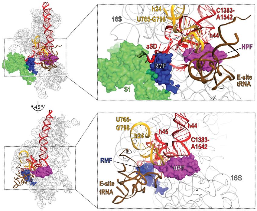
Figure 4. Mapping of 16S rRNA fragments present in 70S ribosomes. (A, B) Primer extension analysis with RNA purified from 70S fractions of WT and
HF cultures in exponential phase (expPh) and after 5 days of incubation (120 h), using primers e (panel A) and f (panel B). Red arrows indicate major
5 -ends that are exclusively present in HF samples, black arrows indicate 5 -ends present in both WT and HF samples. Light red and gray arrows
mark additional 5 -ends in HF and both strains, respectively. For full gels see Supplementary Figure S6. (C, D) RT-PCR analysis of RNA from samples
described in (A, B). Shown are end sections of chromatograms obtained by sequencing with primer RT a (C) and RT b (D). Marked bases indicate
the last base that could be assigned to 16S rRNA unambiguously. Red background indicates sequences corresponding to the 3 -linker used in the RT-
PCR that are ambiguous due to a variable 3 -end of the fragment. For 3 -end sequencing workflow and extended results see Supplementary Figure S7.
(E) Schematic summary of the mapped 16S rRNA fragments accumulating in 70S ribosomes after 5 days of starvation in MOPS MM. Red bars mark
fragments exclusively or prevalently found in HF-70S (fragments I, III, V and VI) or HF-30S (fragment VII); gray bars mark fragments found in both
WT- and HF-70S (fragments II and IV). (F) Overlay of mapped degradation regions with the secondary structure of E. coli 16S rRNA (adapted from
http://rna.ucsc.edu/rnacenter/images/figs/ecoli 16s.pdf). The 16S domains (5 domain, 5 ; Central domain, C; 3 major domain, 3 M; 3 minor domain,
3 m) are indicated. The gap between fragment I and III is coloured in orange; the missing 3 -terminus of fragments I and V is coloured in red. Inserts depict
close-ups of selected regions. Light blue, magenta, green and brown background indicates residues that are interacting (according to Beckert et al. 2018) or
in close proximity to RMF, HPF, ribosomal protein S1 or E-site tRNA in the 100S dimer, respectively. (G) 16S rRNA degradation sites in proximity to the
binding site of RMF in the hibernating ribosome (PDB ID: 6H4N). The 3 -terminal region missing in fragments I and V (red) and h24 (orange) are in close
proximity to RMF (dark blue) and ribosomal protein S1 (light green) at the mRNA exit channel and a tRNA in the E-site (brown). Inset shows close-up
of the indicated region. Ribosomal proteins other than S1 have been omitted for the sake of clarity. (H) Top view of the 30S subunit of the hibernating
ribosome with bound HPF (magenta) (PDB ID: 6H4N) interacting with h44, h45 and h24. Colour scheme as in (G).10 Nucleic Acids Research, 2021
S7A-D). Consistent with the results of the Northern blot
A
analysis, RT-PCR analysis of fragments I and V yielded a
single product with the 3 -terminus corresponding to nu-
cleotide 1382 (Figure 4D; Supplementary Figure S7E and
F). In summary, we mapped the five prevalent fragments
found in HF-70S particles to nucleotides 1–1382 (frag- B
ment I), 1–840 (fragment II), 1–764 (fragment III), 842–
1542 (fragment IV) and 799–1382 (fragment V) (Figure 4E).
Comparing the sequences of the 16S rRNA fragments
Downloaded from https://academic.oup.com/nar/advance-article/doi/10.1093/nar/gkab017/6121461 by guest on 19 February 2021
with the known secondary structure of E. coli 16S rRNA
and a cryo-EM structure of the hibernating ribosome (25)
revealed that fragments I and V lack the entire 3 -minor
domain (3 m) consisting of helix 44 (h44) and h45 and in-
cluding the aSD sequence (Figure 4F–H). In addition, ab-
sence of nucleotides 1383–1396 destroys h28 which forms
the bridge between the 3 -major domain (3 M) and the cen- C
tral and 3 -minor domains. The gap between fragments III
and V spans most of h24 (Figure 4F–H). The respective 3 -
and 5 -ends of fragments II (nucleotide 840) and IV (nu-
cleotide 842) are located on h26 and the 3 end of a possible
additional fragment detected by RT-PCR and sequencing
(Supplementary Figure S7C and D) maps to position 999 at
the base of h33 (Supplementary Figure S8A). Both of these
regions are located at the cytosolic face of the 30S subunit:
h26 protrudes from the 16S platform, in close proximity to
the 16S-3 -end and ribosomal protein S1 in its inactive 100S
conformation, while h33 forms part of the 16S beak and re-
mains exposed in the 100S complex (Supplementary Figure
S8B and C). The additional 5 -ends observed by extension
from primer f (Figure 4B) are located between the central
and 3 -major domains and are mostly positioned in the neck Figure 5. YbeY and RNase R are involved in generation and removal of
and platform regions of the 16S rRNA (Supplementary Fig- accumulating fragments. (A) Schematic of 16S rRNA and DNA oligonu-
ure S8A and B). cleotide probes used for Northern blot analysis in (B) and (C). For the
sequences of the indicated probes see Supplementary Table S2. (B) North-
ern blot analysis of 16S rRNA fragments found in total RNA of hiberna-
tion factor mutants and RNase mutants after five days of starvation. The
YbeY and RNaseR are involved in the degradation of the 16S schematics on the right show detected fragments. Grey bars indicate frag-
rRNA 3 -end ments common to both WT and HF, red bars indicate fragments only
present in HF. Pink bars indicate overhang in fragments I* and V* of
The observed fragmentation of 16S rRNA in HF–70S rnr and ybeY mutants. For additional probes and magnified selected
raises the question which ribonuclease(s) are involved in areas in the range of fragment I and fragment V see Supplementary Figure
the generation of the accumulating fragments. Fragments S9. (C) As described in (B). RNA was resolved on 6% urea–PAGE gels for
II and IV are likely generated by endonucleolytic cleavage detection of RNA in a lower size range. The schematic on the right shows
fragments VI and VII mapped by urea–PAGE and Northern blot analysis.
at positions 840–842. Fragments III and V are likely gener- For additional probes see Supplementary Figure S9.
ated by an initial endonuclease cleavage at position 799, fol-
lowed by removal of helix 24 (765–798), either by additional
endo- or exonuclease activity. Removal of the 16S 3 -end ap-
pears to occur by an endonuclease cleavage event resulting from five day old cultures of WT and HF strains as well
in fragment VI (Figure 3C). as the mutants lacking ybeY or rnr. Both the rnr and in
The 3 -end of the 16S rRNA is a hotspot for degradation particular the HFrnr mutant exhibited highly increased
mechanisms during quality control of ribosome biogenesis overall accumulation of 16S rRNA fragments roughly cor-
and starvation-induced ribosome degradation (56–58). One responding to fragments I-VII found in HF (Figure 5). In-
of the ribonucleases implicated in processing and quality triguingly, both fragments I and V appeared to be slightly
control of the 16S 3 -terminus is the single-strand specific larger than in WT and HF (thus designated I* and V*),
endoribonuclease YbeY (56,57,59). Importantly, YbeY and and could be detected with a probe complementary to re-
the exonuclease RNase R jointly degrade assembled 70S gion 1378–1396 (probe g/h; Figure 5B; Supplementary Fig-
containing misprocessed or otherwise compromised 30S ure S9B), indicating that they are at least 20 nucleotides
subunits in a highly interdependent manner (56). We there- longer than their counterpart in WT and HF, and still
fore speculated that YbeY and RNase R may be involved in contain h28 of the 16S rRNA (Figure 4F).
the generation of 16S fragments and/or the degradation of Deletion of ybeY led to the accumulation of a 17S rRNA
ribosomes containing fragmented 16S rRNA. To test this fragment, likely due to the known 16S processing defect in
we generated rnr or ybeY deletion mutants both in a WT strains lacking ybeY (56,57,59) (Figure 5B; Supplementary
and HF background and analysed total RNA extracted Figure S9B). Both ybeY and HFybeY exhibited in-Nucleic Acids Research, 2021 11
creased accumulation of fragments I–V. Fragments I and during the first 16 h of growth arrest. Continued incubation
V were prevalently present as I* and V* but to some extent led to more gradual degradation at similar rates in wild-
also as their short form (I/V). Strikingly, we could not de- type and HF (Figure 2A). This is consistent with recently
tect fragments VI and VII, which approximately correspond published results showing the degradation of rRNA in re-
to the 3 -terminus missing in fragments I and V (probe i; sponse to starvation for different nutrients, including glu-
Figure 5C). In summary, deletion of rnr and ybeY strongly cose (7,8,51). Protection of ribosomes through hibernation
increased the accumulation of 16S rRNA fragments cor- could therefore be important during growth state transi-
responding to fragments I–V observed in HF–70S parti- tions. This hypothesis is further supported by the transition-
cles. Mutants lacking RNase R exclusively accumulate frag- induced expression of hibernation factors (Supplementary
Downloaded from https://academic.oup.com/nar/advance-article/doi/10.1093/nar/gkab017/6121461 by guest on 19 February 2021
ments I* and V*, which, in contrast to fragments I and V, Figure S1) and the high level of ribosome dimers during the
contain the 16S region corresponding to h28. Most notably, first 16 h of incubation (Figure 2B).
deletion of ybeY leads to complete disappearance of both We detected and mapped several distinct rRNA frag-
3 -terminal fragments (VI and VII). In conclusion, YbeY is ments (fragments I-VI) within assembled HF–70S ribo-
necessary for the generation of fragments corresponding to somes (Figures 3 and 4). These results strongly suggests
the 3 -end observed in the absence of HFs. Both YbeY and that HF cells contain several subpopulations of 70S ribo-
RNase R participate in the generation of fragments I and somes, most importantly 70S containing intact 16S rRNA,
V, although degradation at the 3 -terminus can occur in the 70S with 16S rRNA lacking the entire 3 -minor domain
absence of either one of the RNases. (fragment I, 1–1382) and 70S harbouring a split 16S con-
sisting of fragment III (1–764) and fragment V (799–1382).
In addition, a fraction of 70S containing fragment I or V
DISCUSSION
appears to carry a remnant part of the missing 3 -minor do-
Here we have investigated the physiological role of hiberna- main (fragment VI).
tion factors. The triple hibernation factor mutant showed a 16S rRNA contains multiple sites for inter-subunit
significantly delayed regrowth upon nutrient replenishment bridges, which partly overlap with the missing regions in the
after prolonged growth arrest (Figure 1C–F). Many bac- fragmented 16S species. In particular h24, missing in the
teria, including E. coli, live in habitats with highly fluctu- gap between fragment III and V (765–798) of HF, con-
ating nutrient availability. This feast-or-famine lifestyle re- tains conserved residues around loop 790, including bridge
quires rapid adaptability and a prolonged growth lag af- B2b, that are important for 30S assembly, ribosome func-
ter nutrient replenishment constitutes a severe fitness de- tion and 70S association (64–66). Similarly, the 3 -minor
fect in a competitive environment (10,60). A similar re- domain absent in fragments I and V comprises multiple
growth phenotype was observed in some gram-positive bac- residues forming inter-subunit bridges, including A1418
teria lacking the lHPF-mediated hibernation mechanism (bridge B5), A1483 (B3), G1486 (B3) and T1495 (B2a). Free
(10,14,42,61,62). We show here that in E. coli the two hi- 30 subunits originating from such 70S particles are highly
bernation mechanisms, RMF–HPF-mediated 100S dimer- unlikely to re-enter the translation cycle due to the lack of
ization and RaiA–70S stabilization, appear to have overlap- h24, h44 and h45 as well as the aSD sequence, which are es-
ping functions, as only deletion of all three factors led to a sential for the formation of initiation complexes and trans-
significantly delayed regrowth phenotype (Figure 1C–F and lating 70S ribosomes (3).
Supplementary Figure S2C). Importantly, complementation of all three HFs from
In several organisms, hibernation factors maintain via- plasmid almost entirely rescued both the observed re-
bility during stationary phase and gram-positives S. au- growth defect (Supplementary Figure S2B) and the frag-
reus and M. smegmatis lacking their respective lhpf ho- mentation of 16S rRNA in HF (Supplementary Figure
molog exhibit decreased survival during prolonged star- S5). This further supports our conclusion that regrowth
vation (18,63). In E. coli, reports have been inconclusive of HF cells after starvation is delayed due to an over-
(28,54,55). We therefore investigated viability in a defined all decrease of intact 16S rRNA and the accumulation
minimal medium leading to batch culture glucose starva- of translation-incompetent ribosomal particles containing
tion (MOPS MM) as well as abrupt growth arrest by trans- fragmented 16S rRNA.
fer to PBS. Only moderate effects of deleting all three HFs Intriguingly, degradation sites mapped uniquely to
could be observed, even after extended incubation times HF–16S directly overlap with the interaction sites be-
(Figure 1A and B). In addition, no direct correlation be- tween hibernation factors RMF and HPF and 16S rRNA
tween the presence of hibernating ribosomes and cell vi- determined by cryo-EM (25). RMF directly interacts with
ability was observed, given that 100S dimers in wild-type the aSD sequence at nucleotides 1535–1537, and is enclosed
cells nearly disappeared after five days of starvation in by helices 28, 37, and 40. HPF binds at the A- and P-site at
MOPS MM, while the number of viable cells remained the interface between 30S and 50S subunit, contacting sev-
nearly constant for up to nine days (Figures 1A and 2B). eral 16S rRNA helices including h24 and 44 (Figure 4F–
Therefore, ribosome hibernation may not be needed to H). In addition, ribosomal protein S1 has been shown to
maintain viability even after extended periods of stationary adopt a distinct conformation in hibernating ribosomes,
phase. folding into the mRNA exit channel and interacting with
The observed regrowth defect is likely caused by exces- nucleotides 1538–1540 (25). A tRNA present in the E-site
sive breakdown of ribosomes: RNA degradation measured may confer further protection (25). RaiA shares a bind-
by the in vivo RNA stability assay showed an almost two- ing site with HPF and is therefore likely to have a simi-
fold increased degradation in the HF mutant over the WT lar effect (23,26). The short C-terminal extension of RaiAYou can also read