Human Embryonic Stem Cell-Derived Cells Rescue Visual Function in Dystrophic RCS Rats
←
→
Page content transcription
If your browser does not render page correctly, please read the page content below
CLONING AND STEM CELLS
Volume 8, Number 3, 2006
© Mary Ann Liebert, Inc.

     Human Embryonic Stem Cell–Derived Cells Rescue
         Visual Function in Dystrophic RCS Rats

  RAYMOND D. LUND,1 SHAOMEI WANG,1 IRINA KLIMANSKAYA,2 TOBY HOLMES,1
  REBECA RAMOS-KELSEY,2 BIN LU,1 SERGEJ GIRMAN,1 N. BISCHOFF,1 YVES SAUVÉ,3
                            and ROBERT LANZA2

                                                ABSTRACT

Embryonic stem cells promise to provide a well-characterized and reproducible source of re-
placement tissue for human clinical studies. An early potential application of this technology
is the use of retinal pigment epithelium (RPE) for the treatment of retinal degenerative dis-
eases such as macular degeneration. Here we show the reproducible generation of RPE (67
passageable cultures established from 18 different hES cell lines); batches of RPE derived
from NIH-approved hES cells (H9) were tested and shown capable of extensive photorecep-
tor rescue in an animal model of retinal disease, the Royal College of Surgeons (RCS) rat, in
which photoreceptor loss is caused by a defect in the adjacent retinal pigment epithelium.
Improvement in visual performance was 100% over untreated controls (spatial acuity was ap-
proximately 70% that of normal nondystrophic rats) without evidence of untoward pathol-
ogy. The use of somatic cell nuclear transfer (SCNT) and/or the creation of banks of reduced
complexity human leucocyte antigen (HLA) hES-RPE lines could minimize or eliminate the
need for immunosuppressive drugs and/or immunomodulatory protocols.

               INTRODUCTION                              tients over 60 in the United States (Friedman et al.,
                                                         2004). Previous work has shown that freshly har-

H     UMAN STEM CELL DERIVATIVES have been con-
      sidered a promising source of tissue for re-
generative medicine (Banin et al., 2006; Correia et
                                                         vested RPE can be effective in rescuing photore-
                                                         ceptors in the Royal College of Surgeons (RCS) rat,
                                                         an animal model of indirect photoreceptor degen-
al., 2005; Dunnett and Rosser, 2004; Haruta, 2005;       eration, the RCS rat (Li et al., 1996; Sheedlo et al.,
Lanza et al., 2004; Mueller et al., 2005). The ability   1991). In this animal, a mutation in the MERTK
to generate GMP-grade retinal pigment epithelium         gene affects the ability of RPE cells to phagocytose
(RPE) from human embryonic stem (hES) cells un-          photoreceptor outer segments (D’Cruz et al., 2000)
der well-defined and reproducible conditions             and leads to loss of photoreceptors over a several
would provide a scalable and efficacious product         month time frame (LaVail, 2001). There is a small
for use in human conditions such as age-related          group of humans who have been identified with
macular degeneration (ARMD) where photorecep-            orthologous mutations (Gal et al., 2000; Tada et al.,
tor loss is resultant upon RPE dysfunction. ARMD         2006), but the main value of the animal has been
alone affects more than 30 million people world-         its extensive use in exploring conditions under
wide and is the leading cause of blindness in pa-        which photoreceptors can be rescued.

  1Moran Eye Center, University of Utah Health Science Center, Salt Lake City, Utah.
  2Advanced Cell Technology, Worcester, Massachusetts.
  3Ophthalmology and Physiology, University of Alberta, Edmonton, Canada T6G.

                                                      189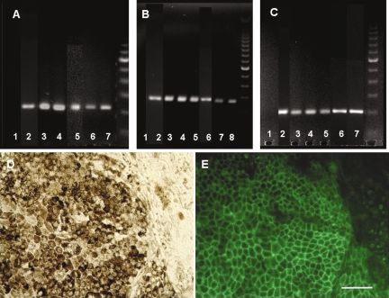
190                                                                                           LUND ET AL.

   A major issue in any cell-based therapy is that      Cell isolation and characterization
of providing a cell source that is readily available,
                                                           hES were cultured as described above and al-
safe, and ethically acceptable and which can be
                                                        lowed to spontaneously differentiate: this resulted
developed commercially in large-scale produc-
                                                        in the appearance of RPE clusters over the course
tion. With this goal in mind, we recently reported
                                                        of 6–8 weeks, from which hES-RPE cells were iso-
the isolation of RPE cells from hES cells which
                                                        lated and subcultured (Klimanskaya et al., 2004).
could be maintained through multiple passages
                                                        Spontaneous differentiation was observed in 7–10-
(30 population doublings) (Klimanskaya et al.,
                                                        day-old cultures, and the cells were switched to dif-
2004). Gene expression profiling demonstrated
                                                        ferentiation medium containing 15% SR in knock-
their higher similarity to primary RPE tissue than
                                                        out high glucose DMEM supplemented with 500
of existing human RPE cell lines D407 and the
                                                        /ml penicillin, 500 g/mL streptomycin, 1% non-
spontaneously generated ARPE19 line. Here we
                                                        essential aminoacids solution, 2 mM GlutaMAX I,
show that new RPE can be re-established in a re-
                                                        0.1 mM -mercaptoethanol, 4 ng/mL bFGF (Invit-
liable and reproducible manner from 18 different
                                                        rogen), 10 ng/mL human LIF (Chemicon), The first
hES lines. We have studied one of these lines to
                                                        pigmented clusters were usually observed in 6–8
show using morphological, behavioral and phys-
                                                        weeks after the initial plating of hES cells, and the
iological techniques that these cells have the
                                                        cells were isolated by hand-picking under the mi-
capacity to support photoreceptor survival and
                                                        croscope after such clusters reached the desired size
preserve visual function after subretinal trans-
                                                        and number. Isolated cells were subcultured in
plantation into RCS rats. The cells did not un-
                                                        knockout high glucose DMEM supplemented with
dergo tumorous transformation after long-term
                                                        500 /mL penicillin, 500 g/mL streptomycin, 1%
transplantation nor did they provoke overt
                                                        non-essential aminoacids solution, 2 mM Gluta-
pathological responses in the host retina.
                                                        MAX I, 0.1 mM -mercaptoethanol, 7% SR, 5% FBS
                                                        on gelatin. The cells were subcultured with 0.25%
                                                        Trypsin/1 mM EDTA (Invitrogen) and passaged at
                   METHODS                              1:3 ratio after the cells re-established the RPE phe-
                                                        notype, usually 2–3 weeks after passaging.
Human embryonic stem cell lines
                                                        Cell injections
   The hES cell lines used in this study were pre-
viously described H1, H7, and H9 (Thomson et               hES-RPE derived from hES cell line WA09 were
al., 1998) (National Institutes of Health–registered    passaged three or four times and pooled together
as WA01, WA07, and WA09), and 15 lines de-              prior to injection. An aliquot of the final cell sus-
rived with the use of private funds (eight of these     pension was frozen and later analyzed by
lines were derived at Harvard University in the         RT-PCR and real-time PCR. At P21–23, 14 dys-
laboratory of Douglas Melton (Cowan et al.,             trophic RCS rats under xylazine-ketamine anes-
2004), and seven were derived at Advanced Cell          thesia received subretinal injections of a suspen-
Technology). The later 15 hES cell lines were de-       sion of cells (2  104cells/eye) via a trans-scleral
rived from human frozen blastocysts or cleaved          approach into the upper temporal retina area as
embryos that were donated by couples who had            previously described (Wang et al., 2005). As con-
completed their fertility treatment. hES cells were     trol material, some rats (n  8) received an injec-
maintained on mitomycin C-treated mouse em-             tion of medium alone. Dystrophic rats that had
bryonic fibroblasts (MEF) in growth medium:             received injections of the RPE cell line, ARPE-19
knockout high glucose DMEM supplemented                 (American Type Culture Collection [ATTC],
with 500 /mL penicillin, 500 g/mL strepto-            Manassas, VA), and non-dystrophic congenic rats
mycin, 1% non-essential aminoacids solution,            were available for comparison. All animals re-
2 mM GlutaMAX I, 0.1 mM -mercaptoethanol,              ceived daily dexamethasone injections (1.6
4 ng/mL bFGF (Invitrogen), 1-ng/mL human LIF            mg/kg, i.p.) for 2 weeks and were maintained on
(Chemicon, Temecula, CA), 8% of Serum Re-               cyclosporine-A (Bedford Labs, Bedford MA) ad-
placement (SR, Invitrogen), and 8% Plasmanate           ministered in the drinking water (210 mg/L; re-
(Bayer). The cells were routinely passaged with         sulting blood concentration: 250–300 g/L)
trypsin at a ratio of 1:3–1:6 every 3–5 days (Kli-      (Prusky et al., 2004) from 1–2 days prior to cell
manskaya and McMahon, 2004).                            injection until animals were euthanized.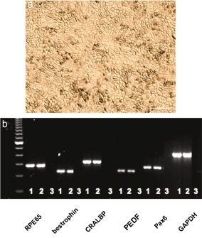
hES-DERIVED CELLS RESCUE VISUAL FUNCTION                                                                191

Electroretinogram responses                             perficial layers of the SC using a modification of
                                                        a procedure we developed in previous work
   The dark adapted electroretinogram (ERG) re-
                                                        (Sauve et al., 2002). For each of 16–20 positions
sponse was recorded as previously described
                                                        recorded over the surface of the SC, a discrete re-
(Pinilla et al., 2004). A double flash protocol was
                                                        ceptive field for that position was localized and
used to isolate cone responses. A conditioning
                                                        the brightness of a flashing spot, 3-degrees-di-
flash was followed 1 sec later by a probe flash.
                                                        ameter projected on a hemisphere, was varied
The role of the conditioning flash in this para-
                                                        with neutral density filters until a response am-
digm is to saturate rods transiently so that they
                                                        plitude was diminished to a level double that of
are rendered unresponsive to the probe flash. The
                                                        background activity.
intensity of the conditioning flash for complete
rod bleaching was set to 1.4 log cd/m2 for all tests.
A mixed b-wave was obtained by presenting the           Histology
probe flash alone, i.e., without being preceded by         Rats were overdosed with sodium pentobarbi-
a conditioning flash. The response to the probe         tal (Sigma, St. Louis, MO) and perfused with
flash (1.4 log cd/m2), preceded by the condition-       phosphate-buffered saline (PBS). The eyes were
ing flash, was taken as reflecting cone-driven ac-      removed and immersed in 2% paraformaldehyde
tivity, and allowed derivation of the rod contri-       for 1 h, infiltrated with sucrose and embedded in
bution. Averages of 3–5 traces (set at 2 min apart      OCT. Coronal sections (10 m) were cut on a
to assure recovery of rod responsiveness) were          cryostat. Five 1-in-5 series were collected. One se-
sufficient to obtain clear responses. Special care      ries was stained with Cresyl violet (CV) for as-
was taken to maintain the electrode placement in        sessing injection site, retinal lamination, and evi-
a consistent position in all animals.                   dence of cellular infiltrates or tumor formation.
                                                        Further series were stained with human-specific
Optomotor acuity thresholds                             nuclear marker—MAB1281 (Chemicon)—for donor
                                                        cells; PCNA for dividing cells and RPE65; be-
   Thresholds to moving stripes of varying spa-
                                                        strophin for RPE cells; and recoverin (a gift from
tial frequency were measured at P90–100. An im-
                                                        Dr. J. McGinnis, University of Oklahoma) for
age of a rotating cylinder covered with a vertical
                                                        photoreceptors. The protocols for processing hu-
sine wave grating was presented in virtual three-
                                                        man-specific nuclear marker followed the manu-
dimensional (3D) space on four computer moni-
                                                        facturers’ data sheets. The secondary antibodies
tors arranged in a square (Douglas et al., 2005;
                                                        were biotinylated anti-mouse or rabbit IgG or
Prusky et al., 2004). Rats standing unrestrained
                                                        conjugated with FITC or Cy3 (Jackson). The cells
on a platform in the center of the square tracked
                                                        were visualized by using Vector Nova RED (Vec-
the grating with reflexive head movements. The
                                                        tor Labs, Burlingham, CA) for human nuclear
spatial frequency of the grating was clamped at
                                                        marker. Photographs were taken by using the Im-
the viewing position by repeatedly recentering
                                                        age-pro-Plus program; montage pictures were
the “cylinder” on the head of the test subject. Acu-
                                                        achieved using Photoshop. For confocal images,
ity threshold was quantified by increasing the
                                                        the pinholes were 75 µm and the width of opti-
spatial frequency of the grating until an optomo-
                                                        cal sections was 1 m. Final images were ob-
tor response could no longer be elicited. The
                                                        tained from the projections of 6–8 single frames.
method, a development of a rotating drum with
                                                        The TIFF images were produced in Adobe Photo-
fixed stripes, has several distinct advantages over
                                                        shop.
the older technology, most important of which
are that individual animals show less variance be-
tween trials, it allows tighter titration of acuity,    RNA extraction and RT-PCR
and it is quicker to perform so animals do not be-        Total RNA from the RPE cells was isolated by
come distracted or go to sleep during testing.          using RNeasy Mini Kit (Qiagen) with the on-col-
                                                        umn DNAase treatment. Gene-specific primer
Luminance threshold recording                           pairs were designed for the following genes:
  After optomotor testing, luminance thresholds         RPE65-F:     ATGGACTTGGCTTGAATCACTT,
were established in selected rats (n  7) by            RPE65-R: GAACAGTCCATGAAAGGTGACA
recording single and multiunit activity in the su-      Bestrophin-F: TAGAACCATCAGCGCCGTC
192 LUND ET AL.
TABLE 1. REPRODUCIBLE GENERATION OF RETINAL denaturation 94°C, 30 sec; annealing 55°C, 30
PIGMENT EPITHELIUM (RPE) LINES FROM sec; extension 72°C, 1 min; final extension 72°C,
HUMAN EMBRYONIC STEM (ES) CELLS
10 min. For negative control, total RNA was ex-
No. RPE lines cluded from the reaction mixture and RNase-
Passage no. established free water was used instead. PCR products were
separated on a 1.5% agarose gel stained with 0.5
ES cell line
WA01 (H1) 30–126 15 mg of ethidium bromide for 30 min, and the flu-
WA09 (H9) 44–67 7 orescence was scanned using a Kodak Image
WA07 (H7) 25 1 Station 4000MM.
Total H lines 23
MA01 6, 9 2
MA03 5–10 4
Statistical analysis
MA04 5–27 5 Average data are presented as mean SEM (or
MA09 7 1
MA14 13 1 mean SD). We used t-test or Mann-Whitney U
MA40 5–27 8 for comparisons unless otherwise indicated
MAJ1 3–17 5 (Statview). Significance is designated as p 0.05.
Total MA lines 26
HUES1 25–43 5
HUES2 30–36 2
HUES3 15–28 3 RESULTS
HUES5 8 1
HUES6 8–12 2 Generation of RPE cell lines
HUES7 7–14 2
HUES8 7–14 2 All 18 hES cell lines studied reliably produced
HUES10 8 1 passageable RPE lines after 6–8 weeks of sponta-
Total HUES lines 18
Total no. RPE lines isolated 67
neous or induced differentiation (Table 1). The
differentiation system did not require co-culture
with animal cells or factors as previously de-
scribed (Haruta et al., 2004). Multiple RPE clus-
Bestrophin-R: TGAGTGTAGTGTGTATGTTGG
ters appeared in adherent cultures and in em-
CRALBP-F: AAATCAATGGCTTCTGCATCATT
bryoid bodies (EB), and were used to establish 67
CRALBP-R: CCAAAGAGCTGCTCAGCAAC
repeatedly passageable (5 passages) RPE lines
PEDF-F: TCTCGGTGTGGCGCACTTCA
with RPE features, such as phenotype and the
PEDF-R: GTCTTCAGTTCTCGGTCTATG
ability to transdifferentiate and differentiate thor-
GADPH-F: CGATGCTGGCGCTGAGTAC
ough multiple passages. Figure 1 shows expres-
GADPH-R: CCACCACTGACACGTTGGC
sion of RPE molecular markers in representative
Reverse transcription–polymerase chain reac- samples of hES-derived RPE lines compared
tion (RT-PCR) was performed with 0.5–1 L of to human fetal RPE cells. Reverse transcrip-
RNA by using a OneStep RT-PCR Kit (Qiagen) tion–polymerase chain reaction (RT-PCR) de-
with the addition of 10 units of RNasin Ribonu- tected the presence of RPE65, bestrophin, and
clease Inhibitor (Promega) under the following PEDF in all samples analyzed (Fig. 1A–E).
conditions in a GeneAmp Perkin Elmer thermo- The appearance and characterization of the
cycler: cDNA synthesis 50°C, 30 minutes; DNA hES-derived RPE cell line used in the animal
polymerase activation 95°C, 15 min; 35 cycles of studies is shown in Figure 2. Differentiation of
FIG. 1. Molecular marker analysis of human embryonic stem cell (hES)–derived retinal pigment epithelium (RPE)
lines. (A-C) Reverse transcription–polymerase chain reaction (panels are composites of gel images from several repre-
sentative experiments). (D,E) Immunofluorescence. (A) Bestrophin: 1—C2C12 cells (negative control); 2—fetal RPE (pos-
itive control); 3—MA03; 4—MA40; 5—WA01; 6—WA09; 7—MAJ1. (B) PEDF: 1—C2C12 cells (negative control); 2—fe-
tal RPE (positive control); 3—MA03; 4—MA40; 5—MA09; 6—WA01; 7—WA09; 8—MAJ1. (C) RPE65: 1—C2C12 cells
(negative control); 2—fetal RPE (positive control); 3—MA03; 4—MA40; 5—MA09; 6—WA01; 7—WA09. (D) Culture of
hES-RPE cells, Hoffman modulation optics. (E) Same field stained with antibodies to bestrophin. Scale bar 100 m.
FIG. 2. Human embryonic stem cell (hES0)–derived retinal pigment epithelium (RPE) used for injection. (a) Hoff-
man modulation optics micrograph of cells 12 days after passaging. Note that the cells are in the process of re-es-
tablished pigmented phenotype. Scale bar 100 m. (b) Expression of RPE molecular markers detected by reverse
transcription–polymerase chain reaction (RT-PCR): 1—hES-RPE; 2—fetal RPE; 3—negative control (no template).
(Panel is a composite of gel images from several representative experiments.)FIG. 1. FIG. 2.
194 LUND ET AL.
hES cell line WA09 (H9) was induced at passages testing luminance threshold responses. We chose
65–66, and hES-RPE were subcultured for several both average and best performers in the opto-
passages prior to transplantation and examining motor test for such assessment. The minimum
in vivo efficacy. Passaging was performed after light intensity that could elicit neuron activation
the cells went through the transdifferentia- at various points across the visual field repre-
tion–differentiation cycle and re-established a sentation was measured by recording unit activ-
mature RPE phenotype, including typical RPE ity from different locations across the superior
“cobblestone” morphology (Fig. 2a). Real-time colliculus (SC). To reduce the data for ease of pre-
RT-PCR assessment of the cells was carried out sentation and statistical analysis, however, it is
at the time of transplantation and compared to presented here as the percentage of the collicular
primary human fetal RPE tissue. Both hES-RPE area (y-axis) from which the responses showed
and fetal RPE displayed high expression of char- visual thresholds less than the value designated
acteristic RPE genes, including RPE65, be- on the x-axis (log units over a background illu-
strophin, CRALBP, and PEDF (Fig. 2b). mination of 0.02 log candela/m2). Asterisks indi-
cate the points at which the curves for grafted and
sham-operated eyes were statistically different (t-
Efficacy of cell line WA09 (H9) after
test, p 0.05). Results were obtained from ani-
transplantation to RCS rat eyes
mals receiving hES-RPE cells (n 7), sham injec-
Eye-pigmented RCS rats received injections of tions (n 5), and no treatment (n 6). In
hES-RPE into the subretinal space of the eye be- non-dystrophic rats, a threshold response of less
tween the RPE and photoreceptor layers at post- than 0.6 log units is recorded. As can be seen in
natal day (P) 21 at an age when photoreceptor de- Figure 3C, by P 100, neurons across the whole vi-
generation had yet to develop. sual field failed to respond with thresholds of 2.7
Electroretinogram (ERG) responses were log units or better in an untreated dystrophic RCS
tested at both P60 and P90 to measure the elec- rat, while responses could be elicited from 18%
trical activity of the outer (a-wave) and inner of the area in sham-injected rats. By comparison,
(b-wave) retina to light flashes. Double flash pre- the cell-injected rats showed 52% of the collicu-
sentations allowed isolation of rod and cone- lar area with thresholds of 2.7 log units or better,
related activity, respectively. At P60, the a-wave with a best point of 1.3 log units.
ERG response is normally lost in RCS rats, and Anatomical examination of the retinas showed
by P90, the b-wave response is severely depleted, that donor hES-RPE cells, identified using human
allowing graft-related effects to be recognized specific nuclear marker, were distributed in the
over background performance (Pinilla et al., subretinal space adjacent to the host RPE layer
2005b). By P60, the hES-RPE-grafted animals (Fig. 4d). Two RPE-specific markers—RPE65 and
achieved significantly better a-wave, b-wave, and bestrophin—stained these cells positively. Hu-
cone mediated responses over untreated and man-specific proliferating cell nuclear antigen
sham-injected animals (Fig. 3A). As with other (PCNA) staining was negative, indicating lack of
studies using ARPE-19 cells (Pinilla et al., 2004, continued cell division among the donor cells.
2005a), there was deterioration of the magnitude There was extensive photoreceptor rescue: 5–7
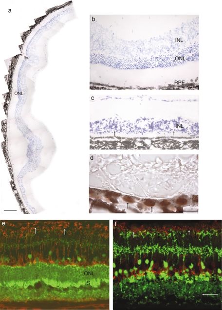
of the responses by P90, but other tests showed cells deep in the outer nuclear layer (ONL; Fig.
continued robust visual performance. One of 4a,b). For comparison, the ONL was 10–12 cells
these was an optomotor test that provided a mea- deep in nondystrophic rats, while in dystrophic
sure of spatial acuity (Douglas et al., 2005; Prusky rats, this layer was reduced to one cell deep at
et al., 2004). Normal non-dystrophic rats gave a P100 (Fig. 4c). Sham injected retinas appeared
figure of approximately 0.6 cycles/degree (c/d), similar to unoperated dystrophic rats except for
while in sham-injected rats, a threshold response a small cluster of remaining photoreceptors im-
of 0.29 0.03 c/d was recorded at P100: un- mediately adjacent to the injection site and a com-
treated animals gave a figure of 0.21 0.03 c/d. parable appearance was seen in graft-protected
By contrast, the cell-grafted rats sustained levels retinas distant from the area of distribution of
of 0.42 0.03 c/d, significantly better than sham- grafted cells. Antibody staining against recoverin
injected rats (p 0.05, t-test). The best animals confirmed the presence of a well-preserved pho-
performed at 0.45 c/d (Fig. 3B). toreceptor layer with inner and outer segments
Animals from each group were selected for evident in the graft-protected area, while distantA ERG at P60
180
160 *
140
Amplitude (uV)
120
100
80 *
60 *
40
20
0
a-hES-RPE a-sham b-hES-RPE b-sham cone cone
hES-RPE b-sham
B Optomotor at P100
0.5
*
0.4
Relative acuity (c/d)
0.3
0.2
0.1
0
hES-RPE Sham Untreated
C Luminance threshold at P100
*
100
*
80
*
60
%
40 hES-RPE
Sham
20 Untreated
0
1.2 1.7 2.2 2.7 3.1 3.6 4.1 4.6
Threshold
FIG. 3. Functional assessment of human embryonic stem cell (hES)–derived retinal pigment epithelium (RPE) after
subretinal transplantation in Royal College of Surgeons (RCS) rats. (A) hES-RPE grafted animals achieved signifi-
cantly better responses over sham controls (p 0.05, t-test) for a-wave (31 27 vs. 6 17 V), b-wave (108 46 V
vs. 36 33 V) and cone b-wave (57 19 vs. 28 13 V). (B) The relative acuity as measured by the optomotor sys-
tem shows that the hES-RPE treated eyes perform significantly better than the medium treated and untreated eyes
(p 0.05, t-test), giving approximately 50% and 100% improvement in visual acuity over the sham and untreated con-
trols, respectively. Non-dystrophic untreated eyes give readings of 0.53–0.6 cycles/degree (c/d). (C) Luminance thresh-
old responses recorded across the superior colliculus, each curve (average SEM) shows the percent of retinal area
(y-axis) where the visual threshold is less than the corresponding value on the x-axis (log units, relative to background
illumination 0.02 cd/m2). Asterisks show the points where the curves for grafted and sham-operated eyes are statis-
tically different (t-test, p 0.05). The curves show that 52% of the area of the superior colliculus (SC) in grafted ani-
mals gave thresholds of 2.7 log units and against shams in which approximately 18% gave thresholds of 2.7 log units.196 LUND ET AL.
from graft, sparsely distributed photoreceptors and other cells that appear to function by deliv-
remained (Fig. 4e,f). There was no indication of ery of growth factors (Wang et al., 2005; Lawrence
extraneous cells in the retina, particularly in the et al., 2004). However, apart from having a mo-
plexiform layers where invasive cells can be eas- lecular profile more closely resembling native
ily recognized even in Cresyl violet–stained sec- RPE than do ARPE-19 cells (Klimanskaya et al.,
tions. None of the retinas showed evidence of 2004), the present cells afford certain advantages
uncontrolled cell proliferation, including tumor over ARPE-19 in that many lines can be gener-
formation. ated. Given the immunoprovocative nature of
RPE cells, the use of hES cells would allow op-
portunity to generate appropriate immuno-
DISCUSSION genetic lines using various HLA-matching strate-
gies. While rescue can be achieved by growth
Our results show that a well-characterized de- factor delivery either by cell-based approaches
rivative of human embryonic stem cells—retinal such as using Schwann cells (Wang et al., 2005) or
pigment epithelium (RPE)—is capable of signifi- cells transfected to deliver specific growth factors
cant rescue of visual function in a clinically rele- (Lawrence et al., 2004) as well as by direct injec-
vant animal model of retinal disease. The cells tion (LaVail et al., 1992), this approach is unlikely
survived long-term (100 days) after transplan- to replace all the functions of RPE cells. Since the
tation into RCS rats, and localized to the subreti- present cells closely resemble RPE cells (Kliman-
nal space without migration into the retina. In ad- skaya et al., 2004), they are more likely to replicate
dition to extensive photoreceptor rescue (5–7 cells a broader range of RPE functions than ARPE-19 or
deep in the outer nuclear layer), the relative acu- by simple factor delivery. The aim here was to
ity as measured by the optomotor system showed demonstrate that the cells remain localized in the
that animals treated with hES-derived RPE per- subretinal space and can support photoreceptor
formed significantly better than sham and un- survival as well as a range of visual functions.
treated controls (50% and 100% improvement in There are numerous advantages of using hES-
visual performance, respectively; visual acuity derived cells as a source of RPE for clinical stud-
was approximately 70% that of normal non-dys- ies. Primary RPE tissue cannot be obtained in
trophic rats). However, the broader role of hES- large enough quantities for wide-scale clinical
derived cells in taking on the full range of RPE use. Furthermore, practical restrictions prevent
cell functions beyond photoreceptor rescue still full safety testing from being performed on every
remains to be explored in vivo. fetal or adult donor source, nor can the functional
Detailed quantitation of rescue was limited to parameters of graft efficacy be systematically as-
functional measures based on previous work in sessed. In contrast, hES-RPE can be derived and
RCS rats. Although rods can be rescued by at least maintained under well-defined and reproducible
two very different cell transplants, they do not conditions using traceable reagents, including
function normally, if at all, but rather deteriora- specific lots of media, sera, growth factors, and
tion of cone-meditated vision was limited (Gir- other culture materials. New and additional banks
man et al., 2005). Therefore, counting cells on the of RPE can be created to test and further optimize
ONL, comprising 97% rods, gives at best an in- yields and functionality. RPE differentiation ap-
direct measure of the degree of vision sustained. pears to be an inevitable event in hES cultures, and
Rescue is in the same order as that achieved pre- we have obtained consistent differentiation of hu-
viously using ARPE-19 cells (Lund et al., 2001) man ES cells to RPE, including long-term hES cul-
FIG. 4. Anatomical rescue of photoreceptors after transplantation of human embryonic stem cell (hES)–derived reti-
nal pigment epithelium (RPE). (a–c) Retinal sections at P100 stained with cresyl violet: extensive photoreceptor res-
cue with hES-RPE graft (a); high-power image of the rescued outer nuclear layer (ONL), which is 5–7 cells deep (b);
distant from graft, the ONL is reduced to a single layer (c). (d) grafted retina section stained with human nuclear
marker (arrows indicate positively stained donor cells). (e,f) Confocal microscopic images of retinal sections double
stained with recoverin (green) and PKCa (red): graft protected area (e) had several layers of ONL with inner seg-
ments, rod bipolar cells have denser terminals, whereas at area distant from graft (f), there is only a single layer of
ONL, and the rod bipolar cell terminals are reduced in density. Scale bar 200 m (a), 20 m (b–f).hES-DERIVED CELLS RESCUE VISUAL FUNCTION 197
198                                                                                                      LUND ET AL.

tures grown either on feeder layers or feeder-free            D’Cruz, P.M., Yasumura, D., Weir, J., et al. (2000). Muta-
on gelatin, fibronectin, laminin, collagen type I and           tion of the receptor tyrosine kinase gene Mertk in the
IV, or in EBs. Importantly, there also remains the              retinal dystrophic RCS rat. Hum. Mol. Genet. 9,
                                                                645–651.
issue of immunogenicity. It has been found that
                                                              Douglas, R.M., Alam, N.M., Silver, B.D., et al. (2005). In-
despite the immunoprivileged status of the eye, al-             dependent visual threshold measurements in the two
logeneic RPE cells can still be rejected, albeit less           eyes of freely moving rats and mice using a virtual-re-
floridly than a typical tissue mismatch allograft               ality optokinetic system. Vis. Neurosci. 22, 677–684.
(Zhang and Bok, 1998). With the further devel-                Dunnett, S.B., and Rosser, A.E. (2004). Cell therapy in
opment of somatic cell nuclear transfer (SCNT),                 Huntington’s disease. Neuro. Rx 1, 394–405.
parthenogenesis, or the creation of banks of re-              Friedman, D.S., O’Colmain, B.J., Munoz, B., et al. (2004).
                                                                Prevalence of age-related macular degeneration in the
duced-complexity human leucocyte antigen (HLA)
                                                                United States. Arch. Ophthalmol. 122, 564–572.
hES cells, RPE lines could be generated to over-              Gal, A., Li, Y., Thompson, D.A., et al. (2000). Mutations
come the problem of immune rejection and the                    in MERTK, the human orthologue of the RCS rat reti-
need for immunoprotective regimens.                             nal dystrophy gene, cause retinitis pigmentosa. Nat.
                                                                Genet. 26, 270–271.
                                                              Girman, S.V., Wang, S., and Lund, R.D. (2005). Time
             ACKNOWLEDGMENTS                                    course of deterioration of rod and cone function in RCS
                                                                rat and the effect of subretinal grafting; a light- and
                                                                dark-adaptation study. Vis. Res. 45; 343–354.
   We would like to acknowledge the excellent                 Haruta, M., Sasai, Y., Kawasaki, H., et al. (2004). In vitro
technical support provided by Tong Lin (ACT),                   and in vivo characterization of pigment epithelial cells
and Elena Budko and Jennifer Hunter (Moran                      differentiated from primate embryonic stem cells. In-
Eye Center). The R.D.L. laboratory was sup-                     vest. Ophthalmol. Vis. Sci. 45, 1020–1025.
ported by grants from the Foundation Fighting                 Haruta, M. (2005). Embryonic stem cells: potential source
Blindness and the Wynn Foundation. R.D.L. is a                  for ocular repair. Semin. Ophthalmol. 20, 17–23.
recipient of a Research to Prevent Blindness Se-              Klimanskaya, I., Hipp, J, Rezai, J.K.A., et al. (2004). De-
                                                                rivation and comparative assessment of retinal pigment
nior Scientific Investigator Award. Work with the
                                                                epithelium from human embryonic stem cells using
HUES lines was initiated at Harvard University                  transcriptomics. Cloning Stem Cells 6, 217–245.
in the laboratory of Douglas Melton with support              Klimanskaya, I., and McMahon, J. (2004). Approaches for
from the Howard Hughes Medical Institution                      derivation and maintenance of human ES cells: detailed
(HHMI) and the Juvenile Diabetes Research                       procedures and alternatives. In Handbook of Stem Cells.
Foundation (JDRF).                                              Vol. 1: Embryonic Stem Cells. R. Lanza, et al., eds. (Else-
                                                                vier/Academic Press, San Diego).
                                                              Lanza R., Blau, H., Melton, D., et al. eds. (2004). Handbook
                                                                of Stem Cells (Elsevier/Academic Press, San Diego).
                   DISCLOSURE                                 LaVail, M.M. (2001). Legacy of the RCS rat: impact of a
                                                                seminal study on retinal cell biology and retinal de-
   I.K., R.K., and R.P. are employees of Advanced               generative diseases. Prog. Brain Res. 131, 617–627.
Cell Technology, a stem cell company in the field             LaVail, M.M., Unoki, K., Yasumura, D., et al. (1992). Mul-
of regenerative medicine. R.D.L., S.W., T.H., B.L.,             tiple growth factors, cytokines, and neurotrophins res-
S.G., N.B., and Y.S. declare that they have no com-             cue photoreceptors from the damaging effects of con-
                                                                stant light. Proc. Natl. Acad. Sci. U.S.A 89, 11249–11253.
peting financial interests.
                                                              Li, N., Fan, W., Sheedlo, H.J., et al. (1996). Photoreceptor
                                                                repair in response to RPE transplants in RCS rats: outer
                                                                segment regeneration. Curr. Eye Res. 15, 1069–1077.
                    REFERENCES                                Lund, R.D., Adamson, P., Sauve, Y., et al. (2001). Sub-
                                                                retinal transplantation of genetically modified human
Banin, E., Obolensky, A., Idelson, M., et al. (2006). Reti-     cell lines attenuates loss of visual function in dystrophic
  nal incorporation and differentiation of neural precur-       rats. Proc. Natl. Acad. Sci. USA 98, 9942–9947.
  sors derived from human embryonic stem cells. Stem          Lawrence, J.M., Keegan, D.J., Muir, E.M., et al. (2004).
  Cells 24, 246–257.                                            Transplantation of Schwann cell line clones secreting
Correia, A.S., Anisimov, S.V., Li, J.Y. et al. (2005). Stem     GDNF or BDNF into the retinas of dystrophic Royal
  cell–based therapy for Parkinson’s disease. Ann. Med.         College of Surgeons rats. Invest. Ophthalmol. Vis. Sci.
  37, 487–498.                                                  45:267–274.
Cowan, C.A., Klimanskaya, I., McMahon, J., et al. (2004).     Mueller, D., Shamblott, M.J., Fox, H.E., et al. (2005). Trans-
  Derivation of embryonic stem cell lines from human            planted human embryonic germ cell–derived neural
  blastocysts. N. Engl. J. Med. 350, 1353–1356.                 stem cells replace neurons and oligodendrocytes in the
hES-DERIVED CELLS RESCUE VISUAL FUNCTION 199
forebrain of neonatal mice with excitotoxic brain dam- Tada, A., Wada, Y., Sato, H., et al. (2006). Screening of the
age. J. Neurosci. Res. 82, 592–608. MERTK gene for mutations in Japanese patients with
Pinilla, I., Lund, R.D., Lu, B., et al. (2005b). Measuring the autosomal recessive retinitis pigmentosa. Mol. Vis. 12,
cone contribution to the ERG b-wave to assess function 441–444.
and predict anatomical rescue in RCS rats. Vision Res. Thomson, J.A., Itskovitz-Eldor, J., Shapiro, S.S., et al.
45, 635–641. (1998). Embryonic stem cell lines derived from human
Pinilla, I., Lund, R.D., and Sauve Y. (2004). Contribution blastocysts. Science 282, 1145–1147.
of rod and cone pathways to the dark-adapted elec- Wang, S., Lu, B., and Lund, R.D. (2005). Morphological
troretinogram (ERG) b-wave following retinal degen- changes in the Royal College of Surgeons rat retina dur-
eration in RCS rats. Vision Res. 44, 2467–2474. ing photoreceptor degeneration and after cell-based
Pinilla, I., Lund, R,D, and Sauve, Y. (2005a). Cone func- therapy. J. Comp. Neurol. 491, 400–417.
tion studied with flicker electroretinogram during pro- Wang, S., Lu, B., Wood, P., et al. (2005). Grafting of ARPE-
gressive retinal degeneration in RCS rats. Exp. Eye Res. 19 and Schwann cells to the subretinal space in RCS
80, 51–59. rats. Invest. Ophthalmol. Vis. Sci. 46:2552–2560.
Prusky, G.T., Alam, N.M., Beekman, S., et al. (2004). Rapid Zhang, X., and Bok, D. (1998). Transplantation of retinal pig-
quantification of adult and developing mouse spatial ment epithelial cells and immune response in the sub-
vision using a virtual optomotor system. Invest. Oph- retinal space. Invest. Ophthalmol. Vis. Sci. 39, 1021–1027.
thalmol. Vis. Sci. 45, 4611–4616.
Sheedlo, H.J., Li, L., and Turner, J.E. (1991). Photorecep- Address reprint requests to:
tor cell rescue at early and late RPE-cell transplantation Dr. Robert Lanza
periods during retinal disease in RCS dystrophic rats.
Advanced Cell Technology
J. Transplant. 2, 55–63.
Sauve, Y., Girman, S.V., Wang, S. et al. (2002). Preserva-
381 Plantation St.
tion of visual responsiveness in the superior colliculus Worcester, MA 01605
of RCS rats after retinal pigment epithelium cell trans-
plantation. Neuroscience 114, 389–401. E-mail: RLanza@advancedcell.comYou can also read