ICO Guidelines for Diabetic Eye Care - www.icoph.org
←
→
Page content transcription
If your browser does not render page correctly, please read the page content below
February 2014

  ICO Guidelines for Diabetic Eye Care

www.icoph.org
ICO Guidelines for Diabetic Eye Care
The International Council of Ophthalmology (ICO) developed the ICO Guidelines for Diabetic Eye Care to serve a
supportive and educational role for ophthalmologists and eye care providers worldwide. They are intended to improve
the quality of eye care for patients around the world.

The Guidelines address the needs and requirements for the following levels of service:
   •   Low-resource or resource-poor settings: Essential or core service for screening and management of DR
   •   Intermediate-resource settings: Mid-Level service
   •   Resource-rich settings: Advanced or state-of-the-art screening and management of DR

The Guidelines are designed to inform ophthalmologists about the requirements for the screening and detection of
diabetic retinopathy, and the appropriate assessment and management of patients with diabetic retinopathy. The
Guidelines also demonstrate the need for ophthalmologists to work with primary care providers and appropriate
specialists such as endocrinologists.

With diabetes and diabetic retinopathy a rapidly increasing problem worldwide, it is vital to ensure that ophthalmologists
and eye care providers are adequately prepared.

The ICO believes an ethical approach is indispensable, as it is the first step toward quality clinical practices. Download
the ICO Code of Ethics at: www.icoph.org/downloads/icoethicalcode.pdf (PDF – 198 KB).

The Guidelines are designed to be a working document and will be updated on an ongoing basis. They were first
released in December 2013. This document was updated and reprinted in February 2014.

The ICO hopes these Guidelines are easy to read, translate, and adapt for local use. The ICO welcomes any feedback,
comments, or suggestions.

Please email us at: info@icoph.org.

2013 Task Force on Diabetic Eye Care                           2014 Diabetic Eye Care Committee
   •   Hugh Taylor, MD, AC, Chairman                               •   Tien Yin Wong, MD, MBBS, PhD, Chairman
   •   Susanne Binder, MD                                          •   Rick Ferris, MD
   •   Taraprasad Das, MD, FRCS                                    •   Neeru Gupta, MD, PhD, MBA
   •   Michel Farah, MD                                            •   Van Lansingh, MD, PhD
   •   Frederick Ferris, MD                                        •   Wanjiku Mathenge, MD, PhD, MBChB
   •   Pascale Massin, MD, PhD, MBA                                •   Eduardo Mayorga, MD
   •   Wanjiku Mathenge, MD, PhD, MBChB                            •   Sunil Moreker, MBBS
   •   Serge Resnikoff, MD, PhD                                    •   Serge Resnikoff, MD, PhD
   •   Bruce E. Spivey, MD, MS, MEd                                •   Hugh Taylor, MD, AC
   •   Juan Verdaguer, MD                                          •   Juan Verdaguer, MD
   •   Tien Yin Wong, MD, PhD
   •   Peiquan Zhao, MD

                     International Council of Ophthalmology | Guidelines for Diabetic Eye Care | Page 1
  Copyright © ICO January 2014. Translation and adaption for non-commercial local use is encouraged, but please credit ICO.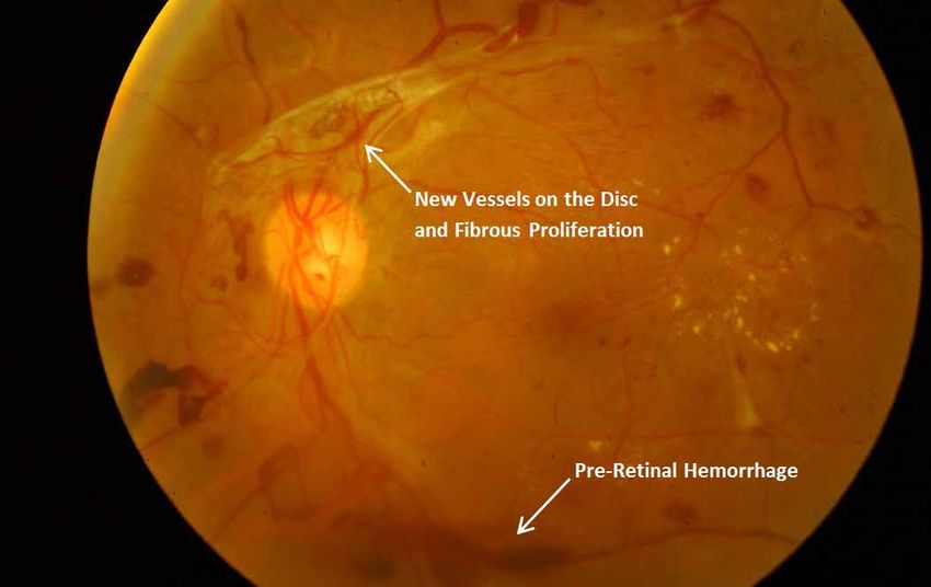
l. Introduction
Diabetes mellitus (DM) is a global epidemic with significant morbidity. Diabetic retinopathy (DR) is the specific
microvascular complication of DM and affects 1 in 3 persons with DM. DR remains a leading cause of vision loss in
working adult populations. Patients with severe levels of DR are reported to have poorer quality of life and reduced
levels of physical, emotional, and social well-being, and they utilize more health care resources.

Epidemiological studies and clinical trials have shown that optimal control of blood glucose, blood pressure, and
blood lipids can reduce the risk of developing retinopathy and slow its progression. Timely treatment with laser
photocoagulation, and increasingly, the appropriate use of intraocular administration of vascular endothelial growth
factor (VEGF) inhibitors can prevent visual loss in vision-threatening retinopathy, particularly diabetic macular edema
(DME). Since visual loss may not be present in the earlier stages of retinopathy, regular screening of persons with
diabetes is essential to enable early intervention.

Epidemiology of Diabetic Retinopathy
In many countries, DR is the most frequent cause of preventable blindness in working-aged adults. In the United States,
an estimated 40% (8% for vision-threatening retinopathy) of persons with type 2 diabetes and 86% (42% for vision-
threatening retinopathy) of persons with type 1 diabetes have DR. High prevalence estimates have also been reported
in other countries. Despite concern about a potential diabetes epidemic in Asia, epidemiologic data for DR in Asian
countries is relatively limited. In Latin America, 40% of diabetic patients had some DR and 17% required treatment. Few
studies of DR have been conducted in Africa.

DR develops with time and is associated with poor control of blood sugar, blood pressure, and blood lipids. The longer
someone has had DM, and the poorer their control, the higher their risk of developing DR. Good control reduces the
annual incidence of developing DR and extends life. However, good control does not necessarily reduce the lifetime risk
of developing DR, so everyone with DM is at risk.

The overall prevalence of DR in a community is also influenced by the number of people diagnosed with early DM:
   •   In resource-rich settings with good health care systems, more people with early DM will have been diagnosed.
       The prevalence of DR in people with newly diagnosed DM will be low, resulting in a lower overall prevalence of
       DR.
   •   In resource-poor settings with less advanced health care systems, fewer people with early DM will have been
       diagnosed. People may be diagnosed with diabetes only when symptomatic or complications have occurred.
       Thus, the prevalence of DR in people with newly diagnosed DM will be high, resulting in a somewhat higher
       overall prevalence of DR.

In general, meta-analysis of large scale studies show that approximately one third of those with DM will have DR, and
approximately one third of those (or 10% of persons with DM) will have vision-threatening DR that requires treatment.

Classification of Diabetic Retinopathy
The classic retinal microvascular signs of DR include microaneurysms, hemorrhages, hard exudates (lipid deposits),
cotton-wool spots (ischemic retina related to accumulations of axoplasmic debris within adjacent bundles of ganglion cell
axons), venous dilation and beading, and intraretinal microvascular abnormalities (i.e., dilated pre-existing capillaries).
(Annex Figures). These signs can be classified into two phases of DR.

Nonproliferative Diabetic Retinopathy
Nonproliferative DR is the early stage of DR. Recognition of nonproliferative retinopathy allows a prediction of risk of
progression, visual loss, and determination of a review interval. Annex Table 1 shows the signs of nonproliferative DR.

Proliferative Diabetic Retinopathy
Proliferative diabetic retinopathy (PDR) is a severe stage of DR and represents an angiogenic response of the retina to
extensive ischemia and capillary closure. Neovascularization has be divided into 2 groups: new vessels on the disc (NVD)
and new vessels elsewhere (NVE). Typically NVE grow at the interface of perfused and nonperfused retina. Annex
Table 2 shows the signs of PDR.

The stages of DR, from nonproliferative to proliferative DR, can be classified using the simple international classification
of DR scale shown in Table 1. DME is an important complication that is assessed separately from the stages of retinopathy,
as it can be associated with any of the DR stages and can run an independent course.

Diabetic Macular Edema
It is important to assess the presence and severity of diabetic macular edema (DME) separately from stages of DR.

                       International Council of Ophthalmology | Guidelines for Diabetic Eye Care | Page 2
    Copyright © ICO January 2014. Translation and adaption for non-commercial local use is encouraged, but please credit ICO.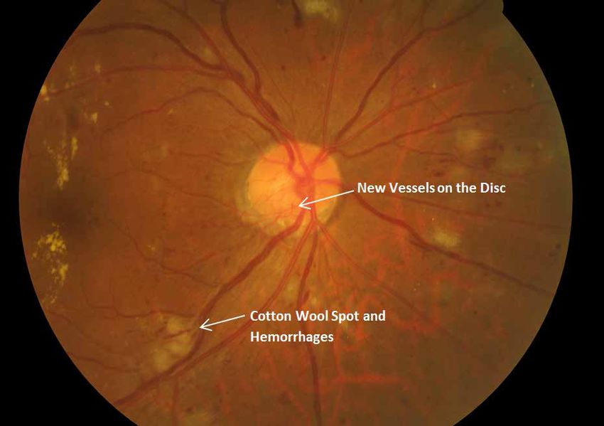
The stages of DR can be classified using the International Classification of DR Scale shown in Table 1. A simplified
grading based on this with referral decision can be used in low-resource settings (Table 2). It is important to remember
that early DME may be first detected by a reduction in visual acuity. An online self-directed course on the grading of
diabetic retinopathy is available at: drgrading.iehu.unimelb.edu.au.

Table 1: International Classification of Diabetic Retinopathy and Diabetic Macular Edema and Referral
Recommendations
                                             Findings Observable on Dilated
         Diabetic Retinopathy                                                                          Referral
                                                    Ophthalmoscopy
    No apparent retinopathy             No abnormalities                                Review in 1–2 years
    Mild nonproliferative DR            Microaneurysms only                             Review in 1–2 years

    Moderate nonproliferative           More than just microaneurysms, but less         Review in 6 months -1 year; or refer
    diabetic retinopathy                than severe nonproliferative DR                 to ophthalmologist

                                        Any of the following:
                                        • Intraretinal hemorrhages (≥20 in each
                                           quadrant);
                                        • Definite venous beading (in 2
    Severe nonproliferative DR                                                      Refer to ophthalmologist
                                           quadrants);
                                        • Intraretinal microvascular abnormalities
                                           (in 1 quadrant);
                                        • and no signs of proliferative retinopathy
                                        Severe nonproliferative DR and 1 or more of
                                        the following:
    Proliferative DR                                                                Refer to ophthalmologist
                                        • Neovascularization
                                        • Vitreous/preretinal hemorrhage

                                             Findings Observable on Dilated
       Diabetic Macular Edema                                                                          Referral
                                                    Ophthalmoscopy#
                                        No retinal thickening or hard exudates in
    DME absent                                                                    Review in 1-2 years
                                        posterior pole
                                        Retinal thickening or hard exudates in
    DME present                                                                Refer to ophthalmologist
                                        posterior pole
                                        Retinal thickening or hard exudates in
    Mild DME                            posterior pole but outside the central
                                        subfield of the macula (diameter 1000 µm)
                                        Retinal thickening or hard exudates within
    Moderate DME                        the central subfield of the macula but not
                                        involving the center point
                                        Retinal thickening or hard exudates
    Severe DME
                                        involving the center of the macula

#
 Hard exudates are a sign of current or previous macular edema. DME is defined as retinal thickening, and this requires
a three-dimensional assessment that is best performed by a dilated examination using slit-lamp biomicroscopy and/or
stereo fundus photography.

                        International Council of Ophthalmology | Guidelines for Diabetic Eye Care | Page 3
     Copyright © ICO January 2014. Translation and adaption for non-commercial local use is encouraged, but please credit ICO.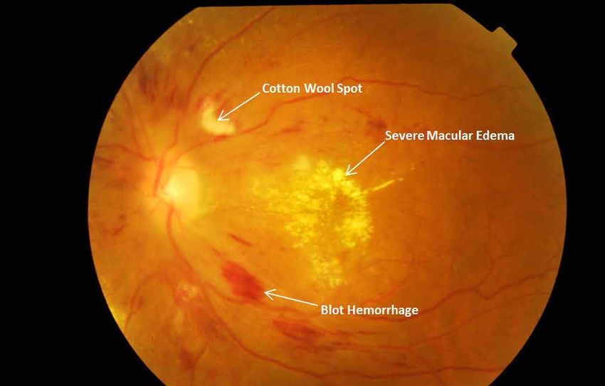
Table 2: Referral Recommendations Based on Simplified Classification of Diabetic Retinopathy and Diabetic
Macular Edema (Low Resource Setting)
             Classification                 Findings Observable on Dilated                         Referral
                                                   Ophthalmoscopy

 No apparent retinopathy or                                                         Review in 1 year for repeat screening
                                          See table 1
 Mild nonproliferative DR                                                           (ophthalmologist not required)

                                                                                    Routine referral within 6 months
 Nonproliferative DR                      See table 1                               if possible (ophthalmologist not
                                                                                    required)
                                                                                    Semi-urgent referral within a few
 Severe nonproliferative DR               See table 1                               months if possible (ideally to an
                                                                                    ophthalmologist)
                                                                                    Urgent referral as soon as possible
 PDR                                      See table 1
                                                                                    (ophthalmologist required)
                                          Retinal thickening or hard exudates       Semi-urgent referral within a few
 DME without center involvement           in the macula but not involving the       months if possible (ideally to an
                                          center of the macula                      ophthalmologist)
                                          Retinal thickening or hard exudates       Urgent referral as soon as possible
 Severe DME with center involvement
                                          involving the center of the macula        (ophthalmologist required)

II. Screening Guidelines

Screening Guidelines
Screening for DR is an important aspect of DM management worldwide. Even if an adequate number of ophthalmologists
are available, using ophthalmologists or retinal subspecialists to screen every person with DM is an inefficient use of
resources.

A screening exam could include a complete ophthalmic examination with refracted visual acuity and state-of-the-art
retinal imaging. However, in a low-resource setting, the minimum examination components to assure appropriate
referral should include a screening visual acuity exam and retinal examination adequate for DR classification. Vision
should be tested prior to pupil dilation. Annex Figure 1 shows an example of the screening process for DR.

The screening vision exam should be completed by trained personnel in any of the following ways, depending on
resources:
   •   Refracted visual acuity examination using a 3- or 4-meter visual acuity lane and a high contrast visual acuity chart.
   •   Presenting visual acuity examination using a near or distance eye chart and a pin-hole option if visual acuity is
       reduced.
   •   Presenting visual acuity examination using a 6/12 (20/40) equivalent handheld chart consisting of at least 5
       standard letters or symbols and a pin-hole option if visual acuity is reduced.

A retinal examination may be accomplished in the following ways:

   •   Direct or indirect ophthalmoscopy or slit-lamp biomicroscopic examination of the retina.
   •   Retinal (fundus) photography (including any of the following: widefield to 30o; mono- or stereo-; dilated or
       undilated).This could be done with or without accompanying optical coherence tomography (OCT) scanning.
       This could also include telemedicine approaches. (Annex Table 3)
   •   For the retinal examination, a medical degree may not be necessary, but the examiner must be well trained to
       perform ophthalmoscopy or retinal photography and be able to assess the severity of DR.

Using adequate information from the visual acuity and retinal examinations, one can decide on an appropriate
management plan, as outlined in Table 2. The plan may be modified based on individual patient requirements.

Patients with less than adequate retinal assessment should be referred to an ophthalmologist unless it is obvious that
there is no DR, or at most, only mild nonproliferative DR (i.e., microaneurysms only). In addition, persons with unexplained
visual-acuity loss should be referred.

As part of a screening exam, persons with diabetes should be asked about their diabetes control, including blood
                       International Council of Ophthalmology | Guidelines for Diabetic Eye Care | Page 4
    Copyright © ICO January 2014. Translation and adaption for non-commercial local use is encouraged, but please credit ICO.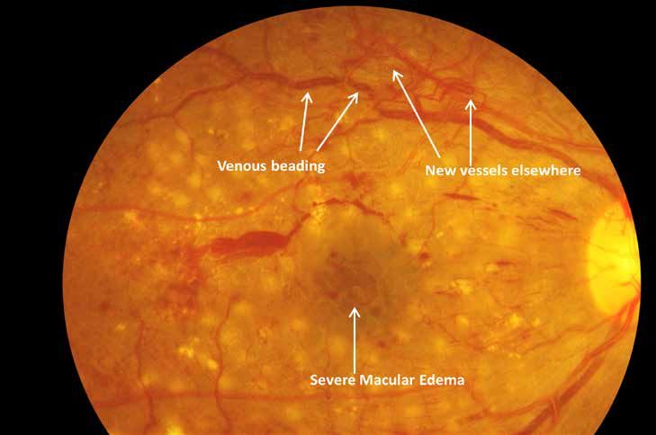
glucose, blood pressure, and serum lipids. In addition, women should be asked if they are or could be pregnant.
Inadequate control and pregnancy may require further appropriate medical intervention.

Referral Guidelines
Minimum referral guidelines are as follows:
   •    Visual acuity below 6/12 (20/40) or symptomatic vision complaints
   •    If DR can be classified according to the International Classification of DR or a Simplified scheme, they should be
        referred accordingly (Table 1 and 2)
   •    If retinal exam or retinal imaging is available but only a less detailed classification of DR is possible:
       »» No retinopathy or only a few small red spots: return for screening exam in 1–2 years
       »» Dot or blot hemorrhages or possible neovascularization: refer to ophthalmologist
       »» White spots in the retina: refer to ophthalmologist
   •    If visual acuity or retinal examination cannot be obtained at the screening examination: refer to ophthalmologist
   •    Patients who have had laser treatment should also be referred for ophthalmic review

III. Detailed Ophthalmic Assessment of Diabetic Retinopathy

1. Initial Patient Assessment
   Detailed patient assessment should include a complete ophthalmic examination, including visual acuity and the
   identification and grading of severity of DR and presence of DME for each eye. The patient assessment should also
   include the taking of a patient history focused on diabetes and its modifiers.
   a. Patient History (Key Elements)
          •   Duration of diabetes
          •   Past glycemic control (hemoglobin A1c)
          •   Medications (especially insulin oral hypoglycemics, antihypertensives, and lipid-lowering drugs)
          •   Systemic history (e.g., renal disease, systemic hypertension, serum lipid levels, pregnancy)
          •   Ocular history
   b. Initial Physical Exam (Key Elements)
          •   Visual acuity
          •   Measurement of intraocular pressure (IOP)
          •   Gonioscopy when indicated (e.g., when neovascularization of the iris is seen or in eyes with increased IOP)
          •   Slit-lamp biomicroscopy
          •   Fundus examination
   c. Fundus Examination Assessment Methods
      Currently, the two most sensitive methods for detecting DR are retinal photography and slit-lamp biomicroscopy
      through dilated pupils. Both depend on interpretation by trained eye health professionals. Other methods are
      listed in Annex Table 2.

Fundus photography has the advantage of creating a permanent record, and for that reason, it is the preferred method
for retinopathy assessment. However, well-trained observers can identify DR without photography and there are many
situations in which that would be the examination of choice.

The use of all instruments requires training and competence but more skill is needed for indirect ophthalmoscopy and
slit-lamp biomicroscopy than for fundus photography. Newer, semi-automatic nonmydriatic fundus cameras can be very
easy to use. Media opacities will lead to image/view degradation and all photographs/images must be reviewed by
trained personnel.

                     International Council of Ophthalmology | Guidelines for Diabetic Eye Care | Page 5
  Copyright © ICO January 2014. Translation and adaption for non-commercial local use is encouraged, but please credit ICO.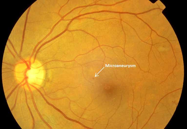
2. Follow-up Examination of Patients with Diabetic Retinopathy
  In general, the follow-up history and examination should be is similar to the initial examination. The assessment of
  visual symptoms, visual acuity, measurement of IOP, and fundus examination are essential.
  a. Follow-up History
        •   Visual symptoms
        •   Glycemic status (hemoglobin A1c)
        •   Systemic status (e.g., pregnancy, blood pressure, serum lipid levels, renal status)
  b. Follow-up Physical Exam
        •   Visual acuity
        •   Measurement of IOP
        •   Gonioscopy when indicated
        •   Slit-lamp biomicroscopy
        •   Fundus examination
  c. Ancillary Tests
        •   Fluorescein angiography is not needed to diagnose DR, proliferative DR or DME, all of which are diagnosed
            by means of the clinical exam.
        •   Fluorescein angiography can be used as a guide for treating DME and as a means of evaluating the
            cause(s) of unexplained decreased visual acuity. Fluorescein angiography can also identify macular capillary
            nonperfusion or sources of capillary leakage resulting in DME as possible explanations for visual loss.
        •   OCT is the most sensitive method to identify sites and severity of DME.
  d. Patient Education
        •   Discuss results or exam and implications.
        •   Encourage patients with DM but without DR to have annual screening eye exams.
        •   Inform patients that effective treatment for DR depends on timely intervention, despite good vision and
            no ocular symptoms.
        •   Educate patients about the importance of maintaining near-normal glucose levels, near-normal blood
            pressure and to control serum lipid levels.
        •   Communicate with the general physician (e.g., family physician, internist, or endocrinologist) regarding
            eye findings.
        •   Provide patients whose conditions fail to respond to surgery and for whom treatment is unavailable
            with proper professional support (i.e., offer referrals for counseling, rehabilitative, or social services as
            appropriate).
        •   Refer patients with reduced visual function for vision rehabilitation and social services.

                      International Council of Ophthalmology | Guidelines for Diabetic Eye Care | Page 6
   Copyright © ICO January 2014. Translation and adaption for non-commercial local use is encouraged, but please credit ICO.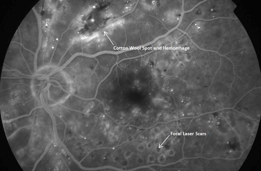
Table 3. Follow-up Schedule and Management for Diabetic Retinopathy Severity According to Resources Available
For all patients regardless of DR severity, optimize medical treatment for glycemic control, hypertension, and dyslipidemia.
    Follow up Schedule            Low-Resource Settings           Intermediate Resource         Resource-Rich Settings
                                                                         Settings
                                Repeat examination              Repeat examination            Repeat examination
 No apparent DR
                                biannually                      biannually                    annually
                                                                Repeat examination
 Mild nonproliferative DR       Repeat examination                                            Repeat examination
                                                                biannually unless annually if
                                biannually                                                    annually
                                                                poor glycemic control
                                                                                              Repeat examination within
 Moderate nonproliferative      Repeat examination              Repeat examination
                                                                                              6-12 months
 DR                             annually                        annually

 Severe nonproliferativeDR      Panretinal                      Panretinal                    Panretinal
 or proliferative DR            photocoagulation                photocoagulation              photocoagulation
                                Focal/grid laser if
                                                                Intravitreal injections of    Intravitreal injections of
 DME                            intravitreal anti-VEGF
                                                                anti-VEGF agents              anti-VEGF agents
                                agents are not available

IV. Treatment of Diabetic Retinopathy
Panretinal laser photocoagulation surgery should be performed in patients with proliferative DR. There are benefits
of early panretinal photocoagulation at the severe nonproliferative DR stage for patients with type 2 diabetes. Other
factors, such as poor compliance with follow up, impending cataract extraction or pregnancy, and status of fellow eye
will help in determining the timing of the panretinal photocoagulation.

1. Panretinal Photocoagulation (PRP)
   a. Pretreatment Discussion with Patients
       •   Patients usually need numerous follow-up visits and may require supplementary laser treatment.
       •   PRP reduces the risk of visual loss and blindness.
       •   Although laser treatment is effective, some patients may still develop vitreous hemorrhage. The hemorrhage
           is caused by the diabetes and not by the laser; it may mean the patient needs more laser treatment.
       •   Laser treatment often reduces peripheral and night vision; treatment may moderately reduce central vision.
           This short-term side effect is compensated by the significant long-term reduction in severe vision loss and
           blindness in laser-treated patients.

   b. Lenses for PRP
       •   The three-mirror Goldmann contact lens has a central opening for treating the posterior pole, and side
           mirrors for treating the mid peripheral and peripheral retina. Disadvantages: small field of view, which requires
           continual manipulation of the lens to complete treatment. Spot size is set at 500µm.
       •   Newer wide-angle contact lenses are often used. Although the image is inverted, there is a large field of
           view allowing for many burns with the field while easily maintaining orientation to the disc and macula. The
           optics of these wide-angle lenses will affect the laser spot size on the retina (Table 4). Wide-angle indirect
           ophthalmoscopy lenses provide an inverted image, but show a large field of view and a magnification of the
           spot in the retina (Table 4). Scatter treatment can be applied to a large area of retina in a single image, and it
           is easy to visualize the disk and the macula.

                     International Council of Ophthalmology | Guidelines for Diabetic Eye Care | Page 7
  Copyright © ICO January 2014. Translation and adaption for non-commercial local use is encouraged, but please credit ICO.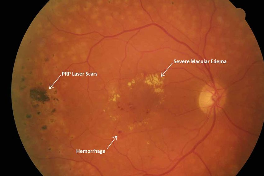
Table 4: Laser Spot Size Adjustment Required for Different Lenses Contact
Lens                      Field of Vision         Axial magnification      Spot magnification      Spot Size Setting for
                                                                                                   ~500 um
Mainster Wide-Field       125°                    0.46                     1.50x                   300µm
Volk TransEquator         120-125°                0.49                     1.43x                   300µm
Volk Quad/Aspheric        130-135°                0.27                     1.92x                   200 to 300µm
Mainster PRP 165          160°                    0.27                     1.96x                   200 to 300 µm

  c. Technique for PRP
       i. The pupil should be fully dilated and topical anesthesia is used. Retrobulbar or subtenons anesthesia to
           reduce pain and decrease eye motion can be employed as necessary.
       ii. The most common wavelengths used are Argon green, blue green (generally avoided currently), and 532
           green laser, using the slit-lamp delivery system. In case of hazy media, Krypton red or diode red laser (814
           nm) can be used. Slit-lamp treatment is most commonly done through a contact lens but can also be
           performed using indirect ophthalmoscopy. For example, when treatment is given under general anesthetic.
       iii. Typical initial settings on the Argon laser would be 500 μm spot size, a 0.1 second exposure and 250-270 mw
            power. The power is gradually increased until a whitish reaction is obtained on the retina. The lesions are
            placed 1 burn width apart. (Table 5)
       iv. A total of 1600-3000 burns are placed in 1 or more sittings, carefully avoiding the macular area and any areas
           of tractional elevation of the retina. The burns are placed 2 to 3 disc diameters away from the center of the
           macula and 1 disc diameter away from the disc, usually outside the arcades and extended peripherally up to
           the equator and beyond.
       v. Laser treatment should not be applied over major retinal veins, preretinal hemorrhages, darkly pigmented
          chorioretinal scars, or within 1 DD (200-300 μm) of center of macula, so as to avoid risk of hemorrhage or
          large scotomas.
       vi. Other considerations:
          •   Additional photocoagulation is needed if there is evidence of worsening of proliferative DR.
          •   Add laser burns in between scars of initial treatment further peripherally and also at the posterior pole,
              sparing the area within 500-1500 μm from the center of the macula.
          •   Favor quadrants with active new vessels or areas with intraretinal microvascular abnormalities where scars
              are more widely spaced and areas of severe ischemia not previously treated, such as the temporal part of
              the posterior pole.
          •   Direct treatment of NVE in between scars is possible.
          •   A subthreshold micropulse diode laser or multi-spot laser can be used.

  d. Panretinal (Scatter) Photocoagulation Technique Following Diabetic Retinopathy Clinical Research Network
     (DRCRNet) Consensus
        Panretinal (scatter) photocoagulation initially consists of 1200 to1600 burns (or the equivalent area treated
        with a multi-spot laser), with a spot size on the retina of approximately 500 µm given over 1 to 3 sittings and
        completed within eight weeks (56) days of initiation. (Table 5)

                      International Council of Ophthalmology | Guidelines for Diabetic Eye Care | Page 8
   Copyright © ICO January 2014. Translation and adaption for non-commercial local use is encouraged, but please credit ICO.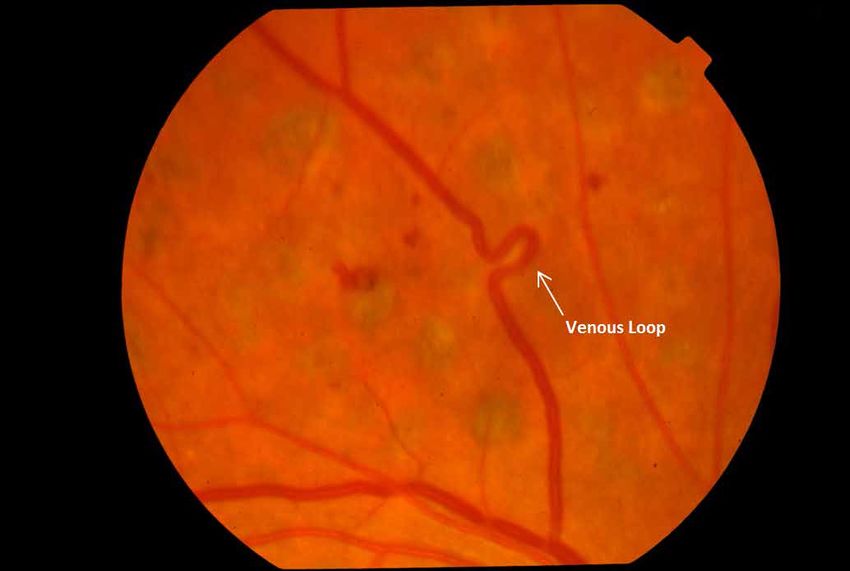
Table 5. The burn characteristics for panretinal photocoagulation:
 Size (on retina):                  500 µm
 Exposure:                          0.1 seconds recommended, 0.05 to 0.2 allowed
 Intensity:                         mild white (i.e. 2+ to 3+ burns)
 Distribution:                      edges 1 burn width apart
 Number of sessions/sittings:       1 to 3
 Nasal proximity to disk:           No closer than 500 µm
 Temporal proximity to              No closer than 3000 µm
 center:
 Superior/inferior limit:           No further posterior than 1 burn within the temporal arcades

 Extent:                            Arcades (~3000 µm from the macular center) to at least the equator

 Total number of burns:             1200 – 1600 There may be instances where 1200 burns are not possible
                                    such as the development of vitreous hemorrhage or inability to complete
                                    a sitting precluding completion of the PRP session. Similarly, there may be
                                    clinical situations in which more than 1600 burns are needed such as initial
                                    difficulty with laser uptake due to media opacity.
 Wavelength:                        Green or yellow (red can be used if vitreous hemorrhage is present)

2. Treatment For Diabetic Macular Edema
   a. Resource-Rich Settings
      i. Optimize medical treatment: Improve glycemic control if HbA1c > 7.5% as well as associated systemic
         hypertension or dyslipidemia.
      ii. Mild or moderate DME without center involvement (e.g., circinate HE ring threatening the center of the
          macula or when no vision loss has occurred in spite of center involvement): Consider focal laser to leaking
          microaneurysms. No treatment is applied to lesions closer than 300 μm from the center of the macula.
      iii. Severe DME with center involvement and associated vision loss*: intravitreal anti- VEGF treatment (e.g., with
           ranibizumab [Lucentis] 0.3 or 0.5mg, bevacizumab [Avastin] 1.25mg, or Aflibercept [Eylea]) 2mg therapy).
           Consideration should be given to monthly injections followed by treatment interruption and re-initiation
           based on visual stability and OCT. Patients should be monitored almost monthly with OCT to consider the
           need for treatment. Typically, the number of injections is 8 the first year, 2 or 3 during the second year, and 1
           to 2 during the third year. Persistent retinal thickening and leaking points: consider laser treatment after 24
           weeks. Treatment with intravitreal triamcinolone may be considered, especially in pseudophakic eyes. (Annex
           Figures 3 and 4). Injections are given 4 mm behind the limbus in the inferotemporal quadrant under topical
           anesthesia using a sterile technique.
      iv. DME associated with proliferative DR: combined intravitreal anti-VEGF therapy and PRP should be
          considered.
      v. Vitreomacular traction or epiretinal membrane on OCT: pars plana vitrectomy may be indicated.

*For eyes with severe DME with center involvement and good visual acuity (20/25 or better), 3 treatment options being
evaluated in an ongoing clinical trial: (1) careful follow-up with anti-VEGF treatment only for worsening DME; (2) anti-
VEGF injections; or (3) laser photocoagulation with anti-VEGF, if necessary.

   b. Intermediate or Low-Resource Settings
      i. Generally similar to above. Focal laser is preferred if intravitreal injection of anti-VEGF agents are not
         available. Bevacizumab (Avastin) is an appropriate alternative to raniziumab (Lucentis) or aflicercept (Eyelea).
         Laser can be applied earlier to areas of persistent retinal thickening in eyes unresponsive to anti- VEGF
         treatment.

                     International Council of Ophthalmology | Guidelines for Diabetic Eye Care | Page 9
  Copyright © ICO January 2014. Translation and adaption for non-commercial local use is encouraged, but please credit ICO.
c. Laser Technique for Macular Edema
i. Focal macular treatment includes focal laser treatment of microaneurysms and grid treatment of areas of
diffuse leakage and focal nonperfusion within 2DD of center of the macula. (Table 6)
ii. Laser parameters used are 50-100 μm spot size, 120-150 mW energy and very light gray intensity of
the burn. Care is taken to demarcate and avoid the foveal avascular zone.
iii. If DME is associated with large areas of macular ischemia, only the areas of retinal thickening are treated.
Table 6. Modified-ETDRS and the Mild Macular Grid Laser Photocoagulation Techniques
Direct/Grid Photocoagulation Mild Macular Grid
Burn Characteristic
(Modified-ETDRS technique) Photocoagulation Technique
Directly treat all leaking
microaneurysms in areas of retinal
Direct treatment thickening between 500 and 3000 µm Not applicable
from the center of the macula (but
not within 500 µm of disc)
Not required, but at least a mild
Change in MA color with direct
gray-white burn should be evident Not applicable
treatment
beneath all microaneurysms
Burn size for direct treatment 50-100 µm Not applicable
Burn duration for direct treatment 0.05 to 0.1 sec Not applicable
Applied to all areas with diffuse Applied to entire area described
Grid treatment leakage or nonperfusion within area below for treatment (including
described below for treatment unthickened retina)
• 500 to 3000 um superiorly, nasally • 500 to 3000 µm superiorly, nasally
and inferiorly from center of and inferiorly from center of
macula macula
Area considered for grid treatment • 500 to 3500 um temporally from • 500 to 3500 µm temporally from
macular center macular center
• No burns are placed within 500 • No burns are placed within 500
um of disc µm of the disc
Burn size for grid treatment 50-100 µm 50 µm
Burn duration for grid treatment 0.05 to 0.1 sec 0.05 to 0.1 sec
Burn intensity for grid treatment Barely visible (light gray) Barely visible (light gray)
200 to 300 total burns evenly
distributed over the treatment area
Burn Separation for Grid Treatment Two visible burn widths apart
outlined above (approx. two to three
burn widths apart)
Wavelength (grid and focal
Green to yellow wavelengths Green
Treatment)
3. Indications for Vitrectomy
a. Severe vitreous hemorrhage of 1–3 months duration and that does not clear spontaneously.
b. Advanced active proliferative DR that persists despite extensive PRP.
c. Traction macular detachment of recent onset.
d. Combined traction-rhegmatogenous retinal detachment.
e. Tractional macular edema or epiretinal membrane involving the macula.
International Council of Ophthalmology | Guidelines for Diabetic Eye Care | Page 10
Copyright © ICO January 2014. Translation and adaption for non-commercial local use is encouraged, but please credit ICO.V. Suggested Indicators for Evaluation of DR Programs
a. Prevalence of diabetic retinopathy related blindness and visual impairment*
b. Proportion of blindness and visual impairment due to DR*
c. Last eye examination for DR among known persons with diabetes (males/females)*
• Never had eye examination for DR
• 0–12 months ago
• 13–24 months ago
• >24 months ago
• Could be simplified as: never/0-12 months ago/>12 months ago
d. Number of patients who were examined for DR during last year
e. Number of patients who received laser and/or anti-VEGF treatment during last year
This absolute number could be used to define ratios such as:
f. Number of patients who received laser and/or anti-VEGF treatments per million general population per year
[equivalent to Cataract Surgical Rate (CSR)]
g. Number of patients who received laser and/or anti-VEGF treatments per number of patients with diabetes in a
given area (hospital catchment area, health district, region, country)
• Numerator: number of laser and/or anti-VEGF treatments during the last year
• Denominator: number of patients with diabetes (population x prevalence of DM; source: IDF Atlas)
h. Number of patients who received laser and/or anti-VEGF treatments per number of persons with vision-
threatening DR in a given area (hospital catchment area, health district, region, country)
• Numerator: number of laser and/or anti-VEGF treatments during the last year
• Denominator: number of patients with vision-threatening DR (population x prevalence of DM x 0.117; source:
IDF Atlas)
* Data available from RAAB surveys
0.117: Estimated average prevalence of vision-threatening DR.
Yau JW, Rogers SL, Kawasaki R, et al. Global Prevalence and Major Risk Factors of Diabetic Retinopathy. Diabetes Care.
Mar 2012;35(3):556-564.
International Council of Ophthalmology | Guidelines for Diabetic Eye Care | Page 11
Copyright © ICO January 2014. Translation and adaption for non-commercial local use is encouraged, but please credit ICO.ICO GUIDELINES FOR DIABETIC RETINOPATHY
ANNEXES
Annex Table 1: Features of Diabetic Retinopathy (also see the photographs continued in the annex)
Feature Description Assessment Considerations
Microaneurysms Isolated, spherical, red dots of They are easiest seen on fluorescein
varying size. They may reflect an angiography
abortive attempt to form a new
vessel or may simply be a weakness
of capillary vessel wall through loss of
normal structural integrity.
Dot hemorrhages Dot hemorrhages cannot always be The term dot hemorrhage/
differentiated from microaneurysms microaneurysm (H/Ma) is often used.
as they are similar in appearance but
with varying size.
Blot hemorrhages Formed where clusters of capillaries The lesion can be seen to be in the
occlude leading to formation of outer plexiform layer on fluorescein
intraretinal blot hemorrhages. angiography where it does not mask
the overlying capillary bed unlike dot
and flame hemorrhages, which lie
more superficially in the retina.
Cotton wool spots These represent the swollen ends of These features are not exclusive to
interrupted axons where build-up of DR and do not in themselves appear
axoplasmic flow occurs at the edge to increase the risk of new vessel
of the infarct. formation. For example, they may
occur in hypertension HIV/AIDS.
Intraretinal microvascular anomalies These are dilated capillary remnants They are easiest seen on fluorescein
following extensive closure of angiography.
capillary network between arteriole
and venule.
Associated features include:
• venous beading ( foci of venous
endothelial cell proliferation that
have failed to develop into new
vessels),
• Venous reduplication (rare),
• Venous loops (thought to
develop due to small vessel
occlusion and opening of
alternative circulation) and
• Retinal pallor and white vessels
Macular changes In nonproliferative Thickening of retina takes place due The appearance of macular edema
retinopathy to accumulation of exudative fluid can be appreciated on stereoscopic
– Macular edema from damaged outer blood-retina examination or inferred by the
– Macrovascular disease barrier (extracellular edema) or as presence of intraretinal exudate.
a result of hypoxia, leading to fluid
accumulating within individual retinal
cells (intracellular edema). It may be
focal or diffuse.
Flame hemorrhage and cotton wool
spot formation. May occur due to
arteriolar occlusion, without capillary
occlusion, which frequently affects
the horizontal nerve fiber layer of the
retina.
Optic disc changes Occasionally swollen optic discs may In diabetic papillopathy, vision is
be seen (diabetic papillopathy) in usually not significantly impaired.
diabetic patients.
International Council of Ophthalmology | Guidelines for Diabetic Eye Care | Page 12
Copyright © ICO January 2014. Translation and adaption for non-commercial local use is encouraged, but please credit ICO.Annex Table 2: Features of Proliferative Diabetic Retinopathy
Feature Description Assessment Considerations
New vessels at the disc (NVD) New vessels at the discs usually arise In order to differentiate NVD from
from the venous circulation on the fine normal small blood vessels note
disc or within 1 disc diameter of the that the latter always taper to an end
disc NVD. and do not loop back to the disc,
while NVD always loop back, may
form a chaotic net within the loop,
and have the top of the loop of wider
diameter than the base.
New vessels elsewhere (NVE) New vessels, which usually occur Not to be confused with intraretinal
along the border between healthy microvascular abnormalities, which
retina and areas of capillary occur within areas of capillary
occlusion. occlusion.
Other sites of new vessels New vessel formation on the iris It is useful to perform gonioscopy in
(NVI) is uncommon but represents such cases to exclude new vessels in
potentially more advanced ischemic the anterior chamber angle (NVA),
changes. New vessel formation on which can lead to neovascular
the anterior hyaloid surface occurs glaucoma.
rarely postvitrectomy if insufficient
laser has been applied to the
peripheral retina.
Fibrous proliferation In proliferative retinopathy, new
vessels grow on a platform of glial
cells
Adapted from British The Royal College of Ophthalmologists Diabetic Retinopathy Guidelines December 2012.
International Council of Ophthalmology | Guidelines for Diabetic Eye Care | Page 13
Copyright © ICO January 2014. Translation and adaption for non-commercial local use is encouraged, but please credit ICO.Annex Table 3: Available Assessment Instruments and Their Advantages and Disadvantages
Technique Advantages Disadvantages Recommendation
Direct ophthalmoscopy #
• Mobile • Requires pupil dilation • Optional for screening
• Inexpensive • Small field • Pupils must be dilated
• Low sensitivity:
even with a trained
practitioner and red
free illumination,
small microvascular
abnormalities may be
difficult to detect
• Less effective than slit-
lamp biomicroscopy
through dilated pupils
• No ability to
retrospectively audit
Indirect ophthalmoscopy# • Mobile • Requires pupil dilation • Optional for screening
• Large field • Even with a trained • Pupils must be dilated
• Relatively inexpensive practitioner and red
free illumination,
small microvascular
abnormalities may be
difficult to detect
• Less effective than slit-
lamp biomicroscopy
through dilated pupils
• No ability to
retrospectively audit
Slit-lamp biomicroscopy • Large field • Requires pupil dilation • Required for
• Immobile ophthalmic
examination
• Requires special lenses
• No ability to
retrospectively audit
Nonmydriatic retinal • Large field • Relatively expensive • Recommended for
photography screening
• Can be used by non- • A dark space is
medically trained staff required for maximum
• No dilation required in pupil dilation
80-90% of cases • Auditable
• Some are portable -
can be transported
to the community in
mobile units
• Can be linked to
computers and images
can be stored for the
long term
• Allows objective
comparison of the
same person, or
between different
groups of people,
examined at different
times or by different
professionals
• Can be used as a
patient education tool,
giving immediacy and
personal relevance
• Readily recalled for
evaluation of screener
performance and audit
of grading
International Council of Ophthalmology | Guidelines for Diabetic Eye Care | Page 14
Copyright © ICO January 2014. Translation and adaption for non-commercial local use is encouraged, but please credit ICO.(Continued) Annex Table 3: Available Assessment Instruments and Their Advantages and Disadvantages
Technique Advantages Disadvantages Recommendation
Nonmydriatic retinal • As above except pupils • As above • Optional
photography used with are dilated for better • Requires pupil dilation
mydriasis quality photos
Mydriatic retinal • Large field • Requires pupil dilation • Desirable in
photography (conventional Expensive ophthalmic center
fundus camera) • Bright flash constricts
the pupil for a long
time
Fluorescein angiography • Only method of • Invasive and needs • Desirable in
assessing capillary general health status ophthalmic center
circulation assessment
• Expensive
• Dilatation needed
Cannot be used by
non-medically trained
staff
OCT • One of the best ways • Expensive • Desirable in
to assess macular • Dilatation needed ophthalmic center
edema (retinal
thickening and • Cannot be used by
intraretinal edema) non-medically trained
staff
Fundus autofluorescence • A form of functional • Role not clearly • Optional high-resource
imaging, giving understood settings
insights into the
metabolic activity of
the retinal pigment
epithelium.
Equipment
Core/essential: for screening, initial assessment, and follow up:
• Nonmydriatic retinal (fundus) photography (recommended for screening).
• Indirect ophthalmoscopy (optional for screening, panoramic view, low magnification). Pupils must be dilated.
• Noncontact biconvex indirect lenses used with the slit lamp (90 D for screening, 78 D for more magnification).
• Direct ophthalmoscopy (optional for screening). Pupils must be dilated.
• Three-mirror contact lens used with slit lamp for stereoscopic and high-resolution images of the macula
(evaluation of macular edema). Pupils must be dilated.
• Slit-lamp biomicroscope.
• Laser equipment: Currently, the most used lasers are (1) The green laser 532 nm, frequency-doubled Nd:YAG
or 514 nm argon laser. The 810 nm infrared laser, or diode laser – this causes deeper burns with a higher rate of
patient discomfort, but tend to be cheaper, is effective, and requires less maintenance.
Desirable in reference centers:
• OCT
• Fluorescein angiography
• Mydriatic retinal photography (large field conventional fundus camera)
• Green lasers are the most used, but the pattern-laser method, with a predetermined multispot treatment cascade
and the 577 nm yellow laser can be used in selected cases
IAPB Standard List of Equipment
The online version of the International Agency for the Prevention of Blindness (IAPB) Standard List provides information
for eye health providers on a carefully evaluated range of eye care technologies, supplies, and training resources suitable
for use in settings with limited resources.
For more information and to get access, please register and log on at IAPB.standardlist.org.
Only registered users have access to the IAPB Standard List catalogue. Please be aware the registration process may
take a few days for approvals to be granted.
International Council of Ophthalmology | Guidelines for Diabetic Eye Care | Page 15
Copyright © ICO January 2014. Translation and adaption for non-commercial local use is encouraged, but please credit ICO.Figure 1: Screening for Diabetic Retinopathy
Diabetes History; Medical History; Current Medication; Biochemical Parameters
Uncorrected Visual Acuity VA
Ophthalmoscopy or Fundus Photography
with current Spectacles
VA> 20/40 VA< 20/40 Diabetic Retinopathy*
None Mild or Severe NPDR, DME,
Moderate or PDR
NPDR
Routine re- Non-urgent Referral for refraction
Urgent Referral
examination and assessment
*Need to optimize medical treatment; glycemic control, hypertension and lipids.
NPDR = non-proliferative diabetic retinopathy
PDR = proliferative diabetic retinopathy
DME=diabetic macular edema
VA=visual acuity
Figure 2: Treatment decision tree of DME based on Center-Involvement and Vision
DME
Assessment: Clinical and OCT
Center involvement?
NO YES
VA 20/40 or worse
NO (indicative of DME)? YES
Treatment failure
Focal Laser treatment Anti-VEGF treatment
DME=diabetic macular edema
VA=visual acuity
International Council of Ophthalmology | Guidelines for Diabetic Eye Care | Page 16
Copyright © ICO January 2014. Translation and adaption for non-commercial local use is encouraged, but please credit ICO.Figure 3: Anti-VEGF treatment decision tree based on the RESTORE study treatment
and re-treatment schedule
Anti-VEGF treatment for DME
Initial treatment with injections given 3 monthly
YES Stable VA achieveda NO
Suspend treatment; return for Continue with 1
monthly follow-up injection per month
Worsening of DMEb
Reinitiate monthly injection
a. VA was considered to have stabilised if there was no (further) improvement in best
corrected visual acuity (BCVA) at the last 2 consecutive visits, or if a BCVA letter
score of 6/6 was observed at the last 2 consecutive visits.
b. Decrease in BCVA and confirmed by OCT and/or other anatomical and clinical
assessments.
VEGF=vascular endothelial growth factor
DME=diabetic macular edema
VA=visual acuity
International Council of Ophthalmology | Guidelines for Diabetic Eye Care | Page 17
Copyright © ICO January 2014. Translation and adaption for non-commercial local use is encouraged, but please credit ICO.Figure 4: Anti-VEGF treatment decision tree based on the DRCR.net re-treatment and
follow-up schedule
Anti-VEGF treatment for DME
Assessment 1 montha after initial injectionsb
DME improvingc
YES NOd
Re-inject and return in 1 month No injectione and return in 1 month
DME worsens or recurs
YES
NO
Double follow-up interval up to 4
monthsf
a. In the DRCR.net study, 4-week, not 1-month, intervals were used.
b. The DRCR.net study required 4 injections of intravitreal ranibizumab every 4 weeks initially;
it is not known whether a different number of injections initially would have worked as well.
DRCR.net also required 2 additional injections at months 5 and 6 if edema persisted and
success had not been met, even in the absence of improvement.
c. Relevant details from the DRCR.net study: 1) DRCR.net “improvement” on Zeiss Stratus
OCT >10% decrease in central subfield thickness; 2) Even if no longer improving on OCT,
injections continued if VA “improvement” (unless 6/6 or better); 3) VA improvement defined
as 5 or more letter increase on Electronic ETDRS Visual Acuity Test.
d. In the DRCR.net study, if focal/grid laser was deferred at baseline, it was added at or after
24 weeks if edema still present and OCT central subfield and vision no longer improving.
e. In the DRCR.net study, all patients received at least 4 injections 4 weeks apart. The decision
to re-inject was at investigator discretion, starting at 16 weeks for “success”, defined as VA
better than 6/6 or OCT central subfieldPhotographs
Figure 1. Mild non-proliferative diabetic retinopathy with microaneurysms
Figure 2. Moderate non-proliferative diabetic retinopathy with hemorrhages, hard exudates and micro aneurysms
International Council of Ophthalmology | Guidelines for Diabetic Eye Care | Page 19
Copyright © ICO January 2014. Translation and adaption for non-commercial local use is encouraged, but please credit ICO.Figure 3. Moderate non-proliferative diabetic retinopathy with moderate macular edema, with hard exudates
approaching the center of the macular.
Figure 4. Moderate non-proliferative diabetic retinopathy with no diabetic macular edema
International Council of Ophthalmology | Guidelines for Diabetic Eye Care | Page 20
Copyright © ICO January 2014. Translation and adaption for non-commercial local use is encouraged, but please credit ICO.Figure 5. Moderate non-proliferative diabetic retinopathy with mild diabetic macular edema
Figure 6. Moderate non-proliferative diabetic retinopathy with severe macular edema
International Council of Ophthalmology | Guidelines for Diabetic Eye Care | Page 21
Copyright © ICO January 2014. Translation and adaption for non-commercial local use is encouraged, but please credit ICO.Figure 7a. Moderate non-proliferative diabetic retinopathy with moderate macular edema
Figure 7b. Fundus Fluorescein Angiogram showing moderate non-proliferative diabetic retinopathy with moderate
macular edema
International Council of Ophthalmology | Guidelines for Diabetic Eye Care | Page 22
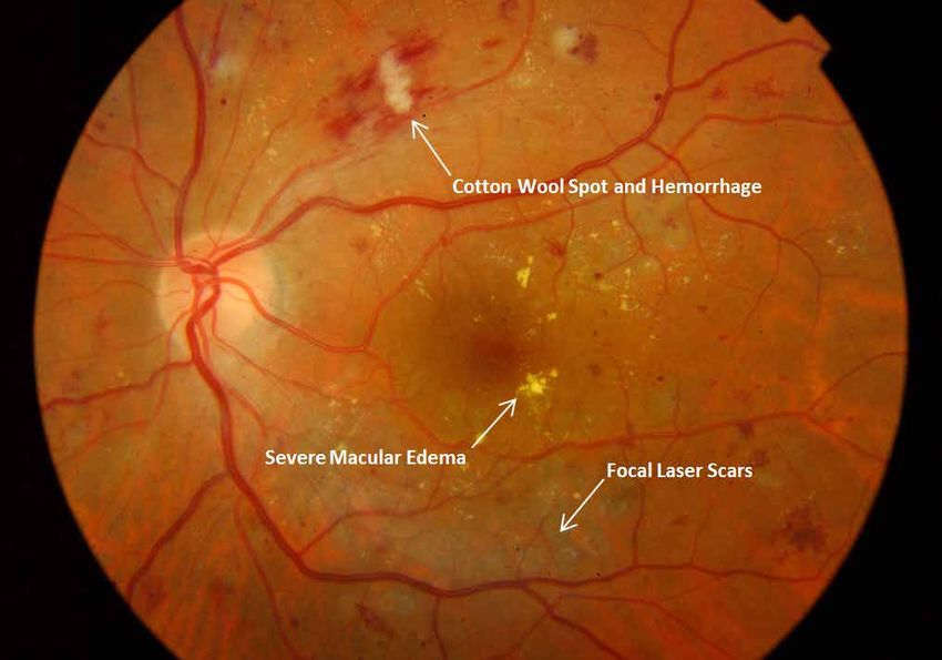
Copyright © ICO January 2014. Translation and adaption for non-commercial local use is encouraged, but please credit ICO.Figure 8. Severe non proliferative diabetic retinopathy with severe diabetic macular edema
Figure 9. Severe non-proliferative diabetic retinopathy with severe diabetic macular edema
International Council of Ophthalmology | Guidelines for Diabetic Eye Care | Page 23
Copyright © ICO January 2014. Translation and adaption for non-commercial local use is encouraged, but please credit ICO.Figure 10. Severe non-proliferative diabetic retinopathy with venous loop
Figure 11. Severe non-proliferative diabetic retinopathy with intra–retinal microvascular abnormality (IRMA)
International Council of Ophthalmology | Guidelines for Diabetic Eye Care | Page 24
Copyright © ICO January 2014. Translation and adaption for non-commercial local use is encouraged, but please credit ICO.Figure 12. Proliferative diabetic retinopathy with venous beading, new vessels elsewhere (NVE) and severe diabetic
macular edema
Figure 13. High risk proliferative diabetic retinopathy with new vessels at the disc
International Council of Ophthalmology | Guidelines for Diabetic Eye Care | Page 25
Copyright © ICO January 2014. Translation and adaption for non-commercial local use is encouraged, but please credit ICO.Figure 14a. High risk proliferative diabetic retinopathy. Pre-retinal hemorrhage before with new vessels on the disc.
Figure 14b. High risk proliferative diabetic retinopathy, with new panretinal photocoagulation (PRP) scars
International Council of Ophthalmology | Guidelines for Diabetic Eye Care | Page 26
Copyright © ICO January 2014. Translation and adaption for non-commercial local use is encouraged, but please credit ICO.Figure 15a. Proliferative diabetic retinopathy. New vessels on the disc and elsewhere
Figure 15b. Proliferative diabetic retinopathy. New vessels on the disc and elsewhere on fluorescein angiogram
International Council of Ophthalmology | Guidelines for Diabetic Eye Care | Page 27
Copyright © ICO January 2014. Translation and adaption for non-commercial local use is encouraged, but please credit ICO.Figure 16a. Diabetic macular edema with panretinal photocoagulation (PRP) (right eye).
Figure 16b. Diabetic macular edema with panretinal photocoagulation (PRP). (left eye)
International Council of Ophthalmology | Guidelines for Diabetic Eye Care | Page 28
Copyright © ICO January 2014. Translation and adaption for non-commercial local use is encouraged, but please credit ICO.Figure 17a. Persistent diabetic macular edema after focal laser treatment
Figure 17b. Persistent diabetic macular edema after focal laser treatment on fundus fluorescein angiogram
International Council of Ophthalmology | Guidelines for Diabetic Eye Care | Page 29
Copyright © ICO January 2014. Translation and adaption for non-commercial local use is encouraged, but please credit ICO.Figure 18a. Proliferative diabetic retinopathy with pre-retinal hemorrhage
New Vessels on the Disc and
Fibrous Proliferation
New Vessels on the Disc
and Fibrous Proliferation
Pre-Retinal Hemorrhage
Pre-Retinal Hemorrhage
Figure 18b. Proliferative diabetic retinopathy with pre-retinal hemorrhage on fundus fluorescein angiogram
International Council of Ophthalmology | Guidelines for Diabetic Eye Care | Page 30
Copyright © ICO January 2014. Translation and adaption for non-commercial local use is encouraged, but please credit ICO.Figure 19. Panretinal (PRP) photocoagulation. First session: inferior retina (laser scars). Second session: superior
retina (fresh burns). Third session will be needed to complete PRP.
International Council of Ophthalmology | Guidelines for Diabetic Eye Care | Page 31
Copyright © ICO January 2014. Translation and adaption for non-commercial local use is encouraged, but please credit ICO.Guidelines for Screening, Assessing, and Treating Diabetic Eye Disease
To create the ICO Guidelines for Diabetic Eye Care, the ICO collected guidelines from around the world for screening,
assessing, and treating diabetic eye disease. This is part of a new initiative to reduce worldwide vision loss related to
diabetes.
View the collected guidelines at: www.icoph.org/taskforce-documents/diabetic-retinopathy-guidelines.html.
In addition to creating a consensus on technical guidelines, as encompassed in the ICO Guidelines for Diabetic Eye
Care, these resources will also be used to focus on:
• Incorporating the critical competencies into ICO curricula and stimulating improved training and continuing
professional development to meet public needs.
• Developing a framework for evaluation of public health approaches and stimulating development,
strengthening, and monitoring of relevant health systems.
Please send questions, comments, or additional resources to: info@icoph.org.
About the ICO
The ICO is composed of 120 national and subspecialty Member societies
from around the globe. ICO Member societies are part of an international
ophthalmic community working together to preserve and restore vision.
Learn more at: www.icoph.org.
Printing Credit
The ICO Guidelines for Diabetic Eye Care were printed for the World Ophthalmology Congress® of the ICO by the
Singapore Eye Research Institute (SERI), Singapore National Eye Center. Learn more about SERI at: www.seri.com.sg.
Photo Credit
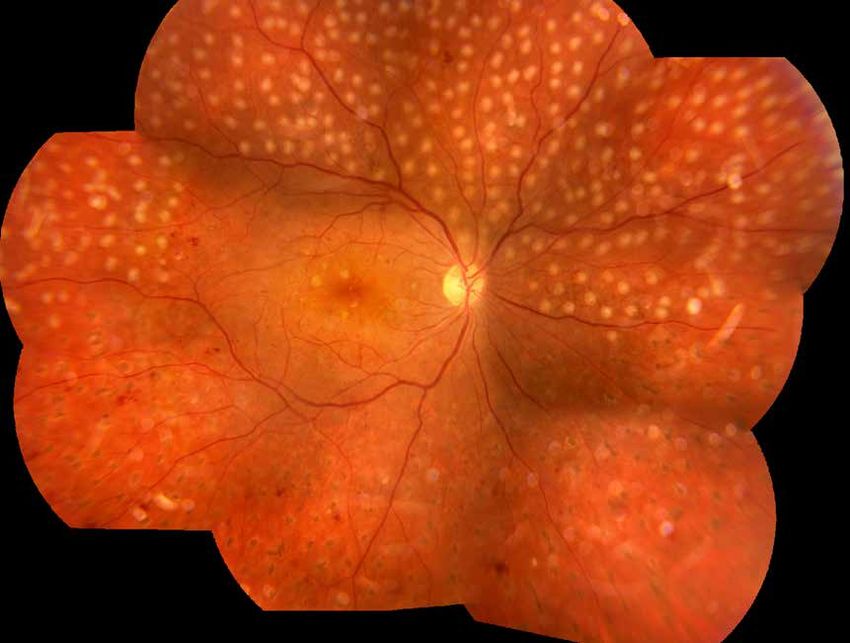
The photos that appear in the ICO Guidelines for Diabetic Eye Care were provided by:
• Pan-American Association of Ophthalmology (Figure 19)
• Singapore Eye Research Institute, Singapore National Eye Center (top and bottom left cover photos and Figures
1-18b)
• University of Melbourne, cover photo (bottom right)
*Photos may be used in translated or adapted versions of the ICO Guidelines for Diabetic Eye Care. They may not
be used for commercial purposes. If the photos are used, credit must be given to the appropriate organization(s).
International Council of Ophthalmology | Guidelines for Diabetic Eye Care | Page 32
Copyright © ICO January 2014. Translation and adaption for non-commercial local use is encouraged, but please credit ICO.ICO Headquarters: ICO International Fellowships Office:
Bruce E. Spivey, MD, President Veit-Peter Gabel, MD, Director for Fellowships
945 Green Street #10 Kuechelstraße 14
San Francisco, California 94133 81375 Munich
United States Germany
Fax: +1 (415) 409-8411 Fax: +49 3212-3200120
Email: info@icoph.org Email: fellowship@icoph.org
Web: www.icoph.org Web: www.icoph.org/fellow
ICO Examinations Office:
David Taylor, FRCOphth, Director for Examinations
11-43 Bath Street
London EC1V 9EL
England
Fax: +44 (0) 20 7608 6947
Email: assess@icoph.org
Web: www.icoexams.org/contact
International Council of Ophthalmology | Guidelines for Diabetic Eye Care | Page 33
Copyright © ICO January 2014. Translation and adaption for non-commercial local use is encouraged, but please credit ICO.You can also read