Identification of some coffee leaf taxa using fluorescence spectroscopy and chemometrics
←
→
Page content transcription
If your browser does not render page correctly, please read the page content below

R ESEARCH         ARTICLE                                                             ScienceAsia 47S (2021): 60–68
doi: 10.2306/scienceasia1513-1874.2021.S013

Identification of some coffee leaf taxa using
fluorescence spectroscopy and chemometrics
Saowaluk Madkoksunga , Plaipol Dedvisitsakula,b , Kanchana Watla-iada,c,d,∗
a
     School of Science, Mae Fah Luang University, Chiang Rai 57100 Thailand
b
     Microbial Products and Innovation Research Unit, School of Science, Mae Fah Luang University,
     Chiang Rai 57100 Thailand
c
     Center of Chemical Innovation for Sustainability, School of Science, Mae Fah Luang University,
     Chiang Rai 57100 Thailand
d
     Tea and Coffee Institute of Mae Fah Luang University, Mae Fah Luang University, Chiang Rai 57100
     Thailand
∗
    Corresponding author, e-mail: kanchana.wat@mfu.ac.th
                                                                                                Received 11 Nov 2020
                                                                                                Accepted 20 May 2021

ABSTRACT: Analytical techniques for identification of coffee taxa are essential for plant breeding and quality control of
products. Rapid technique for discrimination of coffee taxa based on the fluorescence signals from their leaf extracts was
introduced. Five different coffee taxa: Coffea liberica; Coffea congensis; Coffea arabica var. Geisha, a spontaneous hybrid
of C. Arabica and Coffea canephora (Hibrido de timor), a hybrid of Hibrido de timor, and C. arabica var. Cattura; were
investigated based on their fluorescence signals. The individual taxa present different fluorescence spectra. The spectra
obtained from the excitation wavelengths at 300, 330, 390, 420, and 450 nm; and emission wavelengths in the range of
500–790 nm were selected for principal component analysis (PCA) and partial least squares-discriminant analysis (PLS-
DA). It was found that fluorescence signals with excitation wavelength at 300 nm had been successfully implemented for
rapid clustering and identification of some coffee taxa. The PCA score plot presenting natural clustering of data obtained
from the fluorescence spectra tended to agree with the data of chemical contents based on antioxidant activity, total
phenolic content, and total flavonoid content. The Q2 and R2 calculated via leave-one-out cross-validation (LOOCV) of
model obtained from processing of the PLS-DA were 0.6 and 0.8, respectively. It means that the model has potential
for the categorization of coffee taxa based on their leaf extracts without any chemical treatments.

KEYWORDS: fluorescence spectroscopy, coffee leaves, coffee taxa, taxonomic identification, chemometric analysis

INTRODUCTION                                                   using morphological-based techniques [2], molec-
                                                               ular DNA analysis [3], and traditional laboratory-
Coffee species and varieties are important factors             based chemical methods [4]. In addition, there
that affect coffee qualities. The identification of            are many chemical analysis techniques applied for
coffee is very important for the exploitation in coffee        the classification of plant extracts obtained from
plant breeding and quality control of coffee products          various varieties, such as high-performance liq-
as well as the development of taste and favor diver-           uid chromatography-diode array (HPLC-DAD) [5],
sity. There are more than 120 varieties of coffee,             HPLC-diode array detector-time-of-flight-mass spec-
but the two most popular are C. arabica, commonly              trometry (HPLC-DAD-TOF-MS) [6], and nuclear
known as Arabica, and C. canephora known as Ro-                magnetic resonance spectroscopy (NMR) [7]. These
busta [1]. Many scientific reports on taxonomic                techniques could improve performance of discrimi-
analyses of coffee are available on C. arabica and             nation and classification by coupling with chemo-
C. canephora. However, there are limited informa-              metric analysis. However, these methods require
tion of other coffee species and their varieties in the        laborious and time consuming analytical workflow.
scientific literature. More information on genetic di-         Several spectroscopic techniques are successfully
versity of coffee would be beneficial to the develop-          applied for taxonomic identification of living things
ment of coffee taste and favor diversity in the future.        due to their rapidity, cost effectiveness, and non-
The discrimination of coffee taxa has been studied             tedious sample preparation [8, 9]. Recently, the

www.scienceasia.org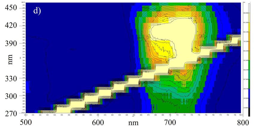
ScienceAsia 47S (2021)                                                                                        61

combinations of FT-NIR spectroscopy and chemo-           MATERIALS AND METHODS
metrics, such SIMCA analysis, have been success-
                                                         Chemicals and reagents
fully used to identify coffee leaves taxa [10]. Be-
sides, Laser-Induced Breakdown Spectroscopy and          Methanol and sodium hydroxide (97%) were pur-
chemometrics have been used to identify coffee           chased from RCI Labscan Company (Thailand).
varieties from their beans [11].         Fluorescence    The L(+)-ascorbic acid standard was obtained from
spectroscopy is one of the rapid techniques im-          POCH (Poland). The 2,2-diphenyl-1-picrylhydrazyl
plemented for taxonomic study in a wide range            (DPPH) and the (±)-catechin hydrate primary ref-
of organisms such as bacteria [12–15], microal-          erence standard was purchased from Sigma Aldrich
gae [16], fungi, and plants [17–19]. Due to the          (US). The gallic acid monohydrate standard was
presence of various autofluorescent molecules, such      purchased from Fluka (Spain). Sodium carbonate
as nicotinamides (NADPH, NAD), pterins, phenols          anhydrous was obtained from Ajax Finechem (Aus-
(hydroxycinnamic acid), alkaloids, flavins (FAD,         tralia) and aluminium chloride hexahydrate (AR
FMN), flavonoids, terpenoids, polyacetylenes, iso-       Grade) was ordered from QReC (New Zealand).
quinolines, chlorophylls, anthocyanins, and antho-       Sodium nitrite (98%) was obtained from LOBA
cyanidins [20], leaves of typical green plants pro-      CHEMIE (India). Folin-Ciocaltue’s phenol reagent
vide numerous differences of fluorescence spectra.       was purchased from MERK (Germany). All reagents
Therefore, fluorescence spectra from leaves were         and chemicals used were of analytical grade.
widely used for fingerprint construction and tax-
onomic identification of plants. There are abun-         Sampling and sample preparations
dances of various fluorescent secondary metabo-          The coffee leaves of four different coffee taxa
lites in coffee leaves, such as chlorogenic acid (5-     (C. liberica, C. arabica var. Geisha, a spontaneous
caffeoyl-quinic acid, 5-CQA)), mangiferin (C2-β-D-       hybrid of C. arabica and C. canephora (Hibrido de
glucoside-1,3,6,7-tetrahydroxyxanthen-9-one), and        timor) named Hibrido de timor hereafter, and a hy-
caffeine (1,3,7-trimethylxanthine) [20]. However,        brid of Hibrido de timor and C. arabica var. Cattura
the literature contains no report on the use of flu-     (named H306/1 ML1/1 hereafter) were collected
orescence spectroscopy for coffee taxonomic study.       for chemical content analyses (antioxidant activity,
Identification of coffee taxa based on their leaves is   total phenolic content, and total flavonoid content).
interesting because leaves could be collected easily     The same four taxa plus C. congensis were collected
all year round and no limitation of fruiting season.     for fluorescence analysis. Those coffee leaves were
     In the present study, cost effective, rapid,        obtained from Mae Lod Royal Agricultural Research
and non-destructive fluorescence spectroscopy com-       Station (the Royal Project Foundation), Chiang Mai,
bined with chemometrics, such as principal com-          Thailand in mid-December (during fruit ripening
ponent analysis (PCA) and partial least squares -        period) 2018 and 2019.
discriminant analysis (PLS-DA), were used for the             The sampling of coffee leaves was carried out
identification of some coffee leaf taxa obtained from    using the minor modified method of Sousa et al [23]
the same planting area [21]. The algorithm of            and Tamimi et al [24]. The five branches were
PCA, an unsupervised method, was processed to un-        selected by counting down from the top of the
derstand the natural clustering of the fluorescence      vertical to the 8th to 12th lateral branch. Then, the
signals of coffee leaf extract without referring to      recently matured leaves from these laterals, usually
class labels. Because coffee leaf contains various       the 3rd or 4th pair back from the branch tip, were
fluorescent secondary metabolites presenting bio-        picked. These leaves should be full-sized with the
logical activities, such as chlorogenic acids (CGA)      same color and texture. Three coffee trees of each
and mangiferin [20, 22], the PCA results obtained        taxon were marked, and at least 15 leaves were
from fluorescence spectra were compared with the         collected per tree.
PCA data from antioxidant activity, total phenolic            All leaf samples were cleaned, dried at 60 °C
content, and total flavonoid content of coffee leaf      in an oven for 3 h, and then ground into powder
samples. Hierarchical cluster analysis (HCA) in the      using a blender. The extraction was performed in
form of dendrogram was also used to demonstrate          triplicate by soaking 0.1 g coffee leaf powder in
the interrelationships between samples. Then, the        1.5 ml of methanol for 12 h followed by shaking the
algorithm of PLS-DA, a supervised method, was            sample solutions for 3 h at 150 rpm in a water bath
performed to construct a model for classification of     shaker (Wisd laboratory instrument, Korea). Then
coffee taxa.                                             the solutions were filtered into 5 ml-volumetric

                                                                                               www.scienceasia.org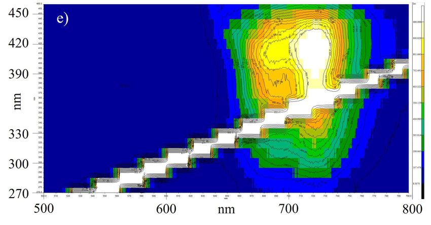
62                                                                                      ScienceAsia 47S (2021)

flasks, adjusted the volume to 5 ml, and stored in      (50 µl). After that, 2.5 M NaOH (250 µl) was added
a refrigerator for further analyses.                    into the mixture solution. The solution volume was
                                                        adjusted to 2 ml by ultra-pure water. The solution
Antioxidant activity assay                              was mixed and incubated for 30 min in a dark
The DPPH radical scavenging activity of the             room. The absorbance of the solution was measured
methanolic extracts obtained from coffee leaf sam-      at 510 nm by using a visible spectrophotometer.
ples were carried out using the minor modified          The calibration standard curve was prepared using
method of Saw et al [25]. Solution of 2,2-diphenyl-     catechin standard in the concentration range of 0
1-picrylhydrazyl (DPPH, 0.3 mM) was prepared in         to 100 ppm. The flavonoids content was presented
methanol. Aliquots of 200 µl of sample extracts         as milligram catechin equivalent per gram dried
were mixed with 1 ml of DPPH solution. Then, the        weight (mgCE/g dried weight).
solution’s volume was adjusted to 4 ml by ultra-
pure water (Millipore Milli-Q™ Reference Ultra-         Measurement of fluorescence spectra
pure Water Purification System), mixed by a vortex      A fluorescence spectrophotometer (LS55 model,
mixer (VM-10 model, Wisd laboratory instrument,         PerkinElmer, USA) equipped with a xenon lamp and
Korea), and then incubated for 30 min in a dark         a photomultiplier detector was used for fluorescence
room. Standard solutions of ascorbic acid in the        spectrum analysis of the leaf extract samples. The
concentration range of 0–0.018 mM were prepared.        excitation wavelength for detecting fluorescence
The absorbance of the solutions was measured at         signal of the studied solution was set from 270 to
515 nm [26] using a visible spectrophotometer           460 nm. The emission wavelength was monitored
(USB4000, Ocean Optics, USA). The DPPH radical          in the range of 500 to 800 nm. All the measure-
scavenging activities of all extracts were calculated   ments were performed in a standard fluorescence
using the ascorbic standard curve. The results          96-well plate with a scan rate at 800 nm/min and
were presented as milligram ascorbic acid equiva-       10 nm bandwidths for the emission and excitation
lent per gram dried coffee leaf weight (mgAAE/g         monochromators.
dried weight).
                                                        Statistical and multivariate analysis
Total phenolic content                                  The spectra of 3D-fluorescence contour plot of all
Analyses of the total phenolic content in coffee leaf   sample extracts were investigated for selecting the
extracts were carried out using the method pro-         excitation wavelengths and the range of emission
posed by Haile et al [27] with minor modification.      wavelength. The excitation wavelengths producing
Briefly, Folin-Ciocalteu reagent (10% w/v) dissolved    different fluorescence intensities in the range of
in methanol was used as reagent. The leaf extract       500–790 nm were selected for further statistical
sample (150 µl) was mixed with the Folin-Ciocalteu      and multivariate analyses. Then, the data were
reagent (125 µl) and incubated for 5 min. Then,         filtered using interquartile range before processing.
the solution was mixed with 1.25 ml of Na2 CO3          Normalization was performed by the sample median
(7% w/v) and adjusted the volume by using ultra-        method. The fluorescence intensities obtained from
pure water to 3 ml. The solution was mixed and          the selected excitation wavelengths of all extracts
then incubated in a dark room for 1 h. After that,      were processed by a PCA to study the natural clus-
the absorbance of the solution was measured at          tering of the samples. Statistical analyses were per-
765 nm using a visible spectrophotometer. Different     formed by mean of the MetaboAnalyst® 4.0 online
concentrations of gallic acid standard solution were    program running by R-script chemometrics [29].
prepared in the range of 0 to 100 ppm. The total        The PCA clustering result obtained from the data
phenolic content was presented as milligram gallic      set of fluorescence spectra was compared with that
acid equivalent per gram dried coffee leaf weight       obtained from the data set of chemical contents
(mgGAE/g dried weight).                                 based on antioxidant activity, total phenolic content,
                                                        and total flavonoid content of coffee leave samples.
Total flavonoid content                                      Then, hierarchical cluster analysis (HCA) in the
Analyses of total flavonoids content in coffee leaf     form of a dendrogram was also used to study the
extracts were carried out using the method pro-         interrelationships between samples. Two impor-
posed by Phuyal et al [28] with minor modification.     tant parameters were considered for performing
The leaf extract sample (200 µl) was mixed with         HCA. The first one was similarity measure includ-
0.55 M of AlCl3 (100 µl) and, then, 3.0 M of NaNO2      ing Euclidean distance, Pearson’s correlation, and

www.scienceasia.org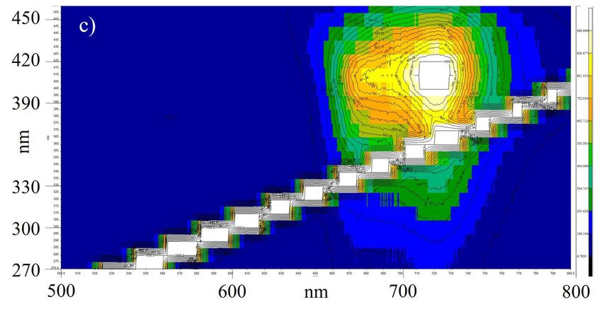
ScienceAsia 47S (2021)                                                                                                                                 63

Spearman’s rank correlation. The other parameter                                                                          a)
was clustering algorithms including average link-                                              0.45
age, complete linkage, single linkage, and Ward’s                                              0.40

                                                            Antioxidant activity (mgAAE/g)
linkage.                                                                                       0.35
    Finally, partial least squares - discriminant anal-                                        0.30
ysis (PLS-DA) was processed to develop a model                                                 0.25
for classification of the membership. Leave-one-                                               0.20
out cross validation (LOOCV) was used for cross                                                0.15
validation. The quality assessments of Q2 and R2
                                                                                               0.10
calculated via cross-validation (CV) were used for
                                                                                               0.05
the estimation of the qualitative measurement of the
                                                                                               0.00
model consistency between the predicted and the                                                        H306/1 ML 1/1    HDT    Liberica       Geisha
original data [30]. In addition, Variable Importance
                                                                                                                         b)
in Projection (VIP) values obtained from sum of                                                 70.0

                                                           Total phenolic contents (mgGAE/g)
squares of the PLS loadings, which consider the
                                                                                                60.0
amount of explained Y -variation in each dimen-
sion [31], were also calculated.                                                                50.0

                                                                                                40.0
RESULTS AND DISCUSSION
                                                                                                30.0
Multivariate analysis of antioxidant activity,
total flavonoid content, and total phenolic                                                     20.0

content                                                                                         10.0

The chemical contents, including antioxidant activ-                                              0.0
                                                                                                        H306/1 ML 1/1   HDT    Liberica       Geisha
ity, total flavonoid content, and total phenolic con-
tent, found in four coffee leaf extracts (C. liberica,                                                                    c)
                                                                                               0.30
C. arabica var. Geisha, Hibrido de timor, and hybrid
                                                           Total flavonoid contents (mgCE/g)

H306/1 ML1/1) are shown in Fig. 1. The chemical                                                0.25
contents found in individual coffee leaf taxa seem
                                                                                               0.20
to be different. The 2D-scores plot between the
selected principal components (PCs) obtained from                                              0.15
the PCA using the chemical contents of the samples
are shown in Fig. 2. The result showed that the                                                0.10

C. liberica, C. arabica var. Geisha, and Hibrido de                                            0.05
timor samples could be clustered clearly according
to their chemical contents. The literature reported                                            0.00
                                                                                                       H306/1 ML 1/1    HDT    Liberica       Geisha
that the coffee beans of C. liberica presented higher
chemical contents (based on the result of the total       Fig. 1 Antioxidant activities (a), total phenolic content
phenol assay, the DPPH assay, and the ferric re-          (b), and total flavonoid content (c) found in four coffee
ducing antioxidant power (FRAP) assay) than that          leaf extracts: C. liberica (Liberica), C. arabica var. Geisha
of C. robusta and C. arabica [32]. However, the           (Geisha), Hibrido de timor (HDT), and a hybrid H306/1
H306/1 ML1/1 (hybrid coffee taxon) could not be           ML1/1.
clustered clearly from the C. liberica and C. arabica
var. Geisha. Therefore, the data set from other
chemical factors might be required for classification     and H306/1 ML1/1) are shown in Fig. 3. The
of hybrid coffee taxa.                                    contour line patterns of each coffee leaf extract
                                                          were different. These fluorescence results might be
Fluorescence spectral characteristics of coffee           due to the differences of concentration of chemical
leave extracts                                            composition in the extract samples as reported by
Fluorescence spectra with emission spectra in hor-        Yisak et al [32]. Besides, it could be observed
izontal axis and excitation spectra in vertical axis      that excitation wavelength at 420 nm provided high
of the methanolic coffee leaf extracts (C. liberica,      intensity of fluorescence signal. The fluorescence
C. congensis, C. arabica var. Geisha, Hibrido de timor,   intensities of each sample obtained from excitation

                                                                                                                                      www.scienceasia.org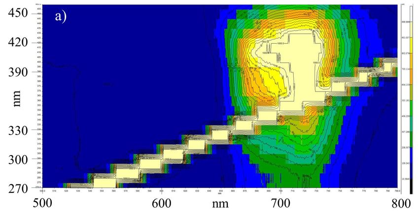
64                                                                                                 ScienceAsia 47S (2021)

Fig. 2 The 2D (a) and 3D-scores plot (b) between the selected principal components (PCs) obtained from the principal
component analysis (PCA) according to the chemical contents of the samples: C. liberica (+), C. arabica var. Geisha
( × ), Hibrido de timor (4), and hybrid H306/1 ML1/1 (3).

Fig. 3 Contour plots of the fluorescence spectra with emission spectra in horizontal axis and excitation spectra in
vertical axis of the methanolic coffee leaf extracts: C. liberica (a), C. congensis (b), C. arabica var. Geisha (c), Hibrido
de timor (d), and a hybrid H306/1 ML1/1 (e).

www.scienceasia.org
ScienceAsia 47S (2021) 65
tained from excitation wavelength at 300 nm of
individual coffee leaf taxa could be performed us-
ing partial least squares-discriminant analysis (PLS-
DA). Variable Importance in Projection (VIP) values
obtained from sum of squares of the PLS load-
ings and performance of classification are shown
in Fig. 5. The VIP values presented that the fluo-
rescence intensities obtained from emission wave-
lengths from 580 to 620 nm had influence on
classification of coffee taxa. The results might be
affected by alkaloids found in coffee, such as caf-
feine, theobromine, and trigonelline, as their max-
imum UV-visible excitation bands were obtained
at 272.89 nm, 272.73 nm, and 264.59 nm, re-
spectively [32, 33]. The amount of those alkaloids
in green coffee beans is influenced by numerous
factors such as coffee variety, genetic properties of
the cultivars, and environment [34]. Moreover,
secondary metabolites in coffee leaves that might
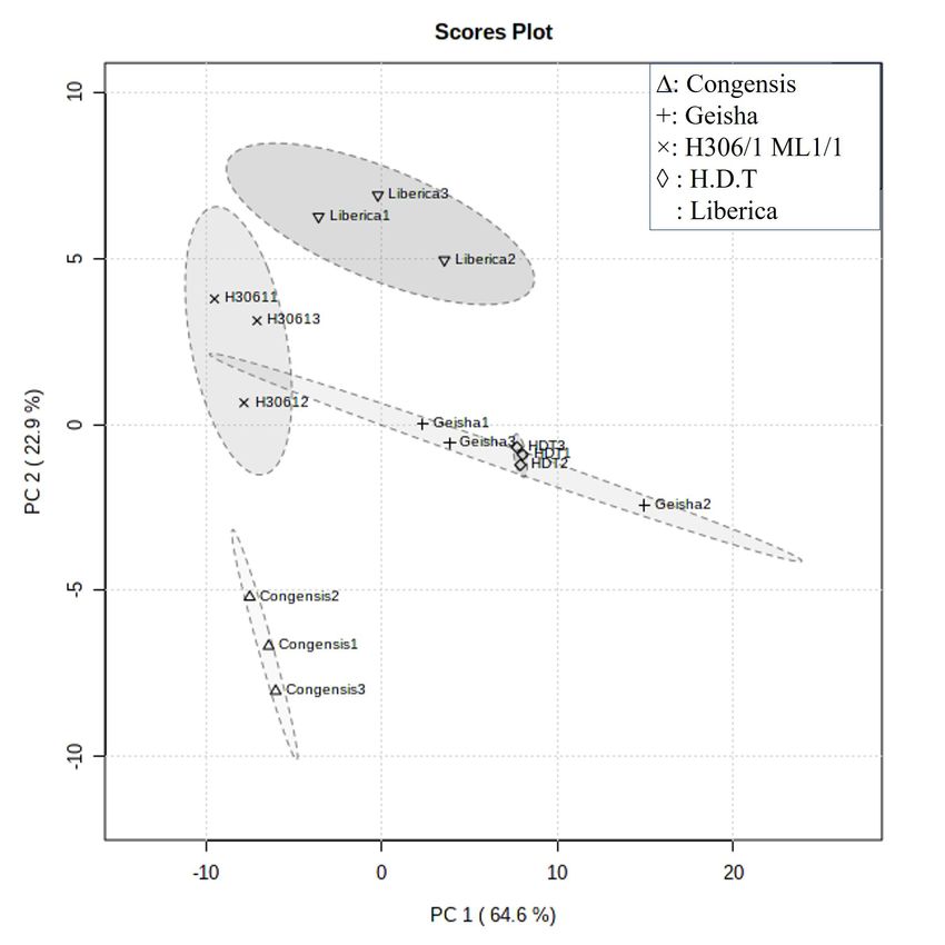
Fig. 4 The scores plot between the selected principal influence the clustering result are chlorogenic acid
components (PC1 × PC2) of raw spectra obtained from (5-CQA) and mangiferin (phenolic compounds) be-
using excitation wavelengths at 300 nm: C. liberica (Ï), cause both compounds exhibit a broad spectral
C. congensis (4), C. arabica var. Geisha (+), Hibrido de range with peaks at about 425 and 520 nm, re-
timor (3), and hybrid H306/1 ML1/1 ( × ). spectively. Other compounds of influence might
be flavins, flavonoids, some terpenoids, polyacety-
lene, isoquinoline, and some alkaloids because these
wavelength at 300, 330, 390, 420, and 450 nm compounds emit fluorescence in the red region of
and the emission range from 500 to 790 nm were the visible spectrum [20]. The quality assessments
selected to perform multivariate analysis for distin- of Q2 and R2 calculated via Leave-one-out cross-
guishing coffee taxa, based on the differences of validation (LOOCV) were 0.6 and 0.8, respectively
fluorescence intensity. (Fig. 5). The red star in Fig. 5 indicates the best
classifier. The literature suggests that R2 value of
Multivariate analysis of fluorescence spectra 0.67 is substantial model [35]. Although Q2 has no
A PCA of the selected fluorescence intensities was standard of comparison or critical value for inferring
processed via the MetaboAnalyst® 4.0 (web on- significance, an empirically inferred value of ¾ 0.4
line) to examine the natural clustering of the coffee is acceptable for a biological model [36]. The Q2 is
taxa. The fluorescence intensities obtained from the close to the R2 when the PLS built on a training set is
combination of emission wavelengths and excitation applied to a test set. It means that PLS model works
wavelengths were assigned as data sets. The scores independently of the specific data used to train the
plot between the selected PCs of individual spectra PLS model. Therefore, there is a tendency to use
obtained from using several excitation wavelengths the fluorescence spectra of methanolic-coffee leaf
was considered. It was found that C. liberica, extracts obtained from the excitation wavelength at
C. arabica var. Geisha, hybrid H306/1ML 1/1, and 300 nm, and the emission range from 500 to 790 nm
C. congensis could be clustered clearly using fluo- for identification of coffee taxa.
rescence intensity from the excitation wavelength An optimized dendrogram of raw fluorescence
at 300 nm, except Hibrido de timor coffee that spectra of coffee leaf extracts (emission wavelength
showed overlapping with C. arabica var. Geisha at 300 nm), using Euclidean distance as similarity
(Fig. 4). Using fluorescence spectra obtained from measure and average linkage as clustering algo-
excitation wavelength at 300 nm tended to separate rithm is shown in Fig. 6. The groups of C. liberica,
the data set as compared with the results of chemical hybrid H306/1ML 1/1, and C. congensis were well
contents, which could separate only three coffee separated. However, the groups of C. arabica var.
taxa. Geisha and Hibrido de timor were overlapped. The
The model using fluorescence intensities ob- natural clustering of the samples presented as a den-
www.scienceasia.org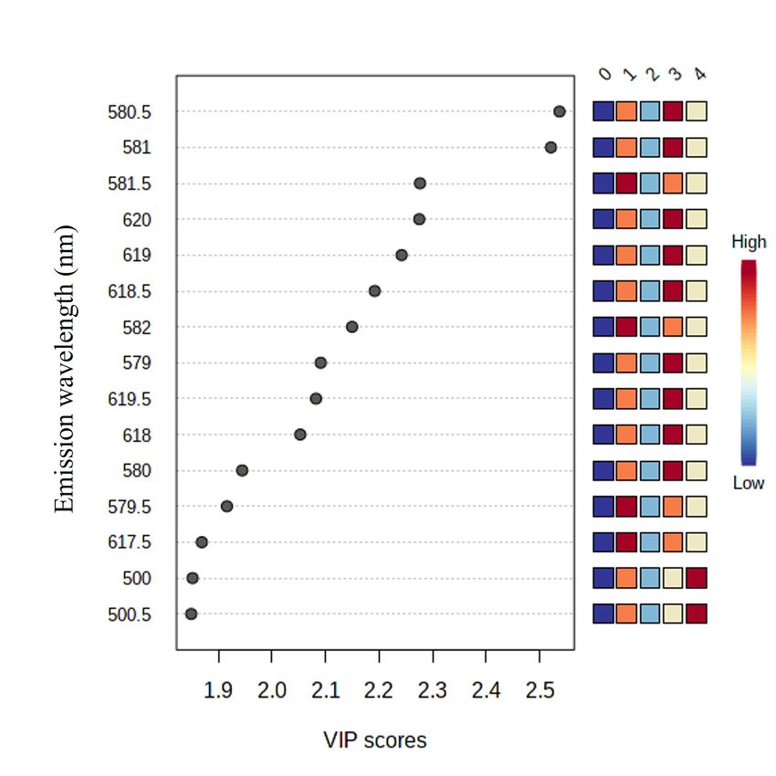
66                                                                                                      ScienceAsia 47S (2021)

                                                                                      Accuracy          R2       Q2
                                                            1.0
                                                            0.9
                                                            0.8
                                                            0.7

                                              Performance
                                                            0.6                                  *
                                                            0.5
                                                            0.4
                                                            0.3
                                                            0.2
                                                            0.1
                                                            0.0
                                                                       1                  2                      3
                                                                                 Number of components

Fig. 5 Variable importance in projection (VIP) values obtained from sum of squares of the PLS loadings (a) and
performance of classification; (b) C. congensis (group 0), C. arabica var. Geisha (group 1), hybrid H306/1 ML1/1
(group 2), Hibrido de timor (group 3), and C. liberica (group 4).

                                                                  orescence spectroscopy coupled with chemometric
       0
       1
                                                HDT3
                                                                  techniques for data clustering and identification is
       2                                        HDT2
                                                                  preferred because the analytical methods are chem-
       3                                        HDT1              ical and time consuming. On the other hand, the
       4
                                                Geisha3           fluorescence technique requires only a few easy
                                                Geisha1           steps of sample extraction. In addition, fluorescence
                                                Geisha2           spectrum is represented from various autofluores-
                                                Liberica3         cent metabolites in sample. It is proved to be simple,
                                                Liberica1         high sensitivity, and less sample consumption [37].
                                                Liberica2

                                                Congesis3
                                                                  CONCLUSION
                                                Congesis3

                                                Congesis3         Multivariate analysis of the data of chemical con-
                                                H306/1 1          tents and fluorescence spectra for simple classifi-
                                                H306/1 2
                                                                  cation of some coffee leave taxa extracted with
                                                H306/1 3
                                                                  methanol was studied. Principal component anal-
                                                                  ysis according to chemical contents obtained from
  15                  10       5              0                   antioxidant activity, total flavonoid content, and
                                                                  total phenolic content could cluster clearly between
Fig. 6 Clustering result shown as dendrogram (distance            Hibrido de timor, C. liberica and C. arabica var.
measure using Euclidean and clustering algorithm using            Geisha. The PCA score plot of fluorescence spectra
average): C. congensis (0), C. arabica var. Geisha (1),           obtained from excitation wavelength at 300 nm
hybrid H306/1 ML1/1 (2), Hibrido de timor, HDT (3),               also separated clearly between C. liberica, hybrid
and C. liberica (4).                                              H306/1ML 1/1, and C. congensis. It agrees with the
                                                                  result of the hierarchical cluster analysis presented
                                                                  as a dendrogram. Due to the PLS model works
drogram agrees with the PCA scores plot between                   independently on specific data, there is a tendency
the selected principal components (PC1 × PC2) of                  to use the fluorescence spectra of methanolic ex-
raw spectra obtained from excitation wavelength at                tracts obtained from the excitation wavelength at
300 nm. This result might be due to genetic factors.              300 nm and the emission range from 500 to 790 nm
Both C. arabica var. Geisha and Hibrido de timor                  for coffee classification study. Fluorescence spec-
coffee originated from same Arabica variety.                      troscopy coupled with chemometric techniques is
    Multivariate analysis of both the chemical con-               recommended as screening methods for coffee taxa
tents and the fluorescence spectra obtained from                  identification because it requires a few easy steps of
coffee leaf extracts could be applied for study of                sample extraction. It is proved to be simple, high
the natural clustering in data. However, the flu-                 sensitivity, and less sample consumption.

www.scienceasia.org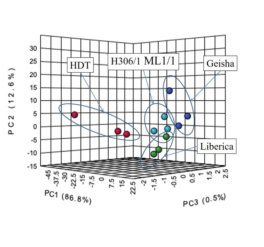
ScienceAsia 47S (2021)                                                                                                  67

Acknowledgements: This work was financial supported                 croorganisms by capture and intrinsic fluorescence
by the National Research Council of Thailand, NRCT                  detection. Biosens Bioelectron 18, 521–527.
(17549). We would also thank the Mae Lod Royal Agricul-       13.   Gorbunov MY, Shirsin E, Nikonova E, Fadeev VV,
tural Research Station, Royal Project Foundation, Chiang            Falkowski PG (2020) A multi-spectral fluorescence
Mai (Thailand) for coffee leaf samples and Mr. Jakkarach            induction and relaxation (FIRe) technique for phys-
Sa-udon for coffee leaf sampling and fruitful discussion.           iological and taxonomic analysis of phytoplankton
                                                                    communities. Mar Ecol Prog Ser 644, 1–13.
                                                              14.   Tourkya B, Boubellouta T, Dufour E, Leriche F (2009)
                                                                    Fluorescence spectroscopy as a promising tool for
REFERENCES
                                                                    a polyphasic approach to pseudomonad taxonomy.
 1. Chamyuang S, Owatworakit A, Intatha U, Duangphet                Curr Microbiol 58, 39–46.
    S (2021) Coffee pectin production: An alternative         15.   Meyer JM, Geoffroy VA, Baida N, Gardan L, Izard
    way for agricultural waste management in coffee                 D, Lemanceau P, Palleroni NJ (2002) Siderophore
    farms. ScienceAsia 47S, 90–95.                                  typing, a powerful tool for the identification of flu-
 2. Anthony F, Bertrand B, Quiros O, Wilches A, Lasher-             orescent and nonfluorescent pseudomonads. Appl
    mes P, Berthaud J, Charrier A (2001) Genetic diver-             Environ Microbiol 68, 2745–2753.
    sity of wild coffee (Coffea arabica L.) using molecular   16.   MacIntyre HL, Lawrenz E, Richardson TL (2010)
    markers. Euphytica 118, 53–65.                                  Taxonomic discrimination of phytoplankton by spec-
 3. Mishra MK, Huded AKC, Jingade P (2020) Assess-                  tral fluorescence. In: David JS, Ondrej P, Michael AB
    ment of the suitability of molecular SCoT markers               (eds) Chlorophyll a Fluorescence in Aquatic Sciences:
    for genetic analysis of coffee species. Botanica 26,            Methods and Applications, Springer, Dordrecht, pp
    184–196.                                                        129–169.
 4. Martín MJ, Pablos F, González AG (1998) Discrim-          17.   Pollastrini M, Holland V, Brüggemann W, Bruelheide
    ination between arabica and robusta green coffee                H, Dănilă I, Jaroszewicz B, Bussotti F (2016) Tax-
    varieties according to their chemical composition.              onomic and ecological relevance of the chlorophyll
    Talanta 46, 1259–1264.                                          a fluorescence signature of tree species in mixed
 5. Japon-Lujan R, Ruiz-Jiménez J, Luque de Castro MD               European forests. New Phytol 212, 51–65.
    (2006) Discrimination and classification of olive tree    18.   Aslam I, Afridi MSK (2018) Pharmacognostic char-
    varieties and cultivation zones by biophenol con-               acterization of Beaumontia grandiflora (Roxb.) Wall.
    tents. J Agric Food Chem 54, 9706–9712.                         leaf for taxonomic identification for quality control
 6. Talhaoui N, Gómez-Caravaca AM, Roldan C, Leon                   of a drug. J Appl Res Med Aromat Plants 8, 53–59.
    L, De la Rosa R, Fernandez-Gutierrez A, Segura-           19.   Tyystjärvi E, Keränen M, Koski A, Nevalainen O, Aro
    Carretero A (2015) Chemometric analysis for the                 EM (1998) Chlorophyll fluorescence can be used to
    evaluation of phenolic patterns in olive leaves from            identify plant species automatically. In: Shaul M, Wei
    six cultivars at different growth stages. J Agric Food          MZ, Vladimir C, Garab G (eds) Photosynthesis: Mech-
    Chem 63, 1722–1729.                                             anisms and Effects, Springer Netherlands, Dordrecht,
 7. Amargianitaki M, Spyros A (2017) NMR-based                      pp 3857–3860.
    metabolomics in wine quality control and authenti-        20.   Talamond P, Verdeil JL, Conéjéro G (2015) Secondary
    cation. Chem Bio Technol Agric 4, ID 9.                         metabolite localization by autofluorescence in living
 8. Kucharska-Ambrożej K, Karpinska J (2020) The ap-               plant cells. Molecules 20, 5024–5037.
    plication of spectroscopic techniques in combina-         21.   Pawar HA, Kamat SR (2014) Chemometrics and its
    tion with chemometrics for detection adulteration of            application in pharmaceutical field. J Phys Chem
    some herbs and spices. Microchem J 153, ID 104278.              Biophys 4, ID 169.
 9. Khamchum C, Punsuvon V, Kasemsumran S, Sut-               22.   Tunnicliffe JM, Cowan T, Shearer J (2015) Chloro-
    tiwijitpukdee N (2013) A feasibility study of oil               genic acid in whole body and tissue-specific glucose
    content and fatty acid composition of seed powder               regulation. In: Preedy VR (ed) Coffee in Health and
    and seed oil of Pongamia pinnata by near infrared               Disease Prevention, Academic Press, UK, pp 777–785.
    spectroscopy. ScienceAsia 39, 384–391.                    23.   Sousa JS, Neves JCL, Martinez HEP, Alvarez VHV
10. Mees C, Souard F, Delporte C, Deconinck E, Stoffelen            (2018) Relationship between coffee leaf analysis and
    P, Stévigny C, De Braekeleer K (2018) Identifica-               soil chemical analysis. Rev Bras Cienc Solo 42.
    tion of coffee leaves using FT-NIR spectroscopy and       24.   Tamimi YN, Silva JA, Yost RS, Hue NV (1997)
    SIMCA. Talanta 177, 4–11.                                       Adequate nutrient levels in soils and plants in
11. Zhang C, Shen T, Liu F, He Y (2018) Identification              Hawaii (general guide). In: Agronomy and Soils AS-
    of coffee varieties using laser-induced breakdown               3, CTAHR, University of Hawaii at Manoa, Honolulu,
    spectroscopy and chemometrics. Sensors 18, ID 95.               USA.
12. Mason HY, Lloyd C, Dice M, Sinclair R, Ellis Jr W,        25.   Saw AKC, Yam WS, Wong KC, Lai CS (2015) A com-
    Powers L (2003) Taxonomic identification of mi-                 parative study of the volatile constituents of south-

                                                                                                         www.scienceasia.org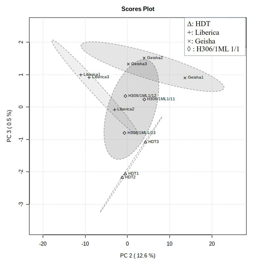
68                                                                                               ScienceAsia 47S (2021)

      east asian Coffea arabica, Coffea liberica and Coffea         prediction (VIP) and of the selectivity ratio (SR)
      robusta green beans and their antioxidant activities.         variable selection methods in the analysis of three
      J Essent Oil Bear Plants 18, 64–73.                           different data sets. J Chemom 29, 528–536.
26.   Brand-Williams W, Cuvelier M, Berset C (1995) Use       32.   Yisak H, Redi-Abshiro M, Chandravanshi BS (2018)
      of a free radical method to evaluate antioxidant              New fluorescence spectroscopic method for the si-
      activity. LWT Food Sci Technol 28, 25–30.                     multaneous determination of alkaloids in aqueous
27.   Haile M, Bae HM, Kang WH (2020) Comparison of                 extract of green coffee beans. Chem Centl J 12, 59.
      the antioxidant activities and volatile compounds of    33.   Mubarak A, Croft KD, Bondonno CP, Din NS (2019)
      coffee beans obtained using digestive bio-processing          Comparison of liberica and arabica coffee: chloro-
      (elephant dung coffee) and commonly known pro-                genic acid, caffeine, total phenolic and DPPH radical
      cessing methods. Antioxidants 9, ID 408.                      scavenging activity. Asian J Agric Biol 7, 130–136.
28.   Phuyal N, Jha PK, Raturi PP, Rajbhandary S (2020)       34.   Navarra G, Moschetti M, Guarrasi V, Mangione MR,
      Total phenolic, flavonoid contents, and antioxi-              Leone M (2017) Simultaneous determination of caf-
      dant activities of fruit, seed, and bark extracts of          feine and chlorogenic acids in green coffee by UV/Vis
      Zanthoxylum armatum DC. Sci World J 2020, ID                  spectroscopy. J Chem 2017, ID 6435086.
      8780704.                                                35.   Peng DX, Lai F (2012) Using partial least squares in
29.   Chong J, Wishart DS, Xia J (2019) Using Metabo-               operations management research: A practical guide-
      Analyst 4.0 for comprehensive and integrative                 line and summary of past research. J Oper Manag 30,
      metabolomics data analysis. Curr Protoc Bioinformat-          467–480.
      ics 68, ID 86.                                          36.   Worley B, Powers R (2013) Multivariate analysis in
30.   Westerhuis JA, Velzen EJ, Hoefsloot HC, Smilde                metabolomics. Curr Metabolomics 1, 92–107.
      AK (2008) Discriminant Q2 (DQ 2) for improved           37.   Gaddam RR, Narayan R, Raju KVSN (2016) Fluo-
      discrimination in PLSDA models. Metabolomics 4,               rescence spectroscopy of nanofillers and their poly-
      293–296.                                                      mer nanocomposites. In: Ponnamma D, Rouxel D,
31.   Farrésa M, Platikanova Y, Tsakovskib L, Taulera R             Thomas S (eds) Spectroscopy of Polymer Nanocompos-
      (2015) Comparison of the variable importance in               ites, Elsevier, Oxford, United Kingdom, pp 158–180.

www.scienceasia.org
You can also read