Immunohistochemical Changes after Metoclopramide Administration in Rat Brain Cells - MDPI
←
→
Page content transcription
If your browser does not render page correctly, please read the page content below
Proceedings

Immunohistochemical Changes after Metoclopramide
Administration in Rat Brain Cells †
Seren Gülşen Gürgen 1,*, Ayşe Tuç Yücel 1, Nurcan Umur 1 and Gülce Naz Yazıcı 2
 1 School of Vocational Health Service, Manisa Celal Bayar University, Manisa 45140, Turkey;
   aysetuc@gmail.com (A.T.Y.); nurcanumur@gmail.com (N.U.)
 2 Faculty of Medicine, Erzincan University, Erzincan 24100, Turkey; gulcenazyazici.ank@gmail.com

 * Correspondence: serengurgen@yahoo.com
 † Presented at the 2nd International Cell Death Research Congress, Izmir, Turkey, 1–4 November 2018.

 Published: 6 December 2018

 Abstract: Metoclopramide, used as an anti-emetic drug in clinical practice, has recently also begun
 being used to establish hyperprolactinemic effects in the breastfeeding. The purpose of this study
 was to investigate the potential side-effects of metoclopramide applied in the lactation period in the
 central nervous system of infant rats. 18 female albino Wistar rats that had just given birth were
 divided into 3 groups together with their pups: Healthy controls, low-dose metoclopramide (10
 mg/kg, twice per day i.p.) and a high-dose metoclopramide group (45 mg/kg, twice per day i.p.).
 Brain tissues from 6 pups from each mother were harvested at the end of the 21st day.
 Immunohistochemical technique was performed using dopamine D2 receptor (DRD2), brain
 derived neurotrophic factor (BDNF) and neural growth factor (NGF), markers of extrapyramidal
 reaction in the brain, as signal molecules. Based on immunohistochemical results, DRD2 expression
 decreased only in the external pyramidal layer neurons in the high-dose infant group. Strong BDNF
 reaction was determined in pyramidal neurons in all layers in the control infant group, and
 decreased reaction was observed in the high- and low-dose groups. No significant difference was
 observed in NGF expression between the three groups. Since high-dose metoclopramide caused a
 decrease in DRD2 expression in the external pyramidal layer in the prefrontal cortex, and since both
 high and low doses reduced BDNF expression, care needs to be taken with the use of
 metoclopramide in the lactation period due to the possibility of extrapyramidal reactions in infants.

 Keywords: brain; extrapyramidal reactions; metoclopramide

1. Introduction
      Metoclopramide (Metpamid), sulpiride (Zegerid), domperidone (Motilium), chlorpromazine
Metoclopramide easily passes the blood-brain barrier and can cause side-effects such as
extrapyramidal system movement disorders (extrapyramidal reactions) [1]. Metoclopramide is used
to increase lactation by breastfeeding mothers and is known to pass into milk. A milk/plasma ratio
of 1.8–1.9 has been reported [2]. Metoclopramide has been observed to lead to fatigue, headache,
anxiety, intestinal disorders and particularly depression and extrapyramidal side-effects (1%) in
mothers [3]. In contrast to the side-effects in the mother, whether it causes side-effects in the newborn
is still the subject of debate. According to some authors, no side-effects are observed, while others
regard metoclopramide passing into milk as a risk [4]. The purpose of this study was to investigate
distribution of expression of dopamine receptor D2 (DRD2), an extrapyramidal reaction marker,
brain-derived neurotrophic factor (BDNF) and neural growth factor (NGF) in the cerebral cortex and
serum of newborn pups of mother rats administered metoclopramide during the lactation period.

Proceedings 2018, 2, 1545; doi:10.3390/proceedings2251545                  www.mdpi.com/journal/proceedings
Proceedings 2018, 2, 1545 2 of 4
2. Materials and Methods
Young female Wistar Albino rats that had just given birth were used. All animal experiments
were carried out in accordance with the European Communities Council Directive of 24 November
1986 (86/609/EEC) and were approved by the Animal Care Committee of Celal Bayar University.
The study began with 18 young female Wistar-albino rats and their 1-day-old pups. Mother rats
in the lactation period were administrated metoclopramide (Sigma-Aldrich, St. Louis, MO, USA) for
hyperprolactinemia. Mother rats and their pups were divided into 3 experimental groups containing
6 rats each. Experimental groups consisted of a control group, a low-dose metoclopramide group (10
mg/kg, 21 days, 2 doses i.p. per day) and a high-dose metoclopramide group (45 mg/kg, 21 days, 2
doses i.p. per day). All infants were sacrificed at 10:00 a.m. at the end of the 21st day. Brain tissues
were rapidly excised and placed in 10% neutral formalin for histological investigation.
Immunohistochemical Methods: At the end of the experiment, pups’ brain prefrontal cortex
tissues were harvested. Tissue samples processed for embedding in paraffin wax according to routine
protocols. Paraffin blocks of the tissue were cut in 5 µm sections and prepared for
immunohistochemical staining. The slides were stained with primary antibodies DRD2 (Mouse
monoclonal, Santa Cruz, CA, USA), BDNF (Rabbit polyclonal antibody, Santa Cruz, CA, USA) and
NGF (Mouse monoclonal antibody, Abcam, Cambridge, MA, USA) using immunohistochemical
methods. The slides were analyzed under an Olympus (CX41, Tokyo, Japan) light microscope. Two
observers blinded to the experimental information evaluated the immunolabeling scores
independently. The staining intensity of the slides with their immunohistochemical protocol was
graded semi-quantitatively, and the HSCORE was calculated using the following equation: HSCORE
= ΣPi (i + 1), where i is the intensity of staining with a value of 1, 2 or 3 (weak, moderate or strong,
respectively), and where Pi is the percentage of stained cells for each intensity, varying from 0 to
100%. The study groups were analyzed using Kruskal-Wallis nonparametric tests. All statistical
analyses were performed using SPSS version 15.0 for Windows software.
3. Results
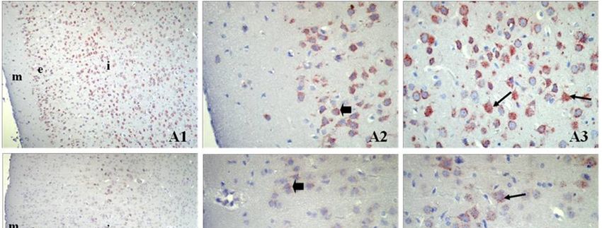
Infant Brain Tissue DRD2, NGF and BDNF Immunochemistry Findings Examination of the
infant prefrontal cortex region revealed no DRD2 expression in the molecular layer in the control
group. However, strong cytoplasmic and granular form expression was observed in the pyramidal
neurons in the external pyramidal layer (p < 0.05) (Figure 1(A1,A2)). Similarly, quite powerful
cytoplasmic and granular form immunoreaction was observed in pyramidal neurons in the internal
pyramidal layer (p < 0.05) (Figure 1(A3)). In the low-dose metoclopramide group, expression was
weak in the molecular and external pyramidal layer of the prefrontal cortex, while moderately and
granular form reaction was noted in some neurons in the internal pyramidal layer (Figure
1(B1,B2,B3)). In the high-dose group, quite strong granular form DRD2 expression was determined
in the internal pyramidal cell layer (p < 0.05), but less than that of the control group in the external
pyramidal layer of the prefrontal cortex (Figure 1(C1,C2,C3)). BDNF expression in the prefrontal
cortex was quite strong in neurons in the external pyramidal layer in the control group, and moderate
in the other two groups (p < 0.05). (Figure not showen). Expression ranging from weak to moderate
was observed in NGF in all groups. There was no significant difference between the groups (p > 0.05).
(Figure not showen) (Table 1).Proceedings 2018, 2, 1545 3 of 4
Figure 1. Prefrontal cortex DRD2 immunostaining. Control (A), low-dose metoclopramide (B), high-
dose metoclopramide (C). m: molecular layer, e: external pyramidal layer, i: internal pyramidal layer
(1) ×100, : external pyramidal neurons, : internal pyramidal neurons, external pyramidal layer (2),
internal pyramidal layer (3). ×400.
Table 1. DRD2, BDNF, NGF H-score levels in the brain prefrontal cortex of infant rats in the control
and low- and high-dose metoclopramide groups.
DRD2 BDNF NGF
H-Score
EP DRD2 * IP DRD2 * EP BDNF * EP NGF
Control 227.3 ± 21.4 235.3 ± 10.6 297.3 ± 31.1 176.7 ± 7
Low-dose 121 ± 3.8 150.7 ± 5.2 168.6 ± 8.6 123 ± 19.3
High-dose 131.3 ± 5.2 218 ± 7.5 164.6 ± 12.1 118.5 ± 5.6
EP: External pyramidal layer, IP: Internal pyramidal layer, DRD2: Dopamine D2 receptor, BDNF:
Brain derived neurotrophic factor, NGF: Neural growth factor * p < 0.05 one-way analysis of variance
ANOVA, data are expressed as mean ± SD.
4. Discussion
Metoclopramide is known to pass into milk [2]. Some authors describe metoclopramide as an
“inverse neuroleptic” [5]. Some authors classify metoclopramide as a neuroleptic but as an
antipsychotic agent with a weak effect [6]. In this study DRD2 decreasing in both internal and external
pyramidal neurons in the low-dose metoclopramide group and in the external pyramidal neurons in
particular in the high-dose group shows that this derives from metoclopramide’s dopamine D2
receptor antagonistic effect [7]. BDNF, which shares a high degree of structural homology with NGF,
is a member of the neurotropin family of molecules, such as neurotropin-3 (NT-3) and neurotropin-
4 (NT-4). NGF has been shown to reduce neuron degeneration in rats and to increase peripheral nerve
regeneration [6]. Antipsychotic drugs are also known to alter neurotropin levels in the brain and to
cause various side-effects (such as extrapyramidal and anticholinergic symptoms, tardive dyskinesia
and hyperprolactinemia). Additionally, recent studies using animal models of schizophrenia haveProceedings 2018, 2, 1545 4 of 4
shown that NGF and BDNF are abnormally regulated in the central nervous system. However, when
we examined the distribution in the brain of the NGF and BDNF signal molecules using
immunohistochemistry on the basis of metoclopramide’s classification as a neuroleptic but weak
antipsychotic agent [6], we determined that the BDNF molecule decreased in the high- and low-dose
groups compared to the control group. This is in agreement with studies reporting that antipsychotic
drugs cause a decrease in BDNF expression in the brain.
5. Conclusions
In conclusion, since metoclopramide at high doses causes a decrease in DRD2 expression in
pyramidal neurons in the internal pyramidal layer in the prefrontal cortex, and since it also reduces
BDNF expression in the prefrontal cortex at both high and low doses, we conclude that care should
be taken over the use of metoclopramide in the lactation period due to potential extrapyramidal
reactions.
Conflict Interests: The authors declare no conflict of interest.
References
1. Miller, L.G.; Jankovic, J. Metoclopramide-induced movement disorders. Clinical findings with a review of
the literature. Arch. Int. Med. 1989, 149, 2486–2492.
2. Kauppila, A.; Arvela, P.; Koivisto, M.; Kivinen, S.; Ylikorkala, O.; Pelkonen, O. Metoclopramide and breast
feeding: Transfer into milk and the newborn. Eur. J. Clin. Pharmacol. 1983, 25, 819–823.
3. Ehrenkranz, R.A.; Ackerman, B.A. Metoclopramide effect on faltering milk production by mothers of
premature infants. Pediatrics 1986, 78, 614–620.
4. Gupta, A.P.; Gupta, P.K. Metoclopramide as a lactogogue. Clin. Pediatr. 1985, 24, 269–272.
5. Stanley, M.; Lautin, A.; Rotrosen, J.; Gershon, S.; Kleinberg, D. Metoclopramide: Antipsychotic efficacy of
a drug lacking potency in receptor models. Psychopharmacology 1980, 71, 219–225.
6. Nakra, B.R.; Bond, A.J.; Lader, M.H. Comparative psychotropic effects of metoclopramide and
prochlorperazine in normal subjects. J. Clin. Pharmacol. 1975, 15, 449–454.
7. Vallone, D.; Picetti, R.; Borrelli, E. Structure and function of dopamine receptors. Neurosci. Biobehav. Rev.
2000, 24, 125–132.
© 2018 by the authors. Licensee MDPI, Basel, Switzerland. This article is an open access
article distributed under the terms and conditions of the Creative Commons Attribution
(CC BY) license (http://creativecommons.org/licenses/by/4.0/).You can also read