In Situ-Based Gels for Nose to Brain Delivery for the Treatment of Neurological Diseases - MDPI
←
→
Page content transcription
If your browser does not render page correctly, please read the page content below
pharmaceutics

Review
In Situ-Based Gels for Nose to Brain Delivery for the
Treatment of Neurological Diseases
Blessing Atim Aderibigbe
 Department of Chemistry, University of Fort Hare, Alice Campus, Eastern Cape, Alice 5700, South Africa;
 baderibigbe@ufh.ac.za

 Received: 13 January 2018; Accepted: 22 February 2018; Published: 30 March 2018

 Abstract: In situ-based gel drug delivery systems that can bypass the blood-brain barrier, deliver
 the therapeutics to the desired site, reduce peripheral toxicity and control drug release kinetics
 have been developed. Some of the therapeutics used to treat neurological diseases suffer from poor
 bioavailability. Preclinical reports from several researchers have proven that the delivery of drugs
 to the brain via the nose-to-brain route using in situ gels holds great promise. However, safety
 issues on the toxicity of the nasal mucosa, transportation of the drugs to specific brain regions and
 determination of the required dose are factors that must be considered when designing these gels.
 This review will be focused on in situ-based gels that are used for the delivery of therapeutics via the
 nose-to-brain route, preclinical reports and challenges.

 Keywords: nose-to-brain delivery; neurological diseases; drug delivery systems; in situ-based gels;
 brain tumor; Alzheimer’s disease

1. Introduction
     Neurological diseases affect the peripheral and central nervous system. These include
the peripheral nerves, spinal cord, brain, cranial nerves, nerve roots, neuromuscular junction,
muscles, etc. [1]. Some examples of neurological disease are Parkinson’s disease, epilepsy, multiple
sclerosis, Alzheimer’s disease, cerebrovascular diseases, brain tumors, etc. [1]. Neurological diseases
affect millions of people worldwide. WHO (World Health Organization) estimated that 47.5 million
people globally are suffering from dementia, and over 7.7 million new cases are reported every
year [1]. The most common form of dementia is Alzheimer’s disease, which contributes to 70% of
the cases. There are several factors that contribute to neurological disease such as genetics, physical
injuries, infections, ageing, lifestyle, nutrition and environmental factors [2–7]. The treatment of
neurological diseases includes the administration of the therapeutics topically, orally and intravenously
and the use of device-based therapies such as deep brain stimulation, surgeries and rehabilitation [8].
Some approaches involve the direct delivery of the drug via injection into the brain, cerebrospinal fluid
or intranasal delivery. Some of these techniques are unsafe, invasive, local and short lasting [9,10].
Another approach to the treatment of neurological diseases is the repair of the central nervous system,
which involves the reconstruction of the damaged neural tissue. However, this approach is hampered
by neurodegeneration [11]. The blood-brain barrier also acts as a barrier that inhibits the delivery
of some therapeutic agents to the central nervous system and hinders drugs from passing through
the endothelial capillaries to the brain [9]. In order to overcome the aforementioned limitations,
gel-based drug delivery systems that can be administered via nose-to-brain routes have been developed.
This approach is non-invasive, enhances drug absorption with less systemic adverse effects and
bypasses BBB (blood-brain barrier). However, this approach has challenges such as the inability
to know the accurate dose of drug to be administered and naso-mucosal irritation resulting from
preservatives, additives and active ingredients added to the formulation that can cause loss of epithelial

Pharmaceutics 2018, 10, 40; doi:10.3390/pharmaceutics10020040            www.mdpi.com/journal/pharmaceutics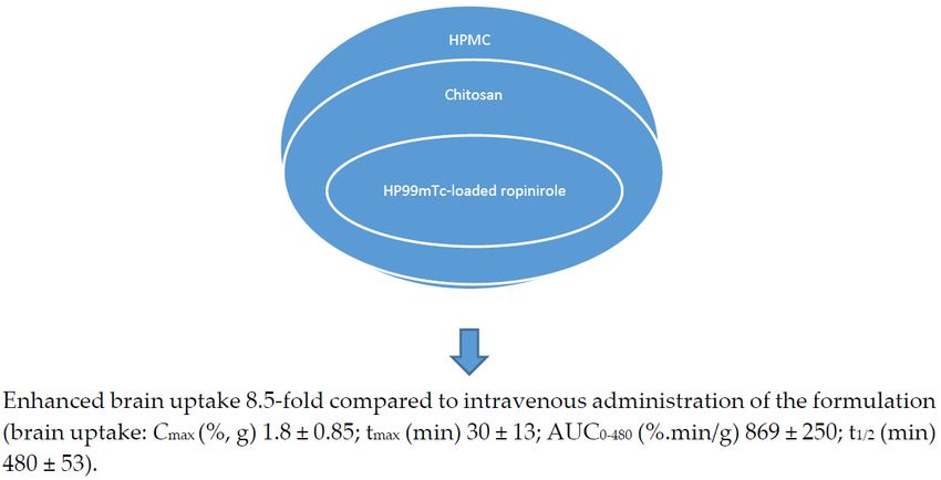
Pharmaceutics 2018, 10, 40                                                                           2 of 17

cell, shrinkage of the mucosal layer and loss of the ciliary layer [12]. Conditions such as allergy, flu,
etc., can also interfere with the drug absorption [12]. This review will be focused on the gel-based drug
delivery systems that are used for the delivery of therapeutics via the nose-to-brain route, preclinical
reports and challenges.

2. Anatomy of the Nose
      The nose is a primary organ that filters particles from the inspired air and provides immunologic
defense. The inspired air comes in contact with the olfactory nerves and provides the sense of smell,
which is closely associated with taste sensation [13]. The external opening of the nose is composed
of nasal bone and cartilage. The nasal cavity extends from the external opening of the nose to the
pharynx [14]. The internal part of the nose is composed of nasal septum that separates the nasal cavity
into the left and right side [13]. The nasal cavity contains epithelium-covered bone. The roof of the
nasal cavity contains a tissue known as the olfactory epithelium, which is useful for the sense of smell
because it contains sensory cells. It protrudes from its surface and is embedded in mucus secreted
by goblet cells in the epithelium. Olfactory sensory neurons are bipolar neurons at the surface of the
epithelium in which their dendrites face the interior space of the nasal cavity. They transmit sensory
signals to the brain [14]. Paranasal sinuses surround the nasal cavity and are characterized by hollow
structures within the bones of the face. They are lined with epithelium and classified as maxillary,
frontal, ethmoidal and sphenoidal sinuses [14]. The maxillary sinuses are the largest paranasal sinuses
and flank the nasal cavity to the left and right. They are connected to the nasal canal via a tiny passage
and allow the passage of air between them [14]. The frontal sinuses are separated from the nasal
cavity by a thin layer of bone. They do not connect to the nasal canal [14]. The ethmoidal sinuses are
also known as air cells. They differ in size and number in different people. The sphenoidal sinuses
are round with varied sizes. The nasal cavity is divided into the vestibular, turbinate and olfactory
regions [14]. The anterior part of the nose is the vestibular region. It is a very narrow part of the nasal
cavity and is composed of vibrissae, which help in filtering particles sizes larger than 10 µm from
the inhaled air [15]. The turbinate region is the principal site for the systematic absorption of drugs
delivered intranasally. It is lined with a pseudostratified columnar epithelium and composed of cells
such as ciliated, non-ciliated, basal and mucus-secreting cells [15]. Non-motile microvilli cover the
ciliated and non-ciliated cells, and they are useful for enhancing the surface area and comprise a region
where drug absorption occurs [15]. Motile cilia cover the cilia cells, and they are responsible for mucus
transport, resulting in mucociliary drug clearance especially in the highly ciliated middle and posterior
regions [15]. In the olfactory epithelium, the nerve cells project into the olfactory bulb of the brain
providing a connection between the brain and the external environment, and this connection is useful
for the transportation of drugs. Mucus covers the epithelium cells and traps unwanted particles.
The mucus contains water, mucin, protein and salts. The proteins it contains are albumin, lactoferrin,
immunoglobulin and lysozyme [15].

2.1. Mechanism of Nose-to-Brain Drug Delivery
     The nasal mucosa has emerged as a useful target tissue for drug delivery when compared to
oral drug administration resulting from its accessibility, high blood flow, large surface area, porous
endothelial membrane and its ability to avoid hepatic first pass metabolism [16–18]. However, the exact
mechanisms of drug delivery for nose-to-brain delivery is not well understood. The pathways
involving the combination of the cerebrospinal fluid, vasculature and lymphatic system have been
reported to be responsible for the transport of bioactive agents from the nasal cavity to the brain
(Figure 1). However, the properties of the therapeutics and delivery system can result in one
predominate pathway [19]. When the drug is deposited on the respiratory epithelium, the drug
is absorbed into systematic circulation and then to the central nervous system. Drug deposited on the
olfactory epithelia is transported to the central nervous system through paracellular or transcellular
transport via olfactory neurons or olfactory epithelial cells. Another route for the transportation of
Pharmaceutics 2018, 10, 40 3 of 17
Pharmaceutics
drug from the2018, 11, xto the brain is via the trigeminal nerves. The olfactory and trigeminal nerve system
nose 3 of 17
connects the brain and nasal cavity. These nerve systems are externally-accessible points of the brain,
major can
which routebeof intranasal
exploited deliverythe
to bypass is BBB
the olfactory
for direct neural pathway
nose-to-brain in which
drug the[20].
delivery drug Theis major
transported
route
via the olfactory nerve axons, to the olfactory bulbs and then into the brain. The
of intranasal delivery is the olfactory neural pathway in which the drug is transported via the olfactory aforementioned
pathway
nerve of drug
axons, delivery
to the across
olfactory the and
bulbs olfactory
then epithelium can be
into the brain. Thefurther classified as:pathway
aforementioned (i) a transcellular
of drug
delivery across the olfactory epithelium can be further classified as: (i) a transcellular pathway,passive
pathway, which involves receptor-mediated endocytosis, fluid phase endocytosis, or which
diffusion;
involves (ii) a paracellular
receptor-mediated pathway fluid
endocytosis, via tight
phasejunctions between
endocytosis, the diffusion;
or passive sustentacular cells of the
(ii) a paracellular
olfactory via
pathway epithelium and olfactory
tight junctions betweenneurons; (iii) the olfactory
the sustentacular nerve
cells of the pathway
olfactory in whichand
epithelium drug uptake
olfactory
is by endocytotic or pinocytotic mechanisms and transportation of the drug
neurons; (iii) the olfactory nerve pathway in which drug uptake is by endocytotic or pinocytotic to the olfactory bulb is
via intracellular axons [20].
mechanisms and transportation of the drug to the olfactory bulb is via intracellular axons [20].
Figure 1.
Figure 1. Schematic
Schematic diagram
diagram of
of the
the mechanisms
mechanisms for
for nose-to-brain
nose-to-brain drug
drug delivery.
delivery.
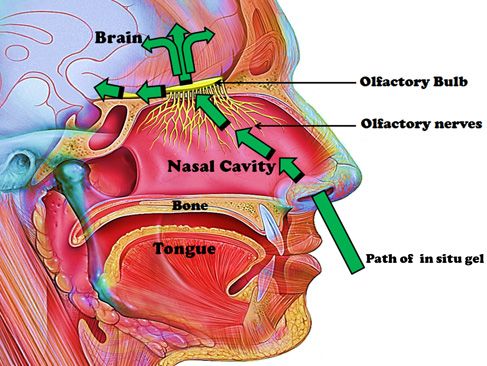
2.2. Anatomical Structures Involved in Nose‐to‐Brain Transport
2.2. Anatomical Structures Involved in Nose-to-Brain Transport
Some anatomical structures are involved in nose-to-brain transport (Figure 2). The nasal mucus
Some anatomical structures are involved in nose-to-brain transport (Figure 2). The nasal mucus
pH is within the range of 5.5–6.5, and it is moved by the cilia on the respiratory mucosa. The amount
pH is within the range of 5.5–6.5, and it is moved by the cilia on the respiratory mucosa. The amount
of mucus in the nasal mucosa can influence the drug absorption [21]. It is the first barrier that drugs
of mucus in the nasal mucosa can influence the drug absorption [21]. It is the first barrier that
administered intranasally must cross before travelling between cells either paracellularly or
drugs administered intranasally must cross before travelling between cells either paracellularly or
transcellularly [22]. Drug administered is transported between the epithelial cells, and these cells can
transcellularly [22]. Drug administered is transported between the epithelial cells, and these cells
be close via several junctions referred to as tight junctions, desmosomes, adhering junctions and gap
can be close via several junctions referred to as tight junctions, desmosomes, adhering junctions and
junctions [22,23]. The unaltered state of these junctions influences the paracellular transport. Some
gap junctions [22,23]. The unaltered state of these junctions influences the paracellular transport.
therapeutics can open these junctions, thereby enhancing nose-to-brain delivery, and this route is
Some therapeutics can open these junctions, thereby enhancing nose-to-brain delivery, and this route is
rapid [22]. It is important to mention that the size of the therapeutics influences the mechanism of
rapid [22]. It is important to mention that the size of the therapeutics influences the mechanism of drug
drug delivery. Therapeutics larger than 20 nm are reported to be transported transcellularly.
delivery. Therapeutics larger than 20 nm are reported to be transported transcellularly. Therapeutics
Therapeutics smaller than 200 nm and in the range of 200–1000 nm undergo caveolae-mediated
smaller than 200 nm and in the range of 200–1000 nm undergo caveolae-mediated endocytosis and
endocytosis and clathrin-mediated endocytosis, respectively. Transcellular drug transport is slow,
clathrin-mediated endocytosis, respectively. Transcellular drug transport is slow, and the bioactive
and the bioactive agents are transported to the olfactory receptor neuron and undergo intraneuronal
agents are transported to the olfactory receptor neuron and undergo intraneuronal transport to the
transport to the olfactory bulb [22,24].
olfactory bulb [22,24].
Bioactive agents can be transported via the olfactory nerve fibers of the olfactory bulb to the CNS
(Central Nervous System). The olfactory bulbs projects to the different regions of the brain such as
the hypothalamus, olfactory tract, amygdala, entorhinal cortex, anterior olfactory nucleus, etc. Intra-
and peri-neural transport occurs via these projections upon intranasal drug administration [22]. The
respiratory region in the nasal cavity is supplied with nerves by projection of the trigeminal nerves,
which are composed of branches such as the ophthalmic, maxillary and mandibular nerves. These
branches enter the brain at the cribriform plate and the lacerated foramen, thereby producing
entrance into the rostral and caudal regions of the brain [22]. The olfactory region’s vascularizationis another feature useful in nose-to-brain transport, and it originates from small branches of the
ophthalmic artery. Drug administered intranasally enters the systemic circulation via this
vascularization, thereby bypassing the BBB to the brain. This is applicable to small and lipophilic
drugs. Drugs can travel via channels between the outermost layer of blood vessels and the basement
membrane 2018,
Pharmaceutics of surrounding
10, 40 tissue by the perivascular pathway [22,25]. This pathway is driven by4bulk
of 17
flow, diffusion and arterial pulsation, resulting in rapid drug distribution in the CNS [25].
Figure 2. Anatomical structures involved in nose-to-brain transport.
Figure 2. Anatomical structures involved in nose-to-brain transport.
3. Neurological Diseases
Bioactive agents canand
be Challenges
transported in Treatment
via the olfactory nerve fibers of the olfactory bulb to
the CNS (Central Nervous System). The olfactory
The treatment of most neurological diseases is challenging bulbs projectsdue toto the
the blood-brain
different regions
barrier,oftheir
the
brain such as
association withthea hypothalamus,
number of genes, olfactory tract,ofamygdala,
the overlap entorhinal
disease-associated cortex,
genes, druganterior olfactory
side effects with
nucleus, etc. Intra- and peri-neural transport occurs via these projections
minimal effects on the disease progression and mechanisms and biomarkers behind neurological upon intranasal drug
administration
diseases not being [22].well
Theunderstood
respiratory[26–28].
region in the nasal cavity is supplied with nerves by projection
of theAlzheimer’s
trigeminal nerves,
disease which are composed
is a neurological of branches
disorder in which suchtheasbrain
the ophthalmic, maxillary
nerve cells are and
destroyed,
mandibular nerves. These branches enter the brain at the cribriform plate and the
resulting in dementia. It is characterized by intracellular neurofibrillary tangles, insoluble β-amyloid lacerated foramen,
thereby producing entrance
(Aβ) peptides/senile plaquesintoand
the the
rostral
lossand
of caudal
differentregions
neuronsof theinbrain [22]. The
the basal olfactory
forebrain region’s
amygdala,
vascularization is another feature useful in nose-to-brain transport,
cortical and hippocampus region of the brain [17]. The risk factors for Alzheimer’s disease and it originates from small
are
branches of the ophthalmic artery. Drug administered intranasally enters the
decreased availability of oxygen resulting in cell death [29], head injury [30], vitamin D deficiencysystemic circulation via
this
[31] vascularization, thereby bypassing
and a high concentration of copperthe andBBB to the brain.levels
homocysteine This is applicable
resulting to small to
in damage andthelipophilic
neurons
drugs. Drugs can travel via channels between the outermost layer of blood
[32]. The current approach in the management of Alzheimer’s disease is symptomatic, which involves vessels and the basement
membrane of surrounding
the counterbalance tissue by the disturbance
of neurotransmitter perivascularof pathway [22,25].
the disease This pathway is
by administration ofdriven by bulk
cholinesterase
flow, diffusion
inhibitors [17]. and
The arterial
United pulsation,
States Food resulting
and Drug in Administration
rapid drug distribution
(FDA) has in the CNS [25].
approved five drugs for
the management of Alzheimer’s disease, which include donepezil, galantamine, rivastigmine,
3. Neurological Diseases and Challenges in Treatment
memantine and the combination of donepezil with memantine. Donepezil, galantamine and
rivastigmine are referred
The treatment of mosttoneurological
as cholinesterase
diseases inhibitors, and they
is challenging duetreat symptoms
to the blood-brain associated with
barrier, their
memory, language,
association etc. They
with a number of act by maintaining
genes, the overlap the levels of acetylcholine,
of disease-associated which
genes, drugthensidecompensates
effects with
for the loss
minimal of the
effects on functioning brain cells [33].
the disease progression and Memantine
mechanismsregulates the activity
and biomarkers of glutamate,
behind neurological an
important
diseases notneurotransmitter,
being well understoodwhich[26–28].
is important in learning and memory. It also protects the cells
against excess glutamate
Alzheimer’s disease by is apartially blocking
neurological MNDA
disorder (N-methyl-D-aspartate)
in which the brain nerve cells receptors [33]. The
are destroyed,
combination
resulting of donepezil
in dementia. It is with memantine
characterized is used to treat
by intracellular moderate totangles,
neurofibrillary severe stages of the
insoluble disease.
β-amyloid
Thesepeptides/senile
(Aβ) drugs suffer from someand
plaques sidetheeffects
loss of(Table 1) [33].
different neurons in the basal forebrain amygdala, cortical
Multiple sclerosis
and hippocampus regionis anofautoimmune
the brain [17]. disease,
The riskandfactors
it affects
forthe central nervous
Alzheimer’s diseasesystem, resulting
are decreased
in the damage of the axon, demyelination and loss of neurological functions
availability of oxygen resulting in cell death [29], head injury [30], vitamin D deficiency [31] [34]. Factors thatand
are
abelieved to contributeoftocopper
high concentration the disease are common levels
and homocysteine childhood infections,
resulting low level
in damage to theofneurons
exposure to
[32].
sunlight
The (vitamin
current D deficiency),
approach smoking of
in the management and genetic factors
Alzheimer’s [34].isThe
disease loss of axons
symptomatic, is reported
which involvestothe
be
initiated by complex
counterbalance inflammatory processes
of neurotransmitter disturbance[34,35].
of theThere is presently
disease no cure for multiple
by administration sclerosis.
of cholinesterase
inhibitors [17]. The United States Food and Drug Administration (FDA) has approved five drugs for the
management of Alzheimer’s disease, which include donepezil, galantamine, rivastigmine, memantine
and the combination of donepezil with memantine. Donepezil, galantamine and rivastigmine
are referred to as cholinesterase inhibitors, and they treat symptoms associated with memory,Pharmaceutics 2018, 10, 40 5 of 17
language, etc. They act by maintaining the levels of acetylcholine, which then compensates for
the loss of the functioning brain cells [33]. Memantine regulates the activity of glutamate, an important
neurotransmitter, which is important in learning and memory. It also protects the cells against excess
glutamate by partially blocking MNDA (N-methyl-D-aspartate) receptors [33]. The combination of
donepezil with memantine is used to treat moderate to severe stages of the disease. These drugs suffer
from some side effects (Table 1) [33].
Table 1. Some drugs used for the treatment of selected neurological diseases.
Drugs Application Route of Administration Side Effects
All stages of Nausea, increased bowel movements, loss
Donepezil Oral
Alzheimer’s disease of appetite
Mild to moderate Nausea, increased bowel movements, loss
Galantamine Oral
Alzheimer’s disease of appetite
Mild to moderate
Rivastigmine Orally and transdermally Dizziness, constipation, headache
Alzheimer’s disease
Moderate to severe
Memantine Oral Dizziness, constipation, headache
Alzheimer’s disease
Nausea, increased bowel movements, loss
Donepezil and Moderate to severe
Oral of appetite, dizziness,
memantine Alzheimer’s disease
constipation, headache.
Relapsing-remitting Subcutaneous and
Beta interferon Myalgia, headache, anemia, nausea
multiple sclerosis intramuscular
First clinical episode of
Glatiramer acetate Subcutaneous Nausea, vomiting, body ache
multiple sclerosis
Relapsing form of
Fingolimod Oral Headache, diarrhea, cough
multiple sclerosis
Relapsing form of
Teriflunomide Oral Liver problems, influenza, nausea, diarrhea
multiple sclerosis
Relapsing form of Burning feeling of the skin, itching, nausea,
Dimethyl fumarate Oral
multiple sclerosis abdominal pain, vomiting
Nausea, diarrhea, constipation, flu-like
Worsening relapsing remitting
Mitoxantrone Intravenous symptoms, cardiac toxicity and
multiple sclerosis
risk of leukemia
Relapsing progressive form of Headache, stomach pain,
Natalizumab Intravenous
multiple sclerosis diarrhea, depression
To treat depression associated Risk of diabetes, weight gain,
Quetiapine Oral
with schizophrenia constipation, dizziness
Risk of diabetes, weight gain,
Risperidone Schizophrenia Oral, intramuscular
constipation, dizziness
Paliperidone Schizophrenia Oral, intramuscular Risk of diabetes, weight gain, headache.
Aripiprazole Schizophrenia Oral, intramuscular Dizziness, nausea, vomiting, sedation
Weight gain, risk of diabetes,
Asenapine Schizophrenia Oral
sedation, akathisia
Suicidal behavior associated Weight gain, sweating, seizures, sedation,
Clozapine Oral
with schizophrenia risk of diabetes
Severe and Weight loss, insomnia, dizziness, headache,
Felbamate Oral
refractory epilepsies ataxia, skin rashes, hepatotoxicity
Dizziness, fatigue, hyperactivity and
Gabapentin Partial seizures Oral
weight gain
Dizziness, headache, diplopia, ataxia,
Lamotrigine Partial seizures Oral, intravenous
skin rash
Drowsiness, insomnia, Irritability
Vigabatrin Partial seizures Oral psychosis, depression, weight
gain, blindness
Leucopenia, fatigue, dizziness, headache,
Levetiracetam Partial seizures Oral
anorexia, psychiatric disturbances
Drowsiness, diplopia, headache, GI distress,
Oxcarbazepine Partial seizures Oral hyponatremia, skin rash,
Stevens–Johnson syndromePharmaceutics 2018, 10, 40 6 of 17
Table 1. Cont.
Drugs Application Route of Administration Side Effects
Diplopia, nystagmus, coarsening of facial
features gingival hyperplasia, hirsutism,
Phenytoin Partial seizure Intravenous skin rashes, Stevens–Johnson syndrome,
agranulocytosis, aplastic
anemia, hepatotoxicity
It is used to treat symptoms of
Parkinson’s syndrome such as Nausea, vomiting, and irregular
Levodopa Oral, intravenous
slow movements and stiff, heart rhythms
rigid body parts
Treat nausea, vomiting, and
irregular heart rhythms, which
Sinemet Oral Dyskinesias
are side effect associated
with levodopa
It mimic dopamine, thus
Nausea, drowsiness, sleep attacks,
Pramipexole helping to produce smooth Oral
hypotension, and hallucinations
voluntary movement
It increase the amount of
Selegiline dopamine in the brain and Nausea, headache, confusion, postural
Oral
hydrochloride reduces the motor symptoms hypotension, hallucinations and insomnia
of Parkinson’s disease
It increase the amount of
Dyskinesias, nausea, diarrhea, sleep
dopamine in the brain and
Tolcapone Oral disturbance, urine dis-coloration
reduce the motor symptoms of
and hallucinations
Parkinson’s disease
Blurred vision, constipation, urinary
Biperiden It reduces tremors and
Oral retention, heat stroke, nervousness, anxiety,
hydrochloride muscle rigidity
confusion, depression, delusions
Multiple sclerosis is an autoimmune disease, and it affects the central nervous system, resulting
in the damage of the axon, demyelination and loss of neurological functions [34]. Factors that are
believed to contribute to the disease are common childhood infections, low level of exposure to sunlight
(vitamin D deficiency), smoking and genetic factors [34]. The loss of axons is reported to be initiated by
complex inflammatory processes [34,35]. There is presently no cure for multiple sclerosis. Two major
strategies that have been reported to be potential routes to the treatment of the disorder are to hinder
the immune system from causing damage that results in the clinical manifestations of the disease and to
establish mechanisms that will result in the CNS being resistant to the deleterious effects of the immune
response, known as neuroprotection [36]. The treatment approach focuses on treating the symptoms,
slowing the progressive forms of the disease and speedy recovery from attacks [34]. Drugs used to
treat the disorder are known as immunomodulatory agents because they modify the immune response,
thereby reducing the deleterious effects mediated by the immune system. However, the mechanisms of
these drugs are not well understood [36]. Some of the drugs used include beta interferons, fingolimod,
glatiramer acetate, teriflunomide, dimethyl fumarate, mitoxantrone, natalizumab, etc. These drugs
suffer from severe side effects, resulting in poor patient compliance such as irritation at the site of
injection, influenza-like symptoms, chest tightness, heart palpitations and breathlessness, heart failure,
leukemia, etc. [34,37]. Other neurological diseases are schizophrenia, epilepsy, Parkinson syndrome,
brain tumor, etc.
Schizophrenia is a chronic mental health disorder, which is characterized by delusions,
hallucinations, disorganized behavior, etc. [38]. It is caused by an excess or a deficiency of
neurotransmitters, which include glutamate, dopamine and serotonin. Dopaminergic pathways
that attribute to the disease are the nigrostriatal pathway in which low dopamine levels affect the
extrapyramidal system, resulting in motor symptoms; excess dopamine in the mesolimbic pathway
that extends from the ventral tegmental area to limbic areas; low mesocortical dopamine levels in
the mesocortical pathway; a decrease in the tuberoinfundibular dopamine in the tuberoinfundibular
resulting in elevated levels of prolactin, amenorrhea and reduced libido [38]. Other factors that are
genetic and environmental also contribute to the disease. The condition is managed using antipsychoticPharmaceutics 2018, 10, 40 7 of 17
drugs, which suffer from adverse effects such as diabetes mellitus, weight gain and hyperlipidemia,
which increase the risk of cardiovascular mortality (Table 1) [38,39]. Other complications associated
with the drugs are dystonia, cataracts, sexual dysfunction, etc. [38].
Epilepsy is a non-contagious and chronic disorder of the brain characterized by recurrent seizures
caused by excess electrical discharges in a group of brain cells [40]. Factors that contribute to the disease
are malaria, birth-related injuries, severe head injury, meningitis, encephalitis, neurocysticercosis, brain
tumor, etc. [40]. Some of the drugs used for the treatment of epilepsy exhibit adverse effects such
as aplastic anemia, hepatitis, allergic rashes, etc. [40]. The main limitation of the drugs used to
treat epilepsy in some patients is pharmacoresistance resulting from mechanisms that are disease
related, genetics and drug related. The disease-related mechanism is responsible for the alterations of
pharmacological targets of antiepileptic drugs in the brains of pharmacoresistant patients leading to
the failure of antiepileptic drugs to block excitatory sodium or calcium currents. The genetic alterations
result from drug efflux transporters leading to poor seizure control, and the drug-related mechanism
is responsible for the reduced efficacy of antiepileptic drugs [41,42].
Parkinson syndrome is characterized by Lewy bodies containing aggregations of the protein
alpha-synuclein and the loss of pigmented melanin-containing neurons in the midbrain [43]. The loss
of pigmented melanin-containing neurons in the midbrain reveals neurodegeneration of dopaminergic
neurons in the substantia nigra characterized by dopamine deficit in the striatum [43].
The treatment of Parkinson’s disease is classified as a symptomatic and neuroprotective therapy.
Presently, there is no proven neuroprotective therapy for the treatment of the disease. In cases of severe
Parkinson’s syndrome disease, when the medication is longer effective, brain surgery, which involves
deep brain stimulation, is performed so as to manage the motor symptoms [44,45]. The drugs that
are used to treat the disease are classified as dopamine precursors, dopamine agonists, monoamine
oxidase B inhibitors and anticholinergics [44,45]. These classes of drugs suffer from some side effects,
as illustrated in Table 1.
A brain tumor is characterized by an abnormal growth of tissue in the brain or central spine,
disrupting the function of the brain, which can be either cancerous or non-cancerous [46]. Tumors are
classified based on where the cells originated. They are classified as benign when they originate from
cells within the brain and do not spread into other tissue; malignant brain tumors that spread into
other tissues and grow rapidly, thus invading the surrounding brain tissue; primary tumors that
start in cells of the brain and can spread to other parts of the brain or to the spine; metastatic brain
tumors that begin in another part of the body and then spread to the brain [46]. The risk factors
are viral infection, chemicals, ionizing radiation and genetic manipulation [47]. Brain tumors are
difficult to treat. Some of the drugs used for the treatment of brain cancer are temozolomide, lomustine,
bevacizumab, carmustine wafer, etc.
4. Nasal Delivery for the Treatment of Neurological Diseases Such as Alzheimer’s Diseases,
Parkinson’s Diseases, Epilepsy, etc.
Gel-based drug delivery systems, which can be used to administer drugs intranasally, have
been developed for the management of neurological diseases. These systems are biodegradable,
biocompatible, can deliver the drugs to the brain, bypass the blood-brain barrier and are potential
therapeutics for the treatment of neurological disorder. The design of these systems, the concentration
of polymers, pore sizes and the rate of degradation influence their efficacy.
4.1. Hydrogels
Hydrogels are hydrophilic, cross-linked networks of water-soluble polymers, which can retain
a large amount of water [48,49]. They can be formulated in various physical forms such as slabs,
films, in situ hydrogels, nanogels, microparticles, nanoparticles, etc. [48]. They can be easily modified
with selected functional groups and exhibit pores with sizes that can be controlled by the density of
crosslinking. Their porosity is useful for the loading and the release of drugs at a rate that is influencedPharmaceutics 2018, 10, 40 8 of 17
by the diffusion coefficient of macromolecules through the hydrogel network [48,49]. Hydrogels are
highly biocompatible, which is attributed to their high water content and their physiochemical
properties, which are similar to the native extracellular matrix [48,49]. The degree of biodegradability
of hydrogels is designed into the hydrogel via selected pathways such as environmental [49]. They can
also be deformed to the shape of the surface to which they are to be delivered [49]. They are sensitive to
external stimuli such as temperature, pH and magnetic field. They are prepared by different methods,
and the method of preparation influences their pore size, rate of degradation, mechanical strength and
drug release mechanism. Due to their unique physicochemical properties, they have been designed for
nose-to-brain delivery.
4.1.1. In Situ Gels
In situ-based gels are systems that exhibit sol-to-gel transition at the site where they are
administered into the body. They are liquid when administered and undergo a sol-to-gel transition
induced by external stimuli such as temperature, pH, ion change and magnetic field or in the biological
environment [48,50]. They exhibit good properties, which make them useful for drug delivery such as:
they are highly compatible with a range of drugs, which are soluble, insoluble, low and high molecular
weight drugs; they are less invasive and can be used to obtain high drug concentrations at the desired
site of action with reduced systemic side effects; biocompatibility; biodegradable and exhibit sustained
drug release over an extended period, thereby enhancing patient compliance [51]. The aforementioned
properties make them useful for nose-to-brain delivery.
In Situ-Based Gels for the Delivery of Anti-Parkinson Drugs
Anti-Parkinson drugs such as levodopa are used for the treatment of Parkinson’s disease, and
its use is limited by its poor bioavailability, which is characterized by its low brain uptake. Its poor
bioavailability is attributed to the irregular gastrointestinal metabolism of the drug before it attaches
to the L-amino acid carrier that transports the drug actively through the duodenum where it enters the
bloodstream [52–55]. Sharma et al. incorporated chitosan nanoparticles loaded with levodopa prepared
by ionic gelation technique using sodium TPP (1 mg/mL) onto a thermo-reversible gel prepared from
Pluronic PF127 (Poloxamer 407) [56] (Table 2). The formulations were characterized, followed by
in vitro drug release studies, which revealed that the formulation obeyed the Hixson-Crowell model
(drug release by dissolution with changes in the surface area and diameter of the formulation).
The addition of polycations enhanced the drug absorption of the formulation on the nasal mucosa by
opening the junctions between epithelial cells and delaying mucociliary clearance. In vivo studies on
Swiss albino rat models further showed that intranasal administration of the chitosan nanoparticles
resulted in an enhanced brain uptake of the drug when compared to the gel formulation, suggesting
that the viscosity of the gel reduced the brain uptake of the drug [56]. Lungare et al. developed
in situ thermoresponsive-based gels by the cold method using Pluronic F127 (Poloxamer 407) as
a thermoreversible polymer and carboxymethylcellulose as a mucoadhesive polymer [57]. It was
loaded with amantadine, an anti-Parkinsonian drug. Increasing the mucoadhesive polymer resulted in
an increased gelation temperature, and increasing amantadine reduced the gelation. A concentration
of 16% of Pluronic F127 was found to be suitable for the sol-to-gel transition of the formulation at
ambient nasal temperatures. These systems are potential therapeutics for the treatment of Parkinson
disease [57]. The formulation was stable, which was evidenced by the repeatable drug release profiles
of the Fickian mechanism (a transport process in which the polymer relaxation time is greater than the
solvent diffusion time), followed by an anomalous drug release mechanism (a combination of diffusion
and erosion controlled drug release) after storage of the formulation at 4 ◦ C for eight weeks. There was
no significant cellular toxicity to the human nasal epithelial cells up to 4 mg/mL and up to 1 mM.
The % drug release from the formulation was in a range of 43–44% in vitro [57,58]. Khan et al. reported
mucoadhesive in situ gel formulation prepared from chitosan and hydroxyl propyl methyl cellulose
for intranasal delivery of ropinirole to the brain [59]. In vivo brain uptake of ropinirole in albinoPharmaceutics 2018, 10, 40 9 of 17
rats following intranasal administration of 99mTc (Technetium 99m)-ropinirole-loaded gel AUC (area
under the curve) (0–480 min) was 8.5-times when compared to the intravenous administration [59]
(Figure 3). Ravi et al. prepared thermosensitive gel for intranasal delivery of rasagiline mesylate,
a drug for the treatment of Parkinson’s disease [60]. The gels were prepared from a combination of
poloxamer 407 and poloxamer 188 in a 1:1 ratio with mucoadhesive polymers, namely: carbopol 934 P
and chitosan. In vivo performance of the formulation in New Zealand white rabbits suggested that the
intranasal administration of the formulation exhibited a better drug bioavailability of six-fold higher
than the oral solution. Nasal mucosal integrity studies indicated maintained integrity of the nasal
mucosa of rats after chronic administration of the formulation. The brain uptake of the formulation
was significantly (p < 0.01) high when compared to the drug solution. Rasagiline mesylate’s poor
bioavailability is attributed to its rapid absorption and first-pass metabolism, which is overcome when
administered intranasally, resulting in extended residence and contact time with nasal epithelium and
enhanced drug absorption from the nasal cavity. High Cmax revealed the rapid absorption of the drug,
and the high2018,
Pharmaceutics AUC x values suggested complete absorption of the drug from the gel formulations9 [60].
11,0–∞ of 17
Rao et al. prepared thermoreversible nasal gels by the cold method from Pluronic F-127 and hydroxy
methyl
Pluronicpropyl
F-127 cellulose,
and hydroxy and methyl
ropinirole, an anti-Parkinson
propyl cellulose, and drug with poor
ropinirole, oral bioavailability,
an anti-Parkinson was
drug with
loaded
poor oralonto the gel [61]. Formulations
bioavailability, was loaded onto exhibited
the gelgelation at the nasalexhibited
[61]. Formulations temperature, and at
gelation thethe
time of
nasal
gelation was less
temperature, andthan
the the
timemucociliary
of gelationclearance
was lesstime.
than The
the nasal residence
mucociliary time of the
clearance formulation
time. The nasal
was influenced
residence bythe
time of theformulation
mucoadhesion and enhanced
was influenced strength
by the of the gel.
mucoadhesion The
and formulations’
enhanced strength exofvivo
the
drug release
gel. The was 56–100%
formulations’ over adrug
ex vivo period of 5 h.
release Histological
was 56–100% overstudya of sheepofnasal
period mucosa revealed
5 h. Histological study that
of
the gelnasal
sheep had amucosa
protective effect when
revealed compared
that the gel had to the free drug,
a protective which
effect when was characterized
compared to thebyfreecellular
drug,
which was
damage. The characterized
brain uptakeby ofcellular
the drug damage. Theadministration
after nasal brain uptake ofwasthe five-fold
drug after nasalcompared
when administration
to the
was five-fold when
administration of thecompared
formulation to the administration
intravenously, of the the
revealing formulation
system asintravenously, revealing
a potential delivery systemthe
system
for as a potentialdrugs.
anti-Parkinsonian delivery system
The drug for anti-Parkinsonian
delivery from the formulationdrugs. Thebrain
to the drugwas
delivery
via the from the
olfactory
formulation
nerves [61]. to the brain was via the olfactory nerves [61].
Figure 3.
Figure 3. Chitosan-based
Chitosan-based in
in situ
situ gels
gels for
for the
the delivery
delivery of
of ropinirole.
ropinirole.
In Situ Gels for the Delivery of Anti-Migraine Drug
In Situ Gels for the Delivery of Anti-Migraine Drug
Sumatriptan succinate is used for the treatment of migraine. It suffers from poor bioavailability,
Sumatriptan succinate is used for the treatment of migraine. It suffers from poor bioavailability,
which is associated with its low retention time in the nasal cavity and its low delivery to the brain via
which is associated with its low retention time in the nasal cavity and its low delivery to the brain
the olfactory pathway. The permeation of sumatriptan across the brain-blood barrier is very poor.
via the olfactory pathway. The permeation of sumatriptan across the brain-blood barrier is very poor.
Galgatte et al. prepared in situ gel in a simulated nasal fluid using deacetylated gellan gum as the
Galgatte et al. prepared in situ gel in a simulated nasal fluid using deacetylated gellan gum as the
gelling agent [62]. The strength of the gel and the rate of drug release from the gel was influenced by
gelling agent [62]. The strength of the gel and the rate of drug release from the gel was influenced by
the concentration of gellan gum in the formulation. The strength of the gel increased with the increase
the concentration of gellan gum in the formulation. The strength of the gel increased with the increase
in the concentration of gellan gum, while the rate of drug release decreased with the increase in the
concentration of the gellan gum. The release mechanism of the drug from the gel followed a Fickian
release model, revealing an erosion diffusion mechanism. Ex vivo permeation was studied using
sheep olfactory nasal mucosa with a thickness of 0.6 mm. Its permeation showed a release of 93% of
the drug over a period of 300 min. The microscopic structure of the mucosa did not reveal cell necrosis
after the application of the formulation. The interaction between the drug and the excipients was notPharmaceutics 2018, 10, 40 10 of 17
in the concentration of gellan gum, while the rate of drug release decreased with the increase in the
concentration of the gellan gum. The release mechanism of the drug from the gel followed a Fickian
release model, revealing an erosion diffusion mechanism. Ex vivo permeation was studied using sheep
olfactory nasal mucosa with a thickness of 0.6 mm. Its permeation showed a release of 93% of the
drug over a period of 300 min. The microscopic structure of the mucosa did not reveal cell necrosis
after the application of the formulation. The interaction between the drug and the excipients was
not significant, suggesting the absence of local irritation to the nasal mucosa. The formulation was
stable at a room temperature of 25 ◦ C and at 4 ◦ C. The concentration of sumatriptan in the plasma
and the brain tissues was higher by nasal in situ gel compared to the oral aqueous solution. In vivo
studies showed that AUC was higher in the plasma and the brain for the formulation administered
by the nasal route when compared to oral administration. The AUC of sumatriptan in brain tissues
was 1.44-times higher when compared to the AUC in plasma when the formulation was administered
intranasally. The results revealed that the permeation of drug molecules across the nasal mucosa to the
brain was via the olfactory pathways (Table 2) [62].
In Situ Hydrogels for the Delivery of Anti-Alzheimer’s Drug
Selected anti-Alzheimer’s drugs have been loaded onto in situ gels, resulting in an enhanced brain
uptake of the drug in vivo. Tao et al. prepared gellan gum-based in situ gel loaded with huperzine
A. The gel was prepared by the precipitation method (Table 2) [63]. Huperzine A uptake by the rat
brain tissues after intranasal administration indicated that the AUC (0–>6 h) value in plasma obtained
after nasal administration was 0.94 compared to the intravenous administration. The AUC (0–>6 h)
of cerebrospinal fluid after nasal administration was 1.3- and 2.- times compared to intravenous and
intragastric administration. The in situ gel significantly increased the distributions of huperzine A in the
rat brain tissues, especially in the cerebrum and hippocampus [63]. Chen et al. loaded curcumin onto
thermosensitive hydrogel for good brain targeting efficiency. The gels were prepared from pluronic
F127 and poloxamer 188 [64]. The gels exhibited shorter gelation time, extended mucociliary transport
time and prolonged curcumin retention in the rat nasal cavity at body temperature. The curcumin
release mechanism from the gel was diffusion and dissolution controlled, respectively, when the
dialysis membrane method and membraneless methods were employed. The nasal mucosal integrity
was maintained over a period of 14 days after application of the formulation. The drug-targeting
efficiencies for curcumin in the cerebrum, cerebellum, hippocampus and olfactory bulb after intranasal
administration of the formulation were 1.82-, 2.05-, 2.07- and 1.51-times, respectively, when compared
to the drug-targeting efficiencies of the drug after intravenous administration of the curcumin solution.
The gel increased the drug uptake and distribution in the rat brain tissue, which was significant in
the cerebellum and hippocampus [64]. Wang et al. prepared thermoreversible in situ nasal gel by the
cold method from poloxamers (P407, P188) and the hydroxypropyl methylcellulose for the delivery of
geniposide [65]. Borneol was employed as a permeation enhancer. The drug content of the formulation
was in the range of 97–100%; the gel strength and mucoadhesive strength of the formulation was
in the range of 25–50 s and 4000–6000 dyn/cm2 , respectively. The in vitro release mechanism of
geniposide was zero-order, and the ex vivo release mechanism of geniposide obeyed the Weibull model
(a mechanism in which the amount of drug release is a function of time), suggesting that the release of
geniposide is influenced by gel corrosion and that the permeation of geniposide is time dependent [65].
The finding revealed the potential of the formulations for the treatment of neurological diseases.
Salatin et al. developed in situ gel from poloxamer 407® for the delivery of rivastigmine hydrogen
tartrate in poly(lactic-co-glycolic acid) nanoparticles [66] A high drug permeation through the sheep
nasal mucosa was observed when compared to the free drug. The formulation was stable; embedding
the drug in nanoparticles enhanced the permeability; and the drug release from the formulation
was sustained. The cellular uptake of the drug from the formulation was time dependent and was
cytocompatible on A549 cells [66]. Abouhussein et al. also investigated brain delivery of rivastigmine
tartrate via mucoadhesive thermosensitive in situ gel intranasally [67]. The mucoadhesive in situPharmaceutics 2018, 10, 40 11 of 17
gel was developed from pluronic F127, HPMC (hydroxypropyl methylcellulose), chitosan, carbopol
934 and NaCMC (sodium carboxymethyl cellulose). In vivo pharmacokinetic and biodistribution
studies using the radiolabeling approach in normal albino mice revealed 84% intranasal permeation
with a good distribution to the brain (0.54% ID/g) when compared to intravenous administration.
Intravenous administration of the drug solution resulted in a high drug distribution to liver and
kidney compared to administration of the formulation intranasally. These findings suggested that
intranasal drug administration reduced drug systemic distribution to different organs, thus resulting in
enhanced drug targeting to the brain, hence overcoming side effects [67]. The Cmax and AUC of brain
concentrations of (0.58% radioactivity/gram) and (84.7% radioactivity.min/gram) were significantly
high. The AUC0-∞ was five-fold greater when administered intranasally when compared to the
free drug solution administered intravenously, suggesting drug transport to the brain was via the
olfactory route. The mucoadhesive nature of the CP 934 polymer used hindered the normal mucociliary
clearance of the formulation [67].
In Situ Gels for the Delivery of Anti-Depressant Drug
Doxepin is an anti-depressant drug. Naik and Nair reported thermoreversible gels prepared
using chitosan and glycerophosphate or poly(ethylene) glycol for the delivery of doxepin to the
brain via intranasal administration. In vivo studies in Swiss albino mice showed a good increase in
activity count and a decrease in immobility time, suggesting good antidepressant activity (Table 2) [68].
The drug solution caused a significant damage to the nasal mucosal tissues, which was characterized
by glandular hyperplasia and severe epithelial hyperplasia. However, the administration of the gel
formulation resulted in mild side effects, which include mild swelling of glands, and there was no
symptom of sluffing of the mucosal epithelium, which was observed in mice in which the drug solution
was administered. The rate of permeation of doxepin from the gel matrix was influenced by its release
profile from the matrix. The drug from the formulation prepared from chitosan, glycerophosphate or
poly(ethylene) glycol permeated at a lower rate when compared to the formulation prepared from
chitosan and glycerophosphate [68]. Fatouh et al. prepared in situ gel loaded with agomelatine,
an antidepressant drug [69]. The drug release from the gel formulations was between 8.9–21% when
compared to the drug release from the drug solution and solid lipid nanoparticles, which was 89%
and 35%, respectively. The in situ gel exhibited significantly higher Cmax , AUC (0–360 min) and
absolute bioavailability of 247 ng/mL, 6677 ng·min/mL and 38%, respectively, when compared to oral
suspension of Valdoxan®(21 ng/mL, 2828 ng·min/mL and 16%, respectively (p < .0001)). The increase
in Cmax , AUC 0–360 min and absolute bioavailability of the gel is due to the increased amount of the
drug that bypasses the hepatic metabolism to reach the systemic circulation; the lipid nature of the
particles, which permits their partition into the lipid bilayer of the nasal epithelial cell membrane
and to penetrate the cells; and the co-surfactants used enhanced the permeability of the membrane
structure by opening of the tight junctions between epithelial cells. Drug concentrations in the brain of
rats after administration of gel formulation revealed a significantly high Cmax and AUC (0–360 min)
(148 ng/mL and 6570 ng min/mL, respectively) compared to the oral suspension (61 ng/mL and
1710 ng·min/mL, respectively) (p < .0001). [69]. Pathan et al. reported ion sensitive in situ nasal gel
loaded with fluoxetine hydrochloride for brain delivery. The gel was prepared from gellan gum and
HPMC (hydroxypropyl methylcellulose) [70]. Ex/in vivo permeation studies revealed that increasing
the concentration of gellan gum from 0.2–0.6% and HPMC from 0.1 to 0.2% decreased the rate of drug
release. The percentage of drug permeation over a period of 240 min from all formulations was between
75% and 94%. The integrity of the epithelial cell in the nasal mucosa treated with the formulation was
maintained, indicating the non-toxic nature of the formulations. In vivo study further revealed that
the administration of the formulation reduced the total immobility period and increased climbing
and swimming behavior [70]. Kaur et al. studied the brain delivery of tramadol hydrochloride using
mucoadhesive thermo-reversible gel [71]. The gels were prepared from chitosan nanoparticles by the
ionic gelation method followed by the addition of the nanoparticles in Pluronic and HPMC-basedPharmaceutics 2018, 10, 40 12 of 17
mucoadhesive thermo-reversible gel. The formulation significantly hindered forced swim-induced
depression by involving anti-oxidant-like effects, significantly increased locomotor activity and body
weight of the rat model in vivo. The nanoparticles further enhanced the delivery of drug to the
brain [71]. Pathan and More developed thermoreversible gel loaded with nortriptyline hydrochloride
for intranasal administration [72]. An increase in the concentration of poloxamer 188 and HPMC
increased the viscosity and mucoadhesive strength and decreased the gelation temperature and drug
percentage permeation. The formulation with 3.6% poloxamer and 0.04% HPMC exhibited a 98%
drug release through sheep nasal mucosa. The formulation was stable over a period of three months,
and the results obtained indicated that the formulation is a potential therapeutic for the treatment of
depression [72]. Haque et al. prepared venlafaxine-loaded alginate nanoparticles for the treatment of
depression by intranasal administration [73]. Pharmacodynamic studies of the formulation for the
antidepressant activity in vivo in adult Wistar rats showed improved swimming and climbing and
reduced immobility (p < 0.01) when compared to the depressed group. The formulation enhanced
the drug concentration in the brain, which is attributed to factors such as: increased absorption time,
reduced nasal mucociliary clearance, increased permeation across nasal mucosa and modulation of
P-gp efflux transporters present on BBB [73]. The brain/blood ratios of the formulation administered
intranasally, drug solution administered intravenously and drug solution administered intranasally
were 0.11, 0.03 and 0.07, respectively, at 30 min, which revealed the potential of the formulation for
nose-to-brain delivery. The brain concentration of drug after intranasal administration of the drug was
743 ng/mL, tmax 60 min, and it was significantly higher (p < 0.05) than the drug solution administered
intravenously (382.91 ng/mL; tmax 30 min) and drug solution administered intranasally (397 ng/mL;
tmax 60 min) [73].
In Situ Gels for the Delivery of Anti-Schizophrenia Drug
Sherje et al. developed in situ gels from carbopol 934 and hydroxypropyl methyl cellulose loaded
with paliperidone for the treatment of schizophrenia (Table 2) [74]. The formulation exhibited good
mucoadhesion with sustained drug release. The nasal mucosa architecture remained unaffected after
treatment with the formulation. The formulation exhibited a high rate of drug permeation through
sheep nasal mucosa, which revealed the role of HP-β-CD (2-Hydroxypropyl)-β-cyclodextrin) as a nasal
absorption enhancer [74].
Table 2. Intranasal administration of bioactive agents via in situ gels.
Bioactive Agents Formulation Composition Neurological Disorder Efficacy References
Levodopa Pluronic F127, chitosan Parkinson’s syndrome Delayed mucociliary clearance [56]
Pluronic F127, No cellular toxicity to human
Amantadine Parkinson’s syndrome [57,58]
carboxymethylcellulose nasal epithelial cells
Chitosan, hydroxyl propyl Enhanced brain uptake of drug
Ropinirole Parkinson’s syndrome [59]
methyl cellulose in vivo
Poloxamer 407, poloxamer
Rasagiline
188, carbopol 934 P Parkinson’s syndrome 6-fold higher drug bioavailability [60]
mesylate
and chitosan
Pluronic F-127 and hydroxy
Ropinirole Parkinson’s syndrome Five-fold brain uptake of the drug [61]
methyl propyl cellulose
High drug concentration in
Sumatriptan Gellan gum Migraine [62]
plasma and brain tissues
Increased drug distributions in
Huperzine A Gellan gum Alzheimer’s the rat brain tissues: the cerebrum [63]
and hippocampus
Improved drug-targeting
efficiencies in the cerebrum,
Curcumin Pluronic F127, Poloxamer Alzheimer’s [64]
cerebellum, hippocampus and
olfactory bulbPharmaceutics 2018, 10, 40 13 of 17
Table 2. Cont.
Bioactive Agents Formulation Composition Neurological Disorder Efficacy References
In vitro release profile of the drug
Poloxamers (P407, P188) and
was zero-order, and the ex vivo
Geniposide hydroxypropyl Alzheimer’s [65]
release mechanism was the
methylcellulose
Weibull model
Enhanced drug permeability and
Poloxamer 407® , sustained drug release profile; the
Rivastigmine poly(lactic-co-glycolic acid) Alzheimer’s cellular uptake of the drug from [66]
nanoparticles the formulation was
time dependent
Pluronic F127, HPMC
Good distribution to the brain
Rivastigmine Chitosan, Carbopol 934 and
Alzheimer’s (0.54% ID/g) when compared to [67]
tartrate sodium carboxymethyl
intravenous administration
cellulose
In vivo studies in Swiss albino
Chitosan and
mice showed a good increase in
Doxepin glycerophosphate, Depression [68]
activity count and a decrease in
poly(ethylene) glycol
immobility time
Pluronic F127, Carbopol,
chitosan, sodium
carboxymethylcellulose, Significantly enhanced brain
Agomelatine Depression [69]
sodium alginate, and uptake in vivo
hydroxypropyl
methylcellulose
Gellan gum and HPMC
Fluoxetine Reduced immobility, increased
(hydroxypropyl Depression [70]
hydrochloride climbing and swimming behavior
methylcellulose)
Increased locomotor activity and
Tramadol
Chitosan, Pluronic, HPMC Depression body weight of the rat model [71]
hydrochloride
in vivo
Nortriptyline
Poloxamer 188 and HPMC Depression Enhanced drug release profile [72]
hydrochloride
Brain uptake was 742.5 ng/mL,
tmax 60 min; improved swimming
Venlafaxine Sodium alginate Depression [73]
and climbing and reduced
immobility in vivo
Carbopol 934 and
High rate of drug permeation via
Paliperidone hydroxypropyl Schizophrenia [74]
sheep nasal mucosa
methyl cellulose
5. Challenges and Future Perspective
The treatment of neurological diseases is challenging because of the number of genes associated,
the progressive nature of the disease and the insufficient knowledge of the mechanisms and biomarkers
associated with these diseases. Due to the aforementioned factors, the approaches used to manage
these diseases include symptomatic and neuroprotective therapy. Most of the drugs used to manage
these diseases suffer from severe side effects, and the drugs are useful at a selected stage of the
disease. In order to overcome the severe side effects associated with some of these drugs and to
improve the brain uptake, some researchers have studied some of these drugs in vivo when loaded
together with nanoparticles onto in situ gels and administered intranasally. The delivery of drugs to
the brain is a challenging and complex approach that requires collaborative research from different
scientists from various fields ranging from the biomedical field to physical and material scientists.
The most challenging task is the design of therapeutics that can target the right subset of diseased
neurons without affecting the healthy neurons [75]. The BBB is permeable to lipophilic molecules with
a molecular weight that is less than 600 Dalton, and the low permeability of BBB is also associated
with low levels of pinocytosis and tight junctions, which are important for maintaining homeostasis in
the central nervous system [22,76,77]. However, therapeutics administered intranasally can bypass
the BBB.
The advantages of the nose-to-brain route for the delivery of therapeutics include the lower risk
of systemic side effects and renal clearing, non-invasiveness, high patient compliance and rapid onsetYou can also read