Indicators of sexual dimorphism in Homo antecessor permanent canines
←
→
Page content transcription
If your browser does not render page correctly, please read the page content below
Journal of Anthropological Sciences
Research articles Vol. 99 (2021), pp. 1 - 18                                          doi. 10.4436/jass.99001
                                                                                          e-pub ahead of print

Indicators of sexual dimorphism in Homo antecessor
permanent canines
Cecilia García-Campos1,2, María Martinón-Torres1,2 , Mario Modesto-Mata1,2, Laura
Martín-Francés1,2,3, Marina Martínez de Pinillos1,2 & José María Bermúdez de Castro1,2

1) Centro Nacional de Investigación sobre la Evolución Humana, Paseo de la Sierra de Atapuerca 3,
  09002, Burgos, Spain
  e-mail: ceciliagc.bio@gmail.com
2) Anthropology Department, University College of London, 14 Taviton Street, London WC1H 0BW, UK
3) Université de Bordeaux, CNRS, MCC, PACEA, UMR 5199 F-33615, Pessac Cedex, France

Summary - One of the main concerns of paleoanthropologists is to make a correct interpretation of
the variability observed in the fossil record. However, the current knowledge about sexual dimorphism in
the human lineage comes mainly from the study of modern human, Neanderthal and pre-Neanderthal
populations, whereas information available about the intrapopulation variability of the groups that
preceded these taxa is still ambiguous. In this preliminary study, Homo antecessor dental sample was
assessed with the aim of trying to evaluate the degree of variability of their permanent canines’ dental tissue
proportions. Microtomographic techniques were here employed in order to measure and compare the crown
volumes and surface areas of their enamel caps and dentine-pulp complexes. Then, the Pearson´s Coefficient
of Variation and the Euclidean Distance were assessed to evaluate of intrapopulation variability of dental
sample. The values obtained were also compared with those of the dental samples from Sima de los Huesos
site (Spain), the Neanderthal site of Krapina (Croatia), as well as from a broad forensic collection of known
sex. Our results showed a marked intrapopulation variability in the dental tissues measurements of the
canines of the individuals H1 and H3 from this site. This variability may be interpreted as an indicator of
sexual dimorphism. If this is the case, H1 may be considered as a male individual, whereas H3 would be
a female. Future discoveries of new fossils in the level TD6.2 of Gran Dolina site might help to confirm or
refute this hypothesis.

Keywords - H. antecessor, Canines, Dimorphism, Enamel, Dentine.

Introduction                                             supraorbital torus, the morphology of the pel-
                                                         vis or the robustness of the muscle attachments
    Sexual dimorphism is an important part of            that may be potentially useful for sex estimation.
the total variability observed in the fossil record      In addition, many of these secondary sexual
(e.g., Stringer 1986; Johanson et al. 1987;              traits are indistinguishable in the skeleton of sub-
Wood 1992; Antón 2003; Skinner et al. 2006).             adult individuals, who have not already reached
However, in most cases the scarcity of fossils           adolescence (Dirkmaat 2012). In this cases, it is
hinders an accurate assessment of the intrap-            not easy to discern whether we are dealing with
opulation variability in extinct groups and, as          a female individual or a male individual who has
a consequence, the sex estimation of isolated            not yet completed his development. Fortunately,
specimens. Likewise, when only small fragments           thanks to their chemical composition, teeth are
of the skeleton are available, it is difficult to        usually found in a good state of conservation in
evaluate certain features such as the size of the        geological deposits. Additionally, these skeletal

Istituto Italiano di Antropologia, founded in 1893 by Giuseppe Sergi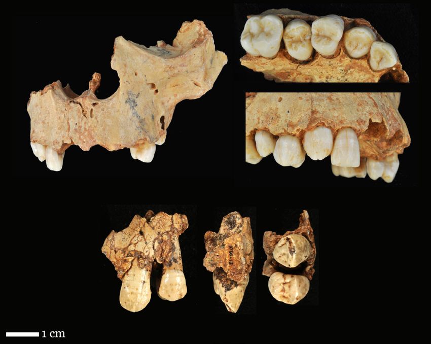
Research Articles               Sexual dimorphism of Homo antecessor canines

structures also offer the advantage of complet-       remains belong, initial studies showed that this
ing their formation early in an individual life.      population exhibited a unique combination of
Therefore, sexual estimation techniques based on      primitive characters (in their dental morphol-
dental features can be especially useful in paleo-    ogy) and derived traits (in their facial morphol-
anthropology for estimating the sex of immature       ogy) which led to the definition of a new spe-
individuals. In particular, methodologies based       cies: Homo antecessor (Bermúdez de Castro et
on permanent canines, the tooth that presents         al. 1997), the oldest species described so far in
the greatest degree of sexual dimorphism in the       the Early Pleistocene of Europe. Later studies
human dentition (e.g., Harris and Bailit 1988;        highlighted the expression of certain features
Hillson 1996; Işcan and Kedici 2003; Peckmann         shared with other Eurasian populations and,
et al. 2015), allow estimating the sex of indi-       in particular, with those that lived during the
viduals from the age of six, which is the age at      Middle and early Later Pleistocene (Martinón-
which the canine crown completes its formation        Torres 2006; Bermúdez de Castro et al. 2015;
(Moorrees et al. 1963).                               Martinón-Torres et al. 2019). This unique
     On the other hand, while the information         mosaic of skeletal and dental characteristics sug-
about modern human populations, Neanderthals          gested that this species might be phylogenetically
and pre-Neanderthals is relatively abundant (e.g.,    close to the divergence between Neanderthals
Wolpoff 1979; Smith 1980; Trinkaus 1980;              and modern humans (Bermúdez de Castro et al.
Arsuaga et al. 1997; Rosas 1997; Lorenzo et al.       2015). In 2020, in an innovative study carried
1998; Rosas et al. 2002; García-Campos et al.         out by Welker and colleagues, the dental enamel
2020) the scarcity and geographically scattered       proteomes of H. antecessor were analyzed. This
pre-Middle Pleistocene fossil record prevents a       research provided evidence that the TD.2 homi-
proper understanding of extinct hominins vari-        nids belonged to a close sister lineage to subse-
ability (e.g. McHenry 1991, 1994; Richmond            quent Middle and Late Pleistocene hominins,
and Jungers 1995; Reno et al. 2003, 2010;             including modern humans, Neanderthals and
Harmon 2009, 2006). In this context, it is par-       Denisovans (Welker et al. 2020). Regarding the
ticularly interesting to attempt an assessment of     paleodemography of this population, the study
the variability of the hominin sample from Gran       of the maxillar, mandibular and dental samples
Dolina-TD6.2 site, from the Sierra de Atapuerca       from this site resulted in a minimum number of
archaeological complex.                               eight individuals, although given the small area
     The TD6.2 level of the Gran Dolina cavity        excavated so far, it is suspected that this num-
has provided a large number of archaeological         ber could be higher (Bermúdez de Castro et al.
and paleontological remains which have allowed        2006). The high percentage (75 %) of imma-
to document the presence of human activity in         ture individuals in the hypodigm of TD6.2 is
this hill range for at least the last million years   noteworthy (Bermúdez de Castro et al. 2015).
(Carbonell et al. 2008; Rodríguez et al. 2011).       Despite the various research lines developed with
This has made of this site one of the most            this fossil sample (e.g., Bermúdez de Castro et al.
important references for Quaternary research          1997, 2003, 2008, 2015; Arsuaga et al. 1999;
(Carbonell et al. 1999). The human fossil remains     Bermúdez de Castro et al. 1999), there were no
found in Gran Dolina-TD6.2 have been studied          studies in which the sexual dimorphism of this
by several authors (e.g., Bermúdez de Castro et       population was assessed. This is mainly because
al. 1997, 1999, 2003, 2008, 2015; Arsuaga et al.      most individuals included Gran Dolina-TD6.2
1999; Lorenzo et al. 1999; Carbonell et al. 2005;     sample has not completed their development,
Gómez-Robles et al. 2012; Martinón-Torres             which complicates their sexual estimation.
et al. 2012, 2019; Martín‐Francés et al. 2018,        In this preliminary study, microtomographic
2020). Concerning the taxonomy and phyloge-           techniques have been employed to analyze two
netic position of the population to which these       human maxillary permanent canines from Gran

2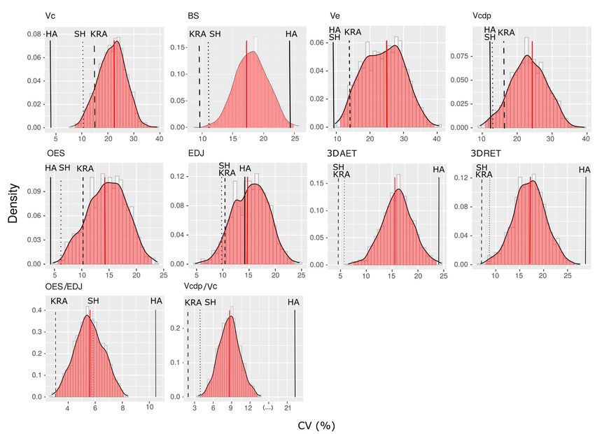
Sexual dimorphism of Homo antecessor canines Research Articles
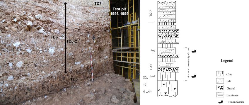
Fig. 1 - Stratigraphy of the Gran Dolina site. On the left, the sub-unit TD6.2 is indicated in the image
of the Gran Dolina site (photo taken by M. A. Martín). On the right, the main lithostratigraphic units
described in the section uncovered of the site (image taken from Campaña et al. 2017).
Dolina-TD6.2 for trying to evaluate the degree This sexually dimorphic pattern has been also
of variability of their dental tissue proportions employed for sex estimation in past populations.
and whether this variability can be attributed to In a study by García-Campos et al. (2020), den-
sexual dimorphism. tal histology was successfully used to estimate the
Several studies have shown that the den- sex and the degree of sexual dimorphism present
tal tissue proportions of permanent teeth in the Middle Pleistocene population of the Sima
are a dimorphic feature, not only in modern de los Huesos (SH) site of the Sierra de Atapuera
humans but also in other hominoid species (Spain), as well as in the dental sample from the
(e.g., Schwartz and Dean 2001; Schwartz et al. Neanderthal site of Krapina (Croatia).
2005; Saunder et al. 2007; Feeney et al. 2010; In order to evaluate the degree of variation
García-Campos et al. 2018a,b, 2020; Sorenti of H. antecessor teeth we employed the Person
et al. 2019). Particularly, female individuals Variability Coefficient (CV) on one hand, and a
tend to have smaller canines and a distinctive Principal Component Analysis and the Euclidean
histological pattern characterized by the rela- Distance assessment on the other. We compared
tive predominance of the enamel component our results with those obtained in previous stud-
and a smaller dentine-pulp complex than the ies about from Sima de los Huesos site (Spain),
males from the same population (Saunder et al. the Neanderthal site of Krapina (Croatia), and
2007; Feeney et al. 2010; García-Campos et al. a large forensic sample of known sex (García-
2018a,b, 2020; Sorenti et al. 2019). In modern Campos et al. 2020).
humans, the differences observed in the volumes
and three-dimensional surfaces of permanent
canines’ dental tissues have turn out to be a use- Materials
ful tool to estimate the sex (García-Campos et
al. 2018a,b) with rates of success comparable to Gran Dolina site is a large cavity, 27 meters
those of other traditional metric and non-metric deep and with a maximum width of 17 meters,
methods based on the cranial and postcranial located in the so-called Trinchera del Ferrocarril
skeleton (e.g., Ateş et al. 2006; Acharya and of the Sierra de Atapuerca archaeological com-
Mainali, 2008; Hassett 2011; Zorba et al. 2013). plex (Burgos, Spain). The stratigraphic sequence
3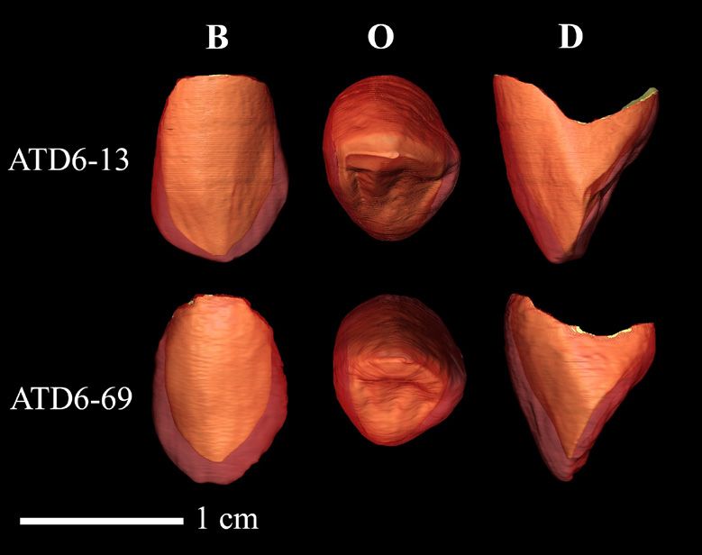
Research Articles               Sexual dimorphism of Homo antecessor canines

Fig. 2 - Permanent canines of Gran Dolina-TD6.2 included in the study. The upper row shows the
maxilla of individual H3 which includes the upper canine ATD6-69, the frontal view on the left and
the lower and lateral views on the right. In the lower row, the left maxillary canine of individual H1,
ATD6-13 is observed (buccal, mesial and occlusal view).

of the Gran Dolina cave is divided into eleven        a final estimated age range of 720- 950 ka for the
levels numbered in an increasing order from the       fossils from this site (Duval et al. 2018). In con-
base to the top: TD1-TD11 (Parés and Pérez-           clusion, taking into account the results obtained
González 1999). In the TD6.2 (Fig. 1) nearly          from all of these studies, as well as the biostrati-
170 human fossils belonging to at least 8 indi-       graphic information from this level (Cuenca-
viduals were found (Bermúdez de Castro et al.         Bescós et al. 1999; Cuenca-Bescós et al. 2015),
1997; Carbonell et al. 2010). Regarding the geo-      the TD6 hominis can be confidently assigned to
chronology of this level, the analysis of ESR dat-    MIS (Marine Isotope Stage) 21.
ing applied to quartz grains from TD6 yielded a           The Gran Dolina-TD6.2 dental sample com-
date that ranges between 600 ± 90 ka and 950 ±        prises 46 permanent and eight deciduous teeth,
90 ka (Moreno et al. 2015). In a new thermolu-        which includes two upper (ATD6-13 and ATD6-
minescence study Arnold and Demuro (2015) a           69) and two lower (ATD6-1 and ATD6-6) per-
weighted mean age of 840 ± 60 ka was obtained         manent canines. The canines ATD6-1, ATD6-6
for this level. Finally, a recent ESR analysis per-   and ATD6-13 belong to individual H1 (the holo-
formed directly on the human remains provided         type of H. antecessor) and the canine ATD6-69,

4
Sexual dimorphism of Homo antecessor canines                                           Research Articles

included within the maxilla ATD6-69, belongs to         scan was performed with two 0.1 mm Copper
individual H3 (Bermúdez de Castro et al. 2015, see      filters, 100-120 kV voltage and 110-140 µA
Fig. 2). In this study, two upper canines (ATD6-        amperage, resulting isometric voxel of 37 μm.
69 and ATD6-13) were analyzed. These teeth were         The subsequent image processing was performed
selected for their good state of conservation. The      using the Amira 6.0.0 software (Visage Imaging,
canine ATD6-69 has not reached the occlusal plane       Inc.). Dental tissues (enamel and dentine-pulp
and therefore does not present wear facet. In addi-     complex) were semi-automatically segmented
tion, the crown of this tooth has a linear hypoplasia   using the Watershed Segmentation Tool and
that is not very marked. On the other hand, the         through manual editing. Following the proto-
canine ATD6-13 presents an oval wear facet on its       col described in García-Campos et al. (2018a,b,
occlusal surface, but it does not reach the dentine.    2019, 2020), we considered the cervical line as
     As a comparative sample, we employed the           the fundamental morphological feature to isolate
specimens analyzed by García-Campos et al.              the crown and the root.
(2020). This sample includes a total of 86 max-              Next, on the virtually isolated crowns, we
illary canines from: the Sima de los Huesos             quantified the following absolute variables: the
site from Spain (n = 16), the Krapina site from         volume of the enamel cap (Ve, in mm3); the vol-
Croatia (n = 12) and a sample of modern humans          ume of the crown dentine including the crown
with different geographical origin (n = 58). We         pulp (Vcdp, in mm3); the surface area of the
considered the results obtained from those teeth        enamel-dentine junction (EDJS, in mm2); the
with a wear degree equal or lower than 3 (Molnar        outer surface of the enamel cap (OES, mm2);
1971) and only one antimere were assessed. The          and the basal surface of the crown (BS, in mm2).
sexual estimations of the fossil samples was taken      These values were subsequently used to compute
from García-Campos et al. (2020) who used               the 3-D average enamel thickness index (3DAET
a combination of two approaches: the Mean               = Ve/EDJS, mm); the 3-D relative enamel thick-
method and a Hierarchical cluster analysis. The         ness index (3DRET = 3DAET/) x 100, scale-
final sex estimation was established through a          free); the crown volume (Vc = Ve + Vcdp, mm3);
comparison of the results of both approaches.           the percentage of crown volume that is dentine
The modern human sample was composed by                 and pulp (Vcdp/Vc = Vcdp/Vc x100, percentage
forensic samples of known sex. For more details         scale); and relative outer enamel complexity ratio
on the sexual estimation techniques see García-         (OES/EDJ, free-scale). Due to ATD6-69 has no
Campos et al. (2020).                                   already finished the root formation the volume
                                                        of the root dentine including the pulp (Vr, mm3)
                                                        was not assessed. These variables were described
Methods                                                 by Olejniczak et al. (2008a,b) and Skinner et
                                                        al. (2008). They were previously employed by
    The isolated tooth TD6-13 was scanned               García-Campos et al. (2018a,b) to estimate the
using the Scanco Medical AG Micro-Computed              sex in modern human samples, reaching accu-
Tomography 80 housed at the Centro Nacional             racy rates of up to 92.3%. Likewise, they were
de Investigación sobre la Evolución Humana              also used to estimate the sex of Neanderthal and
(CENIEH) in Burgos. Scans were performed                pre-Neanderthal fossil samples (García-Campos
employing two 0.1 mm Copper filters and using           et al. 2020).
a voltage of 70 kV and an amperage of 114 µA.                Statistical analyses were performed using the
The resultant slice thickness was 18 micrometers        SPSS software (v. 18.0, SPSS Science, Inc.). A
(μm). ATD6-69, which is included in a maxil-            descriptive statistical analysis was applied con-
lary fragment, was scanned using a Phoenix v/           sidering the data obtained in this study from
tome/x s (GE Measurement & Control) avail-              the maxillary canines of H. antecessor, as well as
able in the same research centre. In this case, the     from SH, Krapina and a modern human sample

                                                                                                        5
Research Articles Sexual dimorphism of Homo antecessor canines
Tab. 1 - Descriptive statistics for the measurements and indices associated included in this study. The
data from SH, KRA and RMH have been obtained from García-Campos et al. (2020). The crown measure-
ments were assessed in the slightly worn upper canines (1-3 wear stage following Molnar, 1971).
MEAN (SD)
SAMPLE SUB- N VC BS VE VCDP OES EDJS 3DAET 3DRET VCDP/ OES/
SAMPLE (mm3) (mm2) (mm3) (mm3) (mm2) (mm2) (mm) (SCALE VC EDJS
FREE) (%) (SCALE
FREE)
ATD6-13 - 371.17 72.30 143.24 227.43 258.80 196.43 0.73 12.01 157.88 1.32
ATD6-69 - 355.49 51.04 165.36 190.13 243.98 159.75 1.04 18.00 114.98 1.53
HA
Total 2 363.33 61.67 154.65 208.69 251.39 178.09 0.89 15.00 136.43 1.43
(11.09) (15.03) (15.15) (26.24) (10.48) (25.94) (0.22) (4.24) (30.33) (0.15)
Females* 3 287.60 46.14 130.35 157.26 208.93 149.64 0.87 16.13 54.71 1.40
(9.68) (2.25) (8.52) (1.62) (5.66) (2.55) (0.05) (0.76) (1.47) (0.02)
Males* 3 364.59 59.39 157.67 206.92 241.87 185.75 0.85 14.37 56.71 1.30
SH (18.59) (4.07) (9.67) (18.17) (16.49) (5.81) (0.04) (0.93) (2.97) (0.12)
Total 9 327.56 51.87 146.81 180.75 226.75 166.39 0.88 15.68 55.10 1.37
(35.34) (6.37) (14.25) (23.62) (17.52) (16.41) (0.05) (1.19) (2.08) (0.08)
Females* 5 379.45 52.58 160.86 218.60 250.26 180.37 0.89 14.81 57.61 1.39
(15.05) (1.98) (7.57) (9.91) (3.15) (3.40) (0.04) (0.65) (1.15) (0.03)
KRA Males* 1 524.26 64.83 215.3 308.96 314.14 229.46 0.94 13.88 58.93 1.37
Total 6 403.59 54.62 169.93 233.66 260.91 188.56 0.90 14.65 57.83 1.39
(60.63) (5.31) (23.23) (37.94) (26.23) (20.27) (0.04) (0.69) (1.16) (0.03)
Females 27 223.94 33.08 109.96 113.98 174.43 115.25 0.95 19.71 50.94 1.51
(35.93) (3.72) (20.04) (18.57) (18.64) (11.32) (0.12) (2.52) (3.36) (0.07)
Males 29 286.86 43.82 128.80 158.07 204.31 142.43 0.90 16.61 55.54 1.43
RMH (57.05) (5.61) (34.77) (26.95) (29.61) (17.81) (0.17) (2.97) (4.64) (0.08)
Total 56 256.52 38.64 119.72 136.81 189.90 129.33 0.92 18.10 53.32 1.47
(57.21) (7.21) (29.93) (32.05) (28.94) (20.24) (0.15) (3.15) (4.66) (0.08)
* We take into account the sexual estimates of the fossil samples by García-Campos et al. (2020).
by Garcia-Campos et al. (2020). The mean and the magnitude of the differences described; nev-
standard deviation of each population were ertheless, we performed other approaches to
assessed, as well as of the male and female sub- evaluate the magnitude of the variability of H.
samples within each group. We take into account antecessor upper canines.
the sexual estimates of the fossil samples and the Firstly, to evaluate the variability of each vari-
actual sex in the case of forensic samples (see able independently, the Pearson´s Coefficient of
García-Campos et al. 2020). Variation (V=(δx/μx) x 100) was then assessed.
Because of the sample size, we could not The CV is considered to be very highly corre-
apply a comparative statistical analysis to assess lated with sexual dimorphism since when the
6Sexual dimorphism of Homo antecessor canines Research Articles
difference between male and females means
increases it causes a proportional increase in
the standard deviation of the pooled-sex sample
(Fleagle et al. 1980). For this reason, this coef-
ficient has previously been used by other authors
to study the internal variability of the fossil
samples (e.g. Kay 1982; Leutenegger and Shell
1987; Arsuaga et al. 1997; Bermúdez de Castro
et al. 2001). In order to avoid that differences
in sample size may interfere in the comparison
of the intra-population variability between mod-
ern human and fossil samples, a bootstrapping
with replacement was employed. It was simu-
lated 1000 random datasets with a sample size
Fig. 3 - Virtual reconstruction of the analysed
of nine individuals from the original modern teeth. In this figure are represented: the upper
human sample using R statistical software. Then, left canine ATD6-13 (H1) and the upper right
we calculated the CV in each dataset. The mean canine ATD6-69 (H3). The right canine ATD6-69
has been mirrored in the image. Views: Buccal
value obtained from the random samples, as well (B), occlusal (O) and distal (D).
as the 95% confidence interval of their distribu-
tion, were compared with the value of the fossil
samples. larger. Despite this, ATD6-69 canine has abso-
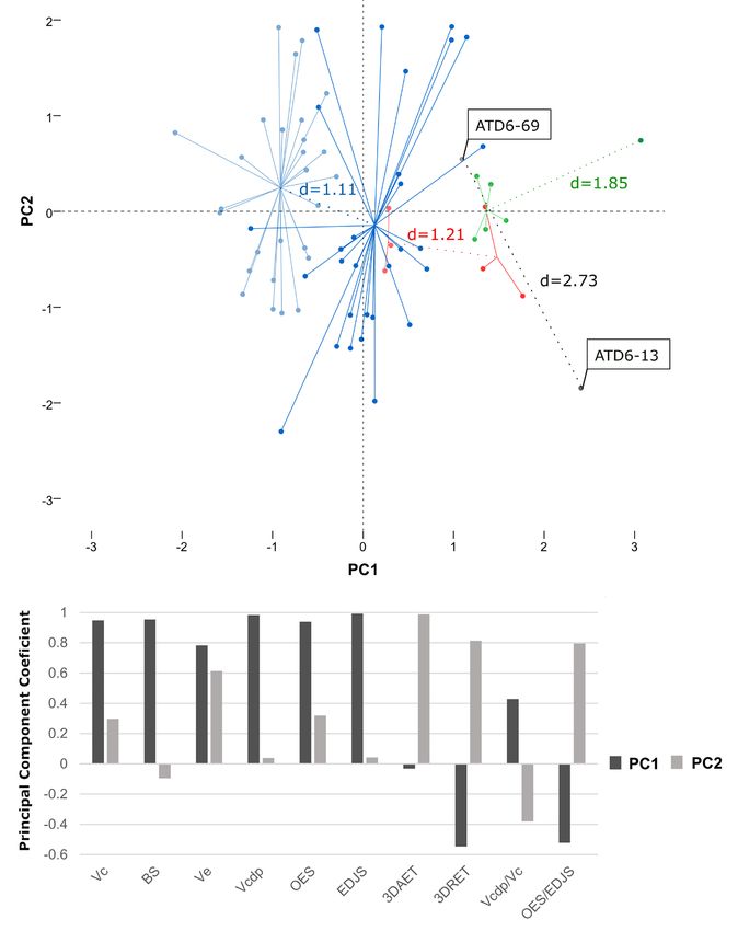
Subsequently, a Principal Component Analysis lutely (Ve) and relatively (3DAET, 3DRET, and
(PCA) was applied to assess the variability of all OES/EDJS) greater enamel cap dimensions than
variables as a whole. We only included absolute ATD6-13. Lastly, ATD6-13 displays a higher
and relative variables measured in teeth with a percentage of crown volume that is a dentine-
wear degree equal or lower than 3 (Molnar, 1971). pulp complex.
Once the PCA results were obtained, the centroids The Pearson´s Coefficient of Variation (CV)
of each group were located and the Euclidean results obtained from H. antecessor (HA), Sima de
Distance (d) between the centroids of the groups los Huesos (SH), Krapina (KRA) and the recent
formed by male and female individuals within each modern human (RMH) samples are presented
population was assessed. The Euclidean Distance in Figure 4. For all absolute variables recorded,
between the canines ATD6-13 and ATD6-69 was except for the crown basal surface (BS), the CV
also calculated. values of the HA maxillary canines fall below the
mean value obtained from the 1000 recent mod-
ern human random samples, but near to the val-
Results ues obtained for SH and KRA. Among them, the
CV values for the variables Vc, Ve and OES fall
The results of the measurement of the den- below to the recent modern humans 95% confi-
tal tissues volumes and surface areas (Tab. 1 and dence interval. The CV value obtained for the BS
Fig. 3) show that the canine ATD6-13 displays of HA dental pieces overtakes the mean of RMH
higher crown dimensions than ATD6-69. The variation, although it falls within the 95% con-
marked canine size variation is also reflected in fidence interval. On the other hand, CV of the
the absolute tissue dimensions. In ATD6-69, the all relative variables recorded (3DAET, 3DRET,
enamel outer surface (OES) and the crown den- Vcdp/Vc, OES/EDJS) in HA maxillary canines
tine-pulp complex dimensions (Vcdp, EDJS) are are clearly higher than the values obtained in SH,
also smaller, partially due to the lower size of its KRA and RMH samples, even surpass the 95%
crown, whereas in ATD6-13 these variables are confidence interval of the RMH variation range.
7Research Articles Sexual dimorphism of Homo antecessor canines
Regarding the results of the Principal 2005). Likewise, genetic alterations of these
Component Analysis, they are provided in Figure genes cause different dental tissue defects (Hu et
5. The PCA analysis generated two principal al. 2012; Cho et al. 2014). On the other hand,
components which explain 90.14% of the total sex hormones not only seem to play an essential
variability observed in the sample. The results role in the development of dental tissues, but also
of the PCA allow us to observe that, in general may be behind of the changes in secondary den-
terms, in each population the female individu- tin deposition produced over the lifetime of the
als are displaced to the left quadrants with regard individual (Zilberman and Smith 2001; Guatelli-
to the male individuals. These quadrants com- Steinberg et al. 2008; Alvesalo 2009; Ribeiro et
prise the negative values for the first compo- al. 2012, 2013; Pentinpuro et al. 2014, 2017).
nent which are obtained when the dental pieces For this reason, the study of the dental tissue
exhibit higher values for the variables 3DAET, volumes and surface areas of permanent canines
3DRET and OES/EDJS, as well as lower values has been previously employed to estimate the
for the absolute variables and the Vcdp/Vc index. sex and degree of sexual dimorphism of modern
ATD6-13 and ATD6-69 appear separated in the human populations (e.g., Saunders et al. 2007;
PCA, being the first displaced to the upper left Feeney et al. 2010; García-Campos et al. 2018a,b,
quadrant. The value for the Euclidean Distance 2020; Sorenti et al. 2019) as well as of some
calculated using the coordinates of the two Middle Pleistocene human groups of Europe,
points that represent the two maxillary canines of such those from Sima de los Huesos (SH) and
Gran Dolina-TD6.2 in the PCA was 2.73. This Krapina (Croatia) (García-Campos et al. 2020).
value is more than the double of those obtained The demographic structure of both SH and
through the comparison of the centroids of the Krapina is characterized by the predominance of
point clouds formed by the female and male subadult and/or juvenile individuals (Bocquet-
individuals from the recent modern human sam- Appel and Arsuaga 1999), which makes difficult
ple (d = 1.11), the Sima de los Huesos sample (d to obtain conclusive sexual estimates from their
= 1.21) or the Krapina sample (d = 1.85). cranial and postcranial remains (Bermúdez de
Castro et al. 2001; Rosas et al. 2002; Arsuaga et
al. 2014). However, the assessment of the den-
Discussion tal tissue proportions of their permanent canines
has allowed not only the confirmation of the sex
Several studies have documented that den- allocation of individuals previously assigned in
tal tissue proportions in the permanent denti- the literature but also to estimate the sex of the
tion are sexually dimorphic not only in modern youngest individuals, which were not assessed in
humans but also in other species of hominoids previous studies (García-Campos et al. 2020). In
(e.g., Schwartz and Dean 2001; Schwartz et al. total, employing this methodology it was possi-
2005; Saunders et al. 2007; Feeney et al. 2010; ble to estimate the sex of 15 out of the 17 indi-
García-Campos et al. 2018a,b, 2020; Sorenti et viduals of the SH sample, as well as of all the
al. 2019). These differences appear to have both Krapina individuals of which permanent canines
a genetic and a hormonal origin (e.g., Alvesalo were available (García-Campos et al. 2020). As
1997, 2009; Zilberman and Smith 2001; in the case of Sima de los Huesos and Krapina
Guatelli-Steinberg et al. 2008; Pentinpuro et samples, the Gran Dolina-TD6.2 population is
al. 2014, 2017). On one hand, the quantitative composed mainly of immature individuals. This
and qualitative differences in the transcriptional is a limitation that must be addressed when car-
products of the amelogenin genes, present on rying out paleodemographic studies on this fossil
both the X and Y chromosomes, influence the sample.
proportions in which hard dental tissues are The sample of H. antecessor included in this
present (Salido et al. 1992; Schwartz and Dean study is composed of two permanent canines: the
8Sexual dimorphism of Homo antecessor canines Research Articles
left maxillary canine ATD6-13, belonging to indi- this individual maxilla may be due to the effect of
vidual H1, holotype of the species (Bermúdez de intersexual variability.
Castro et al. 1997); and the left maxillary canine The analysis of the two upper canines of
ATD6-69 of individual H3. The individual H1 Gran Dolina-TD6.2, belonging to individu-
is identified by a set of isolated permanent teeth, als H1 (ATD6-13) and H3 (ATD6-69), reveals
as well as a fragment on the right side of a man- that the H1 canine has larger crown dimensions
dibular body with the molar series in-situ and a (Tab.1 and Fig. 3). Previous studies have already
small and deteriorated fragment on the left side described differences in size between ATD6-13
of a maxilla with the canine and first premolar and ATD6-69 canines (Bermúdez de Castro
in-situ (Arsuaga et al. 1999, see Fig. 2). The H1 et al. 1999; Martinón-Torres et al. 2019). The
teeth are very large in the context of the variability canine ATD6-69 is in process of eruption, which
known for the European Pleistocene fossil record has prevented that the mesiodistal (MD) and
(Bermúdez de Castro et al. 1999; Martinón- buccolingual (BL) diameters of its crown could
Torres et al. 2019). In particular, the size of the be measured employing traditional techniques.
maxillary canine of H1 is at the upper limit of the However, once the canine ATD6-69 was virtu-
range of variation of the genus Homo (Bermúdez ally reconstructed in this study, it was possible to
de Castro et al. 1999; Martinón- Torres et al. measure the MD and BL diameters of its crown.
2019). For this reason, H1 was previously esti- As it was expected, the values obtained (MD =
mated to represent a young male individual with 8.57 mm, BL = 9.73 mm) are lower than those
an age at death of 12.9 years old according to cur- observed by Bermúdez de Castro et al. (1999)
rent standards (Bermúdez de Castro et al. 1999). in ATD6-13 (MD = 8.9 mm, BL = 11.0 mm).
Likewise, the remarkable size difference between This is consistent with the results obtained from
the teeth of the mandibles ATD6-5 (H1) and the evaluation of the volume and surface areas of
ATD6-96 (H7) makes Carbonell et al. (2005) the crown (Vc, BS, OES, EDJS) of both dental
suggest that the former was probably a male, pieces. Interestingly, despite individual H1 upper
whereas the latter mandible belonged to a female. canine has larger crown dimensions than indi-
On the other hand, the individual H3 is identi- vidual H3, it displays lower volume and relative
fied by the specimen ATD6-69, which consists enamel dimensions (3DAET, 3DRET and OES/
of an important part of the left side of the face, EDJS). This could be potentially attributed to
the alveolar process of the maxilla, the anterior the slight dental wear of individual H1 canine
portion of the palate and the vomer (Arsuaga et (degree of wear 2 according to Molnar, 1971)
al. 1999, see Fig. 2). The incisor, canine, first and whereas the crown of H3 is intact (Bermúdez de
second premolars, first permanent molar on the Castro et al. 1999). However, as it is explained
right side and the first premolar and the com- below, these differences are too large to be only
plete molar series on the left side are preserved explained by the effect of dental wear.
in-situ (Bermúdez de Castro et al. 2006; Martín‐ The results obtained from the Pearson´s
Francés et al. 2018, 2020). Except for the alveo- Coefficient of Variation (CV) show that, for the
lar width, the facial dimensions of ATD6-69 are most of the absolute variables recorded, the CV
small (Arsuaga et al. 1999). This fact has been values of the H. antecessor maxillary canines fall
associated with the age of death of H3, which below the mean value obtained from the 1000
according to current standards, could be between recent modern human random samples, but near
10 and 12 years old (Bermúdez de Castro et al. to the values obtained for SH and KRA (Fig.
1999). However, the results obtained in various 4). However, for all relative variables recorded
studies suggest that the 85% of maxillar size is (3DAET, 3DRET, Vcdp/Vc, OES/EDJS) and
reached by the age of 6 (Enlow and Bang 1965; the area of the crown basal surface (BS), the
Sperber et al. 2001; Vardimon et al. 2010), which CV values of Gran Dolina-TD6.2 teeth are
opens the door to the hypothesis that the size of clearly higher than the values obtained in SH,
9Research Articles Sexual dimorphism of Homo antecessor canines Fig. 4 - Frequency histograms of coefficients of variation (CV, percentage scale) of each absolute variable and associated indices evaluated in upper canines, calculated from 1000 random samples of individuals belonging to the recent modern human sample of the same size that the SH fossil-sam- ple. The vertical red line marks the CV mean value of the whole recent modern human sample and the red shaded area shows 95% confidence interval of this distribution. On the other hand, the black line indicates the CV value Gran Dolina-TD6.2 (HA) hominid dental sample. Finally, the dashed black lines indicate the CV the mean value of the Sima de los Huesos (SH) and Krapina (KRA) sample. KRA and RMH samples, even surpassing the more than double of those obtained through the 95% confidence interval of the RMH variation comparison of the centroids of the point clouds range (Fig. 4). Likewise, as it can be appreciated formed by the female and male individuals from in the scatter plot (Fig. 5), the two canines of the recent modern human sample (d = 1.11), Gran Dolina-TD6.2 appear clearly separated the Sima de los Huesos sample (d = 1.21) or the in the PCA. The canine ATD6-69 falls within Krapina sample (d = 1.85). the positive values of Principal Component 1 The magnitude of the variability observed (PC1), but is closer to the ordinate axis than in the Gran Dolina-TD6.2 dental sample might ATD6-13, which holds higher values for the make us think that the two teeth assessed in this first component. For the Principal Component study may belong to individuals from different 2 (PC2), ATD6-69 falls within the positive taxa. Two- and three-dimensional assessment area, whereas ATD6-13 holds negative values. of dental tissues, and specifically of the enamel The assessment of Euclidean Distance between thickness, has been also used in taxonomic both dental pieces resulted in a value of 2.73, studies to infer the identity and phylogenetic 10
Sexual dimorphism of Homo antecessor canines                                          Research Articles

Fig. 5 - Principal Component Analysis (PCA) applied to the maxillary canines from Gran Dolina -TD6.2 (in
grey), Sima de los Huesos (in red), Krapina (in green) and the modern human (in blue) samples.
Above, the scatter plot represents the first two components of the PCA (PC1 and PC2), which explain
90.14% of the total variability observed in the sample. In the graph, each point appears referenced
concerning the centroid of each sub-sample. The values Euclidean distance (d) between the centroids
of the groups formed by male and female individuals within each population can be also seen in the
scatter plot. Male individuals in the Krapina sample are represented by a single tooth, whose coordi-
nates were employed to calculate the Euclidean distance. The Euclidean distance between the canines
ATD6-13 and ATD6-69 has been also assessed thought the values of their coordinates. Below, the
coefficients of each of the variables and index evaluated for each component can be seen.

                                                                                                     11
Research Articles Sexual dimorphism of Homo antecessor canines
relationships of past hominid species (e.g., Kono of the Neanderthal and modern human lineages
2004; Olejniczak et al. 2008; Smith et al. 2012; (Bermúdez de Castro et al. 2016), which might
Martín-Francés et al. 2018; García-Campos et al. explain the presence of individuals with derived
2019). In particular, the thinly enameled pattern and primitive conditions in their dentition within
has been identified as a distinctive Neanderthal the TD6.2 population. Another possibility is
lineage features (Olejniczak et al. 2008a; Bayle that the differences observed in the dental tissue
et al. 2009; Smith et al. 2012; Buti et al. 2017; proportions of H1 and H3 individuals might
García-Campos et al. 2019). However, neither be due to the presence of sexual dimorphism.
taphonomic nor morphological evidence seem to The differences appreciated between ATD6-13
support this scenario. On the one hand, ATD6- and ATD6-69 concur with the histological pat-
13 and ATD6-69, as well as the other dental and tern that tends to distinguish the dentition of
cranial remains associated with both teeth, were males and females in recent modern humans
discovered in a small excavation area of only 6 (e.g., Feeney et al. 2010; García-Campos et al.
m2, a survey performed in 1994 in the Gran 2018a,b, 2020; Saunders et al. 2007; Sorenti et
Dolina site (Bermúdez de Castro et al. 1999). al. 2019) as well as in other hominoid species
Likewise, all these remains come from the same (Schwartz and Dean 2001; Schwartz et al. 2005;
lithostratigraphic subunit, ATD6.2 (Bermúdez García-Campos et al. 2020). Numerous stud-
de Castro et al. 1997; Carbonell et al. 2010). ies have shown that female individuals tend to
On the other hand, both upper canines share have smaller teeth, with a relative predominance
some morphological traits that characterized the of the enamel component and a smaller dentin-
mosaic pattern typical from H. antecessor denti- pulp complex than male individuals (Schwartz
tion, such as the asymmetry of the occlusal edge, and Dean 2001, 2005; Schwartz et al. 2005;
the reduction mesial cutting edge and the degree Smith et al. 2006; Saunders et al. 2007; Feeney
of inclination of the distal one, or the loss of the et al. 2010; García-Campos et al. 2018a,b, 2020;
cingulum and associated structures (Martinón- Sorenti et al. 2019). Therefore, the presence of
Torres et al. 2019). Therefore, the most parsi- this pattern in Gran Dolina TD6.2 canines,
monious interpretation the results obtained here premolars and molars would reinforce the idea
is that the wide variability observed in the his- that the variability found in the TD6.2 sample is
tological pattern of the two canines from Gran likely due to sexual variation. If this is the case,
Dolina-TD6.2 is result of the intra-population the pattern observed in H1 dentition would be
variability not of the inter-population variability. indicative that we are face on a male individual,
The wide intra-population variability seen while H3 would be a female individual, which
in H. antecessor canines has also been described would support the conclusions obtained by other
in its posterior dentition by Martin-Francés et authors (Bermúdez de Castro et al. 1999, 2006;
al. (2018, 2020). On one hand, Martín-Francés Carbonell et al. 2005).
and colleagues describe that: “The individual Unfortunately, the small sample size of TD6.2
TD6-H1 upper premolars (P 3: ATD6-7 and population prevents us from obtaining conclusive
ATD6-13, and P 4: ATD6-8 and ATD6-9) inferences based on the dental tissue pattern of
exhibit thin enamelled crowns (…) On the con- their permanent canines. The discovery of new fos-
trary, the upper premolars (ATD6-69 specimens) sils at this level of Gran Dolina could help to bet-
belonging to individual H3 exhibit the thick pat- ter understand the intrapopulation variability of
tern in their crowns”. Likewise, similar but less this sample and, therefore, confirm or refute these
marked differences could be appreciated in the hypotheses. In any case, the results obtained in this
molars of both individuals (Martín-Francés et al. study demonstrate, once again, the usefulness of
2018). These authors support that this high vari- the dental tissue proportions of permanent canines
ability would be compatible with H. antecessor for the sexual dimorphism assessment in mod-
species being close to the last common ancestor ern and past human populations. In particular,
12Sexual dimorphism of Homo antecessor canines Research Articles
the study of enamel and dentine dimensions can Alvesalo L (2009) Human sex chromosomes in
be a particularly useful tool in palaeoanthropo- oral and craniofacial growth. Arch Oral Biol
logical contexts, where other bone structures are 54 Suppl 1:S18–24. https://doi.org/10.1016/j.
often fragmented or absent, and especially in those archoralbio.2008.06.004
where their demographic structure has a greater Antón SC (2003) Natural history of Homo erec-
representation of subadult individuals. tus. Am J Phys Anthropol Suppl 37:126–170.
https://doi.org/10.1002/ajpa.10399
Arnold LJ, Demuro M (2015) Insights into TT-
Acknowledgments OSL signal stability from single-grain analyses
of known-age deposits at Atapuerca, Spain.
This study has been supported by the Dirección Gen- Quat Geochronol 30:472–478. https://doi.
eral de Investigación of the Spanish Ministerio de org/10.1016/j.quageo.2015.02.005
Economía y Competitividad (MINECO /FEDER) Arsuaga JL, Carretero JM, Lorenzo C, et al
grant number: PGC2018-093925-B-C31 and The (1997) Size variation in Middle Pleistocene
Leakey Foundation through the personal support of humans. Science 277:1086–1088. https://doi.
G. Getty (2013) and D. Crook (2014-2020) to org/10.1126/science.277.5329.1086
M. M.-T. C. G-C and L. M-F are the recipients Arsuaga JL, Martínez I, Arnold LJ, et al (2014)
of a post-doctoral research grant at the Atapuerca Neandertal roots: Cranial and chronological
Foundation. The micro-CT images were obtained evidence from Sima de los Huesos. Science
in the Laboratory of Microscopy of the CENIEH- 344:1358–1363. https://doi.org/10.1126/
ICTS (Spain) in collaboration with CENIEH staff. science.1253958
We thank all the members of the Atapuerca research Arsuaga JL, Martínez I, Lorenzo C, et al (1999)
team, in particular those who excavate the Gran The human cranial remains from Gran Dolina
Dolina-TD6.2 site, for their dedicated work. We Lower Pleistocene site (Sierra de Atapuerca,
also acknowledge several people for providing access Spain). J Hum Evol 37:431–457. https://doi.
to the modern human sample included. The African org/10.1006/jhev.1999.0309
sample from Sudan was provided by Dr Christopher Ateş M, Karaman F, Işcan MY, Erdem TL (2006)
Dean from the Anatomy Department at University Sexual differences in Turkish dentition. Leg
College London. We are indebted to A. Oettlé, G. Med 8:288–292. https://doi.org/10.1016/j.
Krüger and E.N. L’Abbé for kindly authorizing legalmed.2006.06.003
access to the Pretoria Bone Collection (PBC) of the Bayle P, Braga J, Mazurier A, Macchiarelli R
University of Pretoria, and to Dr. Clément Zanolli (2009) Dental developmental pattern of the
for interceding and making it possible. In the same Neanderthal child from Roc de Marsal: a high-
way, we would like to acknowledge to Dr. Bernardo resolution 3D analysis. J Hum Evol 56:66–75.
Perea Pérez, for authorizing access to the collections https://doi.org/10.1016/j.jhevol.2008.09.002
from Escuela de Medicinal Legal de la Universidad Bermúdez de Castro JM, Arsuaga JL, Carbonell
Complutense de Madrid. E, et al (1997) A hominid from the lower
Pleistocene of Atapuerca, Spain: possible ances-
tor to Neandertals and modern humans. Science
References 276:1392–1395. https://doi.org/10.1126/
science.276.5317.1392
Acharya AB, Mainali S (2008) Are dental in- Bermúdez de Castro JM, Carbonell E, Gómez
dexes useful in sex assessment? J Forensic A, et al (2006) Paleodemografía del hipodigma
Odontostomatol 26:53–59 de fósiles de homininos del nivel TD6 de Gran
Alvesalo L (1997) Sex chromosomes and human Dolina (Sierra de Atapuerca, Burgos): estudio
growth. Hum Genet 101:1–5. https://doi. preliminar. Estudios Geológicos 62:145–154.
org/10.1007/s004390050575 https://doi.org/10.3989/egeol.0662114
13Research Articles Sexual dimorphism of Homo antecessor canines Bermúdez de Castro JM, Martinón-Torres M, Carbonell E, Bermúdez de Castro JM, Parés Martín-Francés L, et al (2015) Homo anteces- JM, et al (2008) The first hominin of Europe. sor: The state of the art eighteen years later. Nature 452:465–469. https://doi.org/10.1038/ Quat Int 433:22-31. https://doi.org/10.1016/j. nature06815 quaint.2015.03.049 Carbonell E, Cáceres I, Ruiz ML et al (2010) Bermúdez de Castro JM, Martinón-Torres M, Cultural cannibalism as a Paleoeconomic sys- Sarmiento S, et al (2003) Gran Dolina-TD6 tem in the European Lower Pleistocene: the versus Sima de los Huesos dental samples from case of level TD6 of Gran Dolina (Sierra de Atapuerca: evidence of discontinuity in the Atapuerca, Burgos, Spain). Curr Anthropol European Pleistocene population? J Archaeol 4:539–549. https://doi.org/10.1086/653807 Sci 30:1421–1428. https://doi.org/10.1016/ Carbonell E, Mosquera M, Rodríguez XP, et al S0305-4403(03)00036-0 (1999) Out of Africa: The Dispersal of the Bermúdez de Castro JM, Pérez-González A, Earliest Technical Systems Reconsidered. Journal Martinón-Torres M, et al (2008) A new early of Anthropological Archaeology 18:119–136. Pleistocene hominin mandible from Atapuerca- https://doi.org/10.1006/jaar.1998.0331 TD6, Spain. J Hum Evol 55:729–735. https:// Cho ES, Kim K-J, Lee K-E, et al (2014) Alteration doi.org/10.1016/j.jhevol.2008.03.006 of conserved alternative splicing in AMELX Bermúdez de Castro JM, Rosas A, Nicolás ME causes enamel defects. J Dent Res 93:980–987. (1999) Dental remains from Atapuerca-TD6 https://doi.org/10.1177/0022034514547272 (Gran Dolina site, Burgos, Spain). J Hum Cuenca-Bescós G, Blain H-A, Rofes J, et al (2015) Evol 37:523–566. https://doi.org/10.1006/ Comparing two different Early Pleistocene jhev.1999.0323 microfaunal sequences from the caves of Bermúdez de Castro JM, Sarmiento S, Cunha Atapuerca, Sima del Elefante and Gran Dolina E, et al (2001) Dental size variation in the (Spain): Biochronological implications and Atapuerca-SH Middle Pleistocene homi- significance of the Jaramillo subchron. Quat nids. J Hum Evol 41:195–209. https://doi. Int 389:148-158. https://doi.org/10.1016/j. org/10.1006/jhev.2001.0491 quaint.2014.12.059 Bocquet-Appel JP, Arsuaga JL (1999) Age Cuenca-Bescós G, Laplana C, Canudo JI Distributions of Hominid Samples at Atapuerca (1999) Biochronological implications of the (SH) and Krapina Could Indicate Accumulation Arvicolidae (Rodentia, Mammalia) from by Catastrophe. J Archaeol Sci 26:327–338. htt- the Lower Pleistocene hominid-bearing lev- ps://doi.org/10.1006/jasc.1998.0370 el of Trinchera Dolina 6 (TD6, Atapuerca, Buti L, Le Cabec A, Panetta D, et al (2017) 3D enam- Spain). J Hum Evol 37:353–373. https://doi. el thickness in Neandertal and modern human org/10.1006/jhev.1999.0306 permanent canines. J Hum Evol 113:162–172. Dirkmaat DC (ed) (2012) A companion to fo- https://doi.org/10.1016/j.jhevol.2017.08.009 rensic anthropology. John Wiley & Sons, Ltd, Campaña I, Benito-Calvo A, Pérez-González A, Chichester, UK. et al. (2017) Pleistocene sedimentary facies of Duval M, Grün R, Parés JM, et al (2018) The the Gran Dolina archaeo-paleoanthropological first direct ESR dating of a hominin tooth site (Sierra de Atapuerca, Burgos, Spain). Quat from Atapuerca Gran Dolina TD-6 (Spain) Int 433:68–84. https://doi.org/10.1016/j. supports the antiquity of Homo antecessor. quaint.2015.04.023 Quat Geochronol 47:120–137. https://doi. Carbonell E, Bermúdez de Castro JM, Arsuaga org/10.1016/j.quageo.2018.05.001 JL, et al (2005) An Early Pleistocene hominin Enlow DH, Bang S (1965) Growth and remodeling mandible from Atapuerca-TD6, Spain. Proc of the human maxilla. Am J Orthod 51:446– Natl Acad Sci USA 102:5674–5678. 464. https://doi.org/10.1016/0002-9416(65) https://doi.org/10.1073/pnas.0501841102 90242-3 14
Sexual dimorphism of Homo antecessor canines                                             Research Articles

Feeney RN, Zermeno JP, Reid DJ, et al (2010)          Harmon EH (2006) Size and shape variation
  Enamel thickness in Asian human canines and            in Australopithecus afarensis proximal fem-
  premolars. Anthropol Sci 118:191–198. htt-             ora. J Hum Evol 51:217–227. https://doi.
  ps://doi.org/10.1537/ase.091006                        org/10.1016/j.jhevol.2006.01.009
Fleagle JG, Kay RF, Simons EL (1980) Sexual di-       Harris EF, Bailit HL (1988) A principal com-
  morphism in early anthropoids. Nature 287:328–         ponents analysis of human odontometrics.
  330. https://doi.org/10.1038/287328a0                  Am J Phys Anthropol 75:87–99. https://doi.
García-Campos C, Martinón-Torres M, Martín-              org/10.1002/ajpa.1330750110
  Francés L, et al (2018a) Contribution of dental     Hassett B (2011) Technical note: estimating sex using
  tissues to sex determination in modern human           cervical canine odontometrics: a test using a known
  populations. Am J Phys Anthropol 166:459–              sex sample. Am J Phys Anthropol 146:486–489.
  472. https://doi.org/10.1002/ajpa.23447                https://doi.org/10.1002/ajpa.21584
García-Campos C, Martinón-Torres M, Martínez          Hillson S (1996) Dental Anthropology.
  de Pinillos M, et al (2018b) Modern humans sex         Cambridge University Press
  estimation through dental tissue patterns of max-   Hu JC-C, Chan H-C, Simmer SG, et al (2012)
  illary canines. Am J Phys Anthropol 167:914–           Amelogenesis imperfecta in two families with
  923. https://doi.org/10.1002/ajpa.23715                defined AMELX deletions in ARHGAP6.
García-Campos C, Martinón-Torres M, Martín-              PLoS One 7:e52052. https://doi.org/10.1371/
  Francés L, et al (2019) Enamel and dentine             journal.pone.0052052
  dimensions of the Pleistocene hominins from         Kay RF (1982) Sivapithecus simonsi, a new species of
  Atapuerca (Burgos, Spain): A comparative               miocene hominoid, with comments on the phylo-
  study of canine teeth. C R Palevol 18:72–89.           genetic status of the ramapithecinae. Int J Primatol
  https://doi.org/10.1016/j.crpv.2018.06.004             3:113–173. https://doi.org/10.1007/BF02693493
García-Campos C, Modesto-Mata M, Martinón-            Kono RT (2004) Molar enamel thickness and distri-
  Torres M, et al (2020) Sexual dimorphism               bution patterns in extant great apes and humans:
  of the enamel and dentine dimensions of the            new insights based on a 3-dimensional whole
  permanent canines of the Middle Pleistocene            crown perspective. Anthropol Sci 112:121–146.
  hominins from Sima de los Huesos (Burgos,              https://doi.org/10.1537/ase.03106
  Spain). J Hum Evol 144:102793. https://doi.         Işcan MY, Kedici PS (2003) Sexual variation in
  org/10.1016/j.jhevol.2020.102793                       bucco-lingual dimensions in Turkish dentition.
Gómez-Robles A, Bermúdez de Castro JM,                   Forensic Sci Int 137:160–164. https://doi.
  Martinón-Torres M, et al (2015) A geomet-              org/10.1016/s0379-0738(03)00349-9
  ric morphometric analysis of hominin lower          Johanson DC, Masao FT, Eck GG, et al (1987)
  molars: Evolutionary implications and over-            New partial skeleton of Homo habilis from
  view of postcanine dental variation. J Hum             Olduvai Gorge, Tanzania. Nature 327:205–
  Evol 82:34–50. https://doi.org/10.1016/j.              209. https://doi.org/10.1038/327205a0
  jhevol.2015.02.013                                  Leutenegger W, Shell B (1987) Variability and sexual
Guatelli-Steinberg D, Sciulli PW, Betsinger TK           dimorphism in canine size of Australopithecus and
  (2008) Dental crown size and sex hormone con-          extant hominoids. J Hum Evol 16:359–367. htt-
  centrations: another look at the development           ps://doi.org/10.1016/0047-2484(87)90066-2
  of sexual dimorphism. Am J Phys Anthropol           Lorenzo C, Arsuaga JL, Carretero JM (1999)
  137:324–333.https://doi.org/10.1002/ajpa.              Hand and foot remains from the Gran Dolina
  20878                                                  Early Pleistocene site (Sierra de Atapuerca,
Harmon E (2009) Size and shape variation in              Spain). J Hum Evol 37:501–522. https://doi.
  the proximal femur of Australopithecus afri-           org/10.1006/jhev.1999.0341
  canus. J Hum Evol 56:551–559. https://doi.          Lorenzo C, Carretero JM, Arsuaga JL, et al (1998)
  org/10.1016/j.jhevol.2009.01.002                       Intrapopulational body size variation and cranial

                                                                                                          15
Research Articles Sexual dimorphism of Homo antecessor canines capacity variation in Middle Pleistocene humans: https://doi.org/10.1006/jhev.1994.1036 the Sima de los Huesos sample (Sierra de Atapuerca, Molnar S (1971) Human tooth wear, tooth Spain). Am J Phys Anthropol 106:19–33. function and cultural variability. Am J https://doi.org/3.0.CO;2-8”>10.1002/(SICI) Phys Anthropol 34:175–189. https://doi. 1096-8644(199805)106:13.0.CO;2-8 Moorrees CF, Fanning EA, Hunt EE Jr (1963) Martín-Francés L, Martinón-Torres M, Martínez Age variation of formation stages for ten perma- de Pinillos M, et al (2020) Ectopic maxillary nent teeth. J Dent Res 42:1490–1502. https:// third molar in Early Pleistocene Homo anteces- doi.org/10.1177/00220345630420062701 sor from Atapuerca-Gran Dolina site (Burgos, Moreno D, Falguères C, Pérez-González A, et Spain). Am J Phys Anthropol 171:733–741. al (2015) New radiometric dates on the low- https://doi.org/10.1002/ajpa.24010 est stratigraphical section (TD1 to TD6) Martín-Francés L, Martinón-Torres M, Martínez of Gran Dolina site (Atapuerca, Spain). de Pinillos M, et al (2018) Tooth crown tis- Quat Geochronol 30:535–540. https://doi. sue proportions and enamel thickness in Early org/10.1016/j.quageo.2015.05.007 Pleistocene Homo antecessor molars (Atapuerca, Olejniczak AJ, Smith TM, Feeney RNM, et al Spain). PLoS One 13:e0203334. https://doi. (2008a) Dental tissue proportions and enamel org/10.1371/journal.pone.0203334 thickness in Neandertal and modern human Martín-Francés L, Martinón-Torres M, Martínez molars. J Hum Evol 55:12–23. https://doi. de Pinillos M, et al 2021. Crown tissue propor- org/10.1016/j.jhevol.2007.11.004 tions and enamel thickness distribution in early Olejniczak AJ, Smith TM, Skinner MM, et al Pleistocene Homo antecessor maxillary premolars (2008b) Three-dimensional molar enamel dis- (Atapuerca, Spain). Unpublished manuscript. tribution and thickness in Australopithecus and Martinón-Torres M (2006) Evolución del apara- Paranthropus. Biol Lett 4:406–410. https://doi. to dental en homínidos: estudio de los di- org/10.1098/rsbl.2008.0223 entes humanos del Pleistoceno de la Sierra de Parés JM, Pérez-González A (1999) Magnetochronology Atapuerca (Burgos). Universidad de Santiago de and stratigraphy at Gran Dolina section, Atapuerca Compostela, Santiago de Compostela. (Burgos, Spain). J Hum Evol 37:325–342. https:// Martinón-Torres M, Bermúdez de Castro JM, doi.org/10.1006/jhev.1999.0331 Gómez-Robles A, et al (2012) Morphological Peckmann TR, Logar C, Garrido-Varas CE, et al description and comparison of the dental re- (2016) Sex determination using the mesio-distal mains from Atapuerca-Sima de los Huesos dimension of permanent maxillary incisors and site (Spain). J Hum Evol 62:7–58. https://doi. canines in a modern Chilean population. Sci org/10.1016/j.jhevol.2011.08.007 Justice 56:84–89. https://doi.org/10.1016/j. Martinón-Torres M, Bermúdez de Castro JM, scijus.2015.10.002 Martínez de Pinillos M, et al (2019) New per- Pentinpuro RH, Lähdesmäki RE, Niinimaa AO, manent teeth from Gran Dolina-TD6 (Sierra de et al (2014) Crown heights in the permanent Atapuerca). The bearing of Homo antecessor on teeth of 45,X and 45,X/46,XX females. Acta the evolutionary scenario of Early and Middle Odontol Scand 72:908–916. https://doi.org/1 Pleistocene Europe. J Hum Evol 127:93–117. 0.3109/00016357.2014.921327 https://doi.org/10.1016/j.jhevol.2018.12.001 Reno PL, McCollum MA, Meindl RS, Lovejoy McHenry HM (1991) Petite bodies of the robust CO (2010) An enlarged postcranial sam- australopithecines. Am J Phys Anthropol 86:445– ple confirms Australopithecus afarensis 454. https://doi.org/10.1002/ajpa.1330860402 dimorphism was similar to modern hu- McHenry HM (1994) Behavioral ecological im- mans. Philos Trans R Soc Lond B Biol Sci plications of early hominid body size. J Hum 365:3355–3363. https://doi.org/10.1098/ Evol 27:77–87. rstb.2010.0086 16
Sexual dimorphism of Homo antecessor canines                                          Research Articles

Reno PL, Meindl RS, McCollum MA, Lovejoy CO              J Phys Anthropol 115:269–283. https://doi.
   (2003) Sexual dimorphism in Australopithecus          org/10.1002/ajpa.1081
   afarensis was similar to that of modern humans.     Schwartz GT, Dean MC (2005) Sexual dimor-
   Proc Natl Acad Sci U S A 100:9404–9409. htt-          phism in modern human permanent teeth. Am
   ps://doi.org/10.1073/pnas.1133180100                  J Phys Anthropol 128:312–317. https://doi.
Ribeiro DC, Brook AH, Hughes TE, et al (2013)            org/10.1002/ajpa.20211
   Intrauterine hormone effects on tooth dimen-        Schwartz GT, Miller ER, Gunnell GF (2005)
   sions. J Dent Res 92:425–431. https://doi.            Developmental processes and canine dimorphism
   org/10.1177/0022034513484934                          in primate evolution. J Hum Evol 48:97–103.
Ribeiro D, Sampson W, Hughes T, et al (2012)             https://doi.org/10.1016/j.jhevol.2004.10.005
   Sexual dimorphism in the primary and perma-         Skinner MM, Gordon AD, Collard NJ (2006)
   nent dentitions of twins: an approach to clarify-     Mandibular size and shape variation in the
   ing the role of hormonal factors. In: Townsend        hominins at Dmanisi, Republic of Georgia. J
   G, Kanazawa E, Takayama H. (eds) New                  Hum Evol 51:36–49. https://doi.org/10.1016/j.
   Directions in Dental Anthropology, University         jhevol.2006.01.006
   of Adelaide Press, Adelaide, p. 46–64.              Skinner MM, Gunz P, Wood BA, Hublin J-J
   10.1017/UPO9780987171870.006                          (2008) Enamel-dentine junction (EDJ) mor-
Richmond BG, Jungers WL (1995) Size varia-               phology distinguishes the lower molars of
   tion and sexual dimorphism in Australopithecus        Australopithecus africanus and Paranthropus
   afarensis and living hominoids. J Hum Evol            robustus. J Hum Evol 55:979–988. https://doi.
   29:229–245.             https://doi.org/10.1006/      org/10.1016/j.jhevol.2008.08.013
   jhev.1995.1058                                      Smith FH 1980. Sexual differences in
Rodríguez J, Burjachs F, Cuenca-Bescós G, et al          European Neanderthal crania with spe-
   (2011) One million years of cultural evolution        cial reference to the Krapina remains.
   in a stable environment at Atapuerca (Burgos,         J Hum Evol 9:359–375. https://doi.
   Spain). Quat Sci Rev 30:1396–1412.                    org/10.1016/0047-2484(80)90048-2
 https://doi.org/10.1016/j.quascirev.2010.02.021       Smith TM, Olejniczak AJ, Reid DJ, et al (2006)
Rosas A (1997) A gradient of size and shape for          Modern human molar enamel thickness and
   the Atapuerca sample and Middle Pleistocene           enamel-dentine junction shape. Arch Oral
   hominid variability. J Hum Evol 33:319–331.           Biol 51:974–995. https://doi.org/10.1016/j.
   https://doi.org/10.1006/jhev.1997.0138                archoralbio.2006.04.012
Rosas A, Bastir M, Martínez-Maza C, Bermúdez           Smith TM, Olejniczak AJ, Zermeno JP, et al
   de Castro JM (2002) Sexual dimorphism in the          (2012) Variation in enamel thickness within the
   Atapuerca-SH hominids: the evidence from the          genus Homo. J Hum Evol 62:395–411. https://
   mandibles. J Hum Evol 42:451–474. https://            doi.org/10.1016/j.jhevol.2011.12.004
   doi.org/10.1006/jhev.2001.0536                      Sorenti M, Martinón-Torres M, Martín-Francés
Salido EC, Yen PH, Koprivnikar K, et al (1992)           L, Perea-Pérez B (2019) Sexual dimorphism of
   The human enamel protein gene amelogenin is           dental tissues in modern human mandibular
   expressed from both the X and the Y chromo-           molars. Am J Phys Anthropol 169:332–340.
   somes. Am J Hum Genet 50:303–316                      https://doi.org/10.1002/ajpa.23822
Saunders SR, Chan AHW, Kahlon B, et al (2007)          Sperber GH, Sperber GHS, Guttmann GD, et
   Sexual dimorphism of the dental tissues in hu-        al. 2001. Craniofacial Development (Book for
   man permanent mandibular canines and third            Windows & Macintosh). PMPH-USA.
   premolars. Am J Phys Anthropol 133:735–             Stringer BC (1986) The credibility of Homo ha-
   740. https://doi.org/10.1002/ajpa.20553               bilis. In: B Wood, H Martin, P Andrews (eds)
Schwartz GT, Dean C (2001) Ontogeny of ca-               Major topics in primate and human evolution.
   nine dimorphism in extant hominoids. Am               Cambridge University Press, Cambridge .

                                                                                                     17
You can also read