Inhibition of c-Kit by tyrosine kinase inhibitors
←
→
Page content transcription
If your browser does not render page correctly, please read the page content below
Published Ahead of Print on November 25, 2014, as doi:10.3324/haematol.2014.117028.
                                       Copyright 2014 Ferrata Storti Foundation.

Inhibition of c-Kit by tyrosine kinase inhibitors
by Allison Galanis and Mark Levis

Haematologica 2014 [Epub ahead of print]

Citation: Galanis A, and Levis M. Inhibition of c-Kit by tyrosine kinase inhibitors.
Haematologica. 2014; 99:xxx
doi:10.3324/haematol.2014.117028

Publisher's Disclaimer.0
E-publishing ahead of print is increasingly important for the rapid dissemination of science.
Haematologica is, therefore, E-publishing PDF files of an early version of manuscripts that
have completed a regular peer review and have been accepted for publication. E-publishing
of this PDF file has been approved by the authors. After having E-published Ahead of Print,
manuscripts will then undergo technical and English editing, typesetting, proof correction and
be presented for the authors' final approval; the final version of the manuscript will then
appear in print on a regular issue of the journal. All legal disclaimers that apply to the
journal also pertain to this production process.
Inhibition of c-Kit by tyrosine kinase inhibitors
Allison Galanis and Mark Levis
Sidney Kimmel Comprehensive Cancer Center, Johns Hopkins
University, Baltimore, MD, USA
Correspondence
Mark Levis MD PhD, Kimmel Cancer Center at Johns Hopkins 1650
Orleans Street, Room 2M44 Baltimore, MD 21287 USA.
Phone: 410-502-3629. Fax: 410-614-1005. E-mail: levisma@jhmi.edu
1
Several small molecule tyrosine kinase inhibitors (TKIs) inhibit c-Kit, an effect associated with
myelosuppression and hair depigmentation. We studied a panel of approved and investigational
TKIs for inhibitory activity against FLT3 and c-Kit, and on hematopoietic progenitor cells.
Potent c-Kit inhibitors such as dasatinib, pazopanib, and quizartinib demonstrated the greatest
disruption of hematopoietic progenitor cells, while sorafenib, which has negligible activity
against c-Kit, demonstrated only minimal disruption. Our data highlights the importance of
determining a therapeutic index between the targeted receptor and c-Kit for TKIs used to treat
malignancies in order to maintain normal hematopoiesis and improve outcomes.
Myelosuppression is a common adverse event in new drug development in oncology. Many
tyrosine kinase inhibitors (TKIs) have activity against c-Kit, a receptor tyrosine kinase (RTK)
which is essential for normal hematopoiesis.1 The c-Kit receptor is an important marker of long-
term hematopoietic stem cells, and it also plays an important role in hair and skin pigmentation.
For example, the W mouse, in which the function of c-Kit is impaired, has white spots, anemia,
and reduced megakorycytes, and c-Kit knockouts in transgenic mice is embryonic lethal
(reviewed in 1). Patients treated with TKIs that inhibit c-Kit, therefore, are at risk for
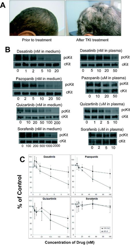
myelosuppression.2 In vivo c-Kit inhibition is also associated with hair depigmentation (Figure
1A).3 Drugs such as pazopanib and sunitinib, which have activity against c-Kit, do not induce
myelosuppression in solid tumor patients when used as single agents. In contrast, dasatinib and
imatinib, which also inhibit c-Kit, have been associated with myelosuppression in patients with
Ph+ leukemia.4 In patients with relapsed/refractory FLT3/ITD AML, treatment with the FLT3
inhibitor quizartinib was associated with myelosuppression, whereas in a similar patient
2
population, a different FLT3 inhibitor, sorafenib, induced no myelosuppression.5, 6 To better

understand the relationship between inhibition of c-Kit, FLT3, and marrow suppression, we

studied a series of different TKIs using bone marrow progenitor cell assays and immunoblots.

Cell lines were cultured as described.2 TF-1 cells were obtained from the American Type

Culture Collection (ATCC; Manassas VA) and grown in RPMI supplemented with GM-CSF

(Invitrogen, Grand Island, NY). Quizartinib was obtained from Ambit Biosciences (San Diego,

CA). Crenolanib was obtained from Arog Pharmaceuticals (Dallas, TX). Dasatinib, pazopanib,

and imatinib were obtained from LC Laboratories (Woburn, MA). Electrophoresis,

immunoblotting, and hematopoietic progenitor cell assays were performed as described.2

Cytokines used included SCF, G-CSF, GM-CSF, IL-3, IL-6, and erythropoietin. Unused

portions of bone marrow from normal donors were collected under an institutional review board-

approved Tumor and Cell Procurement Bank at Johns Hopkins (supported by grant

P30CA006973-44). All donors gave informed consent according to the Declaration of Helsinki.

Dasatinib is a multi-targeted TKI with activity against Bcr-Abl, SRC, and c-Kit.7    The

concentration of dasatinib necessary to inhibit 50% of baseline c-Kit activity (IC50) in TF-1 cells

stimulated with stem cell factor (SCF) is 1.5 nM in culture medium and 30 nM in 100% human

plasma (Figure 1B). The drug has a relatively short half-life of 3-5 hours and steady state

concentrations are in the range of 20-40 nM.7 In progenitor cell assays, dasatinib had a modest

effect on the formation of GM-CFUs (granulocyte-monocyte colony forming units), and a

greater effect on the formation of erythroid colonies (Figure 1C). Dasatinib has been reported to

cause myelosuppression in leukemia patients,4 as well as hair depigmentation.8 Pazopanib, a

                                                 3
TKI approved for use in some solid tumors, is reported to be a potent inhibitor of VEGFR,

PDGFR, and c-Kit.9 We found it to have an IC50 against c-Kit in culture medium of 3.7 nM.

While pazopanib has a seemingly high IC50 against c-Kit in human plasma of 36 µM (due to high

plasma protein binding) (Figure 1B), patients achieve trough drug levels over 50 µM.9 In

progenitor cell assays, pazopanib, at concentrations corresponding to what is routinely achieved

in patients, inhibited erythroid and myeloid progenitor cell activity to a similar degree as

dasatinib (Figure 1C). Hair depigmentation is listed as a common adverse event in the FDA

label for pazopanib, again, indicative of in vivo c-Kit inhibition. While both dasatinib and

pazopanib are potent in vitro inhibitors of c-Kit, of the two drugs, only dasatinib has been

reported to cause myelosuppression as monotherapy.4, 9 Pazopanib is used exclusively to treat

patients with solid tumors (who presumably have intact marrow function). However, when

combined with cytotoxic drugs, pazopanib appears to exacerbate the chemotherapy-induced

myelosuppression.10 Similarly, sunitinib, as monotherapy for solid tumor patients, is not

associated with significant myelosuppression. However, in leukemia patients or in solid tumor

patients in combination with chemotherapy, sunitinib exacerbates myelosuppression.11, 12 A

simple explanation for these findings is that c-Kit inhibition by itself does not induce clinically

significant myelosuppression in the setting of normal bone marrow function.

Inhibition of c-Kit, therefore, correlates with hair depigmentation, inhibition of erythroid

precursor activity in vitro, and, in leukemia patients, myelosuppression. Given the redundant

signaling properties of c-Kit and FLT3,1 simultaneous inhibition of FLT3 and c-Kit could result

in profound myelosuppression. Sorafenib is a potent FLT3 TKI (IC50 in culture medium 3-5 nM)

that has demonstrated efficacy in the treatment of relapsed/refractory FLT3/ITD AML patients.6

                                                  4
There is no reported inhibition of c-Kit by sorafenib, nor have there been any reports of

myelosuppression (even in combination with chemotherapy). These observations are consistent

with the results of our immunoblot (Figure 1B) and with progenitor cell assays (Figure 1C). In

contrast, quizartinib is a potent FLT3 inhibitor (IC50 in culture medium 2 nM; in plasma 18 nM),

and a modestly potent c-Kit inhibitor with an IC50 in culture medium of 28 nM. AML patients

readily achieve micromolar plasma concentrations of this agent,13 and myelosuppression was

observed in leukemia patients treated with quizartinib.5 Quizartinib inhibits both myeloid and

erythroid hematopoietic progenitor cell activity (Figure 1C). Given that FLT3 inhibition alone

(by sorafenib) did not inhibit colony activity, we conclude that quizartinib-induced

myelosuppression is probably mediated through inhibition of c-Kit, rather than inhibition of

FLT3. Interestingly, the most common clinical response to single agent therapy with quizartinib

has been a complete remission with incomplete count recovery (“CRi”).5, 13 The failure to

recover normal hematopoietic function may be due in part to the inhibition of c-Kit by

quizartinib.

While FLT3 inhibition by itself has no effect on hematopoiesis, it possibly still contributes to c-

Kit-induced marrow suppression. Exogenous FLT3 ligand (FL) shifts the dose response to FLT3

inhibitors upward.14 If FLT3 inhibition were contributing to the suppression of hematopoietic

progenitor cell induced by quizartinib, then the addition of FL would be predicted to blunt the

inhibitory effect of quizartinib. In progenitor cell assays, we saw no significant difference in

effect with 200 nM quizartinib with or without exogenous FL (10 ng/mL) (data not shown),

suggesting that FLT3 inhibition does not contribute to marrow suppression from quizartinib.

                                                 5
We conclude that inhibition of c-KIT can translate into clinically significant marrow

suppression, particularly when it occurs in the setting of cytotoxic chemotherapy, or when it is

induced in a patient with a marrow disorder such as leukemia. The more potent c-Kit inhibitors

impair erythroid, and myeloid progenitor cell function, but FLT3 inhibition probably has little

effect on hematopoiesis. Table 1 lists seven TKIs according to their activity against both FLT3

and c-Kit receptors in culture medium and 100% human plasma. Each TKI was ranked

according to its relative potency against c-Kit in vivo, using published pharmacokinetic data

(when available), in vitro potency, the occurrence of hair depigmentation and myelosuppression.

Given the clinical consequences of myelosuppression, the relative difference in inhibitory

activity between the targeted kinase and c-Kit represents an important therapeutic index that

must be accounted for in the development of TKIs. Hair depigmentation can represent a useful

clinical surrogate for this phenomenon.

Authorship

Contribution: A.G. and M.L. designed the study, performed experiments, analyzed the data, and
wrote the manuscript.

The authors declare no conflicts of interest.

Acknowledgements

This work was supported by the NCI Leukemia SPORE P50 CA100632-11.

                                                6
References

1.      Lyman SD, Jacobsen SE. c-kit ligand and Flt3 ligand: stem/progenitor cell factors with
overlapping yet distinct activities. Blood. 1998;91(4):1101-1134.
2.      Galanis A, Ma H, Rajkhowa T, et al. Crenolanib is a potent inhibitor of FLT3 with
activity against resistance-conferring point mutants. Blood. 2014;123(1):94-100.
3.      Moss KG, Toner GC, Cherrington JM, Mendel DB, Laird AD. Hair depigmentation is a
biological readout for pharmacological inhibition of KIT in mice and humans. J Pharmacol Exp
Ther. 2003;307(2):476-480.
4.      Talpaz M, Shah NP, Kantarjian H, et al. Dasatinib in imatinib-resistant Philadelphia
chromosome-positive leukemias. N Engl J Med. 2006;354(24):2531-2541.
5.      Cortes J, Perl A, Dombret H, et al. Final Results of a Phase 2 Open-Label, Monotherapy
Efficacy and Safety Study of Quizartinib (AC220) in Patients 60 Years of Age with FLT3 ITD
Positive or Negative Relapsed/Refractory Acute Myeloid Leukemia. Blood. 2012;120:48a.
6.      Borthakur G, Kantarjian H, Ravandi F, et al. Phase I study of sorafenib in patients with
refractory or relapsed acute leukemias. Haematologica. 2011;96(1):62-68.
7.      Demetri GD, Lo Russo P, MacPherson IR, et al. Phase I dose-escalation and
pharmacokinetic study of dasatinib in patients with advanced solid tumors. Clin Cancer Res.
2009;15(19):6232-6240.
8.      Brazzelli V, Grasso V, Barbaccia V, et al. Hair depigmentation and vitiligo-like lesions in
a leukaemic paediatric patient during chemotherapy with dasatinib. Acta dermato-venereologica.
2012;92(2):218-219.
9.      Hurwitz HI, Dowlati A, Saini S, et al. Phase I trial of pazopanib in patients with advanced
cancer. Clin Cancer Res. 2009;15(12):4220-4227.
10.     Plummer R, Madi A, Jeffels M, et al. A Phase I study of pazopanib in combination with
gemcitabine in patients with advanced solid tumors. Cancer Chemother Pharmacol.
2013;71(1):93-101.
11.     Fiedler W, Serve H, Dohner H, et al. A phase 1 study of SU11248 in the treatment of
patients with refractory or resistant acute myeloid leukemia (AML) or not amenable to
conventional therapy for the disease. Blood. 2005;105(3):986-993.
12.     Crown JP, Dieras V, Staroslawska E, et al. Phase III trial of sunitinib in combination with
capecitabine versus capecitabine monotherapy for the treatment of patients with pretreated
metastatic breast cancer. J Clin Oncol. 2013;31(23):2870-2878.
13.     Cortes JE, Kantarjian H, Foran JM, et al. Phase I Study of Quizartinib Administered
Daily to Patients With Relapsed or Refractory Acute Myeloid Leukemia Irrespective of FMS-
Like Tyrosine Kinase 3-Internal Tandem Duplication Status. J Clin Oncol. 2013;31(29):3681-
3687.
14.     Sato T, Yang X, Knapper S, et al. FLT3 ligand impedes the efficacy of FLT3 inhibitors in
vitro and in vivo. Blood. 2011;117(12):3286-3293.

                                                7
Drug    Target     pFLT3 pcKIT     Steady-    In vivo        Hair          Myelosuppression
               Disease     IC50   IC50       state    c-Kit    depigmentation
                          plasma plasma    plasma inhibition
                                            levels
 Sunitinib     GIST      39 nM 26 nM       82-135      Yes          Yes                 Yes
               RCC                            nM
Pazopanib      RCC        N/A      36 µM    51-95      Yes          Yes                 Yes
             Sarcoma                          uM
Quizartinib    AML       18 nM 2.7 µM         Not      Yes          Yes                 Yes
                                          available
 Dasatinib     CML        N/A      30 nM    20-40     Partial    Occasional             Yes
                                              nM
 Imatinib      CML        N/A      5.4 µM 2.5-5.3     Partial    Occasional             Yes
                                              uM
Crenolanib     AML       48 nM 2.0 µM         Not      No            No                 No
                                          available
 Sorafenib     HCC         484       >60    10-15      No            No                 No
               RCC         nM        µM       uM
Table 1. Relative activity against FLT3 and c-KIT, and myelosuppressive activity of

tyrosine kinase inhibitors. For FLT3, MOLM14 cells were incubated with drug for 1 hour,

lysed, immunoprecipitated for FLT3, and immunoblotted for phospho- and total FLT3. For c-

Kit, TF-1 cells were incubated with drug for 1hr with 20 ng SCF (PeproTech, Rocky Hill NJ)

added to each sample in the last 5 minutes of drug incubation. The treated TF-1 cells were lysed,

immunoprecipitated for c-Kit, and immunoblotted for phospho- and total c-Kit. Densitometry

analysis was performed using Quantity One software (BioRad Inc., Hercules, CA, USA). The

concentration of drug resulting in 50% inhibition from baseline (IC50) was calculated by

regression analysis after linear conversion. Steady state plasma levels were obtained from

published studies. In vivo c-Kit inhibition (most potent to least potent, top to bottom) was

ranked according to the ability of each TKI to achieve sustained drug levels well above the

calculated plasma IC50. The association of each TKI with hair depigmentation and

                                                 8
myelosuppression was determined based on each drug's FDA label and clinical studies

referenced in the table. Abbreviations: GIST: Gastrointestinal stromal tumor; RCC: renal cell

carcinoma; HCC: hepatocellular carcinoma.

                                                9
Figure Legends

Figure 1

(A) An AML patient before treatment (left) and 54 days after treatment (right) with the c-

Kit/FLT3 inhibitor, PLX3397{Burton, 2011 #1126;Burton, 2011 #1126;Burton, 2011

#1126;Burton, 2011 #1126} (This compound is currently being studied in a phase 1 trial- see

NCT01349049; pharmacokinetic data are not yet available for PLX3397, and so it was not

included in this study). (B) TF-1 cells were treated with drug for 1 hour in cell culture medium

or plasma, lysed, and immunoblotted for phospho- and total c-Kit. (C) Normal human bone

marrow was collected and mononuclear cells were isolated. Mononuclear cells were plated in 35

mm dishes at a concentration of 100,000 cells per mL in MethoCult containing various

concentrations of the indicated TKI in quadruplicate. Plates were analyzed 10-14 days later by

morphology for total number of CFU-GM and BFU-E colonies. For each drug, the assay was

performed using three separate marrow samples, and the results were averaged.

                                               10
You can also read


