IPROTEINDB: AN INTEGRATIVE DATABASE OF DROSOPHILA POST-TRANSLATIONAL MODIFICATIONS - BIORXIV
←
→
Page content transcription
If your browser does not render page correctly, please read the page content below
bioRxiv preprint first posted online Aug. 7, 2018; doi: http://dx.doi.org/10.1101/386268. The copyright holder for this preprint
       (which was not peer-reviewed) is the author/funder. All rights reserved. No reuse allowed without permission.

   iProteinDB: an integrative database of Drosophila post-translational
   modifications
   Authors: Yanhui Hu1,2,7, Richelle Sopko1,7, Verena Chung1,2, Romain A. Studer3,4, Sean D. Landry5,
   Daniel Liu2, Leonard Rabinow1, Florian Gnad5, Pedro Beltrao3, Norbert Perrimon1,2,6

   Corresponding author(s): Norbert Perrimon

   Author affiliations:
   1
    Department of Genetics, Harvard Medical School, 77 Avenue Louis Pasteur, Boston, MA 02115
   2
   Drosophila RNAi Screening Center, Harvard Medical School, 77 Avenue Louis Pasteur, Boston,
   MA 02115
   3
    European Molecular Biology Laboratory (EMBL), European Bioinformatics Institute, Wellcome
   Genome Campus, Hinxton, Cambridge CB10 1SD, UK.
   4
    Current affiliation: BenevolentAI, London NW1 1LW, UK.
   5
    Department of Bioinformatics, Cell Signaling Technology Inc., 3 Trask Lane, Danvers, MA
   01923
   6
    Howard Hughes Medical Institute, 77 Avenue Louis Pasteur, Boston, MA 02115
   7
    Authors contributed equally

   Key words: Drosophila, post-translational modification, phosphoproteomics

                                                                       1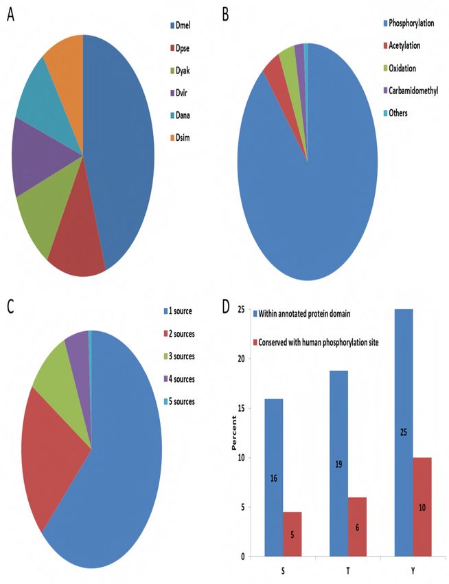
bioRxiv preprint first posted online Aug. 7, 2018; doi: http://dx.doi.org/10.1101/386268. The copyright holder for this preprint
       (which was not peer-reviewed) is the author/funder. All rights reserved. No reuse allowed without permission.

   Abstract
   Post-translational modification (PTM) serves as a regulatory mechanism for protein function,
   influencing stability, protein interactions, activity and localization, and is critical in many
   signaling pathways. The best characterized PTM is phosphorylation, whereby a phosphate is
   added to an acceptor residue, commonly serine, threonine and tyrosine. As proteins are often
   phosphorylated at multiple sites, identifying those sites that are important for function is a
   challenging problem. Considering that many phosphorylation sites may be non-functional,
   prioritizing evolutionarily conserved phosphosites provides a general strategy to identify the
   putative functional sites with regards to regulation and function. To facilitate the identification
   of conserved phosphosites, we generated a large-scale phosphoproteomics dataset from
   Drosophila embryos collected from six closely-related species. We built iProteinDB
   (https://www.flyrnai.org/tools/iproteindb/), a resource integrating these data with other high-
   throughput PTM datasets, including vertebrates, and manually curated information for
   Drosophila. At iProteinDB, scientists can view the PTM landscape for any Drosophila protein and
   identify predicted functional phosphosites based on a comparative analysis of data from
   closely-related Drosophila species. Further, iProteinDB enables comparison of PTM data from
   Drosophila to that of orthologous proteins from other model organisms, including human,
   mouse, rat, Xenopus laevis, Danio rerio, and Caenorhabditis elegans.

                                                                       2
bioRxiv preprint first posted online Aug. 7, 2018; doi: http://dx.doi.org/10.1101/386268. The copyright holder for this preprint
(which was not peer-reviewed) is the author/funder. All rights reserved. No reuse allowed without permission.
Introduction
Post-translational modification is essential for the regulation of many cellular processes. For
example, phosphorylation can serve as a molecular switch for signal transduction (Beurel et al.,
2015; Hunter, 2000; Kockel et al., 2010; Nagini et al., 2018). Based on the annotation of
PhosphoSitePlus (Hornbeck et al., 2012; Hornbeck et al., 2015), the average number of
phosphosites per protein is twelve for the human proteome and seven for the mouse
proteome. Evolutionary studies of protein phosphorylation have suggested that a significant
fraction of these large numbers of phosphosites may be non-functional (Beltrao et al., 2013;
Landry et al., 2009; Studer et al., 2016) and that evolutionarily conserved phosphosites are
often highly relevant for function (Studer et al., 2016), as evidenced, for example, by the
Mitogen Activated Protein Kinase (MAPK) or Extracellular Regulated Kinase (ERK) families (i.e.
ERK/MAPK, JNK, p38). Generally the activation of these kinases requires phosphorylation within
the sequence, TxY, residing within the “T loop” of the catalytic domain by an upstream MAPK-
K/MEK kinase. Upon phosphorylation, the activation loop moves away from the active site,
allowing substrate entry and phosphorylation. The TxY motif is conserved in the vast majority of
MAPK/ERK family members, from yeast to man, allowing, for example, the generation of an
antibody specific for the phosphorylated, active form of MAPK/ERK (Gabay et al., 1997). Other
examples of highly conserved phosphosites include ribosomal protein S6 (rpS6), which is
conserved in essentially all organisms including yeast, plants, invertebrates, and vertebrates.
The physiological roles of phosphorylation at Ser235/236 of rpS6 remained unclear until
genetic approaches abolishing the phosphorylation sites were applied in model organisms
(Meyuhas, 2015). These examples highlight how conservation can illuminate phosphosite
function. Model organisms can play essential roles in the elucidation of the functions of post-
translational modification of highly conserved sites.
Mass spectrometry (MS)-based proteomics is a powerful approach for large-scale identification
and characterization of phosphorylation sites. Three large-scale Drosophila melanogaster
phospho-proteomic datasets have been generated over the past years using MS. Two datasets
were generated from cultured cells (Bodenmiller et al., 2007; Hilger et al., 2009) and one was
generated from embryos (Zhai et al., 2008). Because the coverage of each dataset is limited,
and to further characterize the breadth of phosphorylation in Drosophila, we generated a new
dataset for Drosophila melanogaster and five closely-related species: D. simulans, D. yakuba, D.
ananassae, D. pseudoobscura, and D. virilis. To facilitate the use of this dataset, we built an
online resource, iProteinDB, integrating our data with other large-scale PTM data (Bodenmiller
et al., 2007; Hilger et al., 2009; Zhai et al., 2008) and curated PTM annotations for Drosophila
and other model organisms. At iProteinDB, users are able to align PTM data for any protein of
interest from multiple resources, including data from the six Drosophila species, other model
organisms, and human cells. Additional relevant information, such as disease-related protein
3bioRxiv preprint first posted online Aug. 7, 2018; doi: http://dx.doi.org/10.1101/386268. The copyright holder for this preprint
(which was not peer-reviewed) is the author/funder. All rights reserved. No reuse allowed without permission.
variants, sub-cellular localization, and protein abundance during Drosophila development, is
also provided at iProteinDB.
Methods
Generation of phosphoproteomics data
Pre-larval embryos of mixed sex and age from each of the six Drosophila species were collected.
Since different species develop at different speeds, the timing of collection was different for
each species. Flies were enticed to lay eggs by incubating in the dark on grape juice plates.
Proteins from embryos lysed in 8 M urea were digested with trypsin and separated into 12
fractions by strong cation exchange chromatography. Phosphopeptides were purified with
titanium dioxide microspheres and analyzed via LC-MS/MS on either an LTQ-Orbitrap or
Orbitrap Fusion instrument (Thermo Scientific). SEQUEST was used for spectral matching.
Peptides were filtered to a 1% FDR. Proteins were filtered to achieve a 2% final protein FDR
(final peptide FDR near 0.15%) and a probability-based scoring method was used to assign the
localizations of phosphorylation events (Beausoleil et al., 2006). The reference genomes used
for initial analysis are D. mel r5.53, D. ana r1.3, D. pse r3.1, D. sim r1.4, D. vir r1.2 and D. yak
r1.3 from FlyBase. The sites were re-mapped to D. mel r6.16, D. ana r1.05, D. pse r3.04, D. sim
r2.02, D. vir r1.06, D. yak r1.05 at iProteinDB.
Predicting the probability of phosphorylation
We aligned the phosphorylation sites identified in our datasets from 6 Drosophila species based
on orthologous relationships predicted by OMA (Altenhoff et al., 2018; Altenhoff et al., 2011;
Altenhoff et al., 2015). For each proteome, we assign the probability of a phosphoacceptor
(S+T (together) and Y) to be phosporylated, using a two-step approach. First, we scan each
proteomes to find the kinase specificity of each phosphoacceptor, using NetPhorest (Horn et
al., 2014). This provided 40 scores for kinases specificity for a given region. Then, a support
vector machine algorithm (SVM-light) was trained on each of the six species, using all 40 scores.
We extracted the surrounding region of sites that are detected to be phosphorylated, and they
received an initial score of 1 (positive dataset). The regions surrounding non-detected
phosphosites received an initial score of 0 (negative dataset). We sample the data (2000 for S+T
and 800 for Y) to train the model, and then assign scores to unknown phosphosites based on
the support vector machine output (detected phosphosites by MS always received a score 1,
irrelevant of their prediction).
Comparison of PTM data across major model organisms and human
Orthologous relationships of Drosophila melanogaster proteins to major model organisms
including human, mouse, rat, X. tropicalis, zebrafish and C. elegans were obtained using DIOPT
4bioRxiv preprint first posted online Aug. 7, 2018; doi: http://dx.doi.org/10.1101/386268. The copyright holder for this preprint
(which was not peer-reviewed) is the author/funder. All rights reserved. No reuse allowed without permission.
(release 7) with a DIOPT score of 3 or higher. Protein sequences of the best orthologous genes
based on DIOPT score and each non-redundant isoform of Drosophila melanogaster gene were
aligned using MAFFT (vs 7.305B). Observed PTM sites, conserved phosphorylation sites, domain
and disease-related protein variants are annotated on the aligned protein sequences (Figure 1).
To compare specific PTM sites, the sequence of a sliding window of five amino acids
surrounding the identified phosphosite was extracted and compared across species. The
number of identical amino acids was counted and the percent of identity was calculated by
dividing the number of identical amino acids over the window length. Phylogenetic trees for
protein kinases were generated with Jalview 2.10 (Waterhouse et al., 2009) and illustrated in
iTOL (Letunic and Bork, 2016).
Source of other data sets or tools
Protein information of 6 Drosophila species was obtained from FlyBase
(ftp://ftp.flybase.net/releases/FB2017_03/). Protein information of human, mouse, rat, X.
tropicalis, zebrafish and C. elegans were obtained from RefSeq
(https://www.ncbi.nlm.nih.gov/refseq/). Other high throughput datasets of Drosophila
melanogaster were obtained either from online resources (Phosida:
http://141.61.102.18/phosida/index.aspx and Phosphopep: http://www.phosphopep.org/) or
corresponding supplemental tables of relevant publications (Zhai et al., 2008). The protein
annotation of Swiss-prot and TrEMBL was downloaded from the UniProt FTP site
(http://www.uniprot.org/downloads). Orthologous relationships were obtained from OMA
(https://omabrowser.org/oma/home/) and DIOPT (http://www.flyrnai.org/diopt). Protein
domain annotation of Conserved Domain Database
(https://www.ncbi.nlm.nih.gov/Structure/cdd/cdd.shtml) was extracted from the RefSeq
release files (.gbff files). Kinase motifs were predicted using the API of Scansite3
(http://scansite3.mit.edu/#home). PTM annotation of orthologous genes other than Drosophila
was obtained from PhosphoSitePlus (https://www.phosphosite.org/staticDownloads.action).
Implementation of the online resource
There were several steps involved that process the information and populate the back-end
database of iProteinDB. After downloading data from relevant sources, such as UniProt,
PhophoSitePlus and various publications, the extraction of relevant information was
accomplished with in-house parsers written in Perl and Python. The redundancy of protein
sequences was consolidated and a collection of distinct protein sequences from each
Drosophila species was assembled based on FlyBase genome release (D. mel r6.16, D. ana
r1.05, D. pse r3.01, D. sim r1.04, D. vir r1.02, D. yak r1.03). Since different resources annotate
data based on different genome releases, we synchronized the data from various sources by
mapping the original data (peptides) to the non-redundant protein collection of recent FlyBase
5bioRxiv preprint first posted online Aug. 7, 2018; doi: http://dx.doi.org/10.1101/386268. The copyright holder for this preprint
(which was not peer-reviewed) is the author/funder. All rights reserved. No reuse allowed without permission.
genome release (see above) using the SeqIO interface of BioPython. Once filtered and updated,
the data were then uploaded into a MySQL database, which is currently hosted by the Harvard
Medical School (HMS) Research Computing group.
To display the data, we created a web-based application with PHP and a PHP framework called
Symfony (version 2.6). Several client-side functions rely on JavaScript and AJAX, while some
tabular displays use a jQuery plugin called DataTables.js, which allow for sorting and paging
functionalities within the tables. This web application is also hosted by the HMS Research
Computing group.
Availability
iProteinDB is available for online use without any restrictions at
https://www.flyrnai.org/tools/iproteindb/.
6bioRxiv preprint first posted online Aug. 7, 2018; doi: http://dx.doi.org/10.1101/386268. The copyright holder for this preprint
(which was not peer-reviewed) is the author/funder. All rights reserved. No reuse allowed without permission.
Results
Data integration and quality of six Drosophila phosphoproteomes
Embryos from six Drosophila species were collected, proteins were extracted and digested,
phosphopeptides were isolated, and these samples were then subjected to ionization and
fragmentation for identification and phosphosite determination using a mass-spec based
method described previously (Sopko et al., 2014). The data coverage ranges from 14,915 to
21,750 sites per species (Supplementary Table 1) and motif analysis of the data (Ullah et al.,
2016) shows that the most significant motifs of phosphosites in 6 Drosophila species are quite
similar (Supplementary Figure 1). The orthologous relationships among the six Drosophila
species, as well as other sequenced Drosophila species and mosquito species (Supplementary
Figure 2), were predicted using the OMA algorithm (Altenhoff et al., 2018), which infers
orthologous genes among multiple genomes on the basis of protein sequence. Based on the
multiple-sequence alignment of each orthologous group, the aligned positions were selected,
for which phosphorylation was observed in at least one of the six Drosophila species. Given that
the mass-spec based identification of phosphosites is incomplete, we filled the gap with
machine learning predictions, using a similar approach as in (Studer et al., 2016). A support
vector machine (SVM) algorithm was trained to assign a propensity score of 0-1 to each
corresponding phospho-acceptor residue (serine, threonine, or tyrosine) for each species for
which that residue was not identified as phosphorylated, based on the likelihood of
phosphorylation. This information is available at the iProteinDB resource (see below) to help
researchers interested in identifying evolutionary conserved phosphorylation sites. We next
compared the propensity score with the phosphoproteomics data from other sources. We
found a strong correlation between the propensity score and the chance that a predicted site
was phosphorylated as supported by independent datasets (Supplementary Figure 3a). We also
compared the frequency of phosphorylation among the six Drosophila species with
experimental data for orthologous human proteins. Not surprisingly, phosphorylation sites
conserved among the six Drosophila species were more likely to be reported as phosphorylated
at the corresponding sites in orthologous human proteins. This correlation was more prevalent
for those sites with greater than 50% amino acid similarity between Drosophila and human
orthologs (Supplementary Figure 3b).
We estimated the false negative rate for each of the six Drosophila species by selecting those
sites that are 100% identical (considering eleven amino acid peptides comprising the
phosphosite plus five amino acids upstream and downstream) among all six species and for
which phosphorylation was observed in at least two species. The false negative rate is
estimated to be the percent of the sites that are not covered by the data in each species. For
example, 86% of these sites for Drosophila melanogaster are covered by at least one of the 4
7bioRxiv preprint first posted online Aug. 7, 2018; doi: http://dx.doi.org/10.1101/386268. The copyright holder for this preprint
(which was not peer-reviewed) is the author/funder. All rights reserved. No reuse allowed without permission.
datasets, and/or UniProt annotation so the false negative rate is about 14% while there is only
1 dataset for each of the other 5 Drosophila species, and therefore, the false negative rate is
relatively higher, 44% to 79% (Supplementary Figure 3c).
Integration of phosphoproteomes from other resources
We built the iProteinDB database to store phosphoproteomics data generated by our group
and other large PTM datasets. Other datasets were obtained from the supplemental table of
the original publications (Zhai et al., 2008) or the relevant websites (Bodenmiller et al., 2008;
Bodenmiller et al., 2007; Gnad et al., 2011). Original data were mapped to the same version of
the FlyBase proteome annotation (FB2017_03) and then integrated with our data in iProteinDB.
The information of PTM sites and the score/peptide from the original source are stored and
made available at the iProteinDB website. To compare PTM data across species, we integrated
orthologous relationships of Drosophila species as predicted by OMA (Altenhoff et al., 2018),
the orthologous relationships among major model organisms predicted by DIOPT (Hu et al.,
2011), and PTM data for other species from PhosphoSitePlus (https://www.phosphosite.org)
(Hornbeck et al., 2012; Hornbeck et al., 2015). The subcellular localization annotation and
human disease related protein variants were integrated from UniProt
(https://www.uniprot.org/), whereas protein domain annotation information was integrated
from the National Center for Biomedical Information (NCBI) Conserved Domain Database
(https://www.ncbi.nlm.nih.gov/cdd). Information about protein abundance during Drosophila
development was also integrated from a recent publication (Casas-Vila et al., 2017).
Altogether, iProteinDB covers 168,997 individual PTMs for Drosophila, of which 70,013 (41%)
were observed in Drosophila melanogaster (Figure 1a). 62,239 (89%) of the Drosophila
melanogaster PTM data collected in iProteinDB are phosphorylation sites, covering 8,068
unique proteins and 3,937 genes (Figure 1b). Comparing our Drosophila melanogaster
phosphoproteomics data with that from other sources, we find that 61% of our data overlaps
with one other source and 36% of our data overlaps with at least 2 other sources (Table 1).
Overall, 37% of the phosphorylation data is supported by multiple resources and thus can be
considered high confidence (Figure 1c, Table 1).
Online resource
Users can query Drosophila genes of interest, and choose one isoform if there are multiple non-
redundant isoforms for the gene of the interest. There are three tabs from which to choose
(Figure 2).
1.) Protein detail tab. A user can view the protein sequence from any of the 6 Drosophila
species in FASTA format. PTM sites are color-coded. The amino acid is displayed in red if the
8bioRxiv preprint first posted online Aug. 7, 2018; doi: http://dx.doi.org/10.1101/386268. The copyright holder for this preprint
(which was not peer-reviewed) is the author/funder. All rights reserved. No reuse allowed without permission.
PTM is observed or blue if it was not observed but is predicted to be phosphorylated based on
the data from different Drosophila species. The amino acid is underlined if the phosphorylation
event was observed in more than one Drosophila species. Protein domains are highlighted in
green. A table summarizing all the PTM sites for a given protein, as well as the data sources
from which the PTM information was extracted, is provided, along with detailed information
from the original sources, i.e. the original scores and peptide sequences. A table summarizing
all predicted sites based on data from closely related Drosophila species is provided with a link
to detailed information and multiple sequence alignments. Also indicated in this tab is sub-
cellular localization annotation from UniProt for each phosphoprotein and kinase predicted to
act on individual sites, as identified using ScanSite3 (Obenauer et al., 2003).
2.) Predicted ortholog tab. Users can find a table of the best ortholog candidates for major
model organisms based on DIOPT ortholog predictions (Hu et al., 2011). Multiple sequence
alignments were performed based on the protein sequences of orthologous genes. The
sequences of all the aligned Drosophila phosphosites, over a sliding window of five residues,
were compared to the corresponding sequences of each orthologous gene and a similarity
score was calculated by pair-wise comparison. For example, if 10 of the 11 amino acids
(phosphorylation site plus five amino acids upstream and downstream) are identical between
Drosophila and human sites, the similarity score was assigned as 0.9 (10 divided by 11). Then,
an average similarity score was calculated based on all pairwise combinations at a given site.
All phosphorylation sites with an average similarity score of >0.5 are listed and summarized as
conserved sites. Human disease-related variants annotated at UniProt are also listed, along
with sub-cellular localization annotation of all orthologous proteins from UniProt. Multiple
sequence alignment (MSA) across major model organisms is displayed. For MSAs, observed
PTM sites for all orthologous genes are color-coded, domains are highlighted, and disease
variants are underlined. Conserved sites are bolded. As we hope that iProteinDB will lead to
new discoveries and hypotheses on previously uncharacterized phosphorylation events, we
further integrated information on availability of corresponding antibodies from Cell Signaling
Technology for proteins and sites that are homologous between Drosophila and human to help
users with experimental designs.
3.) Protein abundance tab. Protein expression levels from a comprehensive proteomic study
covering the complete Drosophila melanogaster life cycle (Casas-Vila et al., 2017) are plotted.
On this tab, a user can view the stages of the Drosophila life cycle during which a protein of
interest is expressed.
9bioRxiv preprint first posted online Aug. 7, 2018; doi: http://dx.doi.org/10.1101/386268. The copyright holder for this preprint
(which was not peer-reviewed) is the author/funder. All rights reserved. No reuse allowed without permission.
The Drosophila kinomes and their substrates show significant evolutionary
conservation
The integration of six Drosophila phosphoproteomes along with ortholog information enabled
us to determine the conservation of the Drosophila kinome and to assess the evolutionary
selective pressure on its substrates.
We found that the entire Drosophila melanogaster kinome as defined by Manning and
colleagues(Manning et al., 2002) shows orthologous counterparts in the other Drosophila
species based on the OMA algorithm (Figure 3, Supplementary Figure 4). The few exceptions
such as the absence of an orthologous Abl tyrosine kinase in D. simulans might trace back to
poor genome sequence quality. Consistent with previous observations (Manning et al., 2002) all
Drosophila kinases showed strong evidence for orthologous counterparts in at least one of the
six integrated model organisms. Only Tie-like receptor tyrosine kinase, Ack-like, and Wsck
showed poor or no homology in eukaryotes other than Drosophila species based on DIOPT. The
high conservation of the Drosophila kinome within flies and across other eukaryotes suggests
that the corresponding substrates are also significantly conserved.
To corroborate this hypothesis at the protein level, we determined the proportion of
phosphorylated Drosophila melanogaster proteins that show homologies in other species. We
used the conservation of Drosophila melanogaster proteins, which have not been found to be
phosphorylated as a control to assess significance. We assume that this control set is indeed
enriched for proteins that do not present kinase substrates. While phosphorylated and non-
phosphorylated Drosophila melanogaster proteins have the same proportion of orthologs (99%)
as the close relative Drosophila simulan, the phosphoproteome showed significantly higher
conservation in more distantly related species from Drosophila yakuba (phospho: 99%, control:
97%; p < 0.01 based on two-sided Fisher Exact test) to Caenorhabditis elegans (phospho: 70%,
control: 46%; p < 0.01) (Figure 4a). This suggests that not only the kinome but also its substrates
are more conserved than other proteins. The significant conservation of the identified
phosphoproteome might, however, also be partly driven by an enrichment of highly expressed
proteins in the phosphoset and the presence of potential pseudogenes and predicted proteins
in the control set. We therefore analyzed the conservation of phosphorylated versus non-
phosphorylated residues of identified phosphoproteins, and found that phosphorylated
residues show significantly higher conservation within all Drosophila species (p < 0.01) but not
in more distant species (Figure 4b). Similar trends have been reported for other eukaryotic
phosphoproteomes. For example, human phosphosites have been shown to have significantly
higher conservation in mammals and other higher eukaryotes, but not in distant species
including Caenorhabditis elegans or yeast (Gnad et al., 2010). The prevalent localization of
phosphorylation sites in fast evolving loop and hinge regions of proteins (Iakoucheva et al.,
10bioRxiv preprint first posted online Aug. 7, 2018; doi: http://dx.doi.org/10.1101/386268. The copyright holder for this preprint
(which was not peer-reviewed) is the author/funder. All rights reserved. No reuse allowed without permission.
2004) might make it difficult to map the associated site in aligned disordered regions of
distantly related species. In contrast, non-phosphorylated serines, threonines, and tyrosines are
not restricted to localization on the protein surface, and therefore tend to occur in more
structured and slower evolving regions on the protein (Gnad et al., 2007).
In summary, we found significant conservation of the kinome and substrate proteins across all
species. Similarly phosphorylated residues are significantly conserved within Drosophila, but
difficult to trace back in distant species.
Conservation between the Drosophila and the human phosphoproteomes
underlines the utility of using the former as a model system
To assess the utility of the Drosophila phosphoproteome as a model for human phosphorylation
events, we examined the evolutionary conservation of their phosphorylated sites with a focus
on localization in functional domains and association with diseases. Approximately 17% of
identified phosphosites are within annotated protein domains, and 82% of the identified
phosphosites reside in proteins for which the corresponding Drosophila genes are conserved
with human, based on DIOPT prediction using a score of 3 or more as cutoff (Hu et al., 2011).
The corresponding human sites for 23% of these sites are also phospho-acceptors, among
which, 3201 sites have 50% or more sequence identity to human sequence. Of the three
phospho-acceptor residues, serine has the highest percentage of phosphorylation
(Supplementary Figure 5) while phospho-tyrosine has the highest probability of residing within
a defined protein domain and the highest sequence similarity with human orthologs (Figure
1d). Further analysis showed that the sequence identity of PTM sites between human and
Drosophila melanogaster correlates with the probability that the associated phosphorylation
event has also been observed in human cell phospho-proteomic datasets (Figure 5a).
We observed an enrichment of UniProt human disease related variants located proximal to
phosphosites conserved with Drosophila melanogaster. For example, the enrichment p-value
of disease related variants is 4.3*10-10 by Fisher exact test for the phosphosites with 80% or
higher identity between human and Drosophila sites. To further analyze the intersection
between phosphorylation events in Drosophila and disease variants in human, we calculated
the percent of phosphosites that are within 10 amino acids of a disease variant at each identity
cut-off. Our analysis indicates that more highly conserved sites tend to occupy positions
proximal to residues variant in human disease (Figure 5b). For example, phosphosites with 50%
or higher identity are about 2-fold more likely to be located within 10 amino acids of a disease
variant than phosphosites with 20% or higher identity for the phospho-acceptor sites. Analysis
of sites that are phospho-acceptor residues (serine, threonine, or tyrosine) in Drosophila but
are not in human show a similar trend but this correlation was more prevalent for the phospho-
11bioRxiv preprint first posted online Aug. 7, 2018; doi: http://dx.doi.org/10.1101/386268. The copyright holder for this preprint
(which was not peer-reviewed) is the author/funder. All rights reserved. No reuse allowed without permission.
acceptor sites, indicating that the correlation is driven by the phospho-acceptor as well as the
conservation of the surrounding sequence.
Finally we compared all phosphosites in Drosophila melanogaster with their human
orthologous sites. We identified 370 sites that were observed as phosphorylated in Drosophila
and have 100% identity with human phosphosites over a sliding window of five amino acids.
These sites cover 146 human genes, many of which are kinases, including cyclin dependent
kinases, glycogen synthase kinases, mitogen-activated protein kinases, and ribosomal protein
S6 kinases and the insulin receptor (InR) (Supplementary Table 2). For example, human
glycogen synthase kinase 3A and 3B (GSK3A and GSK3B) auto-phosphorylate on a conserved
tyrosine residue (Y279) for maximal activity, and play an important role in multiple signaling
pathways (Beurel et al., 2015; Nagini et al., 2018). Dysregulation of GSK3 has been linked to
various diseases including cancer, in which GSK3 can function as a tumor promoter or
suppressor in different contexts and with different phosphorylation status (Ma, 2014; Nagini et
al., 2018; Sarkar et al., 2015). The S278, Y279 and S282 sites within the protein-kinase domain
of GSK3 have 100% identity with the Drosophila ortholog sgg (shaggy) and the phosphorylation
of these sites has also been observed in Drosophila (Figure 6a). We further uncovered sites
where the phospho-acceptor identity has changed, such as from serine to threonine (Figure 6b,
Supplementary Table 3), suggesting that phosphorylation of these sites may also be conserved
and required for regulation of the protein activity. We identified proteins for which the
phospho-acceptor residues are conserved among Drosophila but absent in human despite the
surrounding sequences being 100% identical (Figure 6c, Supplementary Table 3). These sites
may regulate species-specific functions. Altogether these results indicate the utility of using
Drosophila as a model system to study the function of these sites in signal transduction and the
regulation of associated proteins.
12bioRxiv preprint first posted online Aug. 7, 2018; doi: http://dx.doi.org/10.1101/386268. The copyright holder for this preprint
(which was not peer-reviewed) is the author/funder. All rights reserved. No reuse allowed without permission.
Conclusion
Drosophila melanogaster is one of the most-studied model organisms. Current PTM resources,
such as PhosphoSitePlus (Hornbeck et al., 2012; Hornbeck et al., 2015), dbPTM (Huang et al.,
2016; Lee et al., 2006) and Phospho.ELM (Diella et al., 2004; Diella et al., 2008; Dinkel et al.,
2011), have comprehensive coverage for human, mouse, and rat, but have very limited
coverage for Drosophila. Resources like PHOSIDA (Gnad et al., 2011), PHOSPHOPEP
(Bodenmiller et al., 2008; Bodenmiller et al., 2007) and dbPAF (Ullah et al., 2016) provide large-
scale PTM data for Drosophila genes but are focused on only one or, at most, a few datasets.
We generated a large-scale proteomics dataset of six closely related Drosophila species, made
the data available, and integrated it with literature annotation and other large datasets for
Drosophila melanogaster. This integrated resource allows researchers to obtain a more
comprehensive view of the PTM landscape, taking into consideration all Drosophila proteomic
data, and enabling comparison to orthologous proteins from other model organisms. Many of
the conserved sites reside within kinases themselves, demonstrating that evolution has largely
“optimized” protein kinase architecture and their operation within signaling pathways. We
expect that iProteinDB will serve as a valuable resource to facilitate functional discovery. For
example, iProteinDB can help a scientist identify sites that are critical for regulation that can be
used for example to generate ‘activity-dead’ proteins that can serve as controls in rescue
experiments with phosphomimetic (Pondugula et al., 2009) and temperature-sensitive mutants
(Hsu and Perrimon, 1994).
13bioRxiv preprint first posted online Aug. 7, 2018; doi: http://dx.doi.org/10.1101/386268. The copyright holder for this preprint
(which was not peer-reviewed) is the author/funder. All rights reserved. No reuse allowed without permission.
Acknowledgments
We would like to thank the members of Perrimon laboratory, Gygi laboratory, Drosophila RNAi
Screening Center (DRSC) and Transgenic RNAi Project (TRiP) for helpful input on the project.
Particularly we would like to thank Dr. Stephanie Mohr for helpful suggestions during
manuscript preparation and Mr. Aram Comjean for advice during the resource implementation.
The DRSC is supported by National Institutes of Health (NIH) National Institute of General
Medical Sciences grant R01 GM 067761 (to N.P.). The National Institutes of Health supported
this work (5R01DK088718, 5P01CA120964, 5R01GM084947 and 5R01GM067761). R.S. is a
Special Fellow of the Leukemia and Lymphoma Society. N.P. is a Howard Hughes Medical
Institute investigator.
14bioRxiv preprint first posted online Aug. 7, 2018; doi: http://dx.doi.org/10.1101/386268. The copyright holder for this preprint
(which was not peer-reviewed) is the author/funder. All rights reserved. No reuse allowed without permission.
Figure 1. Database content and statistics
Distribution of 168,997 observed PTMs in the proteomics dataset (A). Representation of
different types of PTMs (B). Overlap of phosphorylation data for Drosophila melanogaster from
five different sources (C). Distribution of phosphorylation sites observed at three phospho-
acceptor residues (serine (S), threonine (T) and tyrosine (Y)) within protein domains and their
conservation based on at least 50% similarity to human sequence, considering a sliding window
of five amino acids (D).
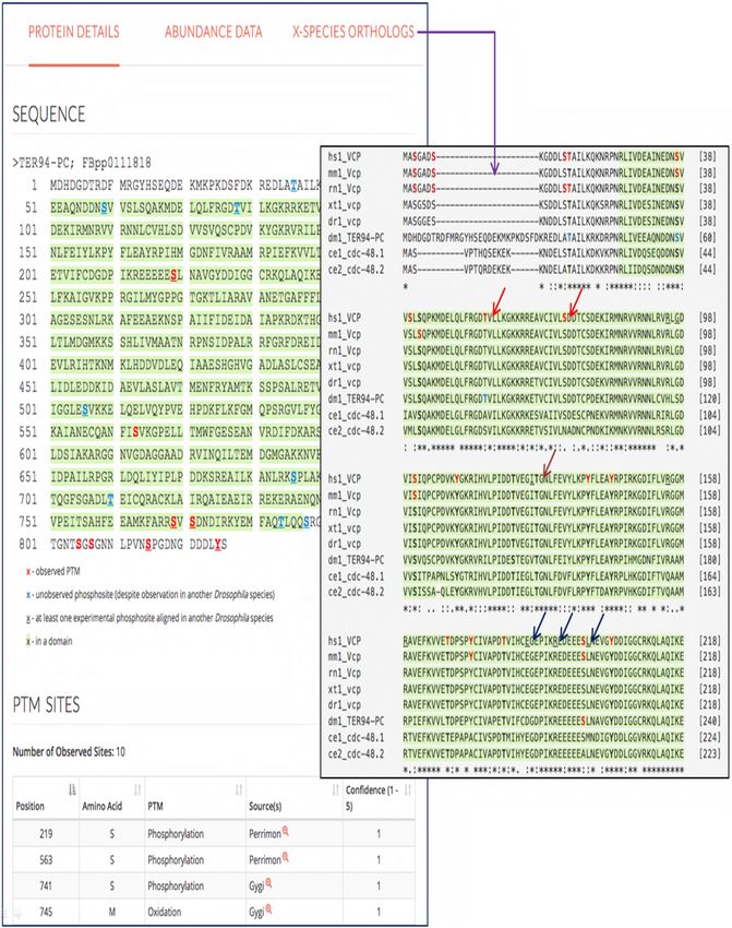
Figure 2. Features of iProteinDB user interface
Observed PTM sites are marked red on the Drosophila melanogaster protein sequence.
Predicted phosphosites based on phospho-proteomic data from five other Drosophila species
are marked in blue. Sites observed in more than one Drosophila species are underlined. The
protein domains are highlighted in green. The data sources of PTMs are summarized. At the
“Predicted Orthologs” page, the multiple sequence alignment of orthologous genes of major
model organisms and human are displayed with observed sites color-coded (red arrows),
conserved sites bolded (brown arrow) and human disease variant mutations underlined (navy
arrows).
Figure 3. Evolutionary relationships among Drosophila melanogaster tyrosine kinases
The core of the plot illustrates the phylogenetic relationships between Drosophila
melanogaster tyrosine kinases estimated by total sequence similarity. The outer circle reflects
the presence of orthologs in other species.
Figure 4. Conservation of phosphorylated proteins and sites
The line plot illustrates the proportions of Drosophila melanogaster phosphoproteins (blue) and
non-phosphoproteins (orange) showing orthologs in other species (A). The line plot shows the
proportions of conserved Drosophila melanogaster phosphosites (blue) and non-
phosphorylated serines, threonines, and tyrosines (orange) across species (B).
Figure 5. Analysis of the conservation of phosphorylation sites of Drosophila melanogaster
Correlations of sequence conservation and observed phosphorylation in Drosophila
melanogaster: 11,619 phosphosites identified in Drosophila melanogaster proteins can be
aligned to phospho-acceptor amino acids of the human orthologs, considering a sliding window
of five amino acids surrounding the identified phosphosite. The probability of the
corresponding phospho-acceptor site having been observed as phosphorylated in human data
correlates with the degree of sequence similarity (A). Correlation of phosphorylation with
disease related protein variants: The chance of the aligned human sites corresponding to the
15bioRxiv preprint first posted online Aug. 7, 2018; doi: http://dx.doi.org/10.1101/386268. The copyright holder for this preprint
(which was not peer-reviewed) is the author/funder. All rights reserved. No reuse allowed without permission.
phosphosites identified in Drosophila locating within 10 amino acids distance to disease
variants correlates with the sequence similarities between human and Drosophila sequences.
The correlation is prevalent for phospho-acceptor sites in human (B).
Figure 6. Examples of phosphosite conservation between human and Drosophila
melanogaster
Examples of phosphosites identified in Drosophila melanogaster (red), also identified as
phosphorylated in human (red), that share 100% identity with human (arrow) and indicated
model organisms (A). Phosphosites where the observed phospho-acceptor residue has changed
(B) and phosphosites where the phospho-acceptors have been lost but the surrounding
sequences are 100% identical (C). The abbreviation of taxonomy name is used to represent
different model organisms (hs - Homo sapiens; mm- Mus musculus; rn - Rattus norvegicus; xt -
Xenopus tropicalis; dr- Danio rerio; dm- Drosophila melanogaster; ce- Caenorhabditis elegans).
16bioRxiv preprint first posted online Aug. 7, 2018; doi: http://dx.doi.org/10.1101/386268. The copyright holder for this preprint
(which was not peer-reviewed) is the author/funder. All rights reserved. No reuse allowed without permission.
References
Altenhoff AM, Glover NM, Train CM, Kaleb K, Warwick Vesztrocy A, Dylus D, de Farias TM, Zile K,
Stevenson C, Long J, Redestig H, Gonnet GH, Dessimoz C. 2018. The OMA orthology database in
2018: retrieving evolutionary relationships among all domains of life through richer web and
programmatic interfaces. Nucleic Acids Res 46:D477-D485.
Altenhoff AM, Schneider A, Gonnet GH, Dessimoz C. 2011. OMA 2011: orthology inference among 1000
complete genomes. Nucleic Acids Res 39:D289-294.
Altenhoff AM, Skunca N, Glover N, Train CM, Sueki A, Pilizota I, Gori K, Tomiczek B, Muller S, Redestig H,
Gonnet GH, Dessimoz C. 2015. The OMA orthology database in 2015: function predictions,
better plant support, synteny view and other improvements. Nucleic Acids Res 43:D240-249.
Beausoleil SA, Villen J, Gerber SA, Rush J, Gygi SP. 2006. A probability-based approach for high-
throughput protein phosphorylation analysis and site localization. Nat Biotechnol 24:1285-1292.
Beltrao P, Bork P, Krogan NJ, van Noort V. 2013. Evolution and functional cross-talk of protein post-
translational modifications. Mol Syst Biol 9:714.
Beurel E, Grieco SF, Jope RS. 2015. Glycogen synthase kinase-3 (GSK3): regulation, actions, and diseases.
Pharmacol Ther 148:114-131.
Bodenmiller B, Campbell D, Gerrits B, Lam H, Jovanovic M, Picotti P, Schlapbach R, Aebersold R. 2008.
PhosphoPep--a database of protein phosphorylation sites in model organisms. Nat Biotechnol
26:1339-1340.
Bodenmiller B, Malmstrom J, Gerrits B, Campbell D, Lam H, Schmidt A, Rinner O, Mueller LN, Shannon
PT, Pedrioli PG, Panse C, Lee HK, Schlapbach R, Aebersold R. 2007. PhosphoPep--a
phosphoproteome resource for systems biology research in Drosophila Kc167 cells. Mol Syst Biol
3:139.
Casas-Vila N, Bluhm A, Sayols S, Dinges N, Dejung M, Altenhein T, Kappei D, Altenhein B, Roignant JY,
Butter F. 2017. The developmental proteome of Drosophila melanogaster. Genome Res
27:1273-1285.
Diella F, Cameron S, Gemund C, Linding R, Via A, Kuster B, Sicheritz-Ponten T, Blom N, Gibson TJ. 2004.
Phospho.ELM: a database of experimentally verified phosphorylation sites in eukaryotic
proteins. BMC Bioinformatics 5:79.
Diella F, Gould CM, Chica C, Via A, Gibson TJ. 2008. Phospho.ELM: a database of phosphorylation sites--
update 2008. Nucleic Acids Res 36:D240-244.
Dinkel H, Chica C, Via A, Gould CM, Jensen LJ, Gibson TJ, Diella F. 2011. Phospho.ELM: a database of
phosphorylation sites--update 2011. Nucleic Acids Res 39:D261-267.
Gabay L, Seger R, Shilo BZ. 1997. In situ activation pattern of Drosophila EGF receptor pathway during
development. Science 277:1103-1106.
Gnad F, Forner F, Zielinska DF, Birney E, Gunawardena J, Mann M. 2010. Evolutionary constraints of
phosphorylation in eukaryotes, prokaryotes, and mitochondria. Mol Cell Proteomics 9:2642-
2653.
Gnad F, Gunawardena J, Mann M. 2011. PHOSIDA 2011: the posttranslational modification database.
Nucleic Acids Res 39:D253-260.
Gnad F, Ren S, Cox J, Olsen JV, Macek B, Oroshi M, Mann M. 2007. PHOSIDA (phosphorylation site
database): management, structural and evolutionary investigation, and prediction of
phosphosites. Genome Biol 8:R250.
17bioRxiv preprint first posted online Aug. 7, 2018; doi: http://dx.doi.org/10.1101/386268. The copyright holder for this preprint
(which was not peer-reviewed) is the author/funder. All rights reserved. No reuse allowed without permission.
Hilger M, Bonaldi T, Gnad F, Mann M. 2009. Systems-wide analysis of a phosphatase knock-down by
quantitative proteomics and phosphoproteomics. Mol Cell Proteomics 8:1908-1920.
Horn H, Schoof EM, Kim J, Robin X, Miller ML, Diella F, Palma A, Cesareni G, Jensen LJ, Linding R. 2014.
KinomeXplorer: an integrated platform for kinome biology studies. Nat Methods 11:603-604.
Hornbeck PV, Kornhauser JM, Tkachev S, Zhang B, Skrzypek E, Murray B, Latham V, Sullivan M. 2012.
PhosphoSitePlus: a comprehensive resource for investigating the structure and function of
experimentally determined post-translational modifications in man and mouse. Nucleic Acids
Res 40:D261-270.
Hornbeck PV, Zhang B, Murray B, Kornhauser JM, Latham V, Skrzypek E. 2015. PhosphoSitePlus, 2014:
mutations, PTMs and recalibrations. Nucleic Acids Res 43:D512-520.
Hsu JC, Perrimon N. 1994. A temperature-sensitive MEK mutation demonstrates the conservation of the
signaling pathways activated by receptor tyrosine kinases. Genes Dev 8:2176-2187.
Hu Y, Flockhart I, Vinayagam A, Bergwitz C, Berger B, Perrimon N, Mohr SE. 2011. An integrative
approach to ortholog prediction for disease-focused and other functional studies. BMC
Bioinformatics 12:357.
Huang KY, Su MG, Kao HJ, Hsieh YC, Jhong JH, Cheng KH, Huang HD, Lee TY. 2016. dbPTM 2016: 10-year
anniversary of a resource for post-translational modification of proteins. Nucleic Acids Res
44:D435-446.
Hunter T. 2000. Signaling--2000 and beyond. Cell 100:113-127.
Iakoucheva LM, Radivojac P, Brown CJ, O'Connor TR, Sikes JG, Obradovic Z, Dunker AK. 2004. The
importance of intrinsic disorder for protein phosphorylation. Nucleic Acids Res 32:1037-1049.
Kockel L, Kerr KS, Melnick M, Bruckner K, Hebrok M, Perrimon N. 2010. Dynamic switch of negative
feedback regulation in Drosophila Akt-TOR signaling. PLoS Genet 6:e1000990.
Landry CR, Levy ED, Michnick SW. 2009. Weak functional constraints on phosphoproteomes. Trends
Genet 25:193-197.
Lee TY, Huang HD, Hung JH, Huang HY, Yang YS, Wang TH. 2006. dbPTM: an information repository of
protein post-translational modification. Nucleic Acids Res 34:D622-627.
Letunic I, Bork P. 2016. Interactive tree of life (iTOL) v3: an online tool for the display and annotation of
phylogenetic and other trees. Nucleic Acids Res 44:W242-245.
Ma T. 2014. GSK3 in Alzheimer's disease: mind the isoforms. J Alzheimers Dis 39:707-710.
Manning G, Plowman GD, Hunter T, Sudarsanam S. 2002. Evolution of protein kinase signaling from
yeast to man. Trends Biochem Sci 27:514-520.
Meyuhas O. 2015. Ribosomal Protein S6 Phosphorylation: Four Decades of Research. Int Rev Cell Mol
Biol 320:41-73.
Nagini S, Sophia J, Mishra R. 2018. Glycogen synthase kinases: Moonlighting proteins with theranostic
potential in cancer. Semin Cancer Biol.
Obenauer JC, Cantley LC, Yaffe MB. 2003. Scansite 2.0: Proteome-wide prediction of cell signaling
interactions using short sequence motifs. Nucleic Acids Res 31:3635-3641.
Pondugula SR, Brimer-Cline C, Wu J, Schuetz EG, Tyagi RK, Chen T. 2009. A phosphomimetic mutation at
threonine-57 abolishes transactivation activity and alters nuclear localization pattern of human
pregnane x receptor. Drug Metab Dispos 37:719-730.
Sarkar D, Leung EY, Baguley BC, Finlay GJ, Askarian-Amiri ME. 2015. Epigenetic regulation in human
melanoma: past and future. Epigenetics 10:103-121.
Sopko R, Foos M, Vinayagam A, Zhai B, Binari R, Hu Y, Randklev S, Perkins LA, Gygi SP, Perrimon N. 2014.
Combining genetic perturbations and proteomics to examine kinase-phosphatase networks in
Drosophila embryos. Dev Cell 31:114-127.
18bioRxiv preprint first posted online Aug. 7, 2018; doi: http://dx.doi.org/10.1101/386268. The copyright holder for this preprint
(which was not peer-reviewed) is the author/funder. All rights reserved. No reuse allowed without permission.
Studer RA, Rodriguez-Mias RA, Haas KM, Hsu JI, Vieitez C, Sole C, Swaney DL, Stanford LB, Liachko I,
Bottcher R, Dunham MJ, de Nadal E, Posas F, Beltrao P, Villen J. 2016. Evolution of protein
phosphorylation across 18 fungal species. Science 354:229-232.
Ullah S, Lin S, Xu Y, Deng W, Ma L, Zhang Y, Liu Z, Xue Y. 2016. dbPAF: an integrative database of protein
phosphorylation in animals and fungi. Sci Rep 6:23534.
Waterhouse AM, Procter JB, Martin DM, Clamp M, Barton GJ. 2009. Jalview Version 2--a multiple
sequence alignment editor and analysis workbench. Bioinformatics 25:1189-1191.
Zhai B, Villen J, Beausoleil SA, Mintseris J, Gygi SP. 2008. Phosphoproteome analysis of Drosophila
melanogaster embryos. J Proteome Res 7:1675-1682.
19bioRxiv preprint first posted online Aug. 7, 2018; doi: http://dx.doi.org/10.1101/386268. The copyright holder for this preprint
(which was not peer-reviewed) is the author/funder. All rights reserved. No reuse allowed without permission.bioRxiv preprint first posted online Aug. 7, 2018; doi: http://dx.doi.org/10.1101/386268. The copyright holder for this preprint
(which was not peer-reviewed) is the author/funder. All rights reserved. No reuse allowed without permission.bioRxiv preprint first posted online Aug. 7, 2018; doi: http://dx.doi.org/10.1101/386268. The copyright holder for this preprint
(which was not peer-reviewed) is the author/funder. All rights reserved. No reuse allowed without permission.(which was not peer-reviewed) is the author/funder. All rights reserved. No reuse allowed without permission.
You can also read