John F. Enders and Measles Virus Vaccine-a Reminiscence
←
→
Page content transcription
If your browser does not render page correctly, please read the page content below
Chapter 1
John F. Enders and Measles Virus Vaccine—a
Reminiscence

S.L. Katz

Contents

References . . . . . . . . . . . . . . . . . . . . . . . . . . . . . . . . . . . . . . . . . . . . . . . . . . . . . . . . . . . . . . . .   10

Abstract Following their initial isolation in cell culture of the virus in 1954, a
succession of investigators under the mentorship of John F. Enders conducted the
research, development, and initial clinical studies responsible for the licensure in
1963 of a successful live attenuated measles virus vaccine. Propagation of the virus
successively in human kidney cells, human amnion cells, embryonated hens’ eggs,
and finally chick embryo cell cultures had selected virus that when inoculated into
susceptible monkeys proved immunogenic without viremia or overt disease, in
contrast to the early kidney cell-passaged material, which in similar monkeys pro-
duced viremia with illness mimicking human measles. Careful clinical studies in
children by the Enders group and then by collaborating investigators in many sites
established its safety, immunogenicity, and efficacy. This Edmonston strain measles
virus became the progenitor of vaccines prepared, studied, and utilized throughout
the United States and many other countries. With appreciation of measles morbid-
ity and mortality, most marked among infants and children in the resource-limited
lands, the vaccine was incorporated into the World Health Organization’s (WHO)
Expanded Programme of Immunization (EPI) in 1974 along with BCG, OPV, and
DTP. Successful efforts to further reduce measles’ burden were launched in 2001
and are continuing as the Measles Initiative (Partnership) under the leadership of
the American Red Cross, International Red Cross, and Red Crescent societies,
Centers for Disease Control (CDC), United Nations Children’s Fund (UNICEF),
WHO, and the United Nations Foundation.

S.L. Katz
Duke University Medical Center, Box 2925, Durham, NC 27710, USA, e-mail: katz0004@
mc.duke.edu

D.E. Griffin and M.B.A. Oldstone (eds.) Measles – History and Basic Biology.                                                                  3
© Springer-Verlag Berlin Heidelberg 2009
4                                                                             S.L. Katz

When in 1954 John F. Enders and his two younger colleagues, Frederick Robbins
and Thomas Weller, received the Nobel Prize in Physiology or Medicine for the
cultivation in cell cultures of polio viruses, he had already returned to his initial
interest in isolating and propagating the virus responsible for measles. Enders was
a unique investigator whose career had followed a less than conventional path. The
scion of a wealthy Connecticut Yankee family, he had attended an elite boys pre-
paratory school, St. Paul’s in Concord, New Hampshire, and then Yale University.
Additionally, he had spent an interim year as a US Navy World War I flight instruc-
tor. Upon university graduation, he was provided a position in the family’s banking
enterprise, responsible for selling real estate. Recognizing his lack of interest and
commitment to such a pursuit, he enrolled in the Graduate School at Harvard
University studying ancient Celtic philology. A fortunate turn of events provided
him a roommate in their rented Brookline apartment, Hugh Ward, a budding
Australian microbiologist. Ward was apprenticed to Hans Zinsser, the eminent
microbiologist, in whose laboratory he studied. Enders visited the laboratory where
he became fascinated by the projects of his roommate and the views he gained
through Ward’s microscope. Abandoning Celtic philology, he joined Zinsser’s
group as a graduate student in microbiology. Here began the career for which he is
widely remembered and appreciated. His early work focused on the role of comple-
ment and antibody in the response to pneumococcal infections. After, and perhaps
because of, the death of his first wife from influenza virus infection, he directed his
investigative efforts to viruses. Early work involved feline panleukopenia, a fatal
disease of cats, mumps virus, and then polio.
    Enders was a very special individual, not fitting the mold of the aggressive goal-
oriented researcher, but more a contemplative, broadly interested investigator who
pursued medical science for enrichment of the field and personal gratification, but
not for audience acclaim. He was an ideal mentor for many young aspirants, most
of whom later succeeded in developing subsequent careers as distinguished scien-
tists. Because he believed that daily and leisurely contact with one’s disciples was
critical to their advancement and the productivity of his laboratory, he never
accepted more than four or five fellows at any time, a marked contrast to many of
his contemporaries. In addition to those from the United States, he enjoyed opening
his laboratory to bright young fellows from abroad (Japan, Iran, the Netherlands,
Sweden, England, Yugoslavia, Belgium, Germany, South Africa, Turkey). On the
daily rounds of the laboratory benches, his question “What’s new?” provided an
effective stimulus to each fellow to have developed something that would then
catch his interest, initiating then a 30- or 60-minute conversation in which the
significance of the findings and the ways in which one might pursue further studies
were discussed. One worked with John Enders not for him (Table 1).
    In 1954, one pediatric fellow who spent a year in the laboratory was dispatched
to a suburban school where an outbreak of measles was reported to be underway.
Thomas Peebles obtained throat swabs and blood specimens from the affected
youngsters and brought them back to the laboratory. In conventional Enders’ fash-
ion, never to waste material and always to utilize available opportunities, cells from
human kidneys had been successfully cultured in vitro. These originated from a
1 John F. Enders and Measles Virus Vaccine—a Reminiscence 5
Table 1 Enders laboratory participants in the research and development of measles virus vaccine
Thomas Peebles Samuel Katz
Kevin McCarthy Ann Holloway
Anna Mitus Donald Medearis
Milan Milovanovic Elizabeth Grogan
neurosurgical procedure then in fashion in which children with hydrocephalus had
a unilateral nephrectomy with a connection then established between the cerebros-
pinal fluid in the subarachnoid space and the ureter of the sacrificed kidney. These
kidneys came to the Enders’ lab, where they were minced, trypsinized, and put into
cell culture with nutrient media. It was in these cells that measles virus was first
successfully cultivated and passaged a number of times (Enders and Peebles 1954).
Because the name of the young student from whom the original virus had been iso-
lated was David Edmonston, this strain of virus has subsequently always been
identified as Edmonston virus. Virus harvested from early passage of these cultures
was inoculated into measles-susceptible monkeys who then developed fever, rash,
viremia, and eventually measles-specific antibodies, both complement-fixing and
virus-neutralizing (Peebles et al. 1957). As ventriculoureteral shunts fell out of
fashion, with the development of improved technology for relief of hydrocephalus,
new sources of human cells were sought. At a neighboring hospital, an obstetrical
institution, 15–20 women were delivered each day of newborns and their placentas
cast aside. Enders, in his customarily frugal but innovative fashion, suggested we
strip the amniotic membrane from these discarded placentas and attempt to prepare
cultures of human amnion cells. One of the fellows went to the obstetrical hospital
to claim a placenta, brought it back to the laboratory, where it was mounted so that
the amniotic membrane could be sterilely removed. The membrane was then
trypsinized, and the resultant cells were dispersed, harvested, and placed in test
tubes and flasks where they were successfully grown. Measles virus after 24
passages in human kidney cells replicated effectively in these human amnion cell
cultures and once again produced an identifiable cytopathic effect (Milovanovic
et al. 1957). With typical Enders’ imaginative approach, he then suggested that if
the virus grew readily in human amnion cells, perhaps it would also replicate in a
nonhuman but similar environment. Therefore after 28 human amnion cell pas-
sages, we moved to embryonated hen’s eggs and inoculated virus intra-amnioti-
cally. The eggs were obtained from a supposedly pathogen-free flock in New
Hampshire. Although there was no visible resultant pathology, fluids harvested
from these infected eggs displayed cytopathology when inoculated back into
human amnion cells, and titers indicated the virus had not merely persisted but had
multiplied (Milovanovic et al. 1957). After six passages in the fertile hen’s eggs, we
prepared cell cultures from trypsinized chick embryo tissue and inoculated virus
into those tubes. Although no effect was seen for the initial passages, after five,
there was visible cytopathology which coincided with demonstrable replication of
the virus in these cultures (Katz et al. 1958). It was 13th passaged chick cell mate-
rial that was inoculated into measles-susceptible monkeys and the results compared6 S.L. Katz with the original early human kidney-cell-propagated virus. In contrast, the chick cell virus produced no rash, no detectable viremia but nonetheless complement-fix- ing and virus-neutralizing antibodies (Enders et al. 1960). In addition to the afore- mentioned studies, chick cell virus was also inoculated directly into the cistern and the cerebral hemispheres of susceptible monkeys. No behavioral changes were noted after this procedure, but the animals were sacrificed and neuropathological studies of the infected cerebral tissue were conducted by veterinary pathologists at the neighboring animal hospital. No histological changes could be identified. In contrast, monkeys similarly injected intracranially with early passaged kidney cell virus developed lesions with local mononuclear cell infiltrates, perivascular cuffing, and demyelination. In another series of experiments, monkeys that had been immu- nized with the chick cell virus were then challenged with the virulent human kidney cell virus and proved completely resistant to infection. After these successful stud- ies in monkeys, the question was how next to proceed to evaluation in humans. Initially, we prepared lots of serum-free vaccine virus carefully scrutinized and tested for any contaminating agents and for sterility, to inoculate one another. Although this was not a test of efficacy, it was a determinant of possible toxicity and safety. With the successful completion of these preliminary studies, we then consid- ered how best to proceed to study the vaccine in susceptible children. At a nearby state Institution for physically and intellectually challenged youngsters, outbreaks of measles occurred every 2 or 3 years, resulting in serious morbidity and a number of deaths. Following discussions with the institutional director, we were able to meet with the parents of several dozen children who had not yet suffered measles. After explaining to them the background of our potential vaccine and our plans for a clini- cal trial, most of them agreed to have their children participate. Using the same materials with which we had inoculated one another in the laboratory, we proceeded to inject subcutaneously a dozen susceptible children with the vaccine and several with sterile tissue culture fluid as placebo. We examined them daily, obtained nasopharyngeal cultures and venous blood samples on alternate days and followed them carefully over the next 3 weeks. Five to 8 days after inoculation, many of them developed fevers that persisted for several days and were then followed by an eva- nescent rash. Throughout this time, they nevertheless remained well and went about their normal activities. No virus was recovered from the throat cultures or blood, but within 2 weeks all had detectable measles virus-neutralizing and complement-fixing antibodies in their sera (Katz and Enders 1959; Katz et al. 1960a). The nursing per- sonnel and others responsible for these children attested to the absence of any appar- ent disability during this time. Buoyed by these initially successful studies, we enlisted colleagues in Denver, New Haven, Cleveland, New York, and Boston to conduct similar studies among home-dwelling children under their care. The suc- cessful completion of these studies resulted in the New England Journal of Medicine reports in 1960 describing the background and development of the vaccine virus and the clinical observations of the vaccinated children (Katz et al. 1960b). Throughout the years of this laboratory and clinical research (1954–1963), the Enders laboratory made available to any and all legitimate investigators who were interested in pursuing related studies varied materials for their use. These included
1 John F. Enders and Measles Virus Vaccine—a Reminiscence 7
virus, cell cultures, and sera. The Enders philosophy was that the more people
working on a problem the sooner solutions would be found. There was never any
intent to patent the virus or to seek monetary return. As a result, within a short
period of time, many university groups pursued measles vaccine investigations and
seven different pharmaceutical firms in the United States and several abroad were
producing their versions of the Edmonston measles virus vaccine (Table 2; Fig. 1).
To attenuate further the clinical results of the initial vaccination (the aforemen-
tioned fever and exanthem), protocols were initiated in which the injection of vac-
cine was accompanied by a simultaneous tiny dose of human immunoglobulin
(0.02 mg/kg body weight), which reduced these manifestations to approximately
Table 2 US Firms that produced measles virus vaccines
Pfizer Lilly
Parke-Davis Lederle
Philips-Roxane Pitman Moore-Dow
Merck (Sharpe and Dohme)a
a
The sole remaining US producer
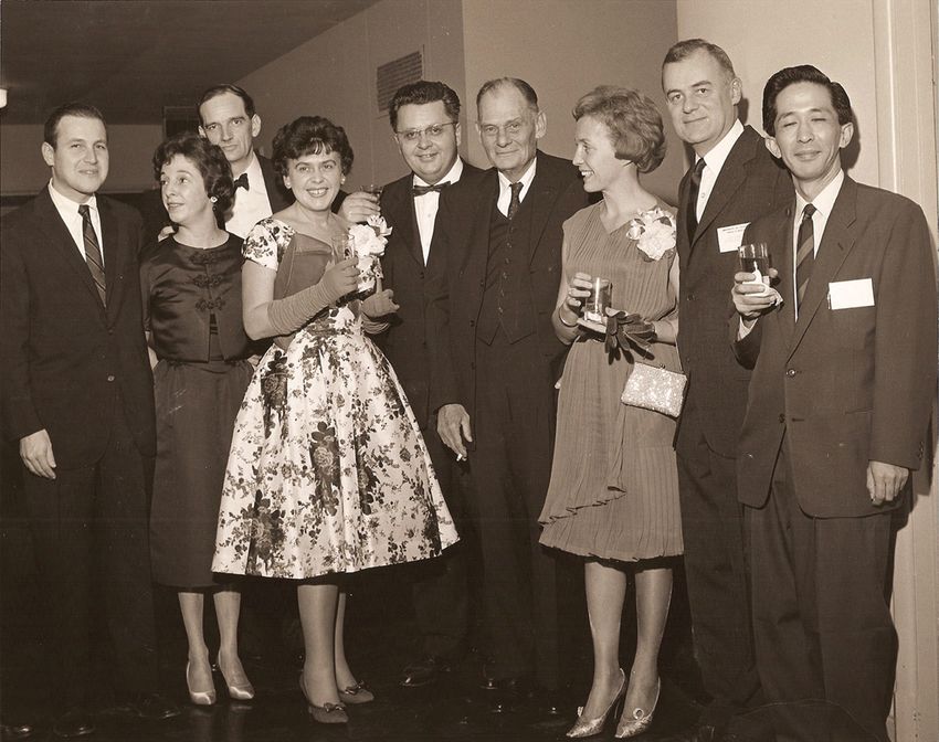
Fig. 1 First International Conference on Measles Immunization. 8 November 1961 at the National
Institutes of Health, Bethesda Maryland. Left to right: Samuel Katz, Ann Holloway, Kevin
McCarthy, Anna Mitus, Milan Milovanovic, John Enders, Gisele Ruckle, Frederick Robbins,
Ikuyu Nagata8 S.L. Katz 10%–15%t of susceptible recipients. A number of investigators (initially Anton Schwarz in 1965 at American Home Products-Pittman Moore Dow and later Maurice Hilleman in 1968 at Merck) further attenuated the Edmonston virus by an increased number of passages in chick embryo fibroblasts at reduced temperature (32°C in contrast to the usual 35°–36°C). Additionally, several firms prepared for- malin-inactivated, alum-precipitated measles vaccine from the Edmonston strain and studied its use in a three-dose schedule (Rauh and Schmidt 1965). The Enders group remained committed to live vaccine, convinced of its advantages over the inactivated preparation (Enders et al. 1962). Both the live attenuated and this inac- tivated vaccine were licensed in the United States on 21 March 1963. Over the ensuing several years, it was discovered that the killed vaccine did not produce enduring immunity and that when recipients were exposed to wild measles, many developed a severe atypical measles infection characterized by high fever, unusual rash beginning most prominently on the extremities, pneumonia with residual pul- monary nodules, and some central nervous system obtundation (Fulginiti et al. 1967; Annunziato et al. 1982). This inactivated vaccine was therefore withdrawn from use in 1967. Fortunately it was not until 1969, 6 years after the licensure of measles virus vaccines, that the responsibility of wild measles virus for subacute sclerosing pan- encephalitis (SSPE) was discovered (Horta-Barbosa et al. 1969; Payne et al. 1969). By then, millions of American children had received live-attenuated measles virus vaccines with no resultant central nervous system complications resembling SSPE, and annual measles cases had been reduced by more than 90%. If the association of measles with SSPE had been appreciated prior to 1963, it is questionable whether licensure of a live-virus vaccine would have been so readily approved. Reassuringly, not only has SSPE become an extreme rarity in the United States and other countries with widespread childhood coverage by measles vaccine, but Bellini and colleagues at CDC have demonstrated that all the few cases identified in recent years are attributable genotypically to wild-type virus distinct from the vaccine strain (Bellini et al. 2005). Early in development of the vaccine, after several presentations at national and international meetings, we began to receive a number of communications from Dr. David Morley, a British pediatrician who was developing child health programs in Nigeria, where he informed us that mortality from measles frequently approached 10%–20%. Of 555 children at his clinic 125 died of measles! He urged us to come to Nigeria and study the vaccine there. Judiciously, however, John Enders cautioned us to wait until the vaccine had proven its safety and efficacy in US youngsters before embarking on such a mission. His concern was that premature studies would be regarded as taking advantage of human guinea pigs rather than as a humane medical mission. Responding eventually to Morley’s entreaties, Katz went in 1960 with Edmonston vaccine provided by Merck, which was then involved in its initial commercial production. The clinical trial was conducted in Imesi-ile, a tiny village outside Ilesha, a larger market town north of Ibadan. When informed of the project, local mothers keenly aware of measles’ morbidity and mortality, eagerly brought their infants and children to participate. Many of these youngsters had malaria,
1 John F. Enders and Measles Virus Vaccine—a Reminiscence                           9

protein malnutrition, and intestinal nematode infestations. Despite these severe
compromises, the initial 26 recipients responded favorably to the vaccine, had no
adverse events, and developed antibodies at the expected time (Katz et al. 1962). A
secondary benefit of this experience was our personal awakening to an awareness
of the serious morbidity and mortality of measles among infants and children in the
resource-limited nations. Our previous perspective had been a rather parochial one,
of measles in the United States where nearly every child by age 7 had acquired the
infection. Complications including otitis media, pneumonia, and gastroenteritis
were common, requiring hospitalization in as many as 20%, but mortality was unu-
sual, approximately one in 500 cases. Progress in the Americas had been remarka-
bly successful, with transmission in the United States halted in 1993 (Katz and
Hinman 2004) and in the entire Western hemisphere by 2002 (de Quadros et al.
2004). The few cases identified since then have been attributable to importations
from countries where measles remains endemic. Although initial success in control
was mainly the result of a single dose schedule, it became apparent that the 5%–
10% of recipients who failed to seroconvert after this administration soon consti-
tuted a significant cluster of susceptibles in whom such a highly transmissible virus
could ignite an outbreak. Therefore, beginning in the early 1990s, a two-dose
schedule became the routine and has been continued worldwide in those nations
where measles vaccination is practiced.
    The experience in Nigeria stimulated our endeavors to place measles vaccine on
the global scene, resulting eventually in its inclusion in the Expanded Program on
Immunization (EPI) of the World Health Organization (WHO). However, there
were still millions of deaths each year and no international effort was initiated,
whereas the global focus was on polio eradication (Katz 2005). However, by the
year 2000, the American Red Cross and International Red Cross and Red Crescent
Societies, joined by the Centers for Disease Control (CDC), the United Nations
Children’s Fund (UNICEF), the United Nations Foundation, and the World Health
Organization (WHO), formed the Measles Partnership (Measles Initiative) with its
goal of reducing measles mortality from 873,000 annually (WHO figures for 1999)
to half in the next 5 years. Remarkably, in the initial 5 years they exceeded their
goal with vaccination of 297 million infants and children (ages 9 months to 5 years)
and a resultant 68% overall decrease in measles mortalities (Wolfson et al. 2007).
Most of this was in sub-Saharan Africa, where only 126,000 deaths were recorded
in 2006 compared to the 506,000 in the first year of the Initiative (Partnership). For
2008–2010, the measles endemic countries of Southeast Asia are the targets of
continuing campaigns.
    Fortunately, measles virus has remained a monotypic agent with remarkably
stable surface proteins that are responsible for induction of immunity. Forty-five
years after introduction of the vaccine in 1963, it continues to provide solid, endur-
ing immunity to vaccine recipients today, neutralizing measles viruses of all line-
ages. Even in those areas where exposure to wild measles viruses have been absent
for many years, antibodies and resultant protection have persisted. An attack of
natural measles conferred lifelong immunity to those who acquired it. Although it
is tempting to predict that successful vaccination with attenuated measles virus will
10 S.L. Katz provide equivalent immunity, it is premature to make such a prediction with the passage of less than five decades since its initial availability. In an era where many individuals are living to their eighties and nineties, the senescence of their immune systems may not maintain what has been assumed to be lifelong immunity. Only by continuing longitudinal studies will the answer to this question be provided. In September 1985, at age 88, John Enders died peacefully at his home while reading poetry. His vision of a measles-free world has come closer to reality than he anticipated, but the challenges of elimination and eradication of so highly trans- missible a virus will continue to confront us for many more years. His legacy, however, endures without challenge. References Annunziato D, Kaplan MH, Hall WW, Ichinose H, Lin JH, Balsam D, Paladino VS (1982) Atypical measles syndrome: pathologic and serologic findings. Pediatrics 70:203–209 Bellini WJ, Rota JS, Lowe LE (2005) Subacute sclerosing panencephalitis: more cases of this dis- eases are prevented by measles immunization than was previously recognized. J Infect Dis 192:1684–1693 de Quadros CA, Izurieta H, Venczel L, Carrasco P (2004) Measles eradication in the Americas: progress to date. J Infect Dis (Suppl 1) 189:S227–S235 Enders JF, Peebles TC (1954) Propagation in tissue culture of cytopathogenic agents from patients with measles. Proc Soc Exp Biol Med 86:277–286 Enders JF, Katz SL, Milovanovic MV, Holloway A (1960) Studies on an attenuated measles-virus vaccine. I. Development and preparation of the vaccine: technics for assay of effects of vacci- nation. New Eng J Med 263:153–159 Enders JF, Katz SL, Holloway A (1962) Development of attenuated measles virus vaccines. A summary of recent investigations. Am J Dis Child 103:335–340 Fulginiti VA, Eller JJ, Downie AW, Kempe CH (1967) Altered reactivity to measles virus: atypical measles in children previously immunized with inactivated measles virus vaccines. JAMA 1075–1080 Horta-Barbosa L, Fucillo DA, Sever JL, Zevan W (1969) Subacute sclerosing panencephalitis: isolation of measles virus from a brain biopsy. Nature 221:974 Katz SL (2005) A vaccine-preventable disease kills half a million children annually. J Infect Dis 192:1679–1680 Katz SL, Enders JF (1959) Immunization of children with a live attenuated measles virus. Am J Dis Child 98:605–607 Katz SL, Hinman AR (2004) Summary and conclusions measles elimination meeting, 16–17 March 2000. J Infect Dis (Suppl 1) 189:S43–S47 Katz SL, Milovanovic MV, Enders JF (1958) Propagation of measles virus in cultures of chick embryo cells. Proc Soc Exp Biol Med 97:23–29 Katz Sl, Enders JF, Holloway A (1960a) Studies on an attenuated measles-virus vaccine. Clinical, virologic and immunologic effects of vaccine in institutionalized children. New Eng J Med 263:159–161 Katz SL, Kempe CH, Black FL, Lepow ML, Krugman S, Haggerty RJ, Enders JF (1960b) Studies on an attenuated measles-virus vaccine. VIII. General summary and evaluation of the results of vaccination. New Eng J Med 263:180–184 Katz SL, Morley DC, Krugman S (1962) Attenuated measles virus vaccine in Nigerian children. Am J Dis Child 103:402–405
1 John F. Enders and Measles Virus Vaccine—a Reminiscence                                   11

Milovanovic MV, Enders JF, Mitus A (1957) Cultivation of measles virus in human amnion cells
   and developing chick embryo. Proc Soc Exp Biol Med 95:120–127
Payne FE, Baublis JV, Itahashi HH (1969) Isolation of measles virus from cell cultures of brain
   from a patient with subacute sclerosing panencephalitis. New Eng J Med 281:585–589
Peebles T, McCarthy K, Enders JF, Holloway A (1957) Behavior of monkeys after inoculation of
   virus derived from patients with measles and propagated in tissue culture. J Immunol
   78:63–74
Rauh LW, Schmidt R (1965) Measles immunization with killed virus vaccine. Am J Dis Child
   109:232–237
Wolfson LJ, Strebel PM, Gacic-Dobo M, Hoekstra EJ, McFarland JW, Hersh BS (2007) Has the
   2005 measles mortality reduction goal been achieved? Lancet 369:191–200
You can also read