JOHN GRISHAM THE TUMOR - Focused Ultrasound Foundation
←
→
Page content transcription
If your browser does not render page correctly, please read the page content below
NO. 1 BESTSELLING AUTHOR

 JOHN
GRISHAM
A NON         -LEGAL           THRILLER

THE TUMOR

F O C U S E D U LT R A S O U N D F O U N D AT I O N

THE TUMOR
A NON         -LEGAL            THRILLER

John Grisham

Focused Ultrasound Foundation . Charlottesville VA

Focused Ultrasound Foundation
    1230 Cedars Court, Suite 206
    Charlottesville VA 22903

    fusfoundation.org

    Text:
    ©2015–2020 John Grisham
    Medical Illustrations:
    © 2015–2020 Anatomical Justice, LLC,
    pp. 16, 17, 18 (fig. 6 & 7)

    All rights reserved under International and
    Pan-American Copyright Convention.

    ISBN 978-1-4951-7940-2

2   JOHN GRISHAM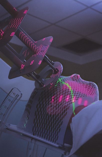
4    Dear Reader

 6 Chapter 1

     The Patient

 9 Chapter 2

     The Tumor

15 Chapter 3

     The Treatment

24 Chapter 4

     The End

29 Chapter 5

     The Alternative

34 Chapter 6
     The End—Revised Version

37 Chapter 7

     The Present

44   Focused Ultrasound Foundation

46   The Ask

THE TUMOR   | Focused Ultrasound Foundation   3
Dear Reader

                                      My knowledge of medicine and medical
                                      research is quite limited. When I was
                                      a student, I drifted away from science
                                      and math, preferring instead subjects
                                      I considered less demanding. I eventually
                                      made it to law school and became a
                                      lawyer. After a brief career suing people
                                      (never a doctor, though), I stumbled
                                      upon fiction and wrote a couple of
                                      books. Others followed, and I happily
                                      shuttered the law office. Because the
                                      books have done well, I have been lucky
enough to dabble in philanthropy. Once you get the reputation of being
generous, a lot of opportunities present themselves.

Seven years ago, my friend and neighbor, Neal Kassell, gave a PowerPoint
presentation on focused ultrasound therapy. Neal is a prominent neurosurgeon
who’s spent his career drilling through skulls and making repairs to brains.
During the PowerPoint, Neal, with great enthusiasm, explained that
focused ultrasound therapy could one day alleviate the need for conventional
brain surgery. Tumors would be destroyed using beams of ultrasound
energy, and afterward the patient would walk out of the operating room
and go home. Not only would the treatment be non-invasive, painless,
quick, and relatively inexpensive, it could also save the patient’s life.

Focused ultrasound therapy is still in its early stages, still experimental, but
there is enough research to date to be very optimistic.

4        JOHN GRISHAM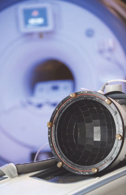
The brain is just the starting point. Tumors in the breast, prostate, pancreas,
liver, kidneys, and bones could be treated on an out-patient basis. Neal loves to
use the example of a man with prostate cancer undergoing focused ultrasound
therapy, then driving himself back to the office for a few hours. Later, he goes
home to celebrate his wedding anniversary with his wife. They share a champagne
toast to growing old together.

This is not science fiction. Around the world, more than 70,000 men with
prostate cancer have been treated with focused ultrasound. Nearly 100,000
women with uterine fibroids (benign tumors of the uterus) have been treated,
thus avoiding hysterectomies and infertility. Clinical trials for tumors of the
brain, breast, pancreas, and liver, as well as Parkinson’s disease and arthritis,
are inching forward at over 360 research sites around the world.

Though focused ultrasound technology is in its infancy, there is great enthusiasm
for its potential to improve quality of life and decrease cost of care. For this
potential, to be fully demonstrated, additional laboratory research and clinical
trials are necessary.

But progress is too slow. There are barriers from regulators, insurance companies,
even many in the medical field.

I have found no other cause, issue, non-profit, or charity that can potentially
save so many lives. One day in the not-too-distant future, you or someone
you love will be diagnosed with a tumor. After the shock, you will think of
focused ultrasound.

   Let’s hope it’s available.

                         THE TUMOR     | Focused Ultrasound Foundation              5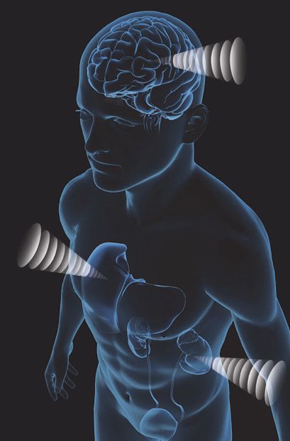
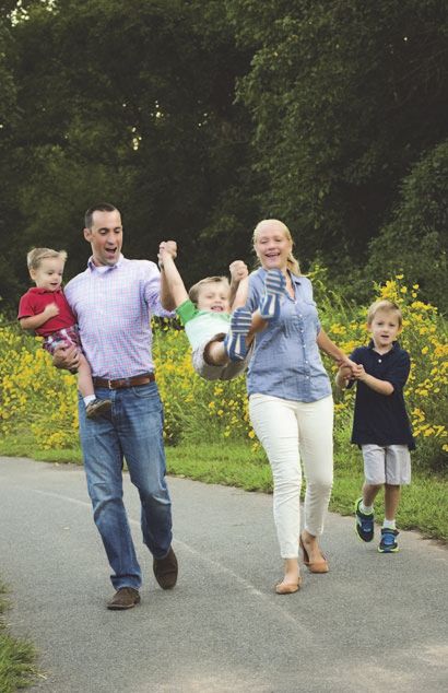
Chapter 1

The Patient

Meet Paul, a 35-year-old banker with a lovely wife, Karen, and three small
children. They enjoy a nice life in the suburbs with lots of friends and
the usual activities—backyard cookouts, swim parties, tee-ball, church on
Sundays. They are active and enjoy great health. Paul’s parents are in
their 60s and also very healthy. Paul gets a complete physical once a year,
jogs twenty miles a week, plays golf and tennis at a nearby club, and
avoids extra pounds. He has an occasional beer, doesn’t smoke, and takes
no medication.

6       JOHN GRISHAM

Chapter 2
The Tumor
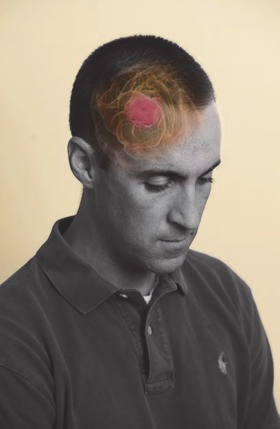
But Paul has a problem. He has a tumor in the right frontal lobe of his
brain, about the size of a hen’s egg.
Looking back, the first symptom was a gradual decrease in his ability to
concentrate at work. Naturally curious and active, he noticed an
uncharacteristic tendency to procrastinate. At times he felt listless and tired.
Then the headaches arrived, and with a fury. He blamed them on stress
and took lots of ibuprofen. As he drove to work one morning, his vision
became so blurred he stopped the car. Karen began to notice mood swings
and a loss of patience with the kids. He grew more irritable, both at
home and at the office. His boss chastised him for barking at a coworker.
He quarreled with Karen over his dour moods and crankiness. She knew
something was changing with her husband and urged him to see a doctor.
He refused.
On a Wednesday morning, as Paul is in the bathroom shaving, Karen hears
a loud thump. She finds him on the floor, shaking in a full-blown grand
mal seizure. She calls 911, and as she waits the seizure stops and he
gradually awakens. He is confused, disoriented—doesn’t recognize Karen
and doesn’t know where he is. The rescue squad arrives. Paul is loaded
into an ambulance and taken to the hospital. In the emergency room,
he is still drowsy and confused and complains of weakness on his left side.
Upon examination, his left hand is very weak and he has difficulty lifting his
left arm and leg. An MR scan reveals the tumor.
He is admitted to the hospital and started on anticonvulsant medication
to prevent further seizures, as well as steroids to decrease the swelling in
his brain around the tumor. Paul and Karen are not shown the MR scan.
A neurosurgeon is consulted.
THE TUMOR | Focused Ultrasound Foundation 9By Thursday morning, the confusion and disorientation are gone, as is the weakness in his left side. He feels much better, briefly, but things will change. When the neurosurgeon arrives early that morning for the initial consultation, he produces the MR scan (opposite). As they stare at it, Paul and Karen are too stunned to speak. The doctor explains that Paul indeed has a tumor in his brain and it appears to be the type known as a glioma. Surgery is needed to remove as much of it as possible and to obtain tissue to determine the type of tumor. They talk about the operation. The doctor covers the risks. For complications like death and paralysis, the risks are very small. The most likely complication will be a weakness on the left side. The surgery will take about three hours, and if all goes well, Paul can expect to go home in three days. 10 JOHN GRISHAM
MR scan of Paul’s tumor

The neurosurgeon explains that gliomas are graded one through four, with
one and two being benign. Three and four are malignant. Grade four,
the most catastrophic, is called a glioblastoma. The life expectancy for
a grade four diagnosis is short. Regardless of treatment—surgery, chemo,
radiation—the average length of survival is about one year. Left untreated
but managed with pain medication only, the patient can expect to live
several months. About 22,000 Americans are diagnosed each year with
glioblastomas; 15,000 die within 12 months. The lucky ones, about one
in ten, live for five years.

Based on the MR scan, the neurosurgeon thinks the odds are about 50–50
that the tumor is benign.

                          THE TUMOR   | Focused Ultrasound Foundation        11
He recommends surgery at the earliest convenient time and it is scheduled
for the following Monday. After the doctor leaves, Paul and Karen attempt
to come to grips with what’s happening. Should they get another opinion?
It seems senseless when staring at the MR scan. There is no doubt about
the tumor. They like their neurosurgeon and a quick search online proves
he’s one of the best. They are in the finest hospital in the city. Surgery is
needed sooner rather than later. There is no time to waste.
Needless to say, the weekend is long and agonizing. Karen gives the bad
news to the family but not to their children. She refuses to believe the
tumor is malignant and is convinced the surgery will go well.
She spends hours online gathering frightening and depressing information
about brain tumors. Ted Kennedy, Susan Hayward, Beau Biden, Lee
Atwater, George Gershwin, Lou Rawls, Bobby Van, Pete Rozelle, Wilma
Rudolph—they are just a few of those who died from a glioblastoma.
On average, they survived about 12 months after being diagnosed.
Opposite
Well-known people who died from a
glioblastoma. On average, they survived
a year after diagnosis.
top, left to right
Lee Atwater, 40
Wilma Rudolph, 54
middle, left to right
George Gershwin, 38
Edward “Ted” M. Kennedy, 77
bottom, left to right
Joseph Robinette “Beau” Biden, III, 46
Susan Hayward, 57
12 JOHN GRISHAMChapter 3
The Treatment
At 6 a.m. on Monday, Paul and Karen are in his hospital room, wide-awake,
fearful but trying to appear brave as they wait for the day to unfold.
At 6:30, Paul is prepped for surgery and two orderlies arrive with a gurney
for the short ride to the operating room. Paul hugs his wife, who’s
overcome with emotion. After he’s gone, she is led by a nurse to the waiting
room. His parents are already there. The nurse tells them the surgery
should be over around noon. As they settle in for a long morning, the
room begins to fill with other anxious families.
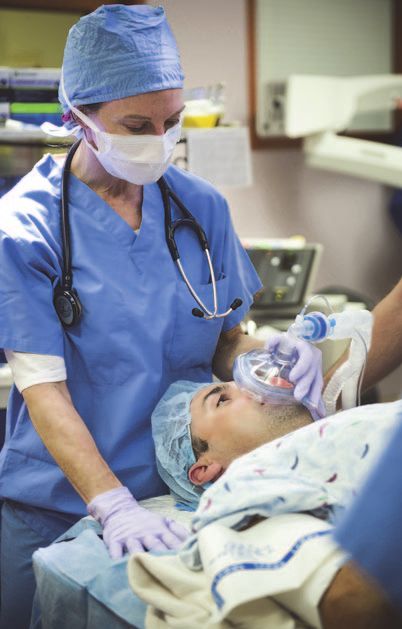
Paul is rolled into the operating room and put to sleep with general anesthesia.
THE TUMOR | Focused Ultrasound Foundation 15His head is shaved and his skull fixed in a three-prong headrest to
immobilize it (fig. 1).
A question-mark-shaped incision
is made from the midpoint of
his forehead just below the
hairline to a point in front of his
right ear.
Burr holes are made in the skull,
and a power saw is used to fashion
a bone flap more or less like the
top of a cookie jar (fig. 2).
The dura mater—a membrane
between the inner table of the
skull and the brain—is cut (fig. 3).
The surface of the frontal lobe is
1 discolored and distorted because
of the tumor.
The tumor is localized with an intraoperative navigation device to
minimize damage to the motor cortex—the portion of the brain
controlling movement of the left side of the body. Under the
magnification of an operating microscope, an incision is made into the
brain, and just beneath this the abnormal tissue is identified.
A portion is cut out and sent to the pathology lab for a preliminary
diagnosis (fig. 4). The tumor is removed by suction and the
bleeding is controlled with coagulation (fig. 5).
16 JOHN GRISHAM2 3
4 5
THE TUMOR | Focused Ultrasound Foundation 176
7
The surgeons are able to remove everything that appears abnormal.
They then suture the dura mater, secure the bone flap with screws (fig. 6),
and staple the skin flap (fig. 7).
After three hours of surgery, they are confident things went as well as
possible. Paul is taken to recovery.
The surgeon goes to the waiting room and meets with Karen and Paul’s
parents. He reports that everything went as expected: the tumor was
removed, there were no complications, and they should be able to see Paul
18 JOHN GRISHAMin about an hour. The initial results of the biopsy are not good—it looks like
a glioblastoma, but it will take several days for the final report.
Meanwhile, Paul is waking up,
and there are problems. He has
profound weakness in the left side
of his body. He cannot lift his left
arm or leg off the bed. He has only
a flicker of motion in his fingers.
The surgeon immediately orders
a CT scan to make sure the
weakness is not caused by
hemorrhaging or a blood clot in
his brain from the surgery. It is not.
The CT scan is unremarkable.
Early Monday evening, Paul and
Karen meet with the neurosurgeon,
who has a preliminary pathology
report; the final one is a few
days away. While the tumor was
successfully removed, it is the type
Post-operative MR scan
of tumor that will likely recur.
The next day another MR scan confirms that there was total removal of all
visible tumor, a rare bit of good news (above).
Chemotherapy and radiation will be necessary to slow its regrowth. As for the
weakness in Paul’s left side, the doctor says it is undoubtedly the result of
surgical manipulations and should get better.
By Tuesday morning, the weakness has improved slightly. Paul is able to lift
his arm but movements of his fingers are slow and his grip is weak. He is able
to stand but can walk only with assistance. Karen stays by his side as the hours
drag on. He wants to discuss what’s on his mind: death, life insurance, his last
THE TUMOR | Focused Ultrasound Foundation 19will and testament, their savings, her future, the kids’ futures. Karen, though,
is simply not ready for this. She doggedly maintains a veneer of optimism.
To Paul, it seems more like denial. She tells him that a lot of friends are eager
to stop by for a visit, but he says no. He looks awful, feels worse, and wants
to see no one, not even his own children.
Early Wednesday morning, one week after his seizure, they meet again with
the neurosurgeon. The pathology report confirms their worst fears:
glioblastoma, grade four. Although the tumor has been removed, it left
behind microscopic portions that extend into the normal brain and cannot be
surgically removed. These remnants of the tumor will almost certainly regrow,
and must be treated with radiation and chemotherapy. When the tumor
returns, there will be the likelihood of more surgery.
With as much professional sympathy as possible, the doctor tells them that,
according to statistics, Paul can expect to live 12 to 14 months. Occasionally
a patient will live 5 to 10 years, but that’s uncommon. He offers his usual,
“Hope for a miracle, but plan for the average.”
“Plan for the average,” Paul repeats after the doctor leaves. Karen pulls the
shades and turns off the lights. They sit in the darkness, holding hands, as
the monitors beep occasionally. When they speak, they discuss the best way
to tell the children.
Thursday morning, the radiation oncologist stops by. He describes
radiation therapy and explains that it will be administered five days a week
for the next six weeks. Among other side effects, Paul will lose his hair
and his face will swell and become disfigured from the steroids he’ll be
given. Later, the neuro-oncologist stops by and they discuss chemotherapy,
which has its own set of unpleasant side effects.
On Friday, Karen brings their children to the hospital to visit. She and Paul
have decided to wait a few months before delivering the bad news. They still
believe in miracles and are praying a lot. After the kids leave, Paul’s parents
Opposite, radiation therapy treatment
THE TUMOR | Focused Ultrasound Foundation 21visit him. With Karen home with the children, he does not pull punches. He’ll be lucky if he’s still living a year from now. Later, alone and in a dark room, Paul opens his laptop and pulls up a calendar for the next 12 months. It’s all there, all planned: the school year, their upcoming vacation, the holidays and birthdays, a golfing trip with his friends, several business trips, his parents’ 40th anniversary. Would he be able to enjoy any of it? Would he even be alive? “Plan for the average” means he should be able to make it to Christmas. What does a father of three young children do to celebrate his last Christmas? Paul thinks about the next 12 months and asks himself many questions. There are no answers. Later that afternoon, he is transferred to a rehabilitation facility to address the weakness in his left side. He cannot raise his left hand to his face, nor can he walk without a cane. Ten days after surgery, he is discharged and taken home. He is instructed to return to the rehab facility three times per week. His left side continues to improve. Being at home lifts his spirits. Friends arrange meals and there is a steady flow of traffic to the house. He tries to eat but his appetite is gone. Two weeks after surgery, Karen drives him to the office where he’s greeted like a hero. He is determined to work at least half a day until he regains his strength, and he assures his colleagues he’ll be back. Paul begins radiation therapy Monday through Friday, five days per week. His hair falls out rapidly from the radiation and, worse, his face begins to swell from the steroids (opposite). The moon face seems to grow each day. He looks terrible. He is constantly fatigued, and his thinking becomes slow and dull from the damage to his brain caused by the radiation. 22 JOHN GRISHAM
Before surgery and radiation therapy           After surgery and radiation therapy

He gives up on the idea of going to the office. His boss promises to cover
for him and keep the paychecks coming. The group medical policy covers
80 percent of the expenses.

His lawyer drafts a new will, not that one is really needed. Paul and Karen
own everything jointly; upon his death, it’s all hers anyway. She certainly
gets the kids. His life insurance policy is for only $250,000. They have
about $40,000 in savings. With three children under the age of eight,
the future is anything but secure. Karen secretly begins checking out
employment opportunities on the internet. Their minister stops by every
other day for a devotion and prayer.

                          THE TUMOR    | Focused Ultrasound Foundation          23
Chapter 4
The End
Six months later, the weakness in Paul’s left side increases dramatically.
He cannot grasp objects with his left hand. He drags his left foot when
walking and cannot move around without assistance. He notices he cannot
concentrate for more than a few seconds. His short-term memory is shot.
An MR scan shows the tumor is back and growing rapidly (below).
It also reveals damage in his brain
compatible with the effects of radiation.
His neurosurgeon offers the option
of another surgery to remove the
recurrent tumor.
Paul and Karen discuss this for several
days. A second operation is more
likely to damage the brain. There is no
certainty that the tumor will not recur
again, and again. They are losing
hope, and their thoughts of miracles
are fading rapidly. Paul could almost
MR showing recurrent tumor throw in the towel, opt for a few final
weeks with pain medication, and
suffer as little as possible to the end. Karen, though, still believes in luck.
The second operation is similar to the first. The visible portions of the
tumor are removed, and Paul’s skull is put back together. When he awakens,
though, the weakness in his left side is much worse. He is transferred to
a rehabilitation center. After three weeks of intense therapy, his condition
does not improve. He can no longer stand without assistance, nor walk
without a walker. His left hand is essentially useless. He is discharged home,
24 JOHN GRISHAMwhere he arrives in a wheelchair. The chemotherapy has been ineffective
and is terminated. Paul takes steroids in an effort to reduce swelling in
his brain.
At this point, Paul begins saying goodbye to his family and friends.
He bravely accepts the fact that his days are numbered, and he wishes to
say farewell on his terms. As bad as he looks, he knows that things will
only get worse.
In a heart-wrenching scene, he and Karen finally tell the children that their
father is about to leave them.
The steroids are not working and are cut off. He’s left with only some
powerful narcotics to deaden the horrible headaches, which occur with
increasing severity.
THE TUMOR | Focused Ultrasound Foundation 25Paul prays for a quick and painless end, but this doesn’t happen. He slowly
deteriorates and becomes increasingly confused and disoriented. He loses
almost all consciousness and his ability to move. He is bedridden and
requires around-the-clock care for feeding and bathing. Karen sleepwalks
through the days and nights, thoroughly drained, but trying gamely to
shield his condition from the children as much as possible. Eight months
after his seizure, Paul has completely checked out, but his heart still manages
to beat. Karen finally begins praying for a merciful end.
Nine months after the first surgery, he passes away, at the age of 36.
The total cost of his treatment and care is
approximately $300,000.
26 JOHN GRISHAMChapter 5
The Alternative
Paul was born in 1980, ten years too early. Had he been born in 1990 and
diagnosed with a brain tumor at the age of 35, in 2025, his story could be
rewritten as follows:
That same Wednesday morning, Karen hears a crash in the bathroom,
and she finds Paul on the floor in a grand mal seizure. He’s taken to the
ER and admitted to the hospital. An MR scan is performed with molecular
imaging, a more advanced scan than was available ten years earlier.
Based on the scan, the neurosurgeon, with virtual certainty, makes a diagnosis
of a glioblastoma and explains the prognosis and the treatment options,
including focused ultrasound therapy. The size and location of Paul’s tumor
make it amenable to treatment with focused ultrasound therapy, which
is what the neurosurgeon recommends. He explains that the tumor in all
probability cannot be cured and will return, but it can be controlled with
repeated treatment, giving Paul additional years with a high quality of life.
Opposite, the focused ultrasound brain
transducer fits over the head and
emits beams of energy that penetrate
the skull to target a tumor.
THE TUMOR | Focused Ultrasound Foundation 291 >
2
Early Friday morning, Paul and Karen walk into the focused ultrasound
therapy suite (fig. 1). He changes into a gown, takes a light sedative, and is
positioned on his back on a table.
His head is securely fixed in a hemispheric focused ultrasound brain transducer
(fig. 2). The transducer is capable of transmitting more than 1,000 intersecting
beams of ultrasound energy through the scalp and skull to the tumor with
a high degree of accuracy and without damaging the adjacent normal tissue.
After the transducer is in place, Paul is inserted into the bore of the MR machine.
30 JOHN GRISHAM3
>
>
4
5
In the adjacent control room, the surgeon manually outlines the tumor,
which is to be destroyed by the focused ultrasound beams (fig. 4). Paul is
awake and feels no discomfort. Karen stays by his side, holding his hand and
chatting with him. The treatment begins. The surgeon is in constant voice
contact with Paul and Karen (fig. 3). He uses continuous images from the
MR scan to guide the precise point where the ultrasound is focused and to
control the delivery of its energy to the target (fig. 5).
THE TUMOR | Focused Ultrasound Foundation 31Before focused ultrasound therapy After focused ultrasound therapy It takes about an hour to kill all of the tumor that can be seen on the MR scan. At the completion of the procedure, another scan is performed to confirm the entire tumor was treated. While this is happening, powerful chemotherapy agents enclosed in microscopic nanoparticles are injected intravenously. These circulate with the blood in every tissue and organ in the body, but the chemotherapy drugs are inactive because they are trapped inside the nanoparticles. After eliminating all of the tumor that can be seen on the MR scan, the surgeon then refocuses the ultrasound to the surrounding brain to activate the nanoparticles, which release their pharmacological payload in the precise area around the tumor where residual microscopic extensions of the tumor have infiltrated. This allows very high concentrations of the drugs to be delivered focally to the brain while minimizing systemic side effects. The remainder of the chemo-laden nanoparticles will be excreted. 32 JOHN GRISHAM
Less than two hours after the treatment began, Paul gets off the table and
walks to the recovery room for observation. There are no complications,
and he is discharged home Friday afternoon. He feels fine. The only
aftershock is some residual clumsiness in his left hand from his initial
seizure, which is decreasing. On Saturday afternoon, he and Karen and the
kids walk down the street for a block party. They have yet to tell their
families and friends about the tumor and the treatment. On Sunday, the
entire family goes to church.

On Monday, Paul is at the office before 8 a.m., eager to catch up after three
days off. He explains to his colleagues that he was in the hospital for “tests,”
but everything is fine. He looks and feels like himself. The weakness in his
left side continues to fade away.

A month after the ultrasound procedure, he undergoes another MR scan.
The scan reveals that the ablated tissue is being absorbed harmlessly by
Paul’s body. The mass is not regrowing due to the efficacy of the focused
chemotherapy treatment that targeted the microscopic extensions of the
malignant tumor. The deadened tissue continues to shrink.

Three years later, Paul again notices difficulty using his left hand. His left
foot occasionally drags. He does not hesitate and calls his doctor immediately.
An MR scan is done and reveals that the tumor is back. The following day,
as an outpatient, he undergoes another focused ultrasound procedure.
The weakness goes away after a month. Four years later, seven years after his
initial diagnosis, the tumor is growing again, and the procedure is done for
the third time.

   Cost to date: approximately $75,000
   Savings: about $225,000 and one life prolonged

                        THE TUMOR     | Focused Ultrasound Foundation            33
Chapter 6 The End—Revised Version The tumor will eventually take Paul’s life. However, focused ultrasound therapy could transform a fatal condition into one that is chronic but manageable. In contrast to the best current treatment circa 2015, the futuristic ultrasound therapy depicted here circa 2025 could potentially be accomplished on an outpatient basis without multiple days of hospitalization, without surgery and its attendant risks of infection and complications like blood clots and brain damage, without the harmful effects of radiation, and with minimal side effects of chemotherapy due to focused drug delivery. The net result could be a dramatic improvement in the quality and longevity of countless lives, and decreased cost of treatment. How many years will it add? At this time, the answer is uncertain. Clinical trials for brain tumors have just begun, and the patients selected for these trials represent the most desperate of cases. Much more research is needed. Researchers believe, with caution, that five additional years are realistic. Perhaps ten. Who wouldn’t bargain for ten more years, especially with a high quality of life? With time, research, and improved technology, neurosurgeons are hopeful that a guy like Paul can live to the age of 45 or even 50, long enough to see his children mature. 34 JOHN GRISHAM
Chapter 7

The Present

Focused ultrasound is a new, revolutionary, groundbreaking, non-invasive
therapeutic technology that has the potential to transform the treatment of
a variety of serious medical disorders in addition to brain tumors, improve
outcomes, and decrease the cost of care. It could become an alternative to,
or complement for, traditional surgery, radiation therapy, and drug delivery.
Focused ultrasound could result in fewer complications, such as damage to
normal tissue, infection, hemorrhage, and pain, as well as shorten recovery
times. By providing safer and more effective therapy, it could reduce death,
disability, and suffering for millions of people around the world.

Focused ultrasound utilizes intersecting beams of high-frequency sound
concentrated accurately and precisely on tissue deep in the body, much as
sunlight passing through a magnifying glass can be focused to burn a hole
in a leaf.

                        THE TUMOR    | Focused Ultrasound Foundation          37
Table 1
Effects at focal point
Variety of effects,
variety of disorders…
■ Thermal ablation
precise heating and destruction
of tissue
■ Focal drug delivery
delivery of very high
concentrations of drugs precisely
where they are needed
■ Blood-brain barrier opening
temporary access of drugs to
the brain
■ Immunomodulation
stimulation of immune response
allowing body to fight cancer
■ Neuromodulation
reversible stimulation or
inhibition of cells in the brain
and nervous system
■ Radiation sensitization
sensitizing tumors to effects of
radiation allowing use of lower
dose to kill cancer cells
■ Stem cell delivery
specific “homing” of stem cells
to targeted tissue
38 JOHN GRISHAMAt the point where the beams converge, the ultrasound energy induces a
variety of biological effects while surrounding structures and tissues remain
undamaged (table 1).
Magnetic resonance or ultrasound imaging is used to identify, guide, and
control the treatment in real time.
In the theoretical example used in this all too common story, focused
ultrasound treatment of the malignant brain tumor controlled but did not
eradicate it. This alternative treatment provided years of high quality of
life to a patient with an ultimately fatal condition. The treatment of brain
tumors is not the only area of medicine in which focused ultrasound
therapy shows promising results. There are many more applications,
including uterine fibroids, prostate cancer, and essential tremor, where
focused ultrasound treatment could potentially cure the disease.
Today, focused ultrasound is in various stages of development for treating
over 120 diseases and conditions, including epilepsy, Alzheimer’s and
Parkinson’s diseases, and tumors of the brain, liver, pancreas, and lung
(table 2). But despite the progress so far, much work remains to be done
before focused ultrasound can be widely used to treat large numbers
of patients.
Unfortunately, it often takes decades for a new therapeutic technology
like focused ultrasound to become widely adopted as a mainstream
standard of care.
THE TUMOR | Focused Ultrasound Foundation 39Table 2
Status of progress by disease
Pre-clinical Outside US FDA
Research Clinical Trials Approval Approval US Reimbursement
Cardiovascular Neurological
Hypertension Essential tremor
Atrial fibrillation Parkinson’s disease
Peripheral artery disease Depression
Varicose veins Neuropathic pain
Arteriovenous malformations OCD
Cardiac hypertrophy ALS/Lou Gehrig’s disease
Congestive heart failure Alzheimer’s disease
Deep vein thrombosis Brain tumors
Cancer pain
Endocrine Disorders Dementia
Thyroid nodules Dystonia
Thyroid cancer Epilepsy
Diabetes Huntington’s disease
Neuroblastoma, pediatric
Gastrointestinal Traumatic brain injury
Liver tumors Addiction
Pancreatic tumors Hydrocephalus
Colorectal tumors Migraine
Esophageal tumors Multiple sclerosis
Stroke
Miscellaneous
Glaucoma Urological
Head & neck tumors Prostate disease
Lung cancer Kidney tumors
Melanoma Kidney stones
Obesity Bladder tumors
Wound healing
Women’s Health
Musculoskeletal Uterine fibroids
Bone metastases Breast cancer
Bone cancer Cervicitis
Multiple myeloma Cervical tumors
Osteoid osteoma Ectopic pregnancy
Plantar fasciitis Endometriosis
Soft tissue cancer Ovarian tumors
Arthritis Polycystic ovary syndrome
Desmoid tumors
Disc degeneration This list includes the most common diseases in
Sacral chordoma the US population. For a complete list of diseases
Osteomyelitis see www.fusfoundation.org.
40 JOHN GRISHAMFocused ultrasound is approved to treat more than 30 conditions in various
countries around the world and approved by the FDA to treat six conditions
in the U.S., including uterine fibroids, prostate cancer, benign prostatic
hyperplasia, bone metastases, Parkinson’s disease, and essential tremor
(Table 2). The Centers for Medicare and Medicaid Services (CMS) in the
U.S. have approved reimbursement for essential tremor.
Table 3
Stakeholders with different agendas
■ Patients
■ Academic research sites
■ Philanthropy
■ Patient advocacy organizations
■ National Institutes of Health
$
■ Venture capital
■ Industry
■ FDA
■ Insurers
■ Medical professionals
Every delay in the availability of focused ultrasound results in unnecessary
death, disability, and suffering for countless people.
There are numerous steps in the complicated process of evolution from an
idea to laboratory research to widespread patient treatment.
It requires the involvement of a large number of organizations that have
different agendas and timelines for decision making (table 3).
THE TUMOR | Focused Ultrasound Foundation 41Table 4
Obstacles
■ Technical and engineering
■ Resistance to new technology
and procedures
■ Evidence generation
■ Cultural and turf issues
■ Financial
■ Regulatory
■ Reimbursement
Regulatory approvals from the government, reimbursement from insurance
companies, and acceptance by physicians are all barriers that must first be
overcome (table 4).
Research is ongoing in more than 360 academic institutions around the world,
including Stanford, University of Virginia, Sunnybrook in Toronto, Royal
Marsden in London, University of Maryland, and Brigham and Women’s.
More than 50 medical device manufacturers, in partnership with public
institutions and patient organizations, are making tremendous progress in
understanding and utilizing the mechanisms by which focused ultrasound
affects tissue. This knowledge is being converted into laboratory studies and
clinical trials which are critical for the ultimate goal—adoption.
The field is growing more rapidly than anticipated, but the amount of
work remaining to move this early-stage research into widespread adoption
is still great. Additional resources are needed to bridge the gap between
research and trials and the treatment of millions of patients with disabling
or life-threatening disorders.
Meet four people whose lives have been dramatically
improved by focused ultrasound therapy.
42 JOHN GRISHAM▲ Six years after her Parkinson’s disease ▲ Elizabeth suffered from uterine diagnosis, Kimberly suffered from fibroids that made it hard for her to leave involuntary shaking and could no longer the house and work with her interior ride her bike or enjoy other forms of design clients. Thanks to a focused exercise. She was successfully treated ultrasound clinical trial, she was able to in a focused ultrasound clinical trial retain her uterus, eliminate her at the University of Maryland. She is symptoms, and get back to living. now back on her bike and says the clock has been turned back on her life. ▲ Peter suffered from tremors since ▲ Canadian teenager Jack was able to his teenage years. As his symptoms resume his favorite sports after having worsened and the uncontrollable shaking focused ultrasound treatment for a made everyday tasks like eating, painful and disabling bone tumor. drinking, and dressing nearly impossible, Before treatment, Jack required heavy he turned to focused ultrasound. doses of pain medications and couldn’t Since the treatment, Peter has a new sleep through the night. After treatment, lease on life and enjoys getting back to his pain went away and he hasn’t the activities he once enjoyed. looked back since.
Focused Ultrasound Foundation

In 2006, Dr. Neal Kassell started the Focused Ultrasound Foundation,
and headquartered it in his hometown of Charlottesville, Virginia. The
Foundation’s mission is simple:

      “To accelerate the development and adoption of focused ultrasound.”

The Foundation is a unique medical research, education, and advocacy
organization whose stated goal is to shorten the time from laboratory research
to widespread patient treatment with focused ultrasound. Its principal
role is to coordinate the activities of the major players: researchers, doctors,
patients, manufacturers, insurers, regulators, and donors. There are now
more than 360 research centers, 105 in the U.S. alone, scattered around
the globe, as well as over 250 clinical trials taking place in 30 countries,
along with 1,000 doctors and scientists involved in research, and 51 private
companies working to perfect the technology.

The Foundation provides resources that are critical to fostering collaboration
and generating evidence of the safety and efficacy of focused ultrasound
treatments. By doing so, it creates a knowledge base that allows medical
professionals, patients, insurers, and countless other organizations to overcome
obstacles, resulting in solutions that help accelerate the development and
adoption of focused ultrasound. The metrics of improving outcomes
of diagnoses and reducing costs of care are hard to calculate. But we can
estimate that the benefits are immense—particularly when applied to an
individual life (table 5).

     The Foundation’s annual budget is $8 million,
     90 percent of which comes from individuals.

44        JOHN GRISHAM
Table 5
Time = Lives
The Foundation
strives to Lives improved
accelerate adoption
Patients
treated
Expected adoption for
new medical device without
the Foundation’s support.
2000 2010 2020 2030
Years to widespread availability
THE TUMOR | Focused Ultrasound Foundation 45The Ask
Focused ultrasound is a new, revolutionary, groundbreaking, non-invasive
therapeutic technology. In the future, focused ultrasound therapy could be
routinely used to treat patients like Paul. Until then, though, millions will
suffer and die. “Acceleration” is the operative word, and the Foundation
needs your help to speed things along.
Visit the Foundation website,
fusfoundation.org, to:
■ find treatment centers
■ learn about clinical trials
■ sign up to stay in touch
■ support our work
■ share this book
And discover how you could help
improve the lives of your friends,
loved ones, children—and maybe
even your own.
Please remember: Time = Lives.
A patient with essential tremor celebrates
the return of the use of his right hand after
undergoing focused ultrasound treatment.
Like | Follow
46 JOHN GRISHAMFocused Ultrasound Foundation
Board of Directors
Neal F. Kassell, MD
Chairman, Focused Ultrasound Foundation
Former Co-chair of Neurosurgery, University of Virginia
Dorothy N. Batten
Founder, iThrive Initiative
Former Director, Landmark Media Enterprises LLC
Eugene V. Fife
Founding Principal, Vawter Capital, LLC
Former Chairman, Goldman Sachs International
John R. Grisham
Author
William A. Hawkins, III
Senior Advisor, EW Healthcare Partners
Former Chairman & CEO, Medtronic
Daniel P. Jordan, PhD
President Emeritus, Thomas Jefferson Foundation, Inc.
Edward J. “Ned” Kelly, III
Former Chairman, Institutional Clients Group, Citigroup Inc.
Syaru Shirley Lin
Adjunct Faculty, Chinese University of Hong Kong
Director, Goldman Sachs Asia Bank
Edward D. Miller, MD
Former CEO, Johns Hopkins Medicine
Frederic H. Moll, MD
Chief Development Officer,
Johnson & Johnson Medical Devices Companies
Co-founder, Auris Health, Inc.
Charles W. “Wick” Moorman IV
Former Chairman & CEO, Norfolk Southern
Former CEO, Amtrak
Steve H. Rusckowski
Chairman, President & CEO, Quest Diagnostics Inc.
Former CEO, Philips Healthcare
Carl P. Zeithaml, PhD
Dean and F. S. Cornell Professor of Free Enterprise,
McIntire School of Commerce, University of Virginia
THE TUMOR | Focused Ultrasound Foundation 47Colophon Photography
Designer Cover, pp. 7, 10, 14, 15, 23, 25, 27, 28,
Anne Chesnut 30, 31, and 35
Charlottesville VA Stephanie Gross, Charlottesville VA
p. 13 (Lee Atwater)
Managing editor Office of the President, 21 January 1989
Sara Coates Myhre
p. 13 (Wilma Rudolph)
Charlottesville VA
Dutch National Archives
Copy editor p. 13 (George Gershwin),
Margo Browning Library of Congress, Prints and
Charlottesville VA Photographs Division, Van Vechten
Collection, reproduction number
Printer LC-USZ62-42534 DLC
Worth Higgins & Associates, Inc. p. 13 (Edward M. Kennedy)
Richmond VA United States Senate official portrait
Stock p. 13 (Beau Biden)
McCoy Silk text & cover Official campaign portrait
p. 13 (Susan Hayward)
Type 20th Century Fox publicity photo
Galliard, Whitney p. 46 The Canadian Press/Frank Gunn
Acknowledgements MR scans
Focused Ultrasound Foundation would pp. 11, 19, 24, 31, 32
like to thank Martha Jefferson Hospital Max Wintermark, MD,
Stanford University School of Medicine
and their Outpatient Care Center for
the generous use of their facilities and
staff in the photography of this book.
Illustrations
Thanks to the following people who pp. 16, 17, 18
Anatomical Justice, LLC
participated in the photography:
Jonathan and Leanna West (Paul and pp. 8, 18, 23
Karen) and their children, Gavin, Deborah A. Dismuke
Isaac, and Nolan; Shane Allen; Matt pp. 37, 38, 40, 41, 42, 45
Eames, PhD; Jeff Elias, MD; Sarah Gray; Anne Chesnut
Thomas E. Huerta; Lee Kassell, MD;
Amber Smith; John Snell, PhD;
Pete Weber; Alexa Witcofsky; and
Kaitlin E. Young.1230 Cedars Court, Suite 206 Charlottesville, VA 22903 fusfoundation.org
You can also read