Journal of the Association of Chartered Physiotherapists in Respiratory Care - Volume 53 Issue 1 2021 - ACPRC
←
→
Page content transcription
If your browser does not render page correctly, please read the page content below
Journal of the Association
of Chartered Physiotherapists
in Respiratory Care
Volume 53 • Issue 1 • 2021

              www.acprc.org.uk
Contents
Introduction 3
Original articles
An evaluation of physiotherapy-led inhalation testing in chronic 4
respiratory disease at a tertiary centre
Rachel Young, Georgia Goode, Annie Jacques, Alex Long, Paul Wilson
Differences in maximal inspiratory pressure when using a standard 20
yoga mat versus a standard yoga block in healthy individuals
Mair Davies, Sue Annetts
A service evaluation on the use of digital chest drains following 30
thoracic surgery on postoperative mobilisation and time on
physiotherapy caseload
Chloe Tait, Leanne McCarthy, Simon Hayward
How culture may influence adherence to ventilatory support systems. 42
A case report of a sikh with amyotrophic lateral sclerosis
Euan Ratcliffe, Robyn Stiger
The use of High-flow oxygen therapy delivered via Airvo™ in the 50
acute setting over a six-month period: A clinical perspective
Elaine Weatherston
Commentary
ACPRC statement and considerations for the teaching of airway clearance 66
techniques in higher education institutions during the COVID-19 pandemic
Jackie Clarke, Amy Bendall, Eleanor Douglas, Liz Buckman, Jane L Cross,
Sheric G Ellum, Lucy Gardiner, Molly Hasmi-Greenwood, Robyn Stiger,
Harriet Shannon
2 Journal of ACPRC • Volume 53 • Issue 1 • 2021 Go to contents page
Introduction We are pleased to present the 2021 Volume 53 Issue 1 for the Journal of the Association of Chartered Physiotherapists in Respiratory Care. Given the current pressures on us all at this present time, we are delighted to have received so many submissions to the ACPRC journal. On behalf of the committee, we would like to thank all of the authors and reviewers who have contributed to the journal. Since the last publication you’ll notice that there has been a change of co-editor. We thank Laura Moth for her contributions to the journal over the last few years; particularly we want to acknowledge her commitment and work on the indexing status for the journal. We also want to welcome Owen to the team; we look forward to working with you! We also want to take this opportunity to highlight the ACPRC conference A Blank Canvas: navigating the future of cardiorespiratory physiotherapy that will be held on Friday 23rd and Saturday 24th April 2021. The conference will be an online event and will bring to- gether clinical expertise, cutting edge research and patient experience within an exciting and interactive programme. It is our intention that all of the abstracts that are accepted for presentation at the conference will be included in a journal supplement that will be pub- lished later in the year. We look forward to seeing you (albeit virtually) at the conference later this year! We really hope you enjoy this issue of the ACPRC Journal and we want to remind that author guidelines are available on the ACPRC website www.acprc.org.uk and we accept submissions throughout the year. Please remember that we also provide members with support through the Research Officer and as editors we are very happy to discuss any po- tential articles ideas with you too. Kind regards Amy Bendall and Owen Gustafson journal@acprc.org.uk 3 Journal of ACPRC • Volume 53 • Issue 1 • 2021 Go to contents page

Original articles
An evaluation of physiotherapy-led inhalation testing
in chronic respiratory disease at a tertiary centre
Rachel Young1, Georgia Goode2, Annie Jacques2,
Alex Long2, Paul Wilson2
Abstract Authors
1All Wales Adult
Background
Cystic Fibrosis Centre,
Inhaled medications improve health outcomes in
University Hospital
chronic respiratory patients. Guidelines and phar-
Llandough, Cardiff and
maceutical licensing agreements recommend a drug
Vale NHS Trust, Penlan
response trial prior to use. Evidence to support this
Road, Llandough,
recommendation is of poor quality; adverse events
CF64 2XX, UK.
are rare, and the trial process has a considerable im-
2St. Bartholomew’s
pact on physiotherapy clinical time and patient expe-
Hospital, Therapies
rience. Current literature suggests using percentage
Department, London
predicted forced expiratory volume in 1 second (FEV1)
EC1A 7BE, UK.
>55% as a predictor of trial success.
Keywords
Aims
Drug reaction
1 To identify the failure rate of inhaled therapy trials
assessment, inhaled
in the chronic respiratory cohort.
therapy, cystic fibrosis,
2 To predict risk factors associated with trial out-
FEV1, pulmonary
come to establish those who could avoid a trial due
function testing.
to low risk stratification.
Correspondence
Methods
author
An evaluation of service was completed which in-
Georgia Goode. Email:
volved a retrospective review of 204 chronic respira-
georgia.goode1@nhs.
tory participants who completed an inhaled therapy
net. Telephone: 077130
trial between September 2017 and September 2019.
99174.
Spirometry and other anthropometric measurements
were recorded at the time of the drug response trial.
Data was analysed using multivariable logistic regres-
sion to identify variables linked to passing inhaled
therapy trials.
4 Journal of ACPRC • Volume 53 • Issue 1 • 2021 Go to contents page
Discussion and conclusion
FEV1% predicted was significantly less for those
that failed (35.5%) compared to those that passed
(53.18%) p = 0.012. Those with an FEV1 predicted >55%
had a higher likelihood to pass p = 0.005, R2 = 0.039.
The evaluation identified a low failure rate (4.9%)
overall to inhalation therapy trials, with the most
significant risk factor for failure being identified as
FEV1 predicted 55% was associated with passing the trial. The authors showed
that by excluding these patients it would reduce the need to test by 83% over the five-year
study period. The study was conducted in a single centre and further research is required
to understand potential risk factors. Taube et al. (2001) also found that patients with more
severe chronic obstructive pulmonary disease (COPD) (FEV1% pred 42 ± 9.5) had more stark
reactions to 3% hypertonic saline compared to 0.9% saline with added histamine release.
The low baseline FEV1% predicted in this study also reflects findings reported by Dennis
et al. (2018) in terms of highlighting those patients that may be more at risk of bronchoc-
onstriction. In selecting patients dependent on their risk factors, it has the potential to re-
duce hospital appointment time and allow early implementation of treatment. Reducing
the number of trials required in all settings would also provide more time to manage dis-
ease-specific physiotherapy issues.
5 Journal of ACPRC • Volume 53 • Issue 1 • 2021 Go to contents page
The aims of the project therefore were to:

1 Identify the failure rate of inhaled therapy trials in the chronic respiratory cohort seen
  at St. Bartholomew’s Respiratory Medicine and Cystic Fibrosis services.
2 Predict risk factors associated with trial outcome to establish those who could avoid
  a trial due to low risk stratification.

Methods and materials
Project design
A service evaluation was completed that utilised retrospective data collection and analy-
sis of inhaled therapy trial results from September 2017 until September 2019. Data was
collected for all adult respiratory patients with chronic respiratory disease at a tertiary
hospital by two senior physiotherapists. The justification for employing a retrospective
review was due to:

1 Consent had been obtained from patients as part of their assessment as part of their
  standard care for the completion of the trial and collection of the measures.
2 Data is routinely recorded and available due to the high level of documentation stand-
  ards around inhaled therapy trials.

The sample included in the evaluation was reflective of the St. Bartholomew’s adult chronic
respiratory disease patient cohort which include the conditions of COPD, asthma, bronchi-
ectasis and CF. All patients who undertook a trial during the timescales for the evaluation
were analysed. Those patients that were excluded were not eligible to have a trial based on
local guidance, that is that the individuals were unable to perform spirometry, and/or had
presented in mid-exacerbation of their condition.

In a previously published study, a power calculation had identified a minimum of 194 pa-
tients was required (p
was administered pre-trial based on clinical examination. The trial was completed by a
competent respiratory physiotherapist requiring >6 months post-qualification experience
that included completion of a respiratory rotation. The trial process usually takes up to 2
hours in total. All measures were repeated immediately after the trial, including FEV1 and
the change was calculated using a percentage change equation (Equation 1).
A failure of the inhalation therapy trial was classified as a FEV1 drop greater than 15% from
initial FEV1 (British Thoracic Society 1997). Patients that failed an inhalation therapy trial
were treated with salbutamol and assessed by the physiotherapist, with all measurements
repeated; and the protocol was that examination by a medic would be requested if the
patient did not return to base line observations and spirometry within 20 minutes of com-
pleting the trial.
Pre FEV1 – Post FEV1 × 100 = percentage (%) constriction
Pre FEV1
Equation 1.
Statistical analysis
Statistical analysis was performed using SPSS version 25.0 (Chicago, IL, USA); character-
istic differences between success and failure groups were identified using independent
t tests (p values documented).
Univariate regression analysis was performed for spirometry (FEV1 and FEV1% predicted),
age, gender, inhaled therapy and disease group as a precursor for multivariate regression
models. FEV1% predicted was grouped into 5% increments from 40% to 60% to ascertain a
risk point for conducting an inhaled therapy trial.
Results
During the two-year review period 204 patients performed an inhalation therapy trial at St.
Bartholomew’s Hospital, with 106 as inpatients and 98 as outpatients (Table 1). The sample
consisted of 114 females (55.9%). Mean age of 43.4 years (SD 19.4), FEV1 of 1.65ℓ (litres)
(SD 0.86) and a FEV1% predicted 52.3% (SD 21.9). Inhaled therapy trials were completed
for antibiotic therapy (n = 132, 64.7%), then hypertonic saline (n = 64, 31.4%) and rhDNase
(n = 8, 3.9%).
The failure rate was calculated as ten patients (4.9%) of the total inhaled therapy trials,
with a statistically significant difference identified in age (42.6 years versus 57.8 years
p = -0.027), FEV1 (1.68ℓ versus 1.06ℓ p = 0.026) and FEV1% predicted (53.18% versus 35.5%
p = 0.012) between patients that passed compared to those that failed respectively. The
regression analysis showed that those with an FEV1 predicted
were more likely to pass the inhaled therapy trial compared to patients without CF (98.0%
versus 83.0% respectively) however this was not statistically significant (p = 0.06).
There were six patients who failed the trial due to being unable to tolerate the medication.
This was due to medication taste or symptoms of coughing or tight chest. All six had an ac-
ceptable change in FEV1. Two of these patients had an FEV1 predicted >55% and these were
for patients with bronchiectasis trialling hypertonic saline 6% and had a small change in
FEV1 post trial (-2%). There were five trials with a significant drop in FEV1 with no symptoms
experienced by the patient, all had an FEV1 less than 55% predicted.
Table 1: Summary of outcomes (mean and standard deviation).
Total Passed Fail p value
N (%) 204 194 (95.1) 10 (4.9)
Demographics
Age (SD) 43.4 (19.4) 42.6 (19.2) 57.8 (18.0) 0.027
Gender (% female) 114 (55.9) 97.4 (n = 111) 2.6 (n = 3) 0.092
Ethnicity (%)
Caucasian 150 (73.5) 143 (95.3) 7 (4.7) 0.831
Asian 36 (17.6) 34 (94.4) 2 (5.6)
Other 18 (8.8) 17 (94.4) 1 (5.6)
Inpatient (%) 106 (52.0) 99 (93.4) 7 (6.6) 0.24
Use of pre-dose bronchodilator (%) 157 (77.0) 153 (97.5) 9 (2.5) 0.191
Medical co-morbidity
CF (%) 98 (48.0) 96 (98.0) 2 (2.0) 0.06
Non-CF (%) 106 (52.0) 88 (83.0) 8 (17.0)
Inhaled drug therapy
HTS (%) 64 (31.4) 61 (95.3) 3 (4.7) 0.743
Antibiotic (%) 132 (64.7) 125 (94.7) 7 (5.3)
rhDnase (%) 8 (3.9) 8 (100) 0
Respiratory function
FEV1 1.65 (0.86) 1.68 (0.87) 1.06 (0.45) 0.026
FEV1% predicted 52.3 (21.9) 53.18 (21.91) 35.5 (12.23) 0.012
SD = standard deviation; CF = cystic fibrosis; HTS = hypertonic saline; FEV1 = forced expira-
tory volume in 1 second.
8 Journal of ACPRC • Volume 53 • Issue 1 • 2021 Go to contents page
Table 2: Grouped FEV1% predicted. Grouped regression Significance R2 FEV1% pred
information relating to a person’s tolerability; the test does not replicate a real-life nebu-
liser routine. Airway clearance techniques are advocated after mucoactive agents and prior
to nebulising an antibiotic to ensure maximum deposition. A more useful assessment of
tolerability than spirometry could be the reaction of the individual during a realistic, com-
bined physiotherapy and nebuliser session, with symptomatic and physiological measure-
ments such as auscultation, respiratory rate and heart rate being recorded.

The results identify potential patient groups that are ‘low risk’ for inhaled therapy trial
failures; if inhalation trials were only carried out on those patients with FEV1 predicted
55%. This suggests this is a very low risk of bron-
choconstriction from inhaled therapy that does not require a full inhalation trial. As previ-
ously mentioned, a change to clinical practice to exclude rhDNase trials would reduce the
burden of hospital appointments and streamline physiotherapy. Outpatient appointments
are known to consume time and money for patients, and for those individuals with chronic
respiratory disease it has been reported to have reduced adherence and attendance due to
treatment burden (Sabaté 2003).

Whilst age initially presented as a statistically significant factor for trial failure, further
multi-variate analysis showed non-significance. The positive correlation between the
two negated age as a significant factor. There was also no statistical significance between
failure rates for the different inhaled medications. Six patients failed due to medication
intolerance but had an acceptable change in FEV1; four of these had an FEV1 predicted
Conclusions
The findings of the evaluation suggest a low failure rate (4.9%) overall to inhalation therapy
trials, with a risk factor for failure being identified as FEV1 predicted
Test dose of inhaled antibiotic, anti-fungal or mucolytic drug
1 Introduction
1.1 This SOP outlines the procedure for assessing the reponse to an intial dose of in-
haled medication by a chartered physiotherapist, ensuring patient care and safety.
1.2 Inhaled medications are used frequently in the management of patients with cystic
fibrosis (CF) and non-CF Bronchiectasis with benefits including, targeted delivery
with a lower doses of antibiotics in comparison to oral or intraveneous antibiotics;
as well as some medications only being available as inhaled medications, such as
mucoactive medications (RhDNase, hypertonic saline, or Mannitol).
2 Rationale
2.1 Physiotherapists are often involved in inhalation therapy education with patients,
regarding medications, devices and routines. This is often due to inhalation thera-
pies being associated with airway clerance techniques (ACT) and breathing pattern
training.
2.2 A bronchoconstriction trial is required for the commonly used inhaled antibiotics
and bronchitol in respiratory patients, as outlined in the summary of product char-
acteristics (SPC).
2.3 RhDNase (Pulmozyme) may cause bronchoconstriction after inhaltion, and it is
therefore recommended that a Inhalation Therapy Response Assessment (ITRA)
is completed prior to long term use in people with CF.
2.4 Hypertonic saline is not classfied as a medication but a medical device. There is
evidence of potential bronchoconstriction with use in people with CF and non-CF
bronchiectasis, and it is therefore recommended that a ITRA is completed prior to
long term use in respiratory patients.
3 Indications
3.1 Identification of the need for inhaled antibiotics, for a respiratory patient, based
on relevant assessment and microbiology results. That is to say, a new growth of
bacterium, mycobacterium, fungus or resistance requiring a change to a current
regime.
3.2 Identification of the need for mucoactive agents, for a respiratory patient, based
on assessment. That includes, a change to sputum rheology, persistant symptoms,
or declining lung function.
4 Precautions
4.1 Previously failed ITRA for a specific medication.
4.2 Any contradictions to performing spirometry, for example, chest pain, sinus surgery,
recent pneumothorax, or hameopytsis.
12 Journal of ACPRC • Volume 53 • Issue 1 • 2021 Go to contents page5 Competencies
5.1 All physiotherapists should complete the Inhaled Therapy Competencies document.
5.2 All physiotherapists band 6 and above working with respiratory patients are eligi-
ble to complete the Inhaled Therapy Compentencies, but will be responsible for the
completion of the document and to seek supervision.
5.3 Physiotherapists at band 5 level working with respiratory patients, must have com-
pleted at least 6 months post qualification working, including a core respiratory
rotation to be eligible to complete the Inhaled Therapy Competencies.
5.4 Competencies will be discussed as part of the physiotherapists annual/rotational
appraisal, with further education, supervision or support provided as required.
6 Outcome Measures
6.1 A full respiratory assessment should be completed for each patient, inclusing oxy-
gen saturations, heart rate (HR), respiratory rate (RR) auscultation, patient reported
symptoms and force expiratory technique spirometry.
6.2 Forced expiratory volume in 1 second (FEV1) will be the primary outcome for those
patients able to perform spirometry testing reliably.
Pre FEV1 – Post FEV1 × 100 = percentage (%) constriction
Pre FEV1
Equation 1.
6.3 In patients unable to perform reliable spirometry all other parameters are to guide
assessment of suitability for further use. (This will include patients identified in the
Precautions section of this document, or children under the age of 5 that are unable
to perform spirometry.)
13 Journal of ACPRC • Volume 53 • Issue 1 • 2021 Go to contents page7 Procedure
ITRA form
Form to be completed by the prescriber, including patient details,
prior to the assessment.
If an outpatient prescription is required this is to be attached to the form.
All patients should be prescribed a short-acting bronchodilator in the event
of a symptomatic decline in FEV1 (>15%).
Collection of medications
Inpatient: Pharmacy should be made aware to facilitate supply of medication
(24-hours notice).
Outpatient: An outpatient prescription should be taken to outpatient pharmacy
at least 2 hours’ prior – ideally a day in advance to avoid delay.
Medication preparation
Confirmation of correct administration including diluents.
Counter signature required by a clinician regularly administering medicines
(for example, nursing staff).
Ensure the correct device for delivery is working (nebuliser or DPI).
Patient preparation
Confirm patient is aware of trial indications and gain consent.
Check patient’s current clinical status, if any changes to discuss
with medical team.
Undertake full respiratory assessment – including auscultation,
respiratory rate and signs of respiratory distress to be documented.
Procedure
Complete spirometry – FEV1/FVC (best of 3 to be noted).
Monitor pulse oximetry and heart rate throughout.
Give medication as prescribed.
Repeat spirometry – FEV1/FVC (best of 3 to be noted).
14 Journal of ACPRC • Volume 53 • Issue 1 • 2021 Go to contents page8 Outcomes of ITRA
Immediate fail Fail Pass
If saturation falls below normal If the post FEV1 has If the post FEV1 has
limit (>3%) for a prolonged reduced by more than reduced by less than
period of time (>10 seconds) 15% (see Equation 1) 10% (see Equation 1)
Indication: disorder breathing Patient should be
pattern or bronchoconstriction monitored closely
If the patient reports nausea, If the post FEV1 has dropped If the post FEV1
light headedness, or chest between 10–15% and is has dropped between
tightness experiencing adverse 10–15% and is not
symptoms (including, experiencing adverse
Indication: disorder breathing
wheeze, increased symptoms
pattern or bronchoconstriction
respiratory rate,
or excessive coughing)
If the patient reports chest
tightness, breathlessness or
wheeziness, of tingling of the
lips, mouth or throat
Indiciation: anaphylaxis
If the post ITRA respiratory If the post ITRA
assessment shows a drop respiratory assessment
of >3% SpO2, increase shows no significant
in HR >10bpm, or RR change to SpO2,
>5bpm and changes to HR, RR or ausculation
ausculation, such as
wheeze or reduced air entry
15 Journal of ACPRC • Volume 53 • Issue 1 • 2021 Go to contents pageRepeat spirometry after 10 minutes
• FEV1 to be re-calculated (Equation 1).
FEV1 drop remains >15% FEV110 Implementation and evaluation
Outcome Method of review Responsibility Frequency
Physiotherarpy Competency Senior Annually
competencies documents physiotherapists
Inhaled medications Inhaled medications Physiotherapy Team Initially 6 months,
trial form completion trial form review then 2 yearly
Incidents and events Datix Physiotherapy Team Initially 6 months,
then 2 yearly
Inhaled medications
trial form review
References
Bathoorn, E., Liesker, J., Postma, D., Koëter, G., van Oosterhout, A. J., & Kerstjens, H. A.
(2007). Safety of sputum induction during exacerbations of COPD. Chest, 131(2), 432–438.
https://doi.org/10.1378/chest.06-2216.
The Nebuliser Project Group of the British Thoracic Society Standards of Care Committee.
Current best practice for nebuliser treatment. (1997). Thorax, 52 Suppl 2, S1–S3.
Brodt, A. M., Stovold, E., & Zhang, L. (2014). Inhaled antibiotics for stable non-cystic fibro-
sis bronchiectasis: A systematic review. The European Respiratory Journal, 44(2), 382–393.
https://doi.org/10.1183/09031936.00018414.
Dennis, B. B., Rinaldi, G., Housley, G., Shah, A., Shah, O. A., & Loebinger, M. R. (2018). The
utility of drug reaction assessment trials for inhaled therapies in patients with chronic lung
diseases. Respiratory Medicine, 140, 122–126. https://doi.org/10.1016/j.rmed.2018.06.008.
Elkins, M. R., & Bye, P. T. (2011). Mechanisms and applications of hypertonic saline. Jour-
nal of the Royal Society of Medicine, 104 Suppl 1(Suppl 1), S2–S5. https://doi.org/10.1258/
jrsm.2011.s11101.
European Lung Foundation. (2018). Testing your lungs: spirometry. Retrieved January 5,
2020, from https://www.europeanlung.org/assets/files/en/publications/spirometry-en.
pdf.
Kellett, F., & Robert, N. M. (2011). Nebulised 7% hypertonic saline improves lung function
and quality of life in bronchiectasis. Respiratory Medicine, 105(12), 1831–1835. https://doi.
org/10.1016/j.rmed.2011.07.019.
O'Neill, K., Moran, F., Tunney, M. M., Elborn, J. S., Bradbury, I., Downey, D. G., Rendall, J.,
& Bradley, J. M. (2017). Timing of hypertonic saline and airway clearance techniques in
adults with cystic fibrosis during pulmonary exacerbation: Pilot data from a randomised
crossover study. BMJ Open Respiratory Research, 4(1), e000168. https://doi.org/10.1136/
bmjresp-2016-000168.
17 Journal of ACPRC • Volume 53 • Issue 1 • 2021 Go to contents pagePasteur, M. C., Bilton, D., Hill, A. T., & British Thoracic Society Bronchiectasis non-CF Guide- line Group (2010). British Thoracic Society guideline for non-CF bronchiectasis. Thorax, 65 Suppl 1, i1–i58. https://doi.org/10.1136/thx.2010.136119. Robinson, M., Hemming, A. L., Regnis, J. A., Wong, A. G., Bailey, D. L., Bautovich, G. J., King, M., & Bye, P. T. (1997). Effect of increasing doses of hypertonic saline on mucociliary clearance in patients with cystic fibrosis. Thorax, 52(10), 900–903. https://doi.org/10.1136/ thx.52.10.900. De Geest, S., & Sabaté, E. (2003). Adherence to long-term therapies: Evidence for ac- tion. European Journal of Cardiovascular Nursing, 2(4), 323. https://doi.org/10.1016/ S1474-5151(03)00091-4. Steinfort, D. P., & Steinfort, C. (2007). Effect of long-term nebulised colistin on lung function and quality of life in patients with chronic bronchial sepsis. Internal Medicine Journal, 37(7), 495–498. https://doi.org/10.1111/j.1445-5994.2007.01404.x. Taube, C., Holz, O., Mücke, M., Jörres, R. A., & Magnussen, H. (2001). Airway response to in- haled hypertonic saline in patients with moderate to severe chronic obstructive pulmonary disease. American Journal of Respiratory and Critical Care Medicine, 164(10), 1810–1815. https://doi.org/10.1164/ajrccm.164.10.2104024. Quanjer, P. H., Stanojevic, S., Cole, T. J., Baur, X., Hall, G. L., Culver, B. H., Enright, P. L., Hankinson, J. L., Ip, M. S., Zheng, J., Stocks, J., & ERS Global Lung Function Initiative. (2012). Multi-ethnic reference values for spirometry for the 3–95-year age range: The global lung function 2012 equations. The European Respiratory Journal, 40(6), 1324–1343. https:// doi.org/10.1183/09031936.00080312. Quon, B. S., Goss, C. H., & Ramsey, B. W. (2014). Inhaled antibiotics for lower airway in- fections. Annals of the American Thoracic Society, 11(3), 425–434. https://doi.org/10.1513/ AnnalsATS.201311-395FR. Wark, P., & McDonald, V. M. (2018). Nebulised hypertonic saline for cystic fibrosis. The Cochrane Database of Systematic Reviews, 9(9), CD001506. https://doi.org/10.1002/14651858. CD001506.pub4. 18 Journal of ACPRC • Volume 53 • Issue 1 • 2021 Go to contents page
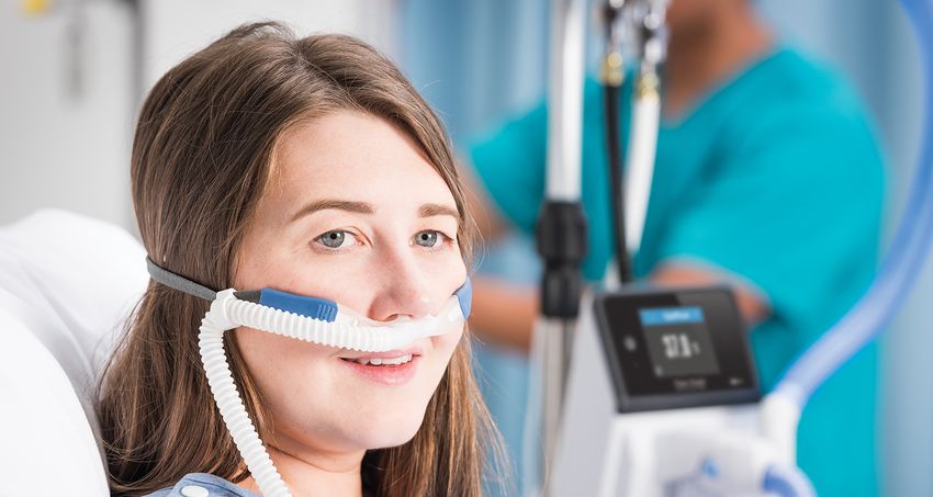
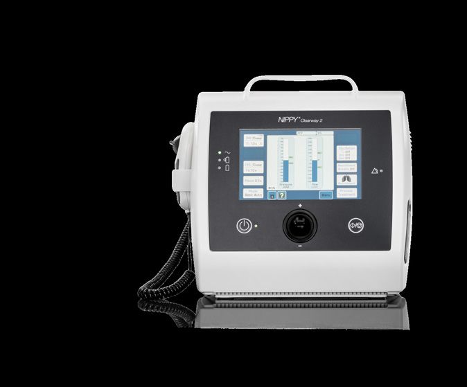
Versatile Airway Clearance
                                                                                                                 SpO2 Monitor

Introducing the new Clearway 2 with TreatRepeat®. The second-generation MI-E device by Breas incorporates
the latest technology to bring effective and accurate treatment for patients requiring airway clearance and
cough assistance. The TreatRepeat function allows clinicians to manually deliver different numbers of insuffla-
tions and exsufflations, and then save the treatment they have just delivered to a profile, to be repeated auto-
matically. This helps to improve accuracy and speed of titration and set-up.

 Feature                                                                                                                       Specification
 Modes:                                                                                                                        Manual, manual with TreatRepeat™, Basic auto, Prog. Timed,
                                                                                                                               Prog. Triggered, IPPB, NIV
 Pressure range:                                                                                                               + 70 / - 70 cmH2O
 Oscillations:                                                                                                                 4 – 20 Hz
 Advanced Synchronisation Technology:                                                                                          Triggered mode, SynchronyBeep™, TreatRepeat™
 Monitoring:                                                                                                                   Inspiratory volume (L), Cough Peak Flow (L/min), SpO2 (%)
 Therapy Delivery Method:                                                                                                      Manual Hand controls/Foot pedal or Automatic
 Prescription Set Up:                                                                                                          4 pre-set prescription options
 Battery:                                                                                                                      Internal (optional), external
 Data Download                                                                                                                 SD card download to PC Software

                          For tutorial videos and other educational material
                          on ventilation and airway clearance, please scan
                          the QR code to go to Education by Breas.

Nippy Clearway is a trademark of Breas Medical Ltd .© 2021 Breas Medical – All rights reserved. Breas Medical reserves the right to make changes in specifications and features shown herein, or discontinue the product described at any time without notice
or obligation. Contact your Breas representative for the most current information. Breas and the Breas logo are trademarks of Breas Medical AB. COR-MAR-MAR-007744-Rev0

                                                                               Unit A2,The Bridge Business Centre                                             For orders and general enquiries
                                                                                          Timothy´s Bridge Road                                                   orders@nippyventilator.com
BREAS.COM                                                        Stratford-Upon-Avon Warwickshire CV37 9HW. UK                                                       Tel: +44 (0)1789 293 460
Differences in maximal inspiratory pressure when using
a standard yoga mat versus a standard yoga block in
healthy individuals
Mair Davies1, Sue Annetts2
Abstract Authors
1Band 5
Background
Physiotherapist,
Yoga, which incorporates posture optimisation and
Cardiff and Vale
specific breathing techniques, is recognised for its
University Health
health benefits including musculoskeletal strength-
Board, Cardiff,
ening, stress relief and respiratory enhancement. It is
CF14 4XW, UK.
therefore becoming an increasingly utilised practice
2Senior Lecturer,
within the medical environment, specifically for those
School of Healthcare
with respiratory dysfunctions such as chronic ob-
Sciences, Cardiff
structive pulmonary disease (COPD). The relationship
University, Cardiff.
between posture and yoga and the potential impact
CF14 4XN, UK.
upon breathing is an important topic to study for its
potential health benefits. This study compares the Keywords
impact of two different yoga seating positions on res- Yoga, position,
piratory muscle strength. maximal inspiratory
pressure, healthy
Methodology
participants.
Eighteen students from Cardiff University between the
ages of 19–34 years were recruited via convenience Correspondence
sampling. Each was in good health, with body mass author
index (BMI) in the normal category and possessed Mair Davies. Email:
no respiratory infection or significant spinal deform- DaviesMB2@outlook.
ities. Participants were required to sit comfortably, com.
cross-legged for 2 minutes in each sitting position,
before three measurements of maximal inspiratory
pressure (MIP) were taken. Analysis was performed
on the highest result in each position using Statisti-
cal Package for the Social Sciences (SPSS) version
25. Comments on comfort levels were given sponta-
neously and, despite not being a primary aim of the
study, provided an interesting discussion point.
20 Journal of ACPRC • Volume 53 • Issue 1 • 2021 Go to contents pageResults and conclusions Sitting on a yoga mat produced higher mean MIP fig- ures (104cm H₂O) than the yoga block (100.39cm H₂O), p = 0.015. Despite statistical significance, the mean difference of 3.61cm H₂O is unlikely to have clinical significance as it is small compared to the observed MIP range (>90 cm H₂O). There does not appear to be a recognised minimum clinically important difference in the literature, making interpretation difficult. Introduction Poor posture has become increasingly prevalent in all demographics, particularly in the sedentary population (Owen 2012). Although extensive research recommends frequent physical activity, people with chronic obstructive pulmonary disease (COPD) remain one of the most inactive populations and spend a significant amount of their time sitting or lying down (Furlanetto et al. 2017). Physiological changes related to COPD, including hyperinfla- tion and thoracic kyphosis, compromise posture causing shoulder protraction, head pro- trusion and slouching (Goncalves et al. 2017). The slouching posture frequently observed in the COPD population compromises diaphragmatic function (Morrow et al. 2016; Albarrati et al. 2018). Habitual maintenance of this posture is also detrimental to spinal health (Castan- haro et al. 2014) and is heavily associated with poor outcomes (Furlanetto et al. 2017). It is therefore important that people with COPD optimise their posture as far as possible. Physiotherapy guidelines encourage the provision of patient education on appropriate postures as a self-management tool to combat the symptoms of dyspnoea and to reduce exertion (Bott et al. 2009). Certain postures can modify the ability of muscles to contract more forcibly in relation to the length tension theory (Kirsch & Stein 2000) thus improving their ability to utilise inspiratory muscles and inhale optimally. Good posture and special- ised breathing techniques present in yoga practice have been associated with improved respiratory function and health outcomes, especially amongst people with COPD (Done- sky-Cuenco et al. 2009; Fulambarker et al. 2012). Yoga can therefore be recommended as an adjunct to other therapies, particularly if the patient’s breathlessness interferes with moderate exercise. Seating aids such as yoga mats and blocks are commonly used to optimise posture and facilitate comfort during yoga practice. Significant changes to body alignment can be ob- served whilst using certain yoga apparatus (Sheeran et al. 2018). Even slight changes in pelvic tilt of only 10 degrees away from neutral impacts on respiratory function (Hwang & Kim 2018). It is therefore important to consider the impact of seating apparatus on body 21 Journal of ACPRC • Volume 53 • Issue 1 • 2021 Go to contents page
alignment when choosing the most beneficial seating position for people with COPD in
relation to pulmonary function during yoga practice. One way of measuring inspiratory
muscle strength and thus general pulmonary function is by calculating MIP.

This study aimed to explore whether there is a statistically significant difference (p
Figure 1a: Position for sitting Figure 1b: Position for sitting on a standard yoga block. on a standard yoga mat. The order of seating positions was randomised to eliminate any bias due to practice effect. This was attained through having the participants select one of the two seating positions from an opaque container. Participants sat for two minutes in each position to regulate their breathing before their MIP was measured using a Micro Respiratory Pressure Me- ter. Data were collected by a single examiner to avoid potential problems arising from inter-rater measurements. As MIP is an effort-dependent measurement the researcher provided standardised verbal encouragement using a specified script. Adhering to ATS and ERS (2002) standardised guidelines, 3 correctly performed manoeu- vres lasting for over 1.5 seconds were attained. A two-minute break in between each measurement was also given in order to regain participant baseline breathing pattern and reduce associated fatigue. Once 3 acceptable measurements in each sitting position, ex- hibiting less than 20% variance of each other, were collected (ATS & ERS 2002), the highest figure for each participant’s sitting position underwent further analysis using SPSS version 25. Following spontaneous feedback regarding perceived comfort level using the apparatus, comments were recorded thereafter to identify any relationships. However, as this was not a part of the original study design, the recording of comfort was limited to stating which apparatus was more comfortable. No attempt was made to attribute a value to the degree of comfort for each sitting position. 23 Journal of ACPRC • Volume 53 • Issue 1 • 2021 Go to contents page
Results
It is particularly important to consider the demographics as lung function can be influ-
enced by age, gender and participant size (Bellemare et al. 2003). Of the 18 participants
successfully recruited (Figure 2), 55% (n = 10) were female. Weight was normally distrib-
uted, and BMI fell within normal parameters (Table 1).
45 students showed
interest at lecture 23 did not respond to
Identification
theatre and supplied invitation email
contact details
3 failed to meet
22 replied to invitation inclusion criteria
Screening email and confirmed on the day:
attendance • 2 BMI >25.
• 1 upper respiratory
tract infection.
1 voluntarily withdrew
Eligibility 19 eligible participants due to personal
circumstances
18 participants took
Included
part in the study
Figure 2: Flow chart for participant selection.
Table 1: Mean, standard deviation (SD) and range of demographic data.
Mean SD Range (Max) (Min)
Age (years) 23.72 4.41 15 34 19
Height (cm) 167.83 10.09 35 188 153
Weight (kg) 64 8.63 32 80 48
BMI (kg/m²) 22.67 1.76 5.6 24.8 19.2
24 Journal of ACPRC • Volume 53 • Issue 1 • 2021 Go to contents pageNormal distribution was assessed and met parametric assumptions. A paired t test was
therefore appropriate and showed that MIP was statistically significantly higher on the
yoga mat compared to the yoga block with a mean difference of 3.61cm H₂O (t = 2.703,
p = 0.015) (Table 2). In addition, all participants reported that sitting on a yoga block com-
pared to a yoga mat was considerably more comfortable. However, this was not quantified.
Table 2: Mean, standard deviation (SD) and range of MIP values in sitting on yoga
mat and sitting on yoga block.
Mean SD Range Max Min
Sitting on standardised yoga mat (cm H₂0) 104.00 27.82 94 166 72
Sitting on standardised yoga block (cm H₂0) 100.39 28.58 96 157 61
Gender subsets largely also followed the same trend of mat MIP values being greater than
those produced on the block however, male values generally exceeded female values.
T tests confirmed that the order of seating positions did not affect results. This further
suggests data consistency overall and within subgroups. There were no significant corre-
lations between MIP and age, height, weight or BMI (suggesting that any differences were
coincidental).
Discussion
This study showed that MIP was significantly greater when measured during sitting on
a yoga mat, compared with a yoga block. Sheeran et al. (2018) reported that yoga block
seating aids induced significant changes to posture and reduced overall flexion in lumbar
spine and pelvic region. This encourages more upright sitting with a slight lumbar lordosis
and anterior pelvic tilt, which is considered the optimal seating position for respiratory
function (Castanharo et al. 2014; Sheeran et al. 2018). It is therefore surprising that MIP
was universally lower on the yoga block compared to the yoga mat which encourages a
posterior pelvic tilt. Additionally, a positive correlation between comfort and inspiratory
muscle strength has previously been identified (Naitoh et al. 2014) which also contradicts
the incidental results of this study where anecdotally, participants were more comfortable
on the yoga block.
Considering the above factors, it might be expected that the block would produce higher
MIP values than the mat. However, this study contradicted this expectation. There is no
obvious physiological explanation for the above findings, but this difference may be ex-
plained by factors which this study had not been designed to formally address. These may
include pelvic tilt, spinal angles, comfort or any other unknown influences. It is also im-
portant to consider whether the difference between positions is of any clinical importance.
The small difference in MIP (only 3.61cm H2O) makes it unlikely to have a significant clinical
impact on the participant.
25 Journal of ACPRC • Volume 53 • Issue 1 • 2021 Go to contents pageMaximal inspiratory pressure is one of the most commonly used measures for respiratory muscle strength and is recommended by ATS & ERS (2002) as a diagnostic tool. Yet, MIP de- vices are less commonly used in clinical practice and are not currently considered suitable as protocol tools in respiratory function testing (Culver et al. 2017). Although MIP is not as reliable as other, invasive, non-volitional methods, data collected in this study shows a stable variation across the range of MIP values. This suggests that the machine performed as expected and that the examiner technique was consistent thus supporting the reliability of the results. There is a wide range of normal MIP figures amongst the general population (Pessoa et al. 2014) and no distinct pattern has been identified by previous studies. However, no min- imum clinically important difference (MCID) has been reported in the literature and it is difficult to estimate whether differences are significant. This study revealed no evident correlations in relation to MIP and age, height, weight or BMI in the sample used. It could therefore be assumed that MIP is not dependant on these variables. Hence these results may be considered applicable to the young, healthy population. However, there may still be a limited applicability of these results to the unwell population such as those with res- piratory, musculoskeletal or neurological disorders for which this study was not intended. Conclusion This study revealed that sitting on a yoga mat yielded greater MIP values (104cm H₂O) than sitting on a standard 5cm yoga block (100.39cm H₂O). Due to the statistically significant difference (p = 0.015) the null hypothesis was rejected. However, as the range of normal MIP values in the studied population is so great (>90cm H₂O), without a reported MCID in existing literature, it is difficult to say whether the difference of 3.61cm H2O between both seating positions has any significance in clinical practice. Furthermore, it is currently un- known whether this information would be transferrable to those with long term respiratory conditions such as COPD where a 3.61cm H₂O difference would represent a greater propor- tional change compared to those with healthy respiratory systems and higher MIP values. The strengths of the study include having a single data collector to avoid inter-rater differ- ences. However, the small size of the study and convenience sampling method of young and healthy participants is not representative of the general population. Further research incorporating measurements of spinal angles, pelvic tilt and perceived comfort might contribute to a fuller understanding of these results. As the clinical applica- bility of this study is unknown, it would also be particularly helpful to repeat this study in people with COPD. Key points • Sitting position influences inspiratory muscle performance and therefore should be considered during yoga practice. 26 Journal of ACPRC • Volume 53 • Issue 1 • 2021 Go to contents page
• In healthy individuals, sitting on a yoga mat produced higher results compared to sit-
ting on a yoga block, despite being no apparent physiological explanation.
• The applicability of MIP measurements to the general population may be hindered by
the apparent lack of recognised MCID values.
Funding and acknowledgements
This research received no specific grant from any funding agency in the public, commercial
or not-for-profit sectors. I would like to acknowledge and thank the 18 participants who
volunteered for this study.
References
Albarrati, A., Zafar, H., Alghadir, A. H., & Anwer, S. (2018). Effect of upright and slouched sit-
ting postures on the respiratory muscle strength in healthy young males. BioMed Research
International, 2018, 3058970. https://doi.org/10.1155/2018/3058970.
American Thoracic Society/European Respiratory Society. (2002). ATS/ERS statement on
respiratory muscle testing. American Journal of Respiratory and Critical Care Medicine,
166(4), 518–624. https://doi.org/10.1164/rccm.166.4.518.
Banerjee, J., Roy, A., Singhamahapatra, A., Dey, P. K., Ghosal, A., & Das, A. (2014). Associa-
tion of Body Mass Index (BMI) with lung function parameters in non-asthmatics identified
by spirometric protocols. Journal of Clinical and Diagnostic Research: JCDR, 8(2), 12–14.
https://doi.org/10.7860/JCDR/2014/7306.3993.
Talaminos Barroso, A., Márquez Martín, E., Roa Romero, L. M., & Ortega Ruiz, F. (2018). Fac-
tors affecting lung function: A review of the literature. Factores que afectan a la función pul-
monar: una revisión bibliográfica. Archivos de Bronconeumologia, 54(6), 327–332. https://
doi.org/10.1016/j.arbres.2018.01.030.
Bellemare, F., Jeanneret, A., & Couture, J. (2003). Sex differences in thoracic dimensions
and configuration. American Journal of Respiratory and Critical Care Medicine, 168(3), 305–
312. https://doi.org/10.1164/rccm.200208-876OC.
Bott, J., Blumenthal, S., Buxton, M., Ellum, S., Falconer, C., Garrod, R., Harvey, A., Hughes,
T., Lincoln, M., Mikelsons, C., Potter, C., Pryor, J., Rimington, L., Sinfield, F., Thompson,
C., Vaughn, P., White, J., & British Thoracic Society Physiotherapy Guideline Develop-
ment Group (2009). Guidelines for the physiotherapy management of the adult, medical,
spontaneously breathing patient. Thorax, 64 Suppl 1, i1–i51. https://doi.org/10.1136/
thx.2008.110726.
Castanharo, R., Duarte, M., & McGill, S. (2014). Corrective sitting strategies: An examination
of muscle activity and spine loading. Journal of Electromyography and Kinesiology, 24(1),
114–119. https://doi.org/10.1016/j.jelekin.2013.11.001.
27 Journal of ACPRC • Volume 53 • Issue 1 • 2021 Go to contents pageCulver, B. H., Graham, B. L., Coates, A. L., Wanger, J., Berry, C. E., Clarke, P. K., Hallstrand, T. S., Hankinson, J. L., Kaminsky, D. A., MacIntyre, N. R., McCormack, M. C., Rosenfeld, M., Stanojevic, S., Weiner, D. J., & ATS Committee on Proficiency Standards for Pulmonary Function Laboratories. (2017). Recommendations for a standardised pulmonary func- tion report. An official American Thoracic Society technical statement. American Journal of Respiratory and Critical Care Medicine, 196(11), 1463–1472. https://doi.org/10.1164/ rccm.201710-1981ST. Donesky-Cuenco, D., Nguyen, H. Q., Paul, S., & Carrieri-Kohlman, V. (2009). Yoga therapy decreases dyspnea-related distress and improves functional performance in people with chronic obstructive pulmonary disease: A pilot study. Journal of Alternative and Comple- mentary Medicine (New York, N.Y.), 15(3), 225–234. https://doi.org/10.1089/acm.2008.0389. Fulambarker, A., Farooki, B., Kheir, F., Copur, A. S., Srinivasan, L., & Schultz, S. (2012). Effect of yoga in chronic obstructive pulmonary disease. American Journal of Therapeutics, 19(2), 96–100. https://doi.org/10.1097/MJT.0b013e3181f2ab86. Furlanetto, K. C., Donária, L., Schneider, L. P., Lopes, J. R., Ribeiro, M., Fernandes, K. B., Hernandes, N. A., & Pitta, F. (2017). Sedentary behavior is an independent predictor of mortality in subjects With COPD. Respiratory Care, 62(5), 579–587. https://doi.org/10.4187/ respcare.05306. Gonçalves, Márcia Aparecida, Francisco, Davi de Souza, Medeiros, Caroline Semprebom de, Brüggemann, Ana Karla Vieira, Mazo, Giovana Zarpellon, & Paulin, Elaine. (2017). Postural alignment of patients with chronic obstructive pulmonary disease. Fisioterapia em Movi- mento, 30(3), 549–558. https://doi.org/10.1590/1980-5918.030.003.ao13. Hwang, Y. I., & Kim, K. S. (2018). Effects of pelvic tilt angles and forced vital capacity in healthy individuals. Journal of Physical Therapy Science, 30(1), 82–85. https://doi. org/10.1589/jpts.30.82. Kirsch, R. &Stein, R. (2000). Neural and muscular properties: Current views and contro- versies. In Winters, J. & Crago, P. (Eds.), Biomechanics and neural control of posture and movement (pp. 39–55). Springer. Liu, Y., Pleasants, R. A., Croft, J. B., Lugogo, N., Ohar, J., Heidari, K., Strange, C., Wheaton, A. G., Mannino, D. M., & Kraft, M. (2015). Body mass index, respiratory conditions, asthma, and chronic obstructive pulmonary disease. Respiratory Medicine, 109(7), 851–859. https:// doi.org/10.1016/j.rmed.2015.05.006. Morrow, B., Brink, J., Grace, S., Pritchard, L., & Lupton-Smith, A. (2016). The effect of po- sitioning and diaphragmatic breathing exercises on respiratory muscle activity in people with chronic obstructive pulmonary disease. The South African Journal of Physiotherapy, 72(1), 315. https://doi.org/10.4102/sajp.v72i1.315. 28 Journal of ACPRC • Volume 53 • Issue 1 • 2021 Go to contents page
Naitoh, S., Tomita, K., Sakai, K., Yamasaki, A., Kawasaki, Y., & Shimizu, E. (2014). The ef- fect of body position on pulmonary function, chest wall motion, and discomfort in young healthy participants. Journal of Manipulative and Physiological Therapeutics, 37(9), 719– 725. https://doi.org/10.1016/j.jmpt.2014.10.005. Owen, N. (2012). Sedentary behavior: Understanding and influencing adults’ prolonged sit- ting time. Preventive Medicine, 55(6), 535–539. https://doi.org/10.1016/j.ypmed.2012.08.024. Pessoa, I. M., Houri Neto, M., Montemezzo, D., Silva, L. A., Andrade, A. D., & Parreira, V. F. (2014). Predictive equations for respiratory muscle strength according to international and Brazilian guidelines. Brazilian Journal of Physical Therapy, 18(5), 410–418. https://doi. org/10.1590/bjpt-rbf.2014.0044. Sheeran, L., Hemming, R., van Deursen, R., & Sparkes, V. (2018). Can different seating aids influence a sitting posture in healthy individuals and does gender matter? Cogent Engineer- ing, 5(1), 1442109. https://doi.org/10.1080/23311916.2018.1442109. 29 Journal of ACPRC • Volume 53 • Issue 1 • 2021 Go to contents page
For Non-Invasive Ventilation,
  think Dräger

        TO FIND OUT MORE VISIT WWW.DRAEGER.COM

        DraegerGlobal   Draeger   @DraegerNews   Draeger
A service evaluation on the use of digital chest
drains following thoracic surgery on postoperative
mobilisation and time on physiotherapy caseload
Chloe Tait1, Leanne McCarthy1, Simon Hayward1
Abstract Authors
1Physiotherapy
Introduction
Department, Blackpool
Chest drains are required following thoracic surgery
Teaching Hospitals
but their presence can cause people pain, limit post-
NHS Foundation Trust,
operative mobility and increase hospital length of stay
Blackpool, UK.
(LOS). The use of portable digital chest drains can pro-
mote early postoperative mobilisation, reduce drain Keywords
duration and hospital LOS. In our hospital, digital Digital chest drains,
chest drains were introduced in February 2017 for use physiotherapy,
with patients following thoracic surgery performed thoracic surgery.
by one thoracic surgeon. Other surgeons continued to
Correspondence
use under water seal (UWS) drains.
author
Aims Chloe Tait. Email:
To explore whether the use of digital chest drains al- chloe.tait1@nhs.net.
lowed earlier postoperative mobilisation, compared Telephone: 01253
with UWS drains. To explore whether the use of digital 953812.
chest drains reduced time on physiotherapy caseload,
chest drain duration and hospital LOS.
Method
A retrospective service evaluation was conducted in
a UK teaching hospital. Data were collected for a six
month period for all patients following thoracic sur-
gery referred to physiotherapy. Data were analysed
using descriptive statistics and statistical tests.
Results
Median day first mobilised postoperatively was statis-
tically significantly shorter for the digital drain group
(day 1) compared to the UWS drain group (day 3) (ob-
served median difference 1, 95% CI 1 to 2 p = 0.0001).
31 Journal of ACPRC • Volume 53 • Issue 1 • 2021 Go to contents pageTime on physiotherapy caseload was statistically sig- nificantly shorter for the digital drain group (4 days) compared to the UWS drain group (5 days) (Observed median difference 1, 95% CI 1 to 2 p = 0.02). There was no statistically significant difference in median chest drain duration between the digital drain group (2 days) and the UWS group (3 days) (observed median difference 1, 95% CI 0 to 0 p = 0.91). Median hospital LOS was shorter for the digital drain group (5 days) compared to the UWS group (6 days) however this dif- ference did not reach statistical significance (observed median difference 1, 95% CI 0 to 1 p = 0.06). Conclusion The use of digital chest drains with inbuilt suction en- abled individuals to mobilise on the first day following thoracic surgery, thereby facilitating earlier liberation from the bed space and reducing the potential for known effects of immobilisation. The use of digital chest drains also facilitated earlier discharge from physiotherapy. In this service evaluation there was no significant difference in chest drain duration or hos- pital LOS between individuals with digital drains and those with UWS drains. Introduction Patients require chest drains following thoracic surgery to remove air and fluid that col- lects in the chest (Lijkendijk et al. 2015; Shoji et al. 2016). Chest drains limit the patient’s ability to move away from their bed space, cause patient’s pain, reduce their independence and pose a potential infection risk (Bertholet et al. 2011). Chest drains are traditionally non-digital and require attachment to wall suction. This causes subjective measurement of air and fluid drainage (Lijkendijk et al. 2015) and often delays postoperative mobilisa- tion (Rathinam et al. 2011; Lijkendijk et al. 2015), lengthening drain duration and hospital length of stay (LOS) (Agostini et al. 2014; Lijkendijk et al. 2015). The use of digital drains allows more accurate measurement of air leak/fluid drainage allowing drains to be re- moved when they meet protocol or when no air leak was reported shortening drain dura- tion (George & Papagiannopoulos 2015; Lijkendijk et al. 2015; NICE 2018). Portable digital drains also enable patients to mobilise earlier postoperatively as they do not need to wait to be detached from wall suction (Rathinam et al. 2011; Lijkendijk et al. 2015; NICE 2018). This facilitates earlier re-expansion of the operated lung reducing the risk of postoperative 32 Journal of ACPRC • Volume 53 • Issue 1 • 2021 Go to contents page
pulmonary complications (PPCs) and aiding earlier drain removal (NICE 2018). Early chest
drain removal helps reduce patients’ pain levels and reduces infection risk (Bertholet et al.
2011). It also allows patients earlier liberation from the bed space and encourages early
patient independence (Bertholet et al. 2011; Agostini et al. 2014; Lijkendijk et al. 2015;
Yeung 2016; NICE 2018). Better lung expansion, reduced risk of PPCs and early postopera-
tive mobilisation can reduce time spend on the physiotherapy caseload. Earlier discharge
from physiotherapy can also encourage earlier independence. Early chest drain removal,
postoperative mobilisation, discharge from physiotherapy and patient independence can
all contribute to a reduction in hospital LOS (Bertholet et al. 2011; Agostini et al. 2014;
Lijkendijk et al. 2015; NICE 2018). Whether the use of digital chest drains reduces the time
taken to mobilise postoperatively, and the time spent on physiotherapy caseload following
thoracic surgery, has not been explored or measured within existing research.
The primary aims were to explore whether the use of digital drains allowed earlier post-
operative mobilisation and reduced time on physiotherapy caseload compared to under
water seal (UWS) drains. Secondary aims were to explore whether the use of digital drains
reduced chest drain duration and hospital LOS compared to UWS drains and reduced hos-
pital LOS.
Method
A service evaluation was conducted exploring retrospective data collected from the 1st Feb-
ruary 2017 to 31st July 2017. This service evaluation was registered with, and approved
by, the Blackpool Teaching Hospitals NHS Foundation Trust’s Research and Development
team. Ethical approval was not required in-line with the trust policy on undertaking service
evaluations.
The use of digital drains commenced in February 2017 at the Lancashire Cardiac Centre
on the cardiac intensive care unit and the cardiothoracic surgical wards. Prior to February
2017 only UWS drains were used following thoracic surgery. From February 2017 all pa-
tients undergoing thoracic surgery performed by one of the thoracic surgeons had a digital
drain and patients undergoing thoracic surgery performed by the other surgeons continued
with UWS drains. The protocol for UWS drains removal was:
• Lobectomy/pneumonectomy: drain to be removed following at least 48 hours with no
air leak for 24 hours.
• Wedge resection: removal after 24 hours if there is no air leak for 12 hours.
• Bullectomy/Pleurodesis: removal after 72 hours if no air leak for 24 hours.
• Biopsy: removal day 1 if no air leak for 12 hours.
• For all other surgeries drains were removed following surgeon approval.
The protocol for digital drain removal was less than 30ml air leak within 12 hours with no
pneumothorax/air airspace and no surgical emphysema on chest radiograph (CXR) and less
than 200mls of fluid drainage within 12 hours. Drain removal was subject to surgeon approval.
33 Journal of ACPRC • Volume 53 • Issue 1 • 2021 Go to contents pageAll patients who received inpatient physiotherapy following thoracic surgery with a chest
drain in situ postoperatively on cardiac intensive care or one of the cardiothoracic wards at
the Lancashire cardiac centre were included in this service evaluation. Patients were iden-
tified from the electronic patient referral system and physiotherapy patient contact sheets.
The following data were retrieved:
• Date of surgery.
• Gender of patient.
• Age of patient.
• Type of surgical incision and surgical procedure.
• Type of chest drain.
• Day first mobilised postoperatively.
• Time spent on physiotherapy caseload (measured in days from day 1 postoperatively
until day discharged from physiotherapy).
• Chest drain duration (number of days chest drain(s) remained in situ postoperatively
measured from day 1 postoperatively until day chest drain removed.
• Hospital LOS (measured in days from day 1 postoperatively until day discharged from
hospital).
Date of surgery, gender and age of the patient, type of surgical incision and surgical pro-
cedure, type of chest drain, day first mobilised and time spent on physiotherapy caseload
were collected from physiotherapy patient contact sheets and the hospital electronic pa-
tient database. Date of chest drain removal was collected from the patient’s routine post-
drain removal CXRs. Hospital discharge date was obtained from the hospital’s electronic
patient database. The time spent on physiotherapists’ caseload was defined as the number
of days patients received treatment from the cardiothoracic physiotherapy team from the
day first assessed by the physiotherapist postoperatively (usually day 1 postoperatively)
until the day the patient was discharged from physiotherapy.
Data were analysed using descriptive statistics and statistical tests. Data were plotted
using histograms to check the normality of the data. Median values were used for all out-
comes due to their skewed distribution. Upper and lower quartiles were used to explore
the spread of the data. The non-parametric Mann Whitney U Test was used to test for sta-
tistically significant differences between data from the digital drain group and the UWS
drain group.
Results
Over the six-month period, from 1st February 2017 to 31st July 2017, 195 patients received
in-patient physiotherapy following thoracic surgery. Two patients died and were excluded
due to incomplete data. A third patient was excluded as they did not have a chest drain
inserted. Of the remaining 192 patients, 110 had a digital drain and 82 had an UWS drain(s)
postoperatively. Table 1 shows the characteristics of patients in the two different drain
34 Journal of ACPRC • Volume 53 • Issue 1 • 2021 Go to contents pageYou can also read