Left Ventricular Global Longitudinal Strain Is Associated With Cardiovascular Outcomes in Patients Who Underwent Permanent Pacemaker Implantation ...
←
→
Page content transcription
If your browser does not render page correctly, please read the page content below
ORIGINAL RESEARCH
                                                                                                                                                published: 30 July 2021
                                                                                                                                       doi: 10.3389/fcvm.2021.705778

                                            Left Ventricular Global Longitudinal
                                            Strain Is Associated With
                                            Cardiovascular Outcomes in Patients
                                            Who Underwent Permanent
                                            Pacemaker Implantation
                                            Dae-Young Kim, Purevjargal Lkhagvasuren, Jiwon Seo, Iksung Cho, Geu-Ru Hong,
                                            Jong-Won Ha and Chi Young Shim*

                                            Division of Cardiology, Severance Cardiovascular Hospital, Yonsei University College of Medicine, Seoul, South Korea

                                            Background: Patients who underwent permanent pacemaker (PM) implantation have
                             Edited by:     a potential risk of left ventricular (LV) systolic dysfunction. However, assessment of
                      Juan R. Gimeno,       LV ejection fraction (LVEF) shows a limited role in identifying subclinical LV systolic
     Hospital Universitario Virgen de la
                                            dysfunction and predicting cardiovascular (CV) outcomes.
                        Arrixaca, Spain

                        Reviewed by:        Methods: We reviewed 1,103 patients who underwent permanent PM implantation
                  Piyush M. Srivastava,     between January 2007 and December 2017. After excluding patients who did not
     University of Melbourne, Australia
              Carmen Muñoz-Esparza,
                                            undergo echocardiograms before or after PM implantation and those with LV ejection
         Ciudad Sanitaria Virgen de la      fraction (LVEF)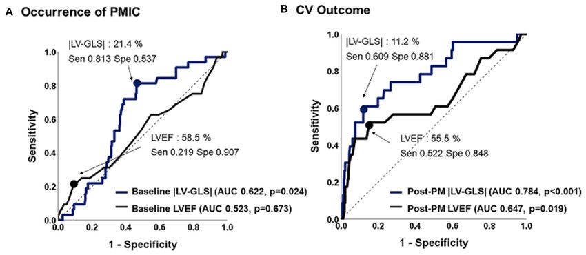
Kim et al.                                                                                               Pacemaker and Global Longitudinal Strain

INTRODUCTION                                                           Follow-Up and Outcomes
                                                                       Patients were scheduled to visit the PM clinic every 6
Patients who have undergone permanent pacemaker (PM)                   months after PM implantation. Follow-up data, including pacing
implantation have a potential risk of left ventricular (LV)            percentage and clinical events, were obtained by reviewing
systolic dysfunction (1–3). PM-induced cardiomyopathy                  medical records. Data of pacing percentage was gathered at the
(PMIC) is generally defined as a decrease in LV systolic               time of the first interrogation after 2 months of PM implantation.
function after right ventricular (RV) pacing with no other             Based on the echocardiographic data between 6 months and
independent triggering factors (4) and is associated with worse        5 years after PM implantation, PMIC was defined as a ≥10%
cardiovascular (CV) outcomes (5). Theoretically, increasing RV         decrease in LVEF compared with baseline echocardiography with
pacing causes LV mechanical dyssynchrony and consequently              resultant LVEF
Kim et al.                                                                                                               Pacemaker and Global Longitudinal Strain

 FIGURE 1 | Representative LV-GLS measurements. (A) LV-GLS was calculated from three standard apical views at baseline echocardiogram (left). The absolute value
 of LV-GLS was 23.0% (right). (B) LV-GLS was assessed after PM implantation. |LV-GLS| was 14.8% (right). LV-GLS, left ventricular global longitudinal strain;
 PM, pacemaker.

borders during the whole cardiac cycle. For analysis of segmental                  regression analysis for adjusting other confounding factors.
strain, we used the 16-segment model that divided the base and                     Predictive values of LV-GLS for CV outcomes were calculated
mid area into six segments (antero-septum, anterior, antero-                       using receiver operating characteristic (ROC) analysis. Clinical
lateral, infero-lateral, inferior, and infero-septal) and the apex                 outcomes were constructed using Kaplan–Meier methods, and
into four segments (septal, inferior, lateral, and anterior), and LV-              comparisons among groups were performed using a log-
GLS was calculated by averaging the values at each segmental                       rank test. The predictors of CV outcomes were evaluated
level as mentioned above. |LV-GLS| was defined as the absolute                     using multivariate nested Cox proportional hazard regression
value of LV-GLS (removing the conventional negative value of                       models. The independence of |LV-GLS| was examined using
LV-GLS data) (Figure 1). The LV-global circumferential strain                      three models. Initially, three subgroups divided by |LV-GLS|
(GCS) was calculated by averaging the values of segmental                          tertile were included in the Cox model as a covariate, with
circumferential strains from the parasternal short-axis view in                    adjustment for age and sex. Then, chronic kidney disease and
the basal-, mid-, and apical-LV levels. |LV-GLS| was defined                       coronary artery disease were included in the second model.
as the absolute value of LV-GCS. We randomly selected 20                           Finally, LVEF and left atrial volume index were included in
patients from the study population and analyzed the intra-                         the last model. Significant differences were considered at P <
and inter-observer reproducibility of LV-GLS measurement by                        0.05. All statistical analyses were performed using SPSS 25.0
Bland–Altman analysis. The intra-class correlation coefficients                    software (IBM Corp., Armonk, NY), and Medcalc statistical
for |LV-GLS| were 0.987 and 0.963 for intra- and inter-observer                    package (Medcalc software, Mariakerke, Belgium) was used for
variation, respectively. The Bland–Altman analysis showed the                      comparison of ROC curves.
limits of agreement (LOA) across a broad range of LV-GLS
values; the bias for intra- and inter-observer measurements of LV-
GLS was 0.47% (range: −0.91 to −0.04%, 95% LOA) and 0.37%                          RESULTS
(range: −0.71 to 0.78%), respectively.                                             Baseline Characteristics
                                                                                   During a mean 44 ± 28 months of follow-up after post-PM
Statistical Analysis                                                               echocardiography, 23 of 300 patients (7.7%) experienced
Continuous variables are presented as mean ± standard                              CV events. Clinical characteristics, medications, and data
deviation (SD), and categorical variables are presented as                         related to PM in patients with or without CV outcomes
frequency and percentage. Comparisons of baseline clinical                         are presented in Table 1. Patients with CV outcomes were
and echocardiographic parameters between the two groups                            older and had a higher prevalence of chronic kidney
were analyzed using Student’s t-test for continuous data and                       disease, coronary artery disease than did those without CV
chi-square (χ2) and Fisher’s exact test for categorical data.                      outcomes. More heart failure medications including renin-
Correlations between pacing percentage and echocardiographic                       angiotensin-aldosterone system blockade and diuretics
variables including the strain of each segment were obtained                       were used by patients with CV outcomes compared
using simple linear regression analysis and multiple linear                        to those without CV outcomes. The ventricular lead

Frontiers in Cardiovascular Medicine | www.frontiersin.org                     3                                             July 2021 | Volume 8 | Article 705778
Kim et al.                                                                                                                                 Pacemaker and Global Longitudinal Strain

TABLE 1 | Baseline characteristics.

                                         Total (n = 300)                  Without CV outcomes (n =277)                          With CV outcomes (n = 23)                       P-value

Age, years                                 67.1 ± 13.2                                66.2 ± 13.8                                         74.5 ± 9.9                              0.005
Male sex, n (%)                             119 (39.7)                                 107 (38.6)                                          12 (52.2)                              0.202
BMI, kg/m2                                  24.3 ± 3.6                                 24.3 ± 3.6                                         25.0 ± 3.6                              0.357
Hypertension, n (%)                         171 (57.0)                                 155 (56.0)                                          16 (69.6)                              0.205
Diabetes mellitus, n (%)                     61 (20.3)                                  54 (19.5)                                           7 (30.4)                              0.210
CKD, n (%)                                    22 (7.3)                                   16 (5.8)                                           6 (26.1)
Kim et al. Pacemaker and Global Longitudinal Strain
TABLE 2 | Echocardiographic characteristics.
Total (n = 300) Without CV outcomes (n = 277) With CV outcomes (n = 23) P-value
Baseline echocardiogram
LVEDD, mm 50.3 ± 4.7 50.2 ± 4.6 51.4 ± 5.5 0.250
LVESD, mm 32.3 ± 4.3 32.2 ± 4.2 33.7 ± 5.4 0.120
LVEF. % 67.8 ± 6.8 67.9 ± 6.7 66.0 ± 7.5 0.191
LV mass index, g/m2 104.4 ± 22.5 103.8 ± 22.3 112.1 ± 25.0 0.130
LA volume index, ml/m2 35.8 ± 12.6 35.4 ± 12.5 40.4 ± 14.0 0.068
e’ velocity, cm/s 6.1 ± 2.4 6.2 ± 2.5 4.7 ± 1.5 0.012
S’ velocity, cm/s 6.7 ± 1.6 6.8 ± 1.6 5.8 ± 1.4 0.007
E/e’ 13.7 ± 6.3 13.3 ± 5.9 19.4 ± 8.7 0.007
|LV-GLS|, % 21.0 ± 5.3 21.2 ± 5.3 18.5 ± 4.7 0.016
|LV-GCS|, % 29.1 ± 7.0 29.2 ± 7.0 27.6 ± 6.8 0.292
Post-PM echocardiogram
Time after PM implantation, years 2.2 ± 1.2 2.2 ± 1.2 2.4 ± 1.4 0.730
LVEDD, mm 49.6 ± 5.1 49.4 ± 5.0 51.7 ± 6.2 0.035
LVESD, mm 33.4 ± 5.7 33.1 ± 5.5 36.4 ± 7.5 0.047
LVEF. % 62.2 ± 10.3 62.7 ± 9.7 55.1 ± 14.1 0.018
PMIC, n (%) 32 (10.7) 22 (7.9) 10 (43.5)Kim et al. Pacemaker and Global Longitudinal Strain
TABLE 3 | Simple correlations between pacing percentage and
echocardiographic variables after PM implantation.
Correlation P-value
coefficient
LVEDD, mm 0.162 0.005
LVESD, mm 0.189 0.001
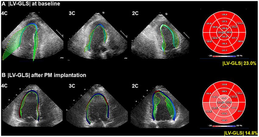
LVEF. % −0.210Kim et al. Pacemaker and Global Longitudinal Strain FIGURE 3 | Predictive values of |LV-GLS| and LVEF for occurrence of PMIC and CV outcome. (A) The |LV-GLS| on baseline echocardiogram revealed significant predictive value for occurrence of PMIC. (B) The |LV-GLS| on post-PM echocardiogram showed a better predictive value for CV outcomes than did LVEF. |LV-GLS|, absolute value of left ventricular global longitudinal strain; LVEF, left ventricular ejection fraction; PMIC, pacemaker-induced cardiomyopathy; PM, pacemaker; CV, cardiovascular. FIGURE 4 | Kaplan–Meier curve according to |LV-GLS| on post-PM echocardiogram. (A) The group of |LV-GLS| ≤ 11.2% revealed a significantly worse CV outcome than the other group (log-rank p < 0.001). (B) The lowest |LV-GLS| group revealed a significantly worse CV outcome than the others (log-rank p < 0.001). |LV-GLS|, absolute value of left ventricular global longitudinal strain; PM, pacemaker; CV, cardiovascular. as RV pacing percentage increased, the global LV mechanical dysfunction by pacing through segmental strain analysis. As function evaluated by |LV-GLS| decreased, and it was more pacing percentage increased, the absolute value of longitudinal obvious when the analysis was performed divided by RV strain in some specific segments decreased. The specific segments pacing site. Previous studies also tried to show the relationship that were highly affected by pacing were those in the entire between pacing percentage and LV-GLS; however, mainly due LV apex. The correlation weakened toward the mid-LV and to the small number of study subjects, they failed to show base, but the absolute value of segmental strain in the inferior a linear correlation (22, 25). Another strength of the present segment and infero-septal segment significantly decreased in study is that it comprehensively shows LV regional mechanical proportion to pacing percentage. These results can be interpreted Frontiers in Cardiovascular Medicine | www.frontiersin.org 7 July 2021 | Volume 8 | Article 705778
Kim et al.                                                                                                                                  Pacemaker and Global Longitudinal Strain

TABLE 4 | Cox regression analysis for CV outcomes.

                                                      Model 1                                              Model 2                                            Model 3

                                     Hazard ratio [95% CI]              P-value           Hazard ratio [95% CI]             P-value           Hazard ratio [95% CI]              P-value

|LV-GLS| > 19.1%                            Reference                                            Reference                                            Reference
14.4 < |LV-GLS| ≤ 19.1%                 5.80 [0.68∼49.84]                0.109              4.06 [0.46∼35.66]                 0.207              4.59 [0.52∼40.13]                 0.169
|LV-GLS| ≤ 14.4%                       14.8 [1.93∼112.70]                0.009             15.18 [1.96∼117.61]                0.009             13.97 [1.72∼113.39]                0.014

Model 1: Adjusted by age and sex.
Model 2: Adjusted by age, sex, CKD, and CAD.
Model 3: Adjusted by age, sex, CKD, CAD, LVEF, and LA volume index.
CI, confidence interval; CKD, chronic kidney disease; CAD, coronary artery disease; LVEF, left ventricular ejection fraction; LA, left atrium; |LV-GLS|, absolute value of left ventricular
global circumferential strain.

based on the pathophysiology of PMIC. During RV pacing,                                         Also, there is a possibility that |LV-GLS| was high because of a
conduction of an electrical wave passes through the myocardium,                                 compensatory increase in stroke volume due to bradycardia in
which is adjacent to the PM lead (6, 26). Therefore, there                                      some patients. As a result, we suggest that, if |LV-GLS| is lower
were significant decreases in strain in the regions close to the                                than the normal reference value before PM implantation, close
pacing area (27). This phenomenon results in dyssynchronous                                     clinical and echocardiographic follow-up should be performed
motion of LV. In patients with dyssynchronous motion due                                        after PM implantation considering the risk of PMIC.
to higher RV pacing burden and subsequent lower LVEF, an
upgrade from PM to cardiac resynchronization therapy might                                      CONCLUSION
be considered to improve LV mechanical dysfunction and to
treat heart failure (5). Considering the cut-off value of |LV-                                  After PM implantation, there were significant regional and
GLS| of post-PM implantation as 11.2%, patients with |LV-                                       global changes in LV mechanical function. On post-PM
GLS| under 11.2% on post-PM implantation echocardiography                                       echocardiogram, reduced |LV-GLS| rather than LVEF is
are recommended to adjust the PM pacing parameters to                                           associated with poor CV outcome. Assessments of LV-GLS
reduce the pacing percentage or consider to converse to                                         by speckle-tracking echocardiography before and after PM
cardiac resynchronization therapy, with aggressive heart failure                                implantation are beneficial for early detection of LV mechanical
medication. In patients with |LV-GLS| in the gray zone between                                  dysfunction and prediction of CV outcomes.
11.2 to 20%, regular echocardiography follow-up to identify
whether |LV-GLS| decreases under 11.2% and manage several risk                                  DATA AVAILABILITY STATEMENT
factors regarding heart failure are required.
                                                                                                The original contributions presented in the study are included
Limitations                                                                                     in the article/Supplementary Material, further inquiries can be
This study has several limitations. First, this study was                                       directed to the corresponding author.
retrospectively designed and comprised patients who were
followed up by regular visits. The interval of follow-up
echocardiogram after PM implantation was not concordant in                                      ETHICS STATEMENT
all patients. To minimize this limitation, the timing of follow-up
echocardiogram was limited to between 6 months and 5 years                                      The studies involving human participants were reviewed and
after PM implantation to evaluate the predictability of future                                  approved by Yonsei University Health System. Written informed
CV events of LV-GLS. Nevertheless, the population might be                                      consent for participation was not required for this study in
biased, and it is possible that the occurrence of clinical events                               accordance with the national legislation and the institutional
was underestimated. Second, echocardiographic examinations                                      requirements. Written informed consent was not obtained from
of patients were not performed using the same equipment,                                        the individual(s) for the publication of any potentially identifiable
which could have produced inconsistency of echocardiographic                                    images or data included in this article.
parameters, especially LV-GLS. However, we used vendor-
independent software and tried to minimize the error of                                         AUTHOR CONTRIBUTIONS
measurement by expert operators. Third, the |LV-GLS| value for
predicting PMIC on the baseline echocardiogram before PM                                        D-YK and CS contributed to the concept and design of this
implantation was 21.4%, which was within the normal range.                                      study. D-YK, PL, JS, IC, G-RH, J-WH, and CS contributed to
According to previous results of a head-to-head comparison of                                   acquisition, analysis, and interpretation of the data. D-YK and CS
LV-GLS among vendors reported by the European Association of                                    contributed to drafting of the manuscript and statistical analysis.
Cardiovascular Imaging/American Society of Echocardiography,                                    G-RH, J-WH, and CS contributed to revision and finalize the
the average |LV-GLS| values measured by TomTec software                                         manuscript. All authors contributed to the article and approved
was 21.5%, trending higher than other software measures. (28).                                  the submitted version.

Frontiers in Cardiovascular Medicine | www.frontiersin.org                                  8                                                    July 2021 | Volume 8 | Article 705778
Kim et al. Pacemaker and Global Longitudinal Strain
SUPPLEMENTARY MATERIAL Supplementary Figure 1 | Kaplan-Meier curve on 2 subgroups according to the
RV pacing percentage divided by 50%. The subgroup with RV pacing under 50%
The Supplementary Material for this article can be found had better event-free survival than those who were not (log rank p = 0.047). RV;
online at: https://www.frontiersin.org/articles/10.3389/fcvm. right ventricle.
2021.705778/full#supplementary-material Supplementary Figure 2 | Kaplan-Meier curve on 2 subgroups according to the
Supplementary Table 1 | Multiple linear regression analysis for post-PM |LV-GLS| degree of |LV-GLS| change between baseline and post-PM echocardiogram. The
and |Apical septal strain|. |LV-GLS|, absolute value of left ventricular global subgroup with the |LV-GLS| reduction under 10% had better event-free survival
longitudinal strain; |Apical septal strain|, absolute value of left ventricular apical than those who were not (log rank p = 0.035). |LV-GLS|; absolute value of left
septal strain; RV, right ventricle; HTN, hypertension; DM, diabetes mellitus. ventricular global longitudinal strain.
REFERENCES pacing-induced cardiomyopathy. Hear Rhythm. (2014) 11:1619–
25. doi: 10.1016/j.hrthm.2014.05.040
1. Thackray SDR, Witte KKA, Nikitin NP, Clark AL, Kaye GC, Cleland JGF. 15. Lang RM, Badano LP, Mor-Avi V, Afilalo J, Armstrong A, Ernande L, et al.
The prevalence of heart failure and asymptomatic left ventricular systolic Recommendations for cardiac chamber quantification by echocardiography
dysfunction in a typical regional pacemaker population. Eur Heart J. (2003) in adults: An update from the American society of echocardiography and
24:1143–52. doi: 10.1016/S0195-668X(03)00199-4 the European association of cardiovascular imaging. Eur Heart J Cardiovasc
2. Yu C-M, Chan JY-S, Zhang Q, Omar R, Yip GW-K, Hussin A, et al. Imaging. (2015) 16:233–71. doi: 10.1093/ehjci/jev014
Biventricular pacing in patients with bradycardia and normal ejection 16. Baumgartner H, Falk V, Bax JJ, De Bonis M, Hamm C, Holm PJ,
fraction. N Engl J Med. (2009) 361:2123–34. doi: 10.1056/NEJMoa0907555 et al. 2017 ESC/EACTS Guidelines for the management of valvular
3. Mumin G, Celiker C. Right ventricular apical and septal pacing: long term heart disease. Eur Heart J. (2017) 38:2739–86. doi: 10.1016/j.rec.201
impacts on ventricular function. Eur Hear J - Cardiovasc Imaging. (2021) 7.12.013
22:432. doi: 10.1093/ehjci/jeaa356.367 17. Risum N, Ali S, Olsen NT, Jons C, Khouri MG, Lauridsen TK, et al. Variability
4. Kiehl EL, Makki T, Kumar R, Gumber D, Kwon DH, Rickard JW, of global left ventricular deformation analysis using vendor dependent and
et al. Incidence and predictors of right ventricular pacing-induced independent two-dimensional speckle-tracking software in adults. J Am Soc
cardiomyopathy in patients with complete atrioventricular block and Echocardiogr. (2012) 25:1195–203. doi: 10.1016/j.echo.2012.08.007
preserved left ventricular systolic function. Hear Rhythm. (2016) 13:2272– 18. Voigt JU, Pedrizzetti G, Lysyansky P, Marwick TH, Houle H, Baumann
8. doi: 10.1016/j.hrthm.2016.09.027 R, et al. Definitions for a common standard for 2D speckle tracking
5. Cho SW, Gwag H. Bin, Hwang JK, Chun KJ, Park KM, On YK, Kim JS, Park echocardiography: consensus document of the EACVI/ASE/Industry Task
SJ. Clinical features, predictors, and long-term prognosis of pacing-induced Force to standardize deformation imaging. Eur Heart J Cardiovasc Imaging.
cardiomyopathy. Eur J Heart Fail. (2019) 21:643–51. doi: 10.1002/ejhf.1427 (2015) 16:1–11. doi: 10.1093/ehjci/jeu184
6. Shim CY. Pacing-induced alterations in left ventricular mechanical 19. Negishi K, Negishi T, Kurosawa K, Hristova K, Popescu BA,
properties: effect of pacing sites. J Cardiovasc Ultrasound. (2011) Vinereanu D, et al. Practical guidance in echocardiographic assessment
19:13. doi: 10.4250/jcu.2011.19.1.13 of global longitudinal strain. JACC Cardiovasc Imaging. (2015)
7. Tantengco MVT, Thomas RL, Karpawich PP. Left ventricular dysfunction 8:489–92. doi: 10.1016/j.jcmg.2014.06.013
after long-term right ventricular apical pacing in the young. J Am Coll Cardiol. 20. Uslan DZ, Tleyjeh IM, Baddour LM, Friedman PA, Jenkins SM, St Sauver JL,
(2001) 37:2093–100. doi: 10.1016/S0735-1097(01)01302-X et al. Temporal trends in permanent pacemaker implantation: A population-
8. Ahmed M, Gorcsan J, Marek J, Ryo K, Haugaa K, Ludwig DR, et al. Right based study. Am Heart J. (2008) 155:896–903. doi: 10.1016/j.ahj.200
ventricular apical pacing-induced left ventricular dyssynchrony is associated 7.12.022
with a subsequent decline in ejection fraction. Hear Rhythm. (2014) 11:602– 21. Seo J, Kim DY, Cho I, Hong GR, Ha JW, Shim CY. Prevalence, predictors,
8. doi: 10.1016/j.hrthm.2013.12.020 and prognosis of tricuspid regurgitation following permanent pacemaker
9. Özdemir K, Altunkaser BB, Daniş G, Özdemir A, Uluca Y, Tokaç M, implantation. PLoS ONE. (2020) 15:1–10. doi: 10.1371/journal.pone.02
et al. Effect of the isolated left bundle branch block on systolic and 35230
diastolic functions of left ventricle. J Am Soc Echocardiogr. (2001) 14:1075– 22. Ahmed FZ, Motwani M, Cunnington C, Kwok CS, Fullwood C, Oceandy
9. doi: 10.1067/mje.2001.115655 D, et al. One-month global longitudinal strain identifies patients who
10. Saito M, Negishi K, Eskandari M, Huynh Q, Hawson J, Moore A, et al. will develop pacing-induced left ventricular dysfunction over time: The
Association of left ventricular strain with 30-day mortality and readmission pacing and ventricular dysfunction (PAVD) Study. PLoS ONE. (2017) 12:1–
in patients with heart failure. J Am Soc Echocardiogr. (2015) 28:652– 14. doi: 10.1371/journal.pone.0162072
66. doi: 10.1016/j.echo.2015.02.007 23. Babu NMS, Srinath SC, Lahiri A, Chase D, John B, Roshan J. Three-
11. Kalam K, Otahal P, Marwick TH. Prognostic implications of dimensional echocardiography with left ventricular strain analyses helps
global LV dysfunction: A systematic review and meta-analysis earlier prediction of right ventricular pacing-induced cardiomyopathy. J Saudi
of global longitudinal strain and ejection fraction. Heart. (2014) Hear Assoc. (2018) 30:102–7. doi: 10.1016/j.jsha.2017.06.001
100:1673–80. doi: 10.1136/heartjnl-2014-305538 24. Sharma AD, Rizo-Patron C, Hallstrom AP, O’Neill GP, Rothbart S, Martins JB,
12. Kim D, Shim CY, Cho YJ, Park S, Lee CJ, Park JH, et al. Continuous positive et al. Percent right ventricular pacing predicts outcomes in the DAVID trial.
airway pressure therapy restores cardiac mechanical function in patients with Hear Rhythm. (2005) 2:830–4. doi: 10.1016/j.hrthm.2005.05.015
severe obstructive sleep apnea: a randomized, sham-controlled study. J Am 25. Tanaka H, Matsumoto K, Hiraishi M, Miyoshi T, Kaneko A, Tsuji T,
Soc Echocardiogr. (2019) 32:826–35. doi: 10.1016/j.echo.2019.03.020 et al. Multidirectional left ventricular performance detected with three-
13. Hwang IC, Cho GY, Yoon YE, Park JJ. Association between global longitudinal dimensional speckle-tracking strain in patients with chronic right ventricular
strain and cardiovascular events in patients with left bundle branch block pacing and preserved ejection fraction. Eur Heart J Cardiovasc Imaging. (2012)
assessed using two-dimensional speckle-tracking echocardiography. J Am Soc 13:849–56. doi: 10.1093/ehjci/jes056
Echocardiogr. (2018) 31:52–63. doi: 10.1016/j.echo.2017.08.016 26. Tops LF, Schalij MJ, Bax JJ. The effects of right ventricular apical pacing on
14. Khurshid S, Epstein AE, Verdino RJ, Lin D, Goldberg LR, ventricular function and dyssynchrony. Implications for therapy. J Am Coll
Marchlinski FE, et al. Incidence and predictors of right ventricular Cardiol. (2009) 54:764–76. doi: 10.1016/j.jacc.2009.06.006
Frontiers in Cardiovascular Medicine | www.frontiersin.org 9 July 2021 | Volume 8 | Article 705778Kim et al. Pacemaker and Global Longitudinal Strain
27. Prinzen FW, Hunter WC, Wyman BT, McVeigh ER. Mapping of regional Publisher’s Note: All claims expressed in this article are solely those of the authors
myocardial strain and work during ventricular pacing: experimental study and do not necessarily represent those of their affiliated organizations, or those of
using magnetic resonance imaging tagging. J Am Coll Cardiol. (1999) 33:1735– the publisher, the editors and the reviewers. Any product that may be evaluated in
42. doi: 10.1016/S0735-1097(99)00068-6 this article, or claim that may be made by its manufacturer, is not guaranteed or
28. Farsalinos KE, Daraban AM, Ünlü S, Thomas JD, Badano LP, Voigt
endorsed by the publisher.
JU. Head-to-head comparison of global longitudinal strain measurements
among nine different vendors: The EACVI/ASE inter-vendor comparison
Copyright © 2021 Kim, Lkhagvasuren, Seo, Cho, Hong, Ha and Shim. This is an
study. J Am Soc Echocardiogr. (2015) 28:1171–81. doi: 10.1016/j.echo.201
open-access article distributed under the terms of the Creative Commons Attribution
5.06.011
License (CC BY). The use, distribution or reproduction in other forums is permitted,
provided the original author(s) and the copyright owner(s) are credited and that the
Conflict of Interest: The authors declare that the research was conducted in the original publication in this journal is cited, in accordance with accepted academic
absence of any commercial or financial relationships that could be construed as a practice. No use, distribution or reproduction is permitted which does not comply
potential conflict of interest. with these terms.
Frontiers in Cardiovascular Medicine | www.frontiersin.org 10 July 2021 | Volume 8 | Article 705778You can also read