Lesion-Symptom Mapping of the Human Cerebellum
←
→
Page content transcription
If your browser does not render page correctly, please read the page content below
Cerebellum
DOI 10.1007/s12311-008-0066-4

Lesion-Symptom Mapping of the Human Cerebellum
D. Timmann & B. Brandauer & J. Hermsdörfer & W. Ilg &
J. Konczak & M. Gerwig & E. R. Gizewski & B. Schoch

# Springer Science + Business Media, LLC 2008

Abstract High-resolution structural magnetic resonance        cerebellar nuclei. Behavioural measures were used which
imaging (MRI) has become a powerful tool in human             represent two main areas of cerebellar function, that is,
cerebellar lesion studies. Structural MRI is helpful to       motor coordination and motor learning. One example are
analyse the localisation and extent of cerebellar lesions     correlations with clinical data which are in good accordance
and to determine possible extracerebellar involvement.        with the known functional compartmentalisation of the
Functionally meaningful correlations between a cerebellar     cerebellum in three sagittal zones: In patients with
lesion site and behavioural data can be obtained both in      cerebellar cortical degeneration ataxia of stance and gait
subjects with degenerative as well as focal cerebellar        was correlated with atrophy of the medial (and intermedi-
disorders. In this review, examples are presented which       ate) cerebellum, oculomotor disorders with the medial,
demonstrate that MRI-based lesion-symptom mapping is          dysarthria with the intermediate and limb ataxia with
helpful to study the function of cerebellar cortex and        atrophy of the intermediate and lateral cerebellum. Similar
                                                              findings were obtained in patients with focal lesions. In
                                                              addition, in patients with acute focal lesions, a somatotopy
Supported by DFG TI 239/5-2, TI 239/8-1 and HE 3592/4-1       in the superior cerebellar cortex was found which is in close
D. Timmann (*) : B. Brandauer : M. Gerwig                     relationship to animal data and functional MRI data in
Department of Neurology, University of Duisburg-Essen,        healthy control subjects. Finally, comparison of data in
Hufelandstrasse 55,                                           patients with acute and chronic focal lesions revealed that
45138 Essen, Germany
                                                              lesion site appears to be critical for motor recovery.
e-mail: dagmar.timmann-braun@uni-duisburg-essen.de
                                                              Recovery after lesions to the nuclei of the cerebellum was
B. Brandauer : J. Hermsdörfer                                 less complete. Another example which extended knowledge
Clinical Neuropsychology Research Group,                      about functional localisation within the cerebellum is
Hospital Munich-Bogenhausen,
                                                              classical conditioning of the eyeblink response, a simple
Munich, Germany
                                                              form of motor learning. In healthy subjects, learning rate
W. Ilg                                                        was related to the volume of the cortex of the posterior
Hertie Institute for Clinical Brain Research,                 cerebellar lobe. In patients with focal cerebellar lesions,
University of Tübingen,
                                                              acquisition of eyeblink conditioning was significantly
Tübingen, Germany
                                                              reduced in lesions including the cortex of the superior
J. Konczak                                                    posterior lobe, but not the inferior posterior lobe. Disor-
School of Kinesiology, University of Minnesota,               dered timing of conditioned eyeblink responses correlated
Minneapolis, MN, USA
                                                              with lesions of the anterior lobe. Findings are in good
E. R. Gizewski                                                agreement with the animal literature. Different parts of the
Department of Neuroradiology, University of Duisburg-Essen,   cerebellar cortex may be involved in acquisition and timing
45138 Essen, Germany                                          of conditioned eyeblink responses in humans. These
                                                              examples demonstrate that MRI-based lesion-symptom
B. Schoch
Department of Neurosurgery, University of Duisburg-Essen,     mapping is helpful to study the contribution of functionally
45138 Essen, Germany                                          relevant cerebellar compartments in motor control and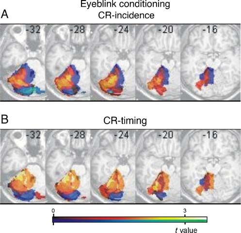
Cerebellum

recovery in patients with cerebellar disease. In addition,        basis and behaviour in patients with focal brain lesions [1,
information about the function of cerebellar cortex and           8, 9]. In patients with degenerative cerebellar disorders,
nuclei can be gained.                                             behavioural data and cerebellar atrophy assessed by conven-
                                                                  tional MRI volumetry have been correlated. Voxel-based
Keywords Human . Cerebellum . Ataxia . Sagittal zone .            morphometry is another option, particularly in disorders with
Somatotopy . Motor learning                                       no obvious abnormalities in structural MRI [10].
                                                                      There are important limitations to infer from correlation
There are different ways to study the function of the human       between a lesion and a behavioural measure on the function
cerebellum. One method which has long been used is to             of a given cerebellar area [11]. The cerebellum is part of
study the impairments in human subjects with cerebellar           a more extended brain circuitry. Thus, it is not meaningful
lesions. The introduction of high-resolution structural brain     to assign a specific behavioural function or deficit to a
imaging has helped to overcome some of the known                  specific lesioned area within the cerebellum, since the
limitations of human lesion studies [1]. Structural magnetic      lesion is part of a wider network involving intact parts
resonance imaging (MRI) in cerebellar lesion studies has          of the cerebellum as well as the cerebellar projections.
different implications.                                           Furthermore, in patients with chronic lesions, plastic
   Firstly, to infer possible cerebellar function based on        changes have taken place. It is likely that both the
lesion localisation and volume, lesions need to be restricted     cerebellum and connected areas change their function in
to the cerebellum alone. Structural MRI is helpful to             an attempt to recover. Recovery has not taken place in
exclude extracerebellar involvement of the central nervous        patients with acute focal lesions. However, effects of
system, for example additional lesions of the brainstem in        temporary malfunctions in connected brain areas due to
subjects with cerebellar stroke or degenerative disorders.        abrupt disconnection have to be considered in this patient
Some shortcomings should, however, be noted. For                  group. Therefore, results of human cerebellar lesion
example, patients with tumours may not present with               studies need to be complemented by animal and human
hydrocephalus at the time of the testing, but tumours are         data using other techniques, for example functional brain
frequently preceded by signs of increased intracranial            imaging and electrophysiological studies including trans-
pressure. The long-term effects of the latter and sudden          cranial magnetic stimulation.
surgical decompression are difficult to control. Further-             Despite these limitations, functionally meaningful corre-
more, cranial MRI cannot detect concomitant disorders of          lations between cerebellar lesion site and behavioural data
the peripheral nervous system. For example, many patients         can be obtained both in subjects with degenerative as well
with cerebellar degeneration show mild accompanying               as focal cerebellar disorders. One example is to correlate
signs of polyneuropathy in the lower limbs. Additional            clinical data with lesion data that are in good accordance
clinical and/or electrophysiological measures are required.       with the known functional compartmentalisation of the
   Secondly, structural MRI can be used to determine the          cerebellum. We used conventional MRI-based volumetry to
localisation and extent of cerebellar lesions. In patients with   quantify the degree of atrophy of the cerebellum and its
cerebellar degeneration volumetric measures have been             three longitudinal subdivisions in patients with cerebellar
applied to quantify the degree of atrophy of the whole            cortical degeneration. Total clinical ataxia rating scores
cerebellum and its subdivisions [2]. In patients with focal       showed a significant negative correlation with the volume
cerebellar lesions, the affected cerebellar lobules are           of the entire cerebellum (normalised to the total intracranial
defined [3], and the cerebellar nuclei can be visualised          volume) [12, 13]. Oculomotor disorders were highly corre-
allowing to analyse which parts of the nuclei are affected        lated with atrophy of the medial cerebellum. Posture and
[4, 5]. Advances of MRI analysis tools and technology             gait ataxia subscores revealed the highest correlations with
including available coils and increasing field strength lead      the medial and intermediate cerebellum. Disorders in limb
to a constant improvement in precision of structural              kinetic functions correlated with atrophy of lateral and
characterisation of the cerebellar lesions [6, 7]. It has to      intermediate parts of the cerebellum. The subscore for speech
be noted that the majority of human cerebellar lesion             disorders showed the highest correlation with the intermediate
models affect the cerebellar cortex. Additional lesions of        cerebellum [13].
the cerebellar nuclei may be present to various extents.              Findings are in good agreement with animal data
However, human lesions affecting primarily the cerebellar         showing that the medial zone is of particular importance
nuclei are exceptionally rare.                                    for control of stance, gait and eye movements and the
   Finally, and being the prime topic of this article, MRI-       intermediate and lateral zones for control of limb move-
based lesion data can be used to correlate cerebellar lesion      ments and speech [14–16]. Atrophy of the intermediate
and behavioural data. Different statistical techniques have       zone, however, was not only correlated with limb ataxia
been introduced to compare lesion site on a voxel-by-voxel        and dysarthria but also with ataxia of stance and gait.
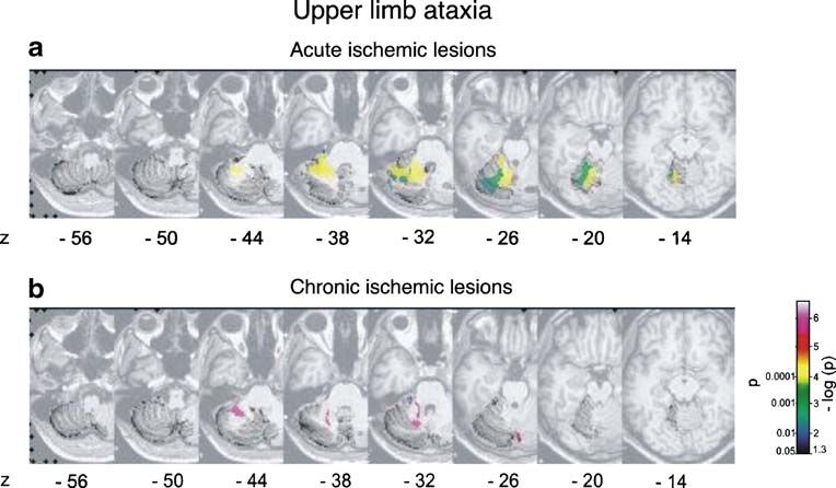
Cerebellum

Likewise, studies of our group in subjects with chronic                        interposed nuclei. In the subgroups with chronic focal
focal cerebellar lesions revealed that postural sway and                       lesions, similar correlations were observed with lesions of
disordered balance control during gait were associated with                    the cerebellar nuclei but no such correlations mentioned for
lesions affecting both the fastigial (that is medial zone)                     the acute group with lesions of the cerebellar cortex
and interposed nuclei (that is intermediate zone) [17, 18].                    (Fig. 1B). The lesion site, therefore, appears to be critical
In the gait study, lesions of the intermediate but not the                     for motor recovery. Findings agree with animal studies
medial zone were associated with abnormalities in the                          showing that a recovery after lesions to the nuclei of the
temporal characteristics of joint coordination patterns in                     cerebellum is often less complete [21].
lower limb control both during leg movements and gait                             Another example with extended knowledge about
[18]. One possible explanation is that lesions of the                          functional localisation within the cerebellum is classical
intermediate zone lead to disordered leg and trunk                             conditioning of the eyeblink response, a simple form of
coordination, which adds to disordered balance control                         motor learning [22]. Most authors agree that some forms of
during stance and gait.                                                        neuronal or synaptic plasticity develop in both cortex and
   In another experiment, the correlation with clinical                        cerebellar nuclei as a result of training [23, 24]. There are,
ataxia rating scores and MRI-defined lesions was per-                          however, ongoing controversies about the relative contri-
formed in patients with acute and chronic focal cerebellar                     butions of cerebellar cortex and nuclei.
lesions using voxel-based lesion-symptom mapping                                  A study of our group found that in healthy human
(VLSM [9]) [19]. In acute patients, a somatotopy in the                        subjects, the number of acquired conditioned eyeblink
superior cerebellar cortex was found which is in close                         responses was significantly related to the volume of the grey
relationship to animal data and functional MRI data in                         matter of the posterior lobe but not to the volume of the grey
healthy control subjects [20]. Upper limb ataxia was                           matter of the anterior lobe, the cerebellar white matter or
correlated with lesions of cerebellar lobules IV–VI, lower                     cerebrum [25]. In a group of patients with cerebellar cortical
limb ataxia with lesions of lobules III and IV, and dysarthria                 degeneration, however, no significant correlations between
with lesions of lobules V and VI.                                              acquisition of conditioned responses and any of the
   Furthermore, in the acute lesions, limb ataxia was                          cerebellar volumes were observed. Floor effects most likely
significantly correlated with lesions of the interposed and                    explained this observation with significantly reduced eye-
part of the dentate nuclei (Fig. 1A) and ataxia of posture                     blink conditioning early in the disease and no possible
and gait with lesions of the fastigial nuclei including part of                further reduction during disease progression.

Fig. 1. VLSM [9] of upper limb ataxia rating score (ICARS; [29]) in A          found within paravermal lobules V and VI. In addition, interposed and
21 subjects with acute and B 33 subjects with chronic ischemic                 dentate nuclei were affected (highest value (yellow area) refers to p<
cerebellar lesions. VLSMs are superimposed on axial slices of the              0.0001, Bonferroni-corrected). B Chronic lesions: statistical significant
cerebellum of a healthy subject normalised to MNI space. Right-sided           correlations were found primarily in areas including the dentate and
lesions are flipped to the left. A Acute lesions: Relating cerebellar lesion   interposed nuclei (p
Cerebellum

                                                                               More detailed information was observed examining
                                                                            patients with focal cerebellar lesions, most of them due to
                                                                            ischemic stroke. Eyeblink conditioning was significantly
                                                                            reduced on the ipsilesional side in subjects with lesions
                                                                            within the common territory of the superior cerebellar
                                                                            artery (SCA; lobule Crus I and above) but within normal
                                                                            limits on the contralesional side. In subjects with lesions
                                                                            restricted to the common territory of the posterior inferior
                                                                            cerebellar artery (PICA; Crus II and below), no significant
                                                                            difference in eyeblink conditioning was found comparing
                                                                            the affected and unaffected side [3]. VLSM [9] analysis
                                                                            revealed that learning rate was significantly reduced in
                                                                            subjects with focal lesions including superior parts of the
                                                                            posterior lobe, particularly lobule HVI (Fig. 2A [26]).
                                                                            Findings are in good accordance with animal data [27].
                                                                               Regarding timing of the conditioned response, we found
                                                                            that conditioned eyeblink responses occurred significantly
                                                                            earlier in subjects with cerebellar lesions in the SCA territory
                                                                            and with cerebellar cortical degeneration but not in subjects
Fig. 2. VLSM [9] of classical eyeblink conditioning in 22 subjects          with PICA infarctions. Corresponding to animal findings of
with unilateral cerebellar lesions. VLSMs related to learning rate (A)      Mauk’s group [28], VLSM analysis revealed that CR onset
and timing (B) of conditioned eyeblink responses are superimposed on
axial slices of the cerebellum of a healthy subject normalised to MNI       was significantly earlier in subjects with cortical lesions
space. Right-sided lesions are flipped to the left. A Relating cerebellar   including parts of the ipsilateral anterior lobe, in particular
lesion sites with learning rate (that is CR-incidences normalised to the    lobule HV (Fig. 2B). Findings suggest that cortical areas of
unaffected side) revealed highest positive t values (that is lowest CR-     the anterior lobe may be involved in conditioned response
incidences) primarily within hemispheral lobule VI (Larsell HVI)
extending to posterior parts of adjacent lobule V (shown in dark            timing and superior parts of the posterior lobe in stimulus
yellow). B Regarding the relationship of lesion sites and timing of         association in humans. This does not speak against an
conditioned responses (that is CR-onset latencies), highest positive t      additional role of the cerebellar nuclei.
values (that is shortest CR-onsets) were found within hemispheral              In summary, lesion-symptom mapping is helpful to study
lobule V (Larsell HV) extending to adjacent lobules IV and anterior
parts of HVI (shown in yellow; adapted from [26])                           the contribution of functionally relevant cerebellar compart-

Fig. 3. Schematic sketch of main findings in patients with focal            adjacent dorsomedial dentate nucleus, whereas lesions of ventrolateral
cerebellar lesions. The figure summarises the results of lesions-symptom    parts of the dentate nucleus were also related to limb ataxia in a third
mapping in our studies on eyeblink conditioning [26], cerebellar ataxia     study [19]. I–X cerebellar lobules according to [30]; dentate, interposed,
rating scores [19], balance in stance [17] and gait [18] and upper and      fastigial cerebellar nuclei; medial, intermediate, lateral cerebellar
lower limb coordination [17, 18]. Note that two studies [17, 18] found a    sagittal zones; CR conditioned eyeblink response
relationship between limb ataxia and lesions of interposed nuclei and
Cerebellum
ments in motor (and non-motor) control and recovery in eration and the relationship to cerebellar atrophy. Clin Neuro-
physiol (in press)
humans with focal and degenerative disorders (Fig. 3).
14. Thach WT, Kane SA, Mink JW, Goodkin HP (1992) Cerebellar
Although the study of participants with focal lesions is output, multiple maps and modes of control in movement
preferable, studies in participants with degenerative disor- coordination. In: Llinas R, Sotelo C (eds) The cerebellum
ders do also lead to meaningful results. In focal lesions, revisited. Springer, New York, pp 283–300
15. Urban PP, Marx J, Hunsche S, Gawehn J, Vucurevic G, Wicht S,
additional information about the function of cerebellar
Massinger C, Stoeter P, Hopf HC (2003) Cerebellar speech
cortex and nuclei can be gained. Application of new representation: lesion topography in dysarthria as derived from
techniques, for example higher resolution structural imag- cerebellar ischemia and functional magnetic resonance imaging.
ing of the cerebellar nuclei using high-Tesla MRI, will Arch Neurol 60:965–972
16. Voogd J, Barmack NH (2005) Oculomotor cerebellum. Prog Brain
improve lesion-symptom mapping in the future Res 151:231–268
17. Konczak J, Schoch B, Dimitrova A, Gizewski E, Timmann D
(2005) Functional recovery of children and adolescents after
References cerebellar tumour resection. Brain 128:1428–1441
18. Ilg W, Giese MA, Gizewski ER, Schoch B, Timmann D (2008)
The influence of focal cerebellar lesions on the control and
1. Rorden C, Karnath HO (2004) Using human brain lesions to infer adaptation of gait. Brain (in press)
function: a relic from a past era in the fMRI age. Nat Rev Neurosci 19. Schoch B, Dimitrova A, Gizewski ER, Timmann D (2006)
5:813–819, Review Functional localization in the human cerebellum based on
2. Makris N, Schlerf JE, Hodge SM, Haselgrove C, Albaugh MD, voxelwise statistical analysis: a study of 90 patients. NeuroImage
Seidman LJ, Rauch SL, Harris G, Biederman J, Caviness VS, 30:36–51
Kennedy DN, Schmahmann JD (2005) MRI-based surface-assisted 20. Grodd W, Hülsmann E, Lotze M, Wildgruber D, Erb M (2001)
parcellation of human cerebellar cortex: an anatomically specified Sensorimotor mapping of the human cerebellum: fMRI evidence
method with estimate of reliability. Neuroimage 25:1146–1160 of somatotopic organization. Hum Brain Mapp 13:55–57
3. Gerwig M, Dimitrova A, Kolb FP, Maschke M, Brol B, Kunnel A, 21. Eckmiller R, Westheimer G (1983) Compensation of oculomotor
Böring D, Thilmann AF, Forsting M, Diener HC, Timmann D deficits in monkeys with neonatal cerebellar ablations. Exp Brain
(2003) Comparison of eyeblink conditioning in patients with Res 49:315–326
superior and posterior inferior cerebellar lesions. Brain 126:71–94 22. Gerwig M, Kolb FP, Timmann D (2007) The involvement of the
4. Dimitrova A, Weber J, Redies C, Kindsvater K, Maschke M, Kolb human cerebellum in eyeblink conditioning. Invited review.
FP, Forsting M, Diener HC, Timmann D (2002) MRI atlas of the Cerebellum 6:38–57
human cerebellar nuclei. Neuroimage 17:240–255 23. Christian KM, Thompson RF (2003) Neural substrates of eyeblink
5. Dimitrova A, Zeljko D, Schwarze F, Maschke M, Gerwig M, conditioning: acquisition and retention. Learn Mem 10:427–455
Frings M, Beck A, Aurich V, Forsting M, Timmann D (2006) 24. De Zeeuw CI, Yeo CH (2005) Time and tide in cerebellar memory
Probabilistic 3D MRI atlas of the human cerebellar dentate/ formation. Curr Opin Neurobiol 15:667–667
interposed nuclei. Neuroimage 30:12–25 25. Dimitrova A, Gerwig M, Brol B, Gizewski ER, Forsting M, Beck
6. Diedrichsen J (2006) A spatially unbiased atlas template of the A, Aurich V, Kolb FP, Timmann D (2008) Correlation of
human cerebellum. Neuroimage 33:127–138 cerebellar volume with eyeblink conditioning in healthy subjects
7. Deoni SC, Catani M (2007) Visualization of the deep cerebellar and in patients with cerebellar cortical degeneration. Brain Res
nuclei using quantitative T1 and rho magnetic resonance imaging 1198:73–78
at 3 Tesla. Neuroimage 37:1260–1266 26. Gerwig M, Hajjar K, Dimitrova A, Maschke M, Kolb FP, Frings
8. Rorden C, Karnath HO, Bonilha L (2007) Improving lesion- M, Thilmann AF, Forsting M, Diener HC, Timmann D (2005)
symptom mapping. J Cogn Neurosci 19:1081–1088 Timing of conditioned eyeblink responses is impaired in cerebel-
9. Bates E, Wilson SM, Saygin AP, Dick F, Sereno MI, Knight RT, lar patients. J Neurosci 25:3919–3931
Dronkers NF (2003) Voxel-based lesion-symptom mapping. Nat 27. Attwell PJ, Rahman S, Yeo CH (2001) Acquisition of eyeblink
Neurosci 6:448–450 conditioning is critically dependent on normal function in
10. Lasek K, Lencer R, Gaser C, Hagenah J, Walter U, Wolters A, cerebellar cortical lobule HVI. J Neurosci 21:5715–5722
Kock N, Steinlechner S, Nagel M, Zühlke C, Nitschke MF, 28. Perrett SP, Ruiz BP, Mauk MD (1993) Cerebellar cortex lesions
Brockmann K, Klein C, Rolfs A, Binkofski F (2006) Morpho- disrupt learning-dependent timing of conditioned eyelid responses. J
logical basis for the spectrum of clinical deficits in spinocerebellar Neurosci 13:1708–1718
ataxia 17 (SCA17). Brain 129:2341–2345 29. Trouillas P, Takayanagi T, Hallett M, Currier RD, Subramony SH,
11. Shallice T (1988) From Neuropsychology to Mental Structure. Wessel K, Bryer A, Diener HC, Massaquoi S, Gomez CM,
Cambridge University Press, Cambridge Coutinho P, Ben Hamida M, Campanella G, Filla A, Schut L,
12. Richter S, Dimitrova A, Maschke M, Gizewski E, Beck A, Aurich Timmann D, Honnorat J, Nighoghossian N, Manyam B (1997)
V, Timmann D (2005) Degree of cerebellar ataxia correlates with International Cooperative Ataxia Rating Scale for pharmacologi-
three-dimensional MRI-based cerebellar volume in pure cerebellar cal assessment of the cerebellar syndrome. The Ataxia Neuro-
degeneration. Eur Neurol 54:23–27 pharmacology Committee of the World Federation of Neurology. J
13. Brandauer B, Hermsdörfer J, Beck A, Aurich V, Gizewski ER, Neurol Sci 145:205–201
Marquardt C, Timmann D (2008) Impairments of prehension 30. Schmahmann JD, Doyon J, Toga AW, Petrides M, Evans AC (2000)
kinematics and grasping faces in patients with cerebellar degen- MRI atlas of the human cerebellum. Academic Press, San DiegoYou can also read