Long COVID or Post-acute Sequelae of COVID-19 (PASC): An Overview of Biological Factors That May Contribute to Persistent Symptoms - Frontiers
←
→
Page content transcription
If your browser does not render page correctly, please read the page content below
REVIEW
                                                                                                                                            published: 23 June 2021
                                                                                                                                   doi: 10.3389/fmicb.2021.698169

                                             Long COVID or Post-acute Sequelae
                                             of COVID-19 (PASC): An Overview of
                                             Biological Factors That May
                                             Contribute to Persistent Symptoms
                                             Amy D. Proal 1 and Michael B. VanElzakker 1,2*
                                             1
                                              PolyBio Research Foundation, Kenmore, WA, United States, 2 Division of Neurotherapeutics, Massachusetts General
                                             Hospital, Harvard Medical School, Boston, MA, United States

                                             The novel virus severe acute respiratory syndrome coronavirus 2 (SARS-CoV-2) has
                                             caused a pandemic of coronavirus disease 2019 (COVID-19). Across the globe, a
                                             subset of patients who sustain an acute SARS-CoV-2 infection are developing a
                                             wide range of persistent symptoms that do not resolve over the course of many
                                             months. These patients are being given the diagnosis Long COVID or Post-acute
                                             sequelae of COVID-19 (PASC). It is likely that individual patients with a PASC diagnosis
                            Edited by:       have different underlying biological factors driving their symptoms, none of which are
                      Martin Michaelis,
   University of Kent, United Kingdom        mutually exclusive. This paper details mechanisms by which RNA viruses beyond just
                       Reviewed by:          SARS-CoV-2 have be connected to long-term health consequences. It also reviews
              Hana Maria Dobrovolny,         literature on acute COVID-19 and other virus-initiated chronic syndromes such as post-
            Texas Christian University,
                                             Ebola syndrome or myalgic encephalomyelitis/chronic fatigue syndrome (ME/CFS) to
                        United States
                 Juan C. De La Torre,        discuss different scenarios for PASC symptom development. Potential contributors
       The Scripps Research Institute,       to PASC symptoms include consequences from acute SARS-CoV-2 injury to one or
                        United States
                                             multiple organs, persistent reservoirs of SARS-CoV-2 in certain tissues, re-activation of
                *Correspondence:
             Michael B. VanElzakker          neurotrophic pathogens such as herpesviruses under conditions of COVID-19 immune
     mvanelzakker@mgh.harvard.edu            dysregulation, SARS-CoV-2 interactions with host microbiome/virome communities,
                                             clotting/coagulation issues, dysfunctional brainstem/vagus nerve signaling, ongoing
                   Specialty section:
         This article was submitted to       activity of primed immune cells, and autoimmunity due to molecular mimicry between
                              Virology,      pathogen and host proteins. The individualized nature of PASC symptoms suggests
               a section of the journal
             Frontiers in Microbiology
                                             that different therapeutic approaches may be required to best manage care for specific
             Received: 20 April 2021
                                             patients with the diagnosis.
             Accepted: 17 May 2021
                                             Keywords: COVID-19, long COVID, SARS-CoV-2, microbiome, vagus, brainstem, infection, virus
            Published: 23 June 2021
                             Citation:
       Proal AD and VanElzakker MB          INTRODUCTION
   (2021) Long COVID or Post-acute
  Sequelae of COVID-19 (PASC): An
                                            The novel virus severe acute respiratory syndrome coronavirus 2 (SARS-CoV-2) has resulted
  Overview of Biological Factors That
         May Contribute to Persistent
                                            in a global pandemic of coronavirus disease 2019 (COVID-19) (Hiscott et al., 2020). Classic
                           Symptoms.        cases of acute COVID-19 are characterized by respiratory symptoms, fever, and gastrointestinal
         Front. Microbiol. 12:698169.       problems (Larsen et al., 2020). However, patients can present with a wide range of other
    doi: 10.3389/fmicb.2021.698169          symptoms, including neurological issues suggesting central nervous system (CNS) involvement

Frontiers in Microbiology | www.frontiersin.org                                   1                                          June 2021 | Volume 12 | Article 698169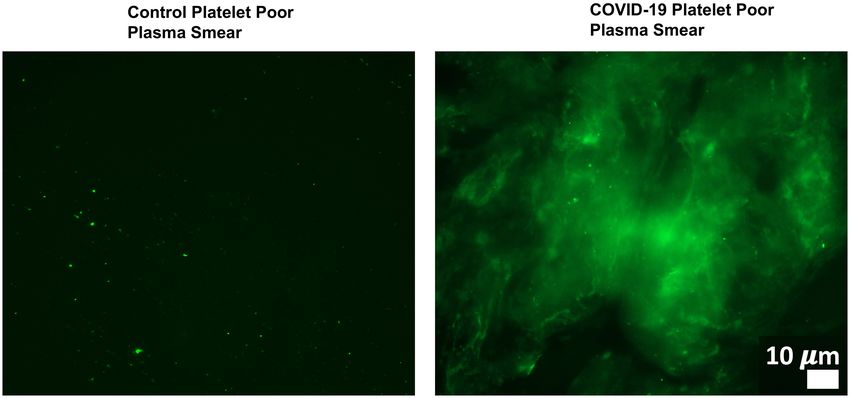
Proal and VanElzakker                                                                                                   Overview of PASC Biology

(Harapan and Yoo, 2021). Acute COVID-19 cases range in length             (Komaroff, 2006). Enteroviruses, several species of which
and severity. Many patients are asymptomatic, while others                can be acquired via respiratory infection, have been identified
require hospitalization and ventilation (Cunningham et al.,               in brain, skeletal muscle, and stomach biopsy specimens of
2021). Overall, an average case of COVID-19 lasts between 1               certain patients with ME/CFS (Gow et al., 1991; McGarry et al.,
and 4 weeks. However, across the globe, a subset of patients who          1994; Richardson, 2001; Chia and Chia, 2008). Other respiratory
sustain an acute SARS CoV-2 infection are developing a wide               pathogens have also been linked to the development of ME/CFS-
range of persistent symptoms that do not resolve over the course          like symptoms (Magnus et al., 2015). For example, one team
of many months (Carfì et al., 2020; Davis et al., 2020; Huang             studied 233 SARS survivors approximately 4 years after initial
C. et al., 2021) (Figure 1). One study of COVID-19 patients               infection, and found that 27.1% met the modified 1994 Centers
who were followed for up to 9 months after illness found that             for Disease Control and Prevention (CDC) criteria for chronic
approximately 30% reported persistent symptoms (Logue et al.,             fatigue syndrome (Lam et al., 2009).
2021). These patients are being given the diagnosis Long COVID,               It is likely that the different pathogens implicated in ME/CFS
post-acute COVID-19 syndrome (PACS), or post-acute sequelae               development are capable of dysregulating host gene expression,
of COVID-19 (PASC).                                                       immunity, and metabolism via similar mechanisms, leading to
   Post-acute sequelae of COVID-19 is being diagnosed in                  similar sets of chronic symptoms in ME/CFS-diagnosed patients
patients who developed severe acute COVID-19, but also in                 (VanElzakker, 2013; Proal and Marshall, 2018). However, a
patients who experienced only mild or asymptomatic cases. For             range of additional biological factors, including changes in host
example, Tabacof et al. (2020) reported a range of long-term              microbiome composition and activity, can also contribute to
symptoms in a cohort of previously confirmed or presumed                  ME/CFS development (Giloteaux et al., 2016; Newberry et al.,
COVID-19 patients whose acute symptoms were largely managed               2018). The same trend may be true of PASC cases, in which SARS-
without the need for hospitalization. Another team documented             CoV-2 infection may instigate or exacerbate different biological
persistent COVID-19 symptoms in 1,407 subjects with confirmed             abnormalities in patients with the diagnosis.
SARS-CoV-2 infection (Huang Y. et al., 2021). Symptoms                        This paper reviews relevant ME/CFS, neuroscience,
included fatigue and muscle weakness, insomnia, palpitations,             microbiology, and single-stranded RNA virus literatures to
chronic rhinitis, dysgeusia, chills, sore throat, and headache. 27%       explore a range of biological factors that may contribute to the
of subjects reported persistent symptoms after 60 days, with              development of chronic symptoms after acute COVID-19, none
patients aged 50 ± 20 years comprising 72% of cases. Women                of which are mutually exclusive. These include consequences
were more likely to report persistent symptoms, and ∼32% of               from acute injury caused by SARS-CoV-2, persistent reservoirs
subjects reporting symptoms at 61+ days after infection were              of SARS-CoV-2 in certain tissues, re-activation of other
asymptomatic at the time of initial SARS-CoV-2 testing.                   neurotrophic pathogens under conditions of COVID-19
   While the development of long-term symptoms following                  immune dysregulation, SARS-CoV-2 interactions with host
SARS-CoV-2 infection is sometimes framed as novel or                      microbiome/virome communities, clotting/coagulation issues,
mysterious, it is actually an expected phenomenon. Most well-             disrupted brainstem/vagus nerve signaling, ongoing activity
studied viral or bacterial pathogens have been connected to               of primed immune cells, and autoimmunity due to molecular
the development of chronic symptoms in a subset of infected               mimicry between pathogen and host peptides.
patients (Bannister, 1988; Rebman and Aucott, 2020; Komaroff
and Bateman, 2021). For example, Ebola virus is associated with a
chronic syndrome that can develop after acute infection (Wilson           ACUTE COVID-19
et al., 2018), with reservoirs of Ebola sometimes identified
in “anatomical sanctuaries” in patient tissue months or years             In order to best understand persistent symptoms arising from
after the virus has cleared from the blood (Lo et al., 2017;              SARS-CoV-2 infection we must first review major trends
Keita et al., 2019).                                                      associated with SARS CoV-2 activity in patients with acute
   Some PASC patients meet the diagnostic criteria for myalgic            COVID-19. SARS-CoV-2 is a positive-sense single-stranded
encephalomyelitis/chronic fatigue syndrome (ME/CFS) – a                   RNA virus (V’kovski et al., 2021). It is one of seven
neuroinflammation-linked condition characterized by a range               coronaviruses capable of infecting humans (Corman et al., 2018).
of debilitating chronic symptoms including severe fatigue,                Compared with other coronaviruses (e.g., HCoV-NL63, HCoV-
musculoskeletal pain, and post-exertional malaise (worsening of           229E, and HCoV-OC43) that are pathogenic to humans but
symptoms following exertion) (Carruthers et al., 2011; Clayton,           generally drive only mild clinical symptoms, SARS-CoV-2 more
2015; Kedor et al., 2021; Komaroff and Bateman, 2021). Overlap            closely resembles MERS-CoV or SARS-CoV (sometimes called
between the PASC and ME/CFS diagnoses is not surprising, since            SARS-CoV-1) in that it is capable of causing severe disease
most cases of ME/CFS begin with a viral infection, or involve             (Zhu et al., 2020).
multiple exposures to viral and bacterial pathogens over time                Many COVID-19 patients are asymptomatic (∼40–45%)
(Rasa et al., 2018).                                                      (Oran and Topol, 2020) or exhibit mild to moderate symptoms
   Pathogens most commonly implicated in ME/CFS                           (Zheng P. et al., 2020). However, approximately 15% progress
development include neurotrophic herpesviruses and                        to severe pneumonia, with ∼5% eventually developing acute
enteroviruses. Several studies have found that active HHV-6               respiratory distress syndrome (ARDS), septic shock and/or
infection is more common in ME/CFS than controls                          multiple organ failure (Cao, 2020; Huang C. et al., 2020).

Frontiers in Microbiology | www.frontiersin.org                       2                                     June 2021 | Volume 12 | Article 698169
Proal and VanElzakker                                                                                                             Overview of PASC Biology

  FIGURE 1 | Most common symptoms remaining after 7 months in 966 respondents from a cohort of suspected and confirmed COVID-19 cases. Results obtained
  via an international web-based survey. Image adapted with permission from Davis et al. (2020).

The most common symptoms of acute COVID-19 are fever,                           genes that can contribute to viral entry into the cell and viral
fatigue, diarrhea, and respiratory symptoms such as cough,                      persistence, including ACE2, TMPRSS2, TMPRSS4, TPCN2,
sore throat and shortness of breath (Larsen et al., 2020).                      CTSL, and NRP1. They found that these genes are expressed in
However, some patients develop neurological manifestations                      neurons, glial cells, and endothelial cells, suggesting their possible
ranging from mild symptoms such as anosmia, dizziness, and                      capacity to support SARS-CoV-2 infection.
headache to more severe cerebrovascular disease, seizures,                          Like all pathogens, SARS-CoV-2 employs a number of
encephalitis, or the Guillain–Barré syndrome (Berlit et al.,                    mechanisms to disable and evade the host immune response
2020). Other extrapulmonary manifestations of COVID-                            (Lucas et al., 2001; Bowie and Unterholzner, 2008; Taefehshokr
19 include acute kidney injury, hyperglycemia, thrombotic                       et al., 2020). These include the ability to replicate within double-
complications, myocardial dysfunction and arrhythmia, acute                     membrane vesicles that are not detected by host pathogen pattern
coronary syndromes, and hepatocellular injury (Gupta et al.,                    recognition receptors (Taefehshokr et al., 2020). SARS-CoV-2
2020; Robba et al., 2020).                                                      also dysregulates the host interferon response (Ribero et al.,
    These diverse COVID-19 symptoms partially reflect the fact                  2020). Interferons are cytokines secreted by host cells in response
that SARS-CoV-2 can infect a wide range of human cell types.                    to viral infection. They bind to cell surface receptors and act
The spike subunit of SARS CoV-2 binds the human angiotensin-                    as transcription factors, regulating the expression of hundreds
converting enzyme 2 (ACE2) receptor to infect and enter host                    of genes whose protein products target viruses at many levels
cells (Hoffmann et al., 2020). Viral cell entry additionally requires           (Acharya et al., 2020). SARS-CoV-2 expresses at least 10 proteins
priming of the spike protein by cellular serine proteases such as               that allow it to either counteract the induction or escape the
TMPRSS2 and TMPRSS4. ACE2 is expressed along the entire                         antiviral activity of interferons (Ribero et al., 2020), allowing
human respiratory system and in brain endothelium and vascular                  the virus to better survive by rendering the host innate immune
smooth muscle cells (Hamming et al., 2004). Single-cell RNA-                    response inefficient.
sequencing studies have also confirmed expression of ACE2 and                       Despite this innate immune disruption, SARS-CoV-2 can
TMPRSS2 in a wide range of cell types including esophageal                      initiate host immune signaling pathways. If the virus is
keratinocytes, renal proximal tubules, pancreatic β-cells, and                  not successfully contained, this results in the production
gastrointestinal epithelial cells (Gupta et al., 2020; Puelles et al.,          of proinflammatory cytokines such as interlukin-6, and the
2020; Qi et al., 2020).                                                         recruitment of neutrophils and myeloid cells (Gubernatorova
    Recent work has further clarified CNS cellular expression                   et al., 2020). This leads to hyperinflammation, and in some
of ACE2 and other genes that may contribute to COVID-19.                        cases, a cytokine storm syndrome (Chen and Quach, 2021).
For example, Matschke et al. (2020) performed an in silico                      Severe COVID-19 can also result in functional exhaustion and
analysis of publicly available datasets to determine which CNS cell             decreased numbers of T lymphocytes, (particularly CD4+ T
types might be prone to SARS-CoV-2 infection. They analyzed                     cells, CD8+ T cells) and natural killer cells (Diao et al., 2020;

Frontiers in Microbiology | www.frontiersin.org                             3                                         June 2021 | Volume 12 | Article 698169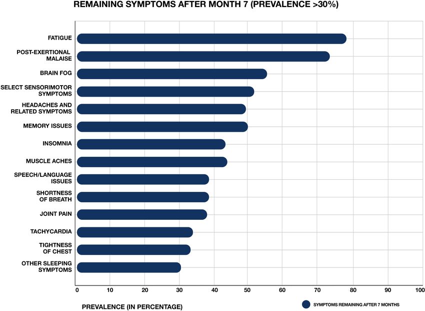
Proal and VanElzakker                                                                                                    Overview of PASC Biology

Zheng M. et al., 2020). Impaired T cell responses can result               cells – including cells infected in a Trojan horse fashion –
from deficient interferon production driven by SARS-CoV-2, as              into the CNS.
interferons promote the survival and effector functions of T cells.           SARS-CoV-2 may also enter the central nervous system via
   SARS-CoV-2 can also drive multi-organ injury via stimulation            the nasal cavity, through the foramina of the cribriform plate,
of clotting cascades (Pretorius et al., 2020a) and related                 and into the olfactory epithelium. There, the virus can exploit the
thromboinflammation, dysregulation of the renin–angiotensin–               close vicinity of olfactory sensory neurons whose axons project
aldosterone system, and endothelial cell damage (Grobler                   into the olfactory bulb of the brain (Montalvan et al., 2020).
et al., 2020; Gupta et al., 2020). Infection-mediated endothelial          Olfactory entry of SARS-CoV-2 into the CNS is now supported
injury and endothelialitis (marked by the presence of activated            by multiple studies. Meinhardt et al. (2021) analyzed the olfactory
macrophages and neutrophils) can trigger excessive thrombin                mucosa, its nervous projections, and several CNS regions in 33
production, inhibit fibrinolysis, and activate complement                  individuals who died from COVID-19. SARS-CoV-2 RNA and/or
pathways in a manner that leads to microvascular dysfunction               protein were identified in anatomically distinct regions of both
and microthrombi deposition.                                               the nasopharynx and brain, including the medulla oblongata of
                                                                           the brainstem (Meinhardt et al., 2021). SARS-CoV-2 RNA levels
                                                                           were highest within the olfactory mucosa sampled directly under
THE NEUROINVASIVE AND                                                      the cribriform plate (n = 20 of 30).
NEUROTROPHIC POTENTIAL OF                                                     Other autopsy studies have identified SARS-CoV-2 RNA
SARS-CoV-2                                                                 or protein in the brainstem of humans and animals (de
                                                                           Melo et al., 2020). Matschke et al. (2020) identified SARS-
Autopsy, animal, and organoid model studies show that, like                CoV-2 RNA or protein in 21 of 40 (53%) of COVID-19
SARS-CoV, SARS-CoV-2 is able to reach and infect cells of the              autopsied brains, with both SARS-CoV-2 RNA and protein
CNS, infect neurons, and produce neuroinflammation (Matschke               detected in 8 of 40 (20%) of brains (Matschke et al.,
et al., 2020; Song et al., 2020; Song et al., 2021). Indeed, SARS-         2020). Immunohistochemistry demonstrated SARS-CoV-2 viral
CoV-2 may be capable of transport up and down nerves and                   proteins in vagal and glossopharyngeal cranial nerves originating
neuronal axons (Lima et al., 2020; Rangon et al., 2020; Song et al.,       from the lower brainstem and in isolated cells of the brainstem’s
2020; Karuppan et al., 2021).                                              medulla oblongata. This is consistent with the brainstem’s
    One pathway by which SARS-CoV-2 may reach the CNS                      relatively high expression of ACE2 (Lukiw et al., 2020). The
is via hematogenous spread from heavily infected airways                   brainstem is also the site of the area postrema circumventricular
and lungs. Systemic inflammation that increases blood brain                organ, itself a site of ACE2 expression (Doobay et al., 2007).
barrier (BBB) permeability would facilitate this kind of                   Matschke et al. (2020) additionally reported diffuse activation
spread. The circumventricular organs are brain structures with             of microglia and infiltration of cytotoxic T lymphocytes in
fenestrated capillaries and high permeability. This normally               brainstem and cerebellum tissue.
allows circulating but non-BBB-crossing mediators to directly                 A smaller study of 18 COVID-19 autopsies focused on
affect brain function. However, during acute infection this                microvascular changes in the olfactory bulb and brainstem.
permeability can also allow for pathogen neuroinvasion. This can           They found infiltrating macrophages and activated astrocytes
occur directly or via a “Trojan horse” mechanism in which host             and microglia in the perivascular spaces of 13 brains. Ex vivo
immune cells infected with intracellular pathogens are actively            11.7 Tesla high-resolution magnetic resonance imaging found
transported into the CNS (Dando et al., 2014; Lauer et al.,                punctate hyperintensities in 9 of 13 patients’ brain tissue,
2018). Perhaps importantly, several circumventricular organs               representing areas of microvascular injury and fibrinogen
have relatively high ACE2 expression (Doobay et al., 2007) likely          leakage. No evidence of SARS-CoV-2 RNA was reported after
due to the fact that angiotensin II is a peptide hormone that does         testing a selection of brain structures from a subset of samples
not easily cross the BBB.                                                  (for example, the medulla of 1 subject and the olfactory bulb of 3
    Like other circumventricular organs, the choroid plexus lacks          subjects) (Lee et al., 2021, Supplementary Table 5).
a tight junction BBB at the endothelium but contains tight                    With these acute COVID-19 trends in mind, we will
junction epithelial cells forming the blood-cerebrospinal fluid            now discuss different biological factors that may contribute
(CSF) barrier. This layer produces CSF and serves an immune                to the development of persistent symptoms in patients with
function, partly by facilitating the exchange of nutrients, waste,         a PASC diagnosis.
and peripheral immune cells between the bloodstream and CSF
(Thompson et al., 2020). Some pathogens have evolved to enter
the brain from blood by exploiting this polarized choroid plexus           SARS-CoV-2 CAN CAUSE INJURY TO
epithelium (Lauer et al., 2018). In a human-pluripotent-stem-              ONE OR MULTIPLE ORGANS OR
cell-derived organoid model, SARS-CoV-2 exhibited tropism for              TISSUES
choroid plexus basal (vascular side) epithelium, despite more
abundant ACE2 on the apical (CSF) side (Pellegrini et al.,                 Long-term symptoms in some PASC patients may be due to
2020). Furthermore, SARS-CoV-2 caused epithelium damage and                consequences from organ or tissue injury caused by SARS-CoV-
barrier leakage, which could facilitate neuroinflammation by               2, or associated clotting or inflammatory processes during acute
allowing increased entry of circulating cytokines and immune               COVID-19 (Del Rio et al., 2020). For example, Guler et al. (2021)

Frontiers in Microbiology | www.frontiersin.org                        4                                     June 2021 | Volume 12 | Article 698169
Proal and VanElzakker Overview of PASC Biology
found that 4 months after SARS-CoV-2 infection, severe overcoming RNAse activity was a central challenge in mRNA
COVID-19 was associated with significant radiological and vaccine development (Pardi et al., 2018). It follows that if inert
functional abnormalities indicative of lung parenchymal and SARS-CoV-2 RNA and/or RNA fragments drive symptoms in
small airway disease. Another early analysis of COVID-19 some PASC patients, the molecular mechanisms of inert RNA
patients upon hospital discharge found that more than a third persistence must be better delineated.
of recovered patients develop lung fibrotic abnormalities (Rai Immune responses can also be used to infer the possible
et al., 2020). Pulmonary fibrosis is characterized by scarring of the persistence of SARS-CoV-2 in certain patients. In a study of
lungs. The condition can present as stable in response to injury or 203 patients, Vibholm et al. (2021) found that after 90 days,
infection, or can become progressive (marked by periods of rapid 5.3% of subjects remained positive for SARS-CoV-2 via RT-
exacerbation) (McDonald, 2021). Both stable and progressive PCR nasopharyngeal testing (Plebani, 2021). While there were
fibrotic lung disease result in excessive deposition of extracellular no differences in SARS-CoV-2 antibody levels between the PCR
matrix molecules such as fibronectin, collagen, and laminin positive and negative subjects, the PCR positive group displayed
in parenchymal lung tissue. This leads to epithelial/endothelial SARS-CoV-2-specific CD8 T-cell responses of significantly
injury and thickened alveolar walls, which can hinder gas increased breadth and magnitude, leading the team to suggest
exchange in the lungs and increase symptoms of fatigue, dyspnea, that such subjects might still harbor replicating virus.
and exercise intolerance. Gaebler et al. (2021) studied COVID-19 antibody responses
SARS-CoV-2 can also drive acute kidney injury (Robbins- in a larger cohort of patients assessed at 1.3 and 6.2 months
Juarez et al., 2020) in a manner that may contribute to PASC after SARS-CoV-2 infection. They found that while IgM and
symptoms. For example, Huang C. et al. (2021) found that IgG anti-spike protein receptor binding domain (RBD) antibody
6 months after acute COVID-19, 35% of patients displayed a titers decreased significantly, levels of RBD-specific memory B
decreased estimated glomerular filtration rate (eGFR). Infection cells remained unchanged. This indicated possible continued
with SARS-CoV-2 is also associated with the development antibody evolution, potentially due to small amounts of SARS-
of pediatric multisystem inflammatory syndrome (MIS-C), CoV-2 antigen or lack of complete viral clearance. To test this
manifestations of which can cause severe organ inflammation possibility, they obtained intestinal biopsy samples from certain
and dysfunction in children, with over 10% of cases showing study subjects. They identified SARS-CoV-2 RNA and protein
acute kidney injury (Belay et al., 2021). More information on in 7 of 14 of the biopsy samples obtained from asymptomatic
MIS-C and other SARS-CoV-2 organ injury-related sequelae can COVID-19 patients with negative nasal-swab PCR, at an average
be found in the excellent review from Nalbandian et al. (2021). of 4 months after acute infection.
However, in some cases, long-term decreased eGFR and MIS- Another team found SARS-CoV-2 RNA in olfactory mucosa
C were not present during acute COVID-19. For example, 13% samples collected from 4 patients with negative nasopharyngeal
of subjects in the Huang C. et al. (2021) study developed new- swab SARS-CoV-2 RNA tests but ongoing loss of smell after
onset reduction of eGFR after displaying normal renal function acute COVID-19 disease (samples were collected 110–196 days
during acute COVID-19. One retrospective study found that 810 after COVID-19 onset) (de Melo et al., 2020). RT-qPCR revealed
of 1,075 COVID-19-associated MIS-C cases were asymptomatic high levels of SARS-CoV-2 RNA in 3 of the olfactory tissue
during their COVID-19 illness (Belay et al., 2021). This suggests mucosa samples, and immunostaining revealed SARS-CoV-2
that biological factors beyond organ injury alone may contribute protein antigens in 3 samples.
to chronic symptoms in such patients. Immunosuppression may facilitate SARS-CoV-2 persistence
(Lancman et al., 2020; Kemp et al., 2021; Tehrani et al.,
2021). One team documented SARS-CoV-2 persistence in an
SARS-CoV-2 APPEARS CAPABLE OF antiphospholipid syndrome patient who was administered a wide
PERSISTENCE IN CERTAIN TISSUES range of immunosuppressive drugs over 154 days (Choi et al.,
2020). Viral infectivity studies confirmed infectious SARS-CoV-
It is also possible that, at least in some PASC patients, 2 virus in nasopharyngeal samples obtained from the patient
SARS-CoV-2 may drive chronic symptoms by persisting in at days 75 and 143 of COVID-19. Examination of the patient’s
certain body sites or tissue reservoirs after acute infection. tissue after death showed the highest SARS-CoV-2 RNA levels in
A growing number of studies show that some patients infected the spleen and lungs. Phylogenetic analysis was consistent with
with SARS-CoV-2 do not successfully clear the virus over long persistent infection and accelerated viral evolution. SARS-CoV-
periods of time (Liotti et al., 2020; Sun et al., 2020; Vibholm 2 mutated in the patient over time to evolve new amino acid
et al., 2021). In such studies, confirmation of SARS-CoV-2 in changes in its spike protein and receptor binding domain.
patient samples is generally assessed via identification of viral A separate report found that late-stage SARS-CoV-2 S
RNA and/or proteins. While identification of SARS-CoV-2 RNA variants isolated from the same patient contained mutations
could technically represent “inert” RNA, the possibility is unlikely that conferred resistance to a common class of SARS-CoV-
because inert RNA in the human body is rapidly degraded 2 neutralizing antibodies isolated from a healthy COVID-19
(Houseley and Tollervey, 2009; Fabre et al., 2014). Nearly every convalescent donor (Clark et al., 2021). Resistance extended to
human cell type, and human tears, saliva, mucus, perspiration, monoclonal antibodies used clinically, and to polyclonal serum
and extracellular spaces express RNAase enzymes that rapidly immunoglobulins obtained from four healthy convalescent
degrade inert RNA (Sorrentino, 2010; Gupta et al., 2013). Indeed, donors. This suggests that in some patients, SARS-CoV-2 may
Frontiers in Microbiology | www.frontiersin.org 5 June 2021 | Volume 12 | Article 698169Proal and VanElzakker Overview of PASC Biology
be able to evade the immune response in a manner that allows Some enteroviruses, including those that drive respiratory
it to drive persistent symptoms. Indeed, some recent COVID- symptoms, also appear capable of persistence and have been
19 variants have spike protein mutations that allow for increased connected to chronic disease (Zhang et al., 2013; Genoni
immune evasion, including resistance to neutralizing antibodies et al., 2017). Enteroviruses of the family picornaviridae include
or escape from HLA-restricted cellular immunity (McCallum the coxsackieviruses, poliovirus, echoviruses, and rhinoviruses
et al., 2021; Motozono et al., 2021; Planas et al., 2021). These (Baggen et al., 2018). These single-stranded RNA viruses cause
mutations could potentially facilitate SARS-CoV-2 persistence in about 10–15 million infections each year in the United States
non-immunocompromised individuals. alone. Several members of the enterovirus family including
Persistence of SARS-CoV-2 in some patients with PASC polioviruses types 1–3 and enterovirus 71/D68 are neurotrophic
symptoms is not unexpected. The literature is replete with and able to drive severe CNS infections (Tang and Holmes, 2017).
examples of single-stranded RNA virus persistence, spanning Several research teams have found enteroviruses in the blood
decades of research on samples obtained from living human and pancreas of patients with type 1 diabetes, and enteroviruses
patients, autopsy studies, and animal studies (Viola et al., 1978; have been associated with increased risk of type 1 diabetes in
Riddell et al., 2007; Doi et al., 2016; Randall and Griffin, prospective studies (Tauriainen et al., 2011; Oikarinen et al.,
2017; Ireland et al., 2020). Persistence of single-stranded RNA 2012). A number of teams have identified enteroviruses and
viruses in the central nervous system has been documented on their proteins in tissue samples obtained from patients with
multiple occasions. In a 1986 paper on the topic, Kristensson ME/CFS or ME/CFS-like symptoms (Yousef et al., 1988; Dowsett
and Norrby explain that “Although it would seem difficult for et al., 1990; Chia et al., 2010; Chia et al., 2015). For example,
RNA viruses to persist in the brain under conditions of normal Chia and Chia found enterovirus VP1 protein and RNA in
immune defense mechanisms, representatives of at least seven stomach biopsy specimens obtained from 165 ME/CFS patients
of the established families of RNA viruses have been shown with chronic abdominal complaints. 82% of ME/CFS specimens
capable of causing persistent infections under these conditions” stained positive for enterovirus VP1 protein, compared to
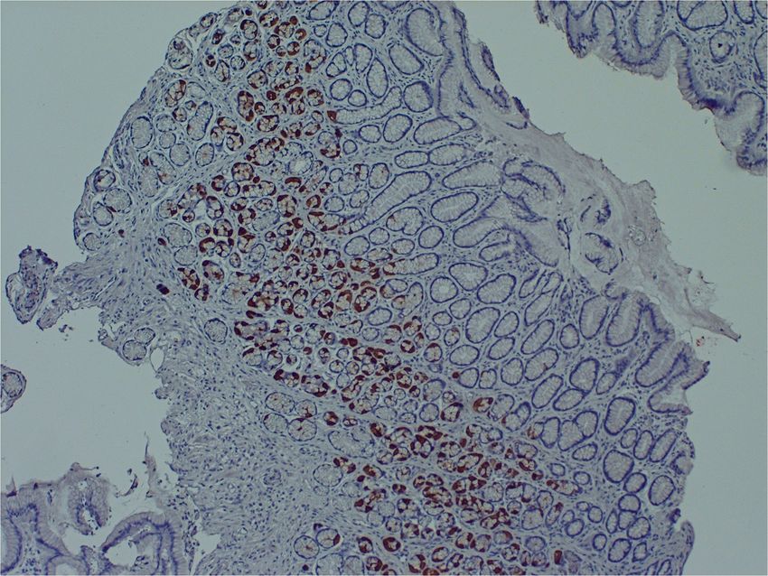
(Kristensson and Norrby, 1986). 20% of control specimens (Chia and Chia, 2008) (Figure 2).
Single-stranded RNA viruses appear to employ a range A non-cytolytic form of enteroviral infection was cultured from
of mechanisms to establish persistent infections (Randall 5 ME/CFS specimens. Positive staining was found in repeat
and Griffin, 2017). For example, hepatitis C virus (HCV) stomach biopsy specimens taken from 6 ME/CFS patients at the
may be able to hijack cellular factors, such as microRNAs onset of symptoms and again 2–8 years later.
that bind its genome, to shield it from degradation by the Enteroviruses have also been found in ME/CFS brain and
50 –30 exoribonuclease Xrn1 (Li et al., 2013). This prevents muscle tissue (Cunningham et al., 1991; Gow et al., 1991;
the innate immune response from recognizing and regulating Richardson, 2001). For example, McGarry et al. (1994) examined
HCV transcription, replication, and genomic RNA abundance. the central nervous system of a woman with ME/CFS who
Modifications (insertions and deletions) at the 50 and/or died by suicide for the presence of enteroviruses. Positive PCR
30 ends of the genomes of other single-stranded RNA sequences with similarity to coxsackievirus B3 were identified
viruses, such as coxsackieviruses and hantaviruses, may in brain samples from the hypothalamus and brainstem, and
also allow such pathogens to establish persistent infections also in muscle and heart samples. No enteroviral sequences were
(Meyer and Southern, 1997; Meyer and Schmaljohn, 2000; identified in any tissue obtained from four control subjects.
Kim et al., 2005). Multiple research teams have identified Zika virus (ZIKV)
Dozens of studies show coronaviruses capable of persistence, RNA in men’s semen more than 6 months after initial infection
with some coronaviruses tied to chronic disease development (Nicastri et al., 2016; Huits et al., 2017; Mead et al., 2018).
(Arbour et al., 1999; Chan et al., 2004). One study found that Other studies, including those in primates, strongly support the
in primates infected with two different coronaviruses, the viruses possibility that, in some people exposed to ZIKV, low-levels of
persisted, replicated, and disseminated in the central nervous the single-stranded RNA virus may persist in a range of tissues
system, leading to demyelination in the brain (Murray et al., and nerves (Aid et al., 2017). For example, in rhesus macaques
1992b). Coronavirus RNA and/or antigen have also been found infected with ZIKV, Hirsch et al. (2017) found ZIKV RNA in
in human multiple sclerosis (MS) brains examined at autopsy, secondary lymphoid tissues, peripheral nervous tissue, joints,
including in both plaque and non-plaque areas of brainstem, and organs of the female reproductive tract up to 35 days post-
cortex, and spinal cord samples (Murray et al., 1992a). infection (the end of the study period).
Other respiratory viruses have been found in patient tissue Many Ebola survivors also endure a range of chronic
long after acute infection. For example, Castro et al. (2020) symptoms — termed post-Ebola syndrome — that include
identified infectious influenza virus A (IAV) in tonsil tissue musculoskeletal pain, fatigue, headaches, and severe vision
removed from children with persistently enlarged tonsils who problems (Wilson et al., 2018). In one large study of Ebola
did not have active respiratory infections at the time of sample survivors, 9.8% of 277 male subjects tested positive for Ebola RNA
collection. Immune cells such as B and T CD8+ lymphocytes in in at least one semen sample (Keita et al., 2019). The probability of
examined tonsil tissue also contained IAV antigens. The team remaining positive for Ebola viral RNA in semen was 93.02% after
concluded that, “Taken together, these results suggest that human 3 months and 60.12% after 6 months. One breast milk sample was
lymphoid tissues can be sites of silent IAV infections with possible positive for Ebola RNA at day 58, and another positive for Ebola
impact on virus shedding to the population” (Castro et al., 2020). RNA 500 days after hospital discharge. Ebola RNA identified in
Frontiers in Microbiology | www.frontiersin.org 6 June 2021 | Volume 12 | Article 698169Proal and VanElzakker Overview of PASC Biology
FIGURE 2 | Enteroviral capsid protein 1 in the stomach biopsy of an ME/CFS patient by immunoperoxidase staining (100×) (Chia et al., 2015). This type of chronic
viral infection is difficult to identify without the use of special techniques such as antibody staining or nucleic acid amplification of biopsies taken from symptomatic
areas, since viral cultures are rarely positive. Original image courtesy of Dr. John Chia.
such body sites appears, as least in some cases, to be viable virus. concluded that their results demonstrate that “what were once
Indeed, a recent Ebola case in Guinea was linked to transmission considered ‘resolved’ RNA viral infections may, in fact, induce
following latent Ebola infection of between 5 and 7 years since diseases later in life that are distinct from those caused by
an original 2014–2016 Makona strain outbreak, rather than new acute infection.”
Ebola virus spillover (Virological, 2021). There is no firm consensus on whether patients harboring
Antibody responses in Ebola survivors also suggest that SARS-CoV-2 RNA or protein for long periods of time remain
Ebola RNA identified in post-Ebola syndrome patients may contagious. Very preliminary data suggest that such patients
indicate the presence of low levels of persistent viable virus. do not infect others. For example, Vibholm et al. (2021) found
Adaken et al. (2021) found that that levels of neutralizing and that ∼5% of their subjects tested positive for SARS-CoV-2
total antibodies continue to fluctuate in the plasma of a high ∼90 days after infection by RT-PCR nasopharyngeal testing,
proportion of Ebola survivors. The authors contend that this but transmission to close contacts was not observed. However,
periodic antibody resurgence might follow periods of low-grade some Ebola outbreaks appear to have been started by post-
Ebola virus replication in body sites/tissues shielded from a full- Ebola patients harboring persistent reservoirs of the virus (Lo
blown immune response. These include the eyes, central nervous et al., 2017; Virological, 2021). The overall topic of RNA virus
system and testes – so-called ‘immunologically privileged sites’ persistence and infectivity must therefore be further studied not
where tolerance to antigens could mitigate inflammation and just for a better understanding of PASC, but for the sake of best
tissue damage, but might allow Ebola itself to persist over long managing the COVID-19 pandemic itself.
periods of time.
Animal model studies that track the activity of viral RNA
over time also suggest that the RNA identified in post- IMMUNE DYSREGULATION PROMOTED
Ebola syndrome and related syndromes may not be inert.
For example, in a mouse model of measles virus (MV)
BY SARS-CoV-2 CAN LEAD TO
neuronal infection, Miller et al. (2019) showed that MV REACTIVATION OF ALREADY
RNA remains detectable in the neurons/CNS of mice long ACQUIRED NEUROTROPHIC
after initial infection, and in the absence of overt disease. PATHOGENS SUCH AS HERPESVIRUSES
Under such conditions, viral replication is suppressed by
the adaptive immune response. However, when the team Another possible scenario for persistent symptom development
experimentally induced depletion of adaptive immune cells in some PASC patients is that SARS-CoV-2 may fully clear from
associated with a loss of T resident memory lymphocytes within patient blood, tissue and nerves after acute infection. However,
the brain, viral transcription and translation recurred. This the virus may dysregulate the host immune response during
correlated with development of a new CNS disease in the acute COVID-19 in a manner that allows previously harbored
mice characterized by severe gait and motor problems that pathogens to reactivate, infect new body sites, and drive new
was distinct from their acute infectious symptoms. The team chronic symptoms.
Frontiers in Microbiology | www.frontiersin.org 7 June 2021 | Volume 12 | Article 698169Proal and VanElzakker Overview of PASC Biology
It is well understood that humans accumulate persistent recognized as cancer-causing or oncogenic viruses (Luo and
viruses over the course of a lifetime. These viruses generally Ou, 2015). These and related viruses such as hepatitis B virus
persist in dormant, latent, or non-cytolytic forms, but may (HBV), HCV, and papillomavirus can drive diseases like cancer
reactivate under conditions of stress or immunosuppression. by expressing proteins that directly modulate human gene
Indeed, people regarded as healthy have been shown to harbor expression, the human immune response, host cell metabolism,
a wide range of persistent viruses in blood, saliva (Wylie et al., and even the host epigenetic environment to promote a range of
2014), or tissue that are capable of activation under such pathological processes (Proal et al., 2017).
conditions (Virgin et al., 2009). For example, EBV can express protein EBNA2. One study
For example, Kumata et al. (2020) took RNA-seq data from found that EBNA2 and its related transcription factors can
the Genomic-Tissue Expression Project: a public resource created bind and activate human genes associated with the development
to study tissue-specific gene expression/regulation from 51 tissue of dozens of chronic conditions including multiple sclerosis,
types collected from 547 healthy individuals at autopsy. They rheumatoid arthritis, type 1 diabetes, and celiac disease (Harley
successfully identified 39 viral species in at least one tissue et al., 2018). In fact, the team demonstrated that EBNA2 directly
(tissue types included brain, pituitary, esophagus, thyroid, heart, binds half of the locations on the human genome known to
breast, lung, kidney, adrenal gland, prostate, nerve, adipose tissue, contribute to lupus risk. EBV protein EBNA3 has been shown
blood vessel, ovary, and uterus). Viruses identified in the various to bind to the human vitamin D nuclear receptor (VDR) to
tissue samples included Epstein-Barr virus (EBV), herpes simplex block activation of its target genes (Yenamandra et al., 2010).
virus (HSV-1), varicella zoster virus (VZV), cytomegalovirus Since the VDR controls the expression of hundreds of human
(CMV), human herpes virus 6-A/B (HHV6-A/B), human herpes genes, including several that regulate key components of the
virus 7 (HHV-7), hepatitis C virus (HCV), human papilloma innate immune response such as TLR-2 and the cathelicidin
virus (HPV), adeno-associated virus and RNA viruses including antimicrobial peptides (Wang et al., 2005), this disruption
respiratory syncytial virus (RSV), and parainfluenza virus 3. can have far reaching negative consequence for overall host
Human coronavirus 229-E was identified in brain, thyroid, heart, immune function.
lung, stomach, adrenal gland, skin and blood samples. The team Epstein-Barr virus can also hijack the metabolism of the
stated: “We found that the human virome includes several viruses cells it infects. For example, Wang et al. (2019) found that,
‘hidden’ by expression/replication in tissues inside the human in infected primary human B cells, EBV upregulated host
body without being abundant in the blood.” mitochondrial 1C metabolism. Expression of EBV proteins, and
Kumata and team also characterized how viruses they not the host cell innate immune response, was required for this
identified associated with human gene expression and immune 1C induction. Indeed, all viruses, and many bacterial and fungal
activity. As a general trend, gene expression and immune pathogens, hijack the metabolism of the cells they infect in order
changes correlated with viral presence in a tissue were associated to gain amino acids, lipids, and other substrates required for
with components of the immune response known to control their own replication and survival (Escoll and Buchrieser, 2018;
pathogen activity. For example, genes associated with “type 1 Thaker et al., 2019; Proal and VanElzakker, 2021). Dozens of
interferon signaling pathway,” “defense response to virus,” and human pathogens capable of persistence modulate the activity
“viral process” were highly upregulated in hepatitis C virus mitochondrial electron chain complexes (Escoll et al., 2019). This
liver tissue samples. leads to bioenergetic and metabolic alterations in infected host
This suggests that persistent viruses are normally kept “in cells that dysregulate oxidative phosphorylation levels and even
check” by the host immune system. However, if the immune regulation of cell death.
response is weakened, challenged, or dysregulated, the same Persistent viruses that activate under conditions of SARS-
viruses may change their gene expression or protein production CoV-2-driven immunosuppression or immune dysregulation
to drive a range of persistent symptoms. For example, more might also infect new body sites and cell types, allowing them
than 90% of humans harbor at least one strain of herpesvirus to drive new symptoms. Both herpesviruses and enteroviruses
(Gacek, 2002), but most infections are kept in latency by host are neurotrophic pathogens, with the herpesvirus active life
interferons (Decman et al., 2005; Le-Trilling and Trilling, 2015). cycle relying on moving through nerves (Steiner et al., 2007;
However, by disabling the host interferon response, (Acharya Huang and Shih, 2015). It follows that under conditions of
et al., 2020), SARS-CoV-2 may allow persistent herpesviruses immunosuppression, they can move out of blood, saliva, or tissue
to take advantage of acute COVID-19. Early studies and case and deeper into the CNS. Once in the CNS, such viruses have
histories demonstrate that herpesviruses are indeed reactivating been shown capable of driving a range of neuroinflammatory
in COVID-19 patients (Chen et al., 2020; García-Martínez et al., processes. For example, HHV6 and HHV7 were recently
2020). For example, Xu R. et al. (2020) reported VZV and HSV- identified in autopsied Alzheimer’s brains, where they regulated
1 reactivation in a patient with severe COVID, which correlated host molecular, clinical, and neuropathological networks in a
with the onset of septic shock. Another team demonstrated manner that contributed to inflammation and neuronal loss
reactivation of HHV-6, HHV-7, and EBV in patients with acute (Readhead et al., 2018). HHV-6 was shown to accelerate
COVID-19 (Drago et al., 2021). neuroinflammation in a non-human primate model of multiple
Herpesvirus infection has been tied to the development sclerosis (Leibovitch et al., 2018).
of many different chronic disease states. For example, EBV, In some cases, even latent viruses express proteins capable
CMV, and Kaposi’s sarcoma-associated herpesvirus (KSHV) are of driving chronic symptoms. For example, elevated cytokine
Frontiers in Microbiology | www.frontiersin.org 8 June 2021 | Volume 12 | Article 698169Proal and VanElzakker Overview of PASC Biology
expression in response to HSV-infected peripheral nerve ganglia and brain tissue (Montoya and Liesenfeld, 2004; Waldman et al.,
was shown to persist even when the virus remains in a latent, 2020) (Figure 3). According to CDC estimates, 11% of the
non-replicating state (Chen et al., 2000). Similarly, SITH-1, a United States population 6 years and older have been infected
protein expressed during HHV-6B latency, has been connected with the persistent parasite (CDC, 2019). A number of research
to HPA axis dysregulation and increased risk of depression teams have now connected T. gondii to the development of
(Kobayashi et al., 2020). conditions such as cancers, epilepsy, Alzheimer’s disease, and
schizophrenia (Ngô et al., 2017). Immunosuppression greatly
facilitates T. gondii’s ability to drive chronic symptoms. For
SARS-CoV-2 MAY IMPACT THE example, Toxoplasma reactivation has been reported in patients
ACTIVITY OF BACTERIA, FUNGI, AND administered immunosuppressive biologics such as anti-TNF
drugs (Baddley et al., 2014; Lewis et al., 2015).
PARASITES
Like viruses, many bacterial, fungal, and parasitic pathogens
also change their activity and/or infect new tissue and the
FUNCTIONAL REDUNDANCY IN
CNS under conditions of immune dysregulation or stress. PATHOGEN-DRIVEN PROCESSES CAN
These include tick-borne bacterial pathogens such as Borrelia FACILITATE CHRONIC SYMPTOM
burgdorferi, Rickettsia, and Bartonella henselae (Rupprecht et al., DEVELOPMENT
2008; Sahni and Rydkina, 2009). Bartonella henselae can drive
blood vessel dysfunction by infecting vascular endothelial cells, It is important to understand that different persistent pathogens
so increased activity of the pathogen in patients additionally capable of reactivating and/or infecting new tissue under
infected with SARS-CoV-2 could aggravate or sustain long-term conditions of SARS-CoV-2 immune dysregulation often modify
vasculature or circulatory symptoms (Beerlage et al., 2012; human gene expression, immunity, and metabolism via similar
Balakrishnan et al., 2016). mechanisms of action. Some even drive disease in a similar
Approximately one third of the world’s population harbors fashion to SARS-CoV-2. This functional redundancy means
Toxoplasma gondii (T. gondii), a parasite that can differentiate that the activity of one pathogen can support the virulence
into a latent form that establishes persistent infection in muscle of the next (a “multiple hit model”) (Proal and Marshall, 2018).
FIGURE 3 | Murine embryonic fibroblasts, 6 h after infection with Toxoplasma gondii tachyzoites. Persistent pathogens such as Toxoplasma may reactivate during
acute COVID-19. Original image courtesy of Dr. Lena Pernas.
Frontiers in Microbiology | www.frontiersin.org 9 June 2021 | Volume 12 | Article 698169Proal and VanElzakker Overview of PASC Biology
For example, SARS-CoV-2 expresses proteins that dysregulate serve as a form of predisposition for COVID-19 development.
the host interferon response (Acharya et al., 2020). However, That is because the life-long need to control the virulence
viruses such as HCV and HSV also downregulate host of such pathogens places a significant burden on the human
interferon signaling to better drive disease (Weber, 2007). immune system. For example, Brodin et al. (2015) measured
Indeed, HSV protein ICP0 directly disrupts interferon signaling hundreds of immune parameters including cytokine responses,
by both blocking the JAK–STAT pathway and downregulating cell population frequencies, and serum proteins in monozygotic
expression of interferon-stimulated genes (Katze et al., 2002). twins discordant for CMV infection (each twin pair included
It follows that patients already harboring HSV at the time one viral seropositive and one viral seronegative sibling).
of SARS-CoV-2 infection may have more trouble mounting CMV discordant monozygotic twins showed greatly reduced
an immune response that fully clears SARS-CoV-2 from all correlations for many immune cell parameters, including
body sites. Conversely, SARS-CoV-2 may fully clear from effector CD8+ cell and gamma-delta T-cell frequency, cell
a patient during acute disease, but not before promoting signaling responses to IL-6 and IL-10 stimulation, and serum
an atmosphere conducive to increased long-term interferon concentrations of IL-6 and IL-10. Overall, non-heritable
dysregulation by HSV. factors determined more than half the variance in 77%
Different persistent pathogens may also work in concert of immune parameters dispersed throughout the immune
with SARS-CoV-2 to sustain a hypoxic environment conducive network, and determined more than 80% of the variance
to long-term oxygen, vasculature and/or related metabolic within 58% of measured immune parameters. The findings
problems in patients with PASC. Hypoxia-inducible factor illustrated “how at least one type of microbial exposure
(HIF-1) is a central regulator of host cell adaptation and can dramatically modulate the overall immune profile of
response to low oxygen levels (Palazon et al., 2014). In healthy individuals.”
a brain organoid model, elevated HIF-1α staining indicated In a separate study, Sylwester et al. (2005) found that
that SARS-CoV-2 induced a locally hypoxic environment in approximately 10% of CD4+ and CD8+ memory T cells in
neural tissues (Song et al., 2020). Many other common viral, CMV seropositive subjects can be directed against the virus.
bacterial and protozoan pathogens also either directly or It follows that an existing infection can impact an individual’s
indirectly enhance HIF-1α stability in a manner that can immune responses to SARS-CoV-2, and that variability in such
sustain a range of chronic disease processes (Werth et al., immune modulation could additionally influence long-term
2010; Zhu et al., 2016). For example, HIF-1 activation occurs PASC symptom development.
during Bartonella henselae infection, and is associated with
increased oxygen consumption, cellular hypoxia, and decreased
ATP levels in infected host cells (Kempf et al., 2005). REACTIVATED VIRUSES AND COVID-19
A hypoxic environment can, in turn, promote the activity ASSOCIATED MYOCARDITIS
of yet other persistent pathogens in a feed-forward fashion.
For example, hypoxia can induce EBV reactivation when HIF- SARS-CoV-2 appears capable of directly infecting the heart
1α binds to EBV’s primary latent-lytic switch gene BZLF1 (Bose and McCarthy, 2020). For example, in a human
(Kraus et al., 2017). engineered heart tissue model Bailey et al. (2020) showed
The high level of functional redundancy by which different that SARS-CoV-2 selectively infects cardiomyocytes in a
persistent pathogens modulate human gene expression, manner that can interfere with heart muscle contraction.
immunity, and metabolism means that no two patients with Ongoing myocardial inflammation has been reported after
chronic symptoms resulting from their activity need harbor recovery from acute COVID-19, even in mildly symptomatic
the exact same mix of pathogens to develop similar sets of or asymptomatic patients. For example, Rajpal et al. (2021)
symptoms. The same is true for location of infection. The ability used cardiac magnetic resonance imaging to demonstrate
of different persistent pathogens to infect the same cell type, that 15% of Ohio State University athletes had myocarditis
the same tissue, the same brain region, or the same nerve could after mild COVID-19. In a separate cohort study of 100
lead to common symptoms in patients harboring different recently recovered COVID-19 patients, cardiac magnetic
organisms. For example, we have previously hypothesized that resonance imaging showed cardiac involvement in 78%
in some patients diagnosed with ME/CFS, viral or bacterial of subjects, and ongoing myocardial inflammation in 60%
pathogens may infect the vagus nerve, which could lead of subjects (Puntmann et al., 2020). The findings were
to similar sets of chronic symptoms in different patients independent of the severity and overall course of acute
(VanElzakker, 2013). COVID-19 illness, preexisting conditions, and time from
initial diagnosis.
Myocarditis identified after acute COVID-19 may be driven
THE ACTIVITY OF PERSISTENT by SARS-CoV-2 or sterile injury to the heart. However, it is
PATHOGENS CAN SERVE AS A FORM worth noting that persistent vasculotrophic virus parvovirus
OF PREDISPOSITION TO COVID-19 B19, enteroviruses such as coxsackie A/B, and herpesviruses
such as HHV6, EBV, and CMV can drive myocarditis (Rose,
It is also worth noting that the activity of persistent pathogens 2016; Tschöpe et al., 2021). It follows that reactivation of
already harbored by a patient infected by SARS-CoV-2 can such pathogens may, either collectively or alone, contribute
Frontiers in Microbiology | www.frontiersin.org 10 June 2021 | Volume 12 | Article 698169Proal and VanElzakker Overview of PASC Biology
to myocarditis or related cardiac inflammatory issues in some concussion, or who may have low levels of persisting or latent
patients with a PASC diagnosis. neurotropic pathogens.
SARS-CoV-2 MAY DYSREGULATE HOST
SARS-CoV-2 OR REACTIVATED MICROBIOME/VIROME BALANCE BY
PATHOGENS MAY INDUCE FACILITATING PATHOBIONT VIRULENCE
PATHOLOGICAL IMMUNE CELL
SIGNALING OR PRIME GLIA Immune dysregulation driven by SARS-CoV-2 might also
promote the collective imbalance of the human body’s microbial
In cases where persistent reservoirs of SARS-CoV-2 or the and viral ecosystems in a manner that could result in PASC
activity of other pathogens might contribute to some PASC symptoms. Humans harbor vast communities of bacteria,
symptoms, such pathogens would be expected to persist as a viruses, fungi, and archea in many body sites including the
“low biomass” infection, in which a relatively small number gut, urinary tract, pancreas, lungs, and oral cavity (Moffatt
of host cells are infected. This is especially likely of ongoing and Cookson, 2017; Neugent et al., 2020; Thomas and Jobin,
CNS infection. 2020). The bacterial, fungal, and archeal components of
However, a low biomass infection can drive serious these ecosystems comprise the human microbiome, with viral
inflammatory symptoms by activating host immune and communities collectively referred to as the human virome
metabolic signaling cascades in a feed-forward fashion. Mast (Scarpellini et al., 2015).
cells and glial cells are particularly well-studied for their ability to Even human blood has been shown to harbor communities
amplify immune signaling cascades associated with low-biomass of organisms, especially in immunocompromised individuals
infection or inflammatory insult. Both cell types play vital roles (Olde Loohuis et al., 2018). For example, Kowarsky et al. (2017)
in the innate immune response toward infection or injury. Upon used massive shotgun sequencing of circulating cell-free DNA
activation by pathogens, toxins, allergens, or injury, mast cells to identify over 3,000 bacteria, viruses, and fungi in blood
degranulate and release multiple proinflammatory substances samples obtained from immunocompromised human patients.
and lipid mediators that can promote inflammatory symptoms. The team was forced to add new branches to the “tree of life” to
For example, mast cells respond directly to influenza A virus classify many of the organisms. They concluded that the newly
infection by releasing proteases, histamine, leukotrienes, antiviral discovered microbes “may prove to be the cause of acute or
chemokines, inflammatory cytokines and other mediators in chronic diseases that, to date, have unknown etiology.”
an effort to control the virus (Graham et al., 2015). However, Under conditions of health, these host microbiome/virome
if viral load becomes too high, or the infection cannot be communities are kept “in check” by a robust host immune
fully contained, the same mast cells can drive a pathological response, and persist in a state of balance or homeostasis.
immune response. However, dozens of chronic conditions are now tied to dysbiosis:
In the central nervous system, mast cells act in close concert a collective imbalance of microbiome/virome ecosystem
with microglia – the brain’s resident macrophage-derived innate composition and dynamics (Belizário and Faintuch, 2018).
immune cells (Silver and Curley, 2013). When microglia or other Conditions characterized by dysbiosis in various body sites
glial cells detect infection, injury, or inflammatory mediators, include gastrointestinal disorders such as irritable bowel
they enter a state of activation in which they change morphology syndrome, Crohn’s disease, and ulcerative colitis, but also a wide
and release their own neuroexcitatory inflammatory mediators. range of neuroinflammatory and metabolic disorders such as
After activating, they retain a “primed” functional state which ME/CFS, Parkinson’s disease, and type 1 and 2 diabetes (Carding
causes an even more robust response to subsequent challenges. et al., 2015; Proal and Marshall, 2018; Baldini et al., 2020).
Glia and mast cell inflammatory signaling cascades are Microbiome/virome dysbiosis of the gut and oral cavity is even
consequently highly sustained by exposure to “multiple hits” being connected to the development of neurological disease
(different inflammatory events that collectively amplify their states including anxiety, depression, autism spectrum disorder,
signaling). For example, mast cells activate in response to and “brain fog”-type symptoms (Rogers et al., 2016; Yang et al.,
SARS-CoV-2, but also play a central role in host defense 2019; Cryan et al., 2020).
against herpes simplex virus infection via production of Microbiome/virome dysbiosis is often characterized by
TNF-α and IL-6 (Aoki et al., 2013). Borrelia burgdorferi significant shifts in organism community composition and
spirochetes additionally induce mast cell activation and diversity that may favor the growth of opportunistic pathogens.
cytokine release (Talkington and Nickell, 1999). Most forms For example, alpha-diversity of the alveolar lung microbiome is
of sterile tissue injury also result in increased mast cell significantly decreased in patients positive for Mycobacterium
activity. Thus, any PASC patient with multiple ongoing tuberculosis (Hu et al., 2020). However, dysbiosis also occurs
inflammatory issues would be expected to suffer from increased when existing commensal members of host ecosystems change
mast cell and glia-related immunopathology. This “primed” their gene expression in a manner that increases their virulence.
state may also be an important part of symptoms like These shifts in virulence can occur on a large-scale, because
sensory sensitivity in some individuals who have survived nearly all bacterial, viral, and fungal organisms in human
an acute neuroinflammatory event such as encephalitis or microbiome/virome communities are pathobionts: they are
Frontiers in Microbiology | www.frontiersin.org 11 June 2021 | Volume 12 | Article 698169You can also read