Lumpy skin disease in Kazakhstan
←
→
Page content transcription
If your browser does not render page correctly, please read the page content below
Tropical Animal Health and Production      (2021) 53:166
https://doi.org/10.1007/s11250-021-02613-6

    REGULAR ARTICLES

Lumpy skin disease in Kazakhstan
Mukhit B. Orynbayev 1,2 & Raikhan K. Nissanova 1,3 & Berik M. Khairullin 1 & Arman Issimov 1,4 & Kunsulu D. Zakarya 1 &
Kulyaisan T. Sultankulova 1 & Lespek B. Kutumbetov 1 & Ali B. Tulendibayev 1 & Balzhan Sh. Myrzakhmetova 1 &
Erbol D. Burashev 1 & Sergazy S. Nurabayev 1 & Olga V. Chervyakova 1 & Aziz K. Nakhanov 1 & Richard A. Kock 5

Received: 9 June 2020 / Accepted: 8 February 2021
# The Author(s) 2021

Abstract
This study describes the registration of the first cases of lumpy skin disease in July 2016 in the Republic of Kazakhstan. In the
rural district of Makash, Kurmangazinsky district of Atyrau region, 459 cattle fell ill and 34 died (morbidity 12.9% and mortality
0.96%). To determine the cause of the disease, samples were taken from sick and dead animals, as well as from insects and ticks.
LSDV DNA was detected by PCR in all samples from dead animals and ticks (Dermacentor marginatus and Hyalomma
asiaticum), in 14.29% of samples from horseflies (Tabanus bromius), and in one of the samples from two Stomoxys calcitrans
flies. The reproductive LSD virus was isolated from organs of dead cattle and insects in the culture of LT and MDBK cells. The
virus accumulated in cell cultures of LT and MDBK at the level of the third passage with titers in the range of 5.5–5.75 log 10
TCID50/cm3. Sequencing of the GPCR gene allowed us to identify this virus as a lumpy skin disease virus.

Keywords Lumpy skin disease . Kazakhstan . Vector-borne disease . Tick . Insect

Introduction                                                                damage, infertility, mastitis, and reduced milk production
                                                                            and up to 20% mortality is reported (Shalaby et al. 2016).
Lumpy skin disease (LSD) mainly infects cattle and is char-                 LSD has been reported in other domestic species and wildlife
acterized by fever, lymphadenitis, edema of subcutaneous cel-               naturally and through experimental infection (Young et al.
lular tissue and viscera, cutaneous nodules (lumps), ocular                 1970; Usadov et al. 2018; EFSA (European Food Safety
discharge, and inflammation of the mucosae (Prozesky and                    Authority). 2015; Greth et al. 1992). LSD antibody has been
Barnard 1982). It is a transmissible disease that is transferred            detected in many species (Hedger and Hamblin 1983; Barnard
by various arthropods (Chihota et al. 2003) and causes signif-              1997; Coetzer 2004) but these might also include reaction to
icant economic losses because of cattle exhaustion, hide                    other capripoxviruses (Davies 1982; Hamblin et al. 1990).
                                                                                The causative agent of lumpy skin disease is a DNA-
                                                                            containing virus of the Capripoxvirus genus, Poxviridae fam-
* Mukhit B. Orynbayev                                                       ily (Tulman et al. 2001). The Capripoxvirus genus consists of
  omb65@mail.ru                                                             three members: sheep pox (SPPV), goat pox (GTPV), and
                                                                            lumpy skin disease (LSDV).
1
     RGE ‘Research Institute for Biological Safety Problems’, Committee
                                                                                The lumpy skin disease virus (LSDV) was identified as the
     of Science, The Ministry of Education and Science of the Republic of   etiological agent for the condition in the 1940s rather than due
     Kazakhstan, Gvardeiskiy, Zhambyl Region, Republic of Kazakhstan        to hypersensitivity to insect bites or plant poisoning as previ-
2
     Kazakh National Agrarian University, Almaty 050010, Republic of        ously described (Macdonald 1931; Von Backstrom 1945).
     Kazakhstan                                                             LSD was apparently confined to Africa until the 1980s when
3
     Kyrgyz National Agrarian University named after K.I.Skryabin,          it emerged in the Middle East and Near Eastern countries
     Bishkek, Kyrgyzstan                                                    (Wainwright et al. 2013). In 2014 and 2016, the disease spread
4
     Sydney School of Veterinary Science, Faculty of Science, University    to Europe and after being first registered in 2014 in Azerbaijan
     of Sydney, Sydney, Australia                                           (Zeynalova et al. 2016) spread into Russia (EFSA (European
5
     Pathobiology and Population Sciences, Royal Veterinary College,        Food Safety Authority) 2017). From there, it spread to the
     Hawkshead Lane Herts AL9 7TA, UK                                       Northern Caucasus (Salnikov et al. 2018; Sprygin et al.
166 Page 2 of 7 Trop Anim Health Prod (2021) 53:166
2018a, b). According to the data of the Information and farms are located separately from the settlements and each of
Analytical Department of the Rosselkhoznadzor in the them has more than a hundred heads of cattle. In total, in 2016
Russian Federation, during years 2016–2017, LSD outbreaks Kurmangazinsky district of Atyrau Province recorded 49,000
were registered in the Republic of Dagestan, in the Republic cattle, kept in 1,042 yards and farms 3,557 cattle were kept in
of in Bashkortostan, as well as on the Kazakhstan neighboring 69 yards and farms of the Makash rural district. All settle-
territories (Volgograd, Saratov, Samara, Astrakhan and ments affected with clinical lumpy skin disease concentrated
Orenburg regions) (Rosselkhoznadzor 2017). in the basin and flood plain of the Volga River running to the
LSD emergence in northwestern Kazakhstan is likely to Caspian Sea. Located in a densely populated region along the
have serious consequences. The livestock industry is re- highway and railway from Atyrau towards the Russian
emerging including the legalization of pastoral livestock live- Federation. The Kurmangazinsky district of the Atyrau
lihoods once banned during the Soviet period. In addition, the Province in the west borders on the Krasnoyarsk district of
region is inhabited by significant numbers of potentially sus- the Astrakhan Province of Russia, in the north with the
ceptible wild animals including the critically endangered saiga Bokeyordinsky district of the West Kazakhstan region, and
antelope (Saiga tatarica tatarica), which Kazakhstan protects, in the east with the Isatai district of the Atyrau Province.
and this population constitutes the major part of the remaining The territory of the Kurmangazinsky district is 20.8 thousand
global population of this species. Various species of endemic square kilometers, which is crossed by many rivers; the largest
insects and ticks occur in the steppe which might constitute a is the Kigach River.
permanent vector, while the wide temperature variations may
restrict the potential for vector competence and completion of Sampling and preparation of tissues for virus
the life cycles of the virus in this region. detection
Our investigation aimed at clarifying the cause of a disease
outbreak in animals in the Atyrau region in 2016, as well as on The Research Institute for Biological Safety Problems
presumptive diagnosis of LSD assessing the level of infection (RIBSP) sent an investigative mission to the region to further
among various species of insects and ticks in an outbreak area. research the disease and samples were collected in
In this article, we describe the first confirmed cases of LSD in July 2016 at a farm of the Kurmaganzy district of Atyrau
Kazakhstan in 2016, earlier reported as LSD based on clinical Province during an outbreak of a disease with unknown
evidence alone to the OIE (WAHIS 2021). The paper reports etiology.
in full the diagnostic and control methods used and preventive Biological samples from animals with clinical signs of the
measures taken to reduce further spread of LSD in the country disease were also collected by state veterinarians as part of
routine epidemiological surveillance. RIBSP investigators
attended at the sampling. For studies, blood samples were
Materials and methods taken from five sick cows and nine samples of internal organs
(lymph nodes, spleen, lungs, skin with nodular lesions) from
LSD affected areas and cattle populations two dead cows. In addition to this, 13 ticks were collected
from infected cattle and 21 horseflies and two biting flies were
In late June and early July 2016, an outbreak of a disease of collected in the infection locus using Vavoua traps (Mihok
unknown etiology was observed in the village Makash in the et al. 1995). Detailed information on the collected specimens
Kurmaganzinsky district of the Atyrau region, located 50 km is shown in Table 1. All collected samples were preserved in
from the border with Russia. The first cases of the disease liquid nitrogen and delivered to the Class III biological safety
reported on June 30, 2016, in 5 animals in the village of laboratory (RIBSP) in Gvardeskiy for diagnosis.
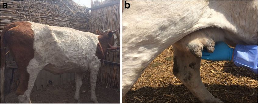
Makash. The animals showed depression, inappetence, fever
of 40 °C, nodule formation on the skin in various parts of the Collection and preparation of samples
body ranging in size from 0.5 to 3 cm, serous discharge from
the eyes, and increased salivation (Fig. 1). Reports of an un- Preparation of samples was carried out according to the pro-
known disease in the rural district continued. As of July 7, cedure described in chapter 1.1 Specimen collection, submis-
2016, clinical signs of the unknown disease were detected in sion and preparation, OIE Terrestrial Manual 2018.
78 cattle from the Makash rural district, and by July 21, 2016,
when the disease was notified to the OIE, 459 cattle had fallen Preparing arthropods for research
ill and 34 died (disease prevalence was 12.9% and mortality
0.96%). Ticks (Dermacentor marginatus and Hyalomma asiaticum),
In the Kurmangazinsky district, all the livestock is privately horseflies (Tabanus bromius), and flies (Stomoxys calcitrans)
owned by citizens who keep animals in 7 livestock enterprises collected in the field were transported to the laboratory and
and several peasant farms. The farm and pasture of peasant then were washed one time in ethanol and two times in sterileTrop Anim Health Prod (2021) 53:166 Page 3 of 7 166
Fig. 1 Clinical signs of lumpy
skin disease
phosphate-buffered saline (PBS) with (penicillin 500 IU/ml, The flasks were checked daily during 7–10 days for cyto-
streptomycin 1 mg/ml, mycostatin 100 IU/ml). Each insect pathic effects (CPE). If no CPE was observed by day 10, the
was transferred to a small chilled porcelain mortar and ground. culture was frozen three times, and the clarified supernatant
To prepare the suspension, 0.3 ml of PBS solution with anti- was inoculated into fresh LT or MDBK culture. The specific-
biotics (penicillin and streptomycin, 100–200 IU/ml) was ity of the CPE was confirmed by PCR.
added. The suspension was frozen and thawed three times,
and then clarified by centrifugation at 600g for 10 min. The DNA extraction
supernatant was used for the study. Pools were prepared from
several individuals of the same arthropod species for virus DNA was extracted from supernatant, blood, and culture sus-
isolation. pension using the “DNeasy® Blood & Tissue Kit (250)”,
QIAGEN, following the manufacturer’s protocol.
Virus isolation in cell culture PCR implementation
Virus isolation was performed as described in chapter 1.2 PCR was performed to confirm the presence of LSDV specific
Virus isolation in cell culture, OIE Terrestrial Manual 2018. nucleic acid using a pair of primers: forward primer, 5′-
Primary lamb testicle (LT) cell culture and MDBK cell line TTTCCTGATTTTTCTTACTAT-3′ and reverse primer, 5′-
were used. One ml of clarified supernatant or blood was inoc- AAATTATATACG TAAATAAC-3′ (El-Nahas et al. 2011).
ulated to a confluent monolayer in a 25 cm2 culture flask at 37 The reaction mixture contained 25 μL 2× AmpliTaq Gold
°C and allowed to adsorb for 1 h. The culture was washed with (Applied Biosystems) buffers, 1 μL each of 20-pM primers,
warm PBS and filled with 10 ml of antibiotic-containing 1 μL of DNA, and up to 50 μL of water. Amplification modes
DMEM medium (penicillin 100 IU/ ml, streptomycin 0.1 were as follows: initial denaturation at 95 °С for 5 min, dena-
mg/ml) and 2% fetal calf serum. turation at 94 °С for 45 s, annealing at 56–60 °С for 45 s,
Table 1 The results of PCR studies and virus isolation in LT cell culture
No. Sample PCR Virus isolation
tested/
positive 1 passage Time of 2 passage Time of 3 passage Time of
tested/ appearance tested/ appearance tested/ appearance
isolated of CPE (d.p.i.) isolated of CPE (d.p.i.) isolated of CPE (d.p.i.)
1 Lymph node 2/2 2/2 №1 (7) 2/2 (3) 2/2 (2)
№2 (8)
2 Spleen 2/2 2/2 (8) 2/2 (3) 2/2 (2)
3 Lungs 2/2 2/2 (7) 2/2 (3) 2/2 (2)
4 Nodules 3/3 3/3 (7) 3/3 (2) 3/3 (2)
5 Blood of sick cattle 5/5 Н.и. – Н.и. – Н.и. –
6 Ticks Dermacentor marginatus 4/4 1/0 – 1/1 №1 (6) 1/1 №1 (3)
7 Ticks Hyalomma asiaticum (Weiss, 1968) 9/9 2/0 – 2/1 №1 (5) 2/2 №1 (2)№2 (4)
8 Horseflies Tabanus bromius 21/3 1/0 – 1/1 №1 (6) 1/1 №1 (3)
9 Flies Stomoxys spp. 2/1 1/0 – 1/1 (7) 1/1 (3)166 Page 4 of 7 Trop Anim Health Prod (2021) 53:166
replication at 72 °С for 2 min, 35 cycles, post-replication at 72 marginatus and Hyalomma asiaticum), in three specimens
°С for 10 min. The size of the resulted amplificons was in line (14.29%) from horseflies (Tabanus bromius), and in one of
with the expected size of the PCR product (192 bp). The the specimens from two flies (Stomoxys calcitrans) (Table 1).
museum strain “Cattle/1986” deposited into the Microbial Positive specimens were used to infect the primary
Collection of the RIBSP served as a positive control. trypsinized lamb testicle cell cultures (LT) and the continuous
A second PCR was carried out on all positive samples to bovine kidney cell line (MDBK). Both infected cell cultures
amplify the GPCR gene for phylogenetic analysis. This was demonstrated the presence of the cytopathogenic agent.
done using primers designed by Le Goff et al. 2009 (Le Goff A cytopathic effect was detected at the first passage level in
et al. 2009). LT cell culture after infection with a suspension from all or-
The DNA amplification of the GPCR gene was performed gans (LN, spleen, lungs, nodules) taken from dead animals.
in a 25 μL 2× AmpliTaq Gold buffers, 1 μL each of 10-pM CPE in cell culture infected with a suspension of
primers, 1 μL of DNA, and up to 50 μL of water. The PCR Dermacentor marginatus, horseflies, and flies (Stomoxys
amplification of the GPCR gene involved an initial denatur- spp.) was observed from the 2 passage level after 6–7 days
ation at 96 °C for 5 min followed by 35 cycles of final dena- of cultivation. During infection, the same cell culture with
turation at 95 °C for 30 s, annealing at 50 °C for the 30 s, and suspensions of Hyalomma asiaticum ticks, only one sample
extension at 72 °C for 30 s as previously described. of two used, had a cytopathic effect in the second passage, and
the second sample in the third passage. The cytopathogenic
Sequencing effect of the virus in the first passage level usually appeared
within 7 to 9 days after infection. In the subsequent passages,
Sequencing was conducted by dideoxysequencing with the these terms were reduced, and at the third passage level, CPE
use of chain-terminating dideoxynucleotides (Sanger tech- was noted for 2–4 days. Distinct CPE with 75–90% affection
nique) in a 16-channel sequencer Genetic Analyzer 3130 xl, of the cellular layer was demonstrated in these cultures on the
Applied Biosystems (USA). POP-7 was used as a polymer for third passage level on the 8th day of cultivation.
capillaries. The terminating DNA products were generated by Suspensions of the internal organs taken from animals post
the method of cyclic sequencing. mortem were used for infecting the MDBK cell culture. The
The evolutionary history was inferred using the neighbor- cytopathic effect of the virus in this cell culture was detected
joining method (Saitou and Nei 1987). The percentage of in the second passage level after 6–7 days of incubation. In the
replicate trees in which the associated taxa clustered together third passage, the timing of the onset of CPE was reduced and
in the bootstrap test (1000 replicates) are shown next to the noted on the third day with the defeat of 70–80% of the mono-
branches (Felsenstein 1985). The tree is drawn to scale, with layer cells.
branch lengths in the same units as those of the evolutionary The virus accumulated in these cultures (LT, MDBK) on the
distances used to infer the phylogenetic tree. The evolutionary third passage level in titers within 5.5–5.75 log 10 TCID50/cm3.
distances were computed using the Tamura-Nei method The specificity of the isolated viruses was confirmed in PCR
(Tamura and Nei 1993) and are in the units of the number of and by electron microscopy. In all specimens, an amplification
base substitutions per site. Evolutionary analyses were con- product sized 192 bp typical for the LSDV was generated. The
ducted in MEGA7 (Kumar et al. 2016). The analysis involved study of the morphology and fine structure of the negatively
32 nucleotide sequences. stained preparations showed viral particles typical for LSDV.
The size of the virions varied within 300 × 370 nm, while the
Electron microscopy bulk mass of particles (90%) was sized 300 × 350 nm.
A virus isolated from the skin nodules of a dead animal was
To study the morphology of the LSDV the specimens of the selected for archiving “Nodulares/Dermatitis/Atyrau-2016”
pathology material (skin, lumps) from cattle and insects were and deposited into the Microbial Collection of the RGE
examined. LSDV preparations were assayed in the electron RIBSP, and later this same virus was used to produce an
microscope (JEM 100 B) with negative contrasting by 4% attenuated candidate vaccine strain.
solution of phosphate-tungstic acid, pH 6.8. Sequencing of the GPCR gene allowed us to identify this
virus as a lumpy skin disease virus.
Results Disease control
Biological samples from sick and dead animals, ticks and After the first cases of the disease, in the Kurmangazy region,
insects in the disease locus were assayed for LSDV. The the veterinary service introduced restrictive measures to localize
capripoxvirus DNA was detected by PCR in all tested samples and eliminate the disease. The movement of all species of do-
of organs from dead animals and ticks (Dermacentor mestic animals was prohibited, and a daily farm visit was carriedTrop Anim Health Prod (2021) 53:166 Page 5 of 7 166
out in order to identify animals with clinical signs of lumpy skin 2017). Until this time, LSD was considered exotic to
disease. After laboratory confirmation of the disease, the affect- Kazakhstan and measures were not taken to prevent or diag-
ed area (Kurmangazy region) was quarantined. All 459 animals nose the disease routinely. Through prompt action, the disease
with clinical signs were culled and the owners were compensat- was apparently eliminated through stamping out policy in the
ed at the market value of the animal. Disinfection of affected epidemic focus in Kazakhstan, and a strategy was developed
sites was completed including 28.116 m2 of land. The remaining to prevent further incursions from infected countries. The con-
live animals were treated with repellents. trol strategy is based on vaccination of the entire susceptible
After confirming the diagnosis and reporting the disease to livestock in Kazakhstan using lumpivax® vaccine
OIE, all the cattle of the Atyrau Province were vaccinated, as manufactured by Kenya Veterinary Vaccines Production
well as animals in the risk zone (threatened zone) of the West Institute (KEVEVAPI).
Kazakhstan and Aktobe Province. Vaccination was carried out The peak activity of blood-sucking insects and ticks in
using the lumpivax® vaccine manufactured by the Kenya Kazakhstan falls in April and October (Faulde n.d.). The
Veterinary Vaccine Manufacturing Institute (KEVEVAPI). emergence of the disease was coincident with a peak of vector
activity, and introduction of LSDV on to the territory of
Kazakhstan was likely either through the movement of infect-
Discussion ed livestock and subsequent transmission of the virus by
blood-sucking insects, as reported elsewhere, or through
The lumpy skin disease virus is considered a serious pathogen movements of vectors, although the latter is less likely as
of transboundary significance (Saegerman et al. 2018). Since vector movements are over relatively small distances, flies
about 2015, the disease has spread from African countries and mosquitos mostly only a few hundred meters unless wind
through the Near East into Europe, Azerbaijan, and Russia, assisted [32, 36]. The transmission pathways and epidemiol-
and caused significant economic losses (Zeynalova et al. ogy of LSD are as yet not fully understood. LSDV has been
2016; Salnikov et al. 2018; Sprygin et al. 2018a, b; detected in biting flies (Stomoxys and Musca spp), Aedes mos-
Saegerman et al. 2018). The disease was previously recorded quitoes, in ticks (Rhipicephalus appendiculatis),
only in tropical and sub-tropical countries. There remain many (Amblyomma hebraeum), and (Rhipicephalus (Boophilus)
gaps in knowledge on the establishment of LSD in temperate decolaratus) (Tuppurainen and Oura 2011; Chihota et al.
latitudes, and what role rising environmental temperatures, 2001; Last 2017; Issimov et al. 2020). Alexandrov
mediated through vectors, will play in LSD epidemiology. (Alexandrov 2017) reported possible mechanical transmission
The first cases of the disease were reported in Russia in of the LSD virus by Tabanus spodopterus females during an
2015 and this led to studies regarding potential arthropod car- outbreak in 2016 in Bulgaria (Alexandrov 2017). Later,
riers of LSDV. Virus DNA was found in 13 species of ixodid Sohier et al. (2019) experimentally demonstrated that
ticks (Gazimagomedov et al. 2017), and in 2017, DNA of the Haematopota spp. (horseflies) can mechanically transmit the
virus was detected in Musca domestica L. (Sprygin et al. LSD virus (Sohier et al. 2019). In our study, several arthropod
2018a, b). At the same time, the infection prevalence of some species including ixodid ticks (Dermacentor marginatus and
of the studied species of ticks reached up to 16.3% in the Hyalomma asiaticum), horseflies (Tabanus bromius), and
epidemic zone (Gazimagomedov et al. 2017). other biting flies (Stomoxys calcitrans) collected in the disease
Disease in Kazakhstan was first reported in early July 2016 focus were assayed as potential transmitters. All tick samples
in the Kurmangazy district of Atyrau Province. LSD diagnosis were positive, and a proportion of horse flies and Stomoxys
from this outbreak is confirmed by the laboratory assays flies (Table 1). The first LSDV isolate in cell culture was
(PCR) of the samples from sick and dead animals, as well as obtained from sampled horseflies (Tabanus bromius) collect-
confirmation of the presence of the virus in the arthropods ed during the outbreak of the disease. These results support the
collected in the epidemic focus. The etiological agent was studies of Sohier et al. (2019), which showed experimentally
isolated in the primary lamb testicle cell culture and in the that horseflies can mechanically transmit LSDV. The mechan-
continuous cell culture MDBK. Further sequencing of the ical transmission of LSDV by ixodid ticks has been shown by
gene GPCR and the phylogenetic analysis showed the close many researchers, but until now, there has been little informa-
genetic relationship of the isolated capripoxvirus with a group tion on the species of ticks and arthropods which might play a
of LSD viruses including those circulating in the areas of the role in the transmission of LSDV. Our studies have shown that
Russian Federation adjacent to Kazakhstan. Emergence of the all individuals sampled, of both species of ticks collected from
disease in the western regions of Kazakhstan from probable the region of the outbreak of the disease, were PCR positive
transboundary sources was confirmed. According to the infor- for LSDV, and the virus was isolated from the pool of ticks of
mation in Russia during May–June 2016, there were 163 LSD both species, through cell culture. The ticks and biting flies
suspected foci including three in the Astrakhan region border- recorded here are widespread throughout western Kazakhstan
ing the Atyrau Province of Kazakhstan (Rosselkhoznadzor and could become vectors of mechanical transmission of the166 Page 6 of 7 Trop Anim Health Prod (2021) 53:166
LSDV among domestic and wild animals if the virus is permission directly from the copyright holder. To view a copy of this
licence, visit http://creativecommons.org/licenses/by/4.0/.
established in the animal population. This opportunistic study
contributes to published data on the vector transmission of the
disease (Tuppurainen et al. 2011). The persistence of the virus
in the region will depend on the completion of life cycles of
LSD across a zone with an extreme climate and this possibility
References
needs to be monitored through appropriate surveillance Alexandrov, T. (2017). Lumpy skin disease situation in Europe.
annually. Preventive and control measures for an effective control-presenta-
Studies have shown that a novel cattle disease in the Atyrau tion. 11th Annual Meeting of EPIZONE, 19-21 September, Paris.
Province of Kazakhstan in 2015 was caused by infection with Barnard, B.J. (1997). Antibodies against some viruses of domestic ani-
mals in southern African wild animals. Onderstepoort Journal of
the lumpy skin disease virus. Virus was also detected among
Veterinary Research, 64, 95–110
arthropod horseflies, biting houseflies, and or ticks suggesting Chihota, C.M., Rennie, L.F., Kitching, R.P., Mellor, P.S., (2001).
the possibility of these species as vectors of LSD in this region. Mechanical transmission of lumpy skin disease virus by Aedes
aegypti (Diptera: Culicidae). Epidemiology and Infection, 126(2),
317–321. https://doi.org/10.1017/S0950268801005179
Code availability Not applicable. Chihota, C.M. Rennie, L.F., Kitching, R.P., Mellor, P.S., (2003).
Attempted mechanical transmission of lumpy skin disease virus by
biting insects. Medical and Veterinary Entomology, 17(3), 294–300.
Author contribution Orynbayev MB: data analysis, writing the manu- https://doi.org/10.1046/j.1365-2915.2003.00445.x
script. Nissanova RK: laboratory analysis. Khairullin BM: data analysis.
Coetzer, J.A.W. (2004). Lumpy skin disease. In J. A. W. Coetzer, & R. C.
Issimov A: data analysis. Zakarya K: research supervision. Sultankulova
Tustin (Eds.). Infectious diseases of livestock. 2nd ed. pp. 1268–
KT: data analysis, writing the manuscript. Kutumbetov LB: research
1276. Cape Town, South Africa: Oxford University Press.
supervision. Tulendibayev AB: laboratory analysis. Myrzakhmetova
Davies, F.G. (1982), Observations on the epidemiology of lumpy skin
BSh: laboratory analysis. Burashev YeD: laboratory analysis.
disease in Kenya. The Journal of hygiene, 88(1), 95–102. https://doi.
Nurabayev SS: fieldwork, laboratory analysis. Chervyakova OV: labora-
org/10.1017/s002217240006993x
tory analysis. Nakhanov AK: laboratory analysis. Kock RA: data analy-
sis, writing the manuscript. EFSA (European Food Safety Authority). (2017). Scientific report on
lumpy skin disease: I. Data collection and analysis. EFSA Journal,
15(4), 4773, 54 pp. doi: https://doi.org/10.2903/j.efsa.2017.4773
Funding The study was implemented under the financial support of the EFSA (European Food Safety Authority). (2015). Scientific opinion on
Ministry of Education and Science and the Ministry of Agriculture of the lumpy skin disease. EFSA Journal, 13(1), 3986.
Republic of Kazakhstan.
El-Nahas, E.M., El-Habbaa, A.S., El-bagoury, G.F., Radwan, M.E.I.
(2011). Isolation and Identification of Lumpy Skin Disease Virus
Data availability All data needed to evaluate the conclusions in the paper from Naturally Infected Buffaloes at Kaluobia, Egypt. Global
are present in the paper. Additional data related to this paper may be Veterinaria, 7, 234-237
requested from the authors. Faulde, M.K. (n.d.) Vector-borne Infectious Diseases in Kazakhstan
https://www.acq.osd.mil/eie/afpmb/docs/dveps/Kazakhstan.pdf
Declarations Felsenstein, J. (1985). Confidence limits on phylogenies: An approach
using the bootstrap. Evolution, 39, 783-791
Gazimagomedov, M., Kabardiev, S., Bittirov, A., Abdulmagomedov, S.,
Ethics approval This study was approved by the Committee for
Ustarov, R., Musaev, Z., Bittirova, A. (2017). Specific composition
Veterinary Control and Surveillance of the Ministry of Agriculture of
of Ixodidae ticks and their role in transmission of nodular dermatitis
the Republic of Kazakhstan. All ethics, field, and laboratory studies were
virus among cattle in the North Caucasus. The 18th Scientific
reviewed and approved by the appropriate committees of the Research
Conference Theory and Practice of the Struggle Against Parasite
Institute for Biological Safety Problems (RIBSP).
Animal Diseases- Compendium 18, 107–110
Greth, A., Gourreau, J.M., Vassart, M., Nguyen-Ba-Vy Wyyers, M.,
Consent to participate Not applicable. Lefevre, P.C. (1992). Capripoxvirus disease in an Arabian Oryx
(Oryx leucoryx) from Saudi Arabia. Journal of Wildlife Diseases,
Consent for publication Not applicable. 28, 295–300. https://doi.org/10.7589/0090-3558-28.2.295
Hamblin, C., Anderson, E.C., Jago, M., Mlengeya, T., Hipji, K. (1990),
Conflict of interest The authors declare no competing interests. Antibodies to some pathogenic agents in free-living wild species in
Tanzania. Epidemiology and Infection, 105(3), 585-94. doi: https://
Open Access This article is licensed under a Creative Commons doi.org/10.1017/s0950268800048226.
Attribution 4.0 International License, which permits use, sharing, adap- Hedger, R.S. and Hamblin, C. (1983). Neutralising antibodies to lumpy
tation, distribution and reproduction in any medium or format, as long as skin disease virus in African wildlife. Comparative Immunology,
you give appropriate credit to the original author(s) and the source, pro- Microbiology and Infectious Diseases, 6, 209–213. https://doi.org/
vide a link to the Creative Commons licence, and indicate if changes were 10.1016/0147-9571(83)90012-7
made. The images or other third party material in this article are included Issimov, A., Kutumbetov, L., Orynbayev, M., Khairullin, B.,
in the article's Creative Commons licence, unless indicated otherwise in a Sultankulova, K., Myrzakhmetova, B., White, P.J. (2020).
credit line to the material. If material is not included in the article's Mechanical transmission of lumpy skin disease virus by stomoxys
Creative Commons licence and your intended use is not permitted by spp (stomoxys calsitrans, stomoxys sitiens, stomoxys indica), dip-
statutory regulation or exceeds the permitted use, you will need to obtain tera: muscidae. Animals, 10, 477; doi:https://doi.org/10.3390/
ani10030477Trop Anim Health Prod (2021) 53:166 Page 7 of 7 166
Kumar, S., Stecher, G. and Tamura, K. (2016). MEGA7: Molecular Sprygin, A., Pestova, Y., Prutnikov, P., Kononov, A. (2018b). Detection
Evolutionary Genetics Analysis version 7.0 for bigger datasets. of vaccine-like lumpy skin disease virus in cattle and Musca
Molecular Biology and Evolution, 33, 1870-1874. domestica L. flies in an outbreak of lumpy skin disease in Russia
Last, R.D. (2017). Lumpy Skin Disease of Springbok. CPD Articles, in 2017. Transboundary and Emerging Diseases, 00:1–8. https://
Hooo-Hooo, Vol 11 No 4, doi.org/10.1111/tbed.12897
Le Goff, С., Lamien, C.E., Fakhfakh, E., Chadeyras, A., Abu-Adulugba, Tamura, K. and Nei, M. (1993). Estimation of the number of nucleotide
E., Libeau, G., Tuppurainen, E., Wallace, D.B., Adam, T., Silber, substitutions in the control region of mitochondrial DNA in humans
R., Gulyaz, V., Madani, H., Caufour, P., Hammami, S., Diallo, A., and chimpanzees. Molecular Biology and Evolution, 10, 512-526
Albina, E. (2009). Capripoxvirus G-protein-coupled chemokine re- Tulman, E.R., Afonso, C.L., Lu, Z., Kutish, G.F., Rock, D.L. (2001).
ceptor: A host-range gene suitable for virus animal origin discrimi- Genome of lumpy skin disease virus. Journal of Virology, p.
nation. Journal of General Virology, 90, 1967–77 7122–7130 DOI: https://doi.org/10.1128/JVI.75.15.7122-7130.
Macdonald, R.A.S. (1931). Pseudo-Urticaria of Cattle. Northern 2001
Rhodesian Department of Health Annual Report. 20–21 Tuppurainen, E. and Oura, C. (2011). Review: Lumpy Skin Disease: An
Mihok, S., Kang’ethe E.K., Kamau, G.K. (1995). Trials of Traps and Emerging Threat to Europe, the Middle East and Asia.
Attractants for Stomoxys spp. (Diptera: Muscidae). Journal of Transboundary and Emerging Diseases, 59, 40-8. https://doi.org/
Medical Entomology, 32(3), 283–289. doi:https://doi.org/10.1093/ 10.1111/j.1865-1682.2011.01242.x.
jmedent/32.3.283 Tuppurainen, E.S.M., Stoltsz, W.H., Troskie, M., Wallace, D.B., Oura,
Prozesky, L., Barnard, B.J.H. (1982). A study of the pathology of lumpy C.A.L., Mellor, P.S., Venter, E.H. (2011). A Potential Role for
skin disease in cattle. Onderstepoort Journal of Veterinary Ixodid (Hard) Tick Vectors in the Transmission of Lumpy Skin
Research,. 49, 167-75. Disease Virus in Cattle. Transboundary and Emerging Diseases,
Rosselkhoznadzor. LSD-infected regions of RF in 2017. http://fsvps.ru 58(2), 93–104. https://doi.org/10.1111/j.1865-1682.2010.01184.x
Saegerman, C., Bertagnoli, S., Meyer, G., Ganière, J.P., Caufour, P., De Usadov, T.R., Morgunov, Yu.P., Zhivoderov, S.P., Balysheva, V.I.,
Clercq, K., Casal, J. (2018). Risk of introduction of lumpy skin Pivov A, E. Yu, Koltsov, A. Yu, Yanzhieva, D.V., Sukher, M.M.,
disease in France by the import of vectors in animal trucks. PLoS Lunitsyn, A.V., Salnikov, N.I. (2018). Lumpy skin disease virus,
ONE. 13(6): e0198506. https://doi.org/10.1371/journal.pone. isolated in 2015 in Russia from cattle, is pathogenic for sheep at
0198506 experimental infection. Sel’skokhozyaistvennaya biologiya
Saitou, N. and Nei, M. (1987). The neighbor-joining method: A new [Agricultural Biology], 53, 2, 438-446
method for reconstructing phylogenetic trees. Molecular Biology Von Backstrom, U. (1945). Ngamiland cattle disease: preliminary report
and Evolution, 4, 406-425 on a new disease, the etiological agent being probably of an infec-
Salnikov, N., Usadov, T., Kolcov, A., Zhivoderov, S., Morgunov, Y., tious nature. Journal of South African Veterinary Medical
Gerasimov, V., Gogin, A., Titov, I., Yurkov, S., Malogolovkin, Association, 16, 29–35
A., Kolbasov, D., Lunitsyn, A. (2018). Identification and character-
Wainwright, S., El Idrissi, A., Mattioli, R., Tibbo, M., Njeumi, F.,
ization of lumpy skin disease virus isolated from cattle in the
Raizman, E. (2013). Emergence of lumpy skin disease in the
Republic of North Ossetia Alania in 2015. Transboundary and
Eastern Mediterranean Basin countries. FAO Empres Watch, 29,
Emerging Diseases, 00:1–5. https://doi.org/10.1111/tbed.12818
1–6
Shalaby, M.A., El-Deeb, A., El-Tholoth, M., Hoffmann, D., Czerny,
World Animal Health Information Database (WAHIS Interface)
C.P., Hufert, F.T., Abd El Wahed, A. (2016). Recombinase poly-
(2021) Lumpy skin disease, Kazakhstan, – Version 1. https://
merase amplification assay for rapid detection of lumpy skin disease
www.oie.int/wahis_2/public/wahid.php/Reviewreport/Review/
virus. BMC Veterinary Research, 12(1), 244. https://doi.org/10.
viewsummary?reportid=20520. Accessed 15 Feb 2021
1186/s12917-016-0875-5
Sohier, C., Haegeman, A., Mostin, L., De Leeuw, I., VanCampe, W., Young, E., Basson, P.A., Weiss, K.E. (1970). Experimental infection of
DeVleeschauwer, A., Tuppurainen, E.S., van den Berg, T., De game animals with lumpy skin disease virus (prototype strain
Regge, N., DeClercq, K. (2019). Experimental evidence of mechan- Neethling). Onderstepoort Journal of Veterinary Research, 37(2),
ical lumpy skin disease virus transmission by Stomoxys calcitrans 79-87.
biting fies and Haematopota spp. Horseflies. Scientific Reports, 9, Zeynalova, S., Asadov, K., Guliyev, F., Vatani, M., Aliyev, V. (2016).
20076 https://doi.org/10.1038/s41598-019-56605-6 Epizootology and molecular diagnosis of lumpy skin disease among
Sprygin, A., Artyuchova, E., Babin, Y., Prutnikov, P., Kostrova, E., livestock in Azerbaijan. Frontiers in Microbiology, 7, 1022. https://
Byadovskaya, O., Kononov, A. (2018a). Epidemiological character- doi.org/10.3389/fmicb.2016.01022
ization of lumpy skin disease outbreaks in Russia in 2016.
Transboundary and Emerging Diseases, 00:1–8. https://doi.org/ Publisher’s note Springer Nature remains neutral with regard to jurisdic-
10.1111/tbed.12889 tional claims in published maps and institutional affiliations.You can also read