Macrophage Activation by a Substituted Pyrimido 5,4-b Indole Increases Anti-Cancer Activity - bioRxiv
←
→
Page content transcription
If your browser does not render page correctly, please read the page content below
bioRxiv preprint first posted online Mar. 18, 2019; doi: http://dx.doi.org/10.1101/581710. The copyright holder for this preprint
     (which was not peer-reviewed) is the author/funder, who has granted bioRxiv a license to display the preprint in perpetuity.
                                        All rights reserved. No reuse allowed without permission.

Macrophage Activation by a Substituted Pyrimido[5,4-b]Indole
Increases Anti-Cancer Activity
Joseph Hardie,a Javier A. Mas-Rosario,b Siyoung Ha,a Erik M. Rizzo,c and Michelle E. Farkasa,b*

a
    Department of Chemistry, University of Massachusetts Amherst, 710 North Pleasant Street, Amherst, Massachusetts, 01003,

USA.
b
    Molecular and Cellular Biology Graduate Program, University of Massachusetts, Amherst, MA.
c
    Department of Biochemistry and Molecular Biology, University of Massachusetts, Amherst, MA

ABSTRACT: Immunotherapy has become a promising new approach for cancer treatment due to the immune system’s ability
to remove tumors in a safe and specific manner. Many tumors express anti-inflammatory factors that deactivate the local
immune response or recruit peripheral macrophages into pro-tumor roles. Because of this, effective and specific ways of acti-
vating macrophages into anti-tumor phenotypes is highly desirable for immunotherapy purposes. Here, the use of a small
molecule TLR agonist as a macrophage activator for anti-cancer therapy is reported. This compound, referred to as PBI1,
demonstrated unique activation characteristics and expression patterns compared to treatment with LPS, through activation of
TLR4. Furthermore, PBI1 treatment resulted in anti-tumor immune behavior, enhancing macrophage phagocytic efficiency
five-fold versus non-treated macrophages. Additive effects were observed via use of a complementary strategy (anti-CD47
antibody), resulting in ~10-fold enhancement of phagocytosis, suggesting this small molecule approach could be used in con-
junction with other therapeutics. 1 2

KEYWORDS: inflammatory, immunotherapy, macrophage, adjuvant, cancer, phagocytosis

1. Introduction                                                                against many cancer types and can increase disease recurrence
                                                                               [1]. As a result, alternative strategies are sought after, such as
Cancer is one of the leading causes of death around the world,                 immunotherapy [2]. Cancer immunotherapy involves utilizing
and fighting it is a focus of research programs globally [1].                  the immune system to eliminate the disease, and is attractive on
However, most cancer therapy regimens continue to rely on                      account of the immune system’s specificity and biocompatibil-
non-specific chemotherapy and radiotherapy to eliminate tu-
mors, which have severe side effects. They are also ineffective

1ABBREVIATIONS:       CD47; cluster of differentiation 47, IL-4; interleukin 4, CS1; CCND3 subset 1, CSF1R, colony stimulating
factor receptor 1, MFEG8; Milk fat globule-EGF factor 8 protein, VEGF; Vascular endothelial growth factor, SEAP; Secreted em-
bryonic alkaline phosphatase, LPS; Lipopolysaccharide, IFN-γ; Interferon-γ
2
    *Address correspondence to farkas@chem.umass.edu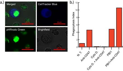
bioRxiv preprint first posted online Mar. 18, 2019; doi: http://dx.doi.org/10.1101/581710. The copyright holder for this preprint
  (which was not peer-reviewed) is the author/funder, who has granted bioRxiv a license to display the preprint in perpetuity.
                                     All rights reserved. No reuse allowed without permission.

ity compared to traditional cancer therapy. Several immuno-                 Because of these issues, there is a need to identify alternative
therapies have been approved clinically, and others have                    macrophage activators. Additionally, therapeutic candidates
reached the clinical trial stage [3].                                       should be amenable to chemical modifications and association
                                                                            with targeted delivery vehicles. To date, there are few immune-
While immunotherapy is alluring, developing immune-based                    modulating compounds approved clinically. One example is the
strategies is challenging. Normally, the immune system detects              TLR7 activator imiquimod, which is approved for the topical
and eliminates pre-oncogenic cells [4], however, cancer cells               treatment of genital warts and basal cell carcinoma [19]. How-
can generate cytokines and receptors for immune evasion and                 ever, in terms of general anti-cancer agents, systemically deliv-
reprogramming [5]. In this manner, tumorigenic cells are able               ered drugs are desirable to provide access to a range of tumor
to escape detection and disable pro-inflammatory behaviors. An              locations and facilitate immune cell recruitment. There have
example of the former is over-expression of “don’t eat me” sur-             been examples of anti-inflammatory antibody blockades uti-
face marker CD47 by cancer cells, preventing phagocytosis [6],              lized to reprogram macrophages for cancer therapy [7,20], but
while in the latter the tumor releases chemo-attractants and anti-          in terms of modifications, antibodies are more difficult to alter
inflammatory signals, such as IL-4, IL-10, CS1, CSF1R, and                  than small organic molecules, and are also linked to uncontrol-
MFG-E8, to reprogram immune cells to perform pro-tumor-                     lable immune-based toxicity [21].
igenic roles [4,7]. These include facilitation of angiogenesis,
epithelial to mesenchymal transition (EMT), and microenviron-               Here, we utilize a small molecule TLR4 activator to induce the
ment remodeling [8]. Once this point is reached, the likelihood             M1 phenotype and enhance anti-oncogenic properties in mac-
of patient survival decreases sharply [9].                                  rophages (Fig. 1). This molecule, a pyrimido[5,4-b]indole re-
                                                                            ferred to as PBI1, was previously identified among a series of
Because of the immune system’s role in cancer progression,                  compounds that activate TLR4 [22]. This molecule was shown
there is great interest in the re-education of immune elements              to bind to TLR4 and induce expression of various pro-inflam-
into anti-cancer entities. Polarization of macrophages into M1              matory cytokines in dendritic cells [22]. Preclinical studies have
(classically activated) phenotypes is important toward refocus-             shown that structurally similar molecules are effective immune
ing the immune system for eliminating cancer [10]. In this im-              adjuvants for influenza therapy [23], however, these com-
mune-stimulating phenotype, macrophages attack and phagocy-                 pounds have not been evaluated in terms of macrophage activa-
tose tumor cells. This facilitates a larger overall attack by the           tion and anti-cancer activity. Our hypothesis is that this small
immune system, resulting in tumor elimination [10]. Macro-                  molecule TLR4 agonist can activate and polarize macrophages
phages also generate reactive oxygen species (ROS) and present              into an anti-cancer phenotype nearly as well as naturally occur-
tumor antigens, which recruit T cells and B cells to the tumor              ring cytokines or existing adjuvants. We demonstrate that PBI1
site [11]. In contrast, immune-suppressing M2 (alternatively                upregulates pro-inflammatory genes in macrophages and in-
activated) phenotypes, or tumor-associated macrophages                      duces M1-associated phenotypic changes and cytokine produc-
(TAMs), contribute to tumor progression, joining the tumor                  tion. Macrophages treated with PBI1 demonstrate enhanced
mass and microenvironment [10]. TAMs release pro-tumor                      anti-tumor activity toward B-cell lymphoma cells as determined
growth factors, such as vascular endothelial growth factor                  by phagocytosis assays. We also show that treatment of M2-like
(VEGF), promote vascularization, remodel the microenviron-                  macrophages with PBI1 results in their re-education of macro-
ment, and silence the immune response [12,13,14]. Overall, re-              phages toward an M1-like phenotype.
programming macrophages into M1-like states and away from
M2/TAM phenotypes has great potential as an anti-cancer im-
munotherapeutic approach [15].

The polarization of macrophages toward M1 phenotypes is a
well-studied phenomenon, with known pathways identified.
Specifically, the interferon gamma (IFN-γ) receptor (IFNGR)
and the toll-like receptor (TLR) class are understood to activate
macrophages into pro-inflammatory roles [16]. The most com-
mon strategies for in vitro M1 macrophage polarization involve              Fig. 1 Illustration of pyrimido[5,4-b]indole (PBI1)-mediated
treatment with IFN-γ (ligand for IFNGR) and lipopolysaccha-                 macrophage activation. Following TLR4 stimulation, resulting
ride (LPS, a TLR4 agonist originating from bacterial cell walls)            M1 macrophages have enhanced levels of TNF-α and iNOS, in-
[16]. While these agonists are useful as research tools, both               creased ROS generation, and phagocytosis efficiency
IFN-γ and LPS have drawbacks that make them non-viable ther-
apeutically. IFN-γ is a small protein, difficult to consistently            2. Materials and Methods
modify and incorporate into delivery vehicles [17], while LPS
is a bacterial cell wall component consisting of a mixture of               All reagents were purchased from Thermo-Fisher Scientific ex-
structures and may be contaminated with other bacterial com-                cept where otherwise noted. All DMSO utilized was cell culture
ponents, resulting in off-target immune effects [18]. Systemic              grade (Sigma). Confocal microscopy images were obtained on
administration of these agents results in immune overstimula-               an Eclipse Ti-E microscope (Nikon, Tokyo, Japan) using a 63X
tion, leading to negative outcomes including septic shock, cyto-            or 20X objective at 25 ºC. Images were acquired and processed
kine storms, and death [18].                                                using NIS-Elements and ImageJ. Flow cytometry analysis was
                                                                            performed on a BD LSRFortessa 5L flow cytometer equipped
                                                                            with FACSDiva (BD Sciences, USA) at the Flow Cytometry
bioRxiv preprint first posted online Mar. 18, 2019; doi: http://dx.doi.org/10.1101/581710. The copyright holder for this preprint
(which was not peer-reviewed) is the author/funder, who has granted bioRxiv a license to display the preprint in perpetuity.
All rights reserved. No reuse allowed without permission.
Core Facility at the University of Massachusetts, Amherst. amplified at 53 ºC for 10 minutes and melted at 80 ºC for 10
RTPCR data was generated using a CFX Connect Real-Time minutes. The cDNA was frozen at -20 ºC and then used for RT-
PCR Detection System (Biorad, Hercules, CA). For assays re- PCR within 1 week. RNA and cDNA were quantified using a
quiring absorbance measurements, a SpectraMax M2 plate NanoDrop 2000 (ThermoFisher). RNA and cDNA contamina-
reader was used (Molecular Devices, San Jose, CA). tion, inhibition and integrity was assessed by analyzing the
A260/A280 ratio, where 1.8 for DNA and 2.0 for RNA was con-
2.1 Cell Culture sidered “pure.”
RAW 264.7 cells were purchased from American Type Culture 2.5 Quantitative RT-PCR
Collection (ATCC, Manassas, VA). Primary immortalized
macrophages were a gift from Prof. Susan Carpenter at the Uni- RT-PCR was performed on cDNA as prepared above using a
versity of California, Santa Cruz. Both types of cells were cul- CFX Connect real-time system (Biorad) with iTaq Universal
tured at 37 °C under a humidified atmosphere containing 5% SYBR Green Supermix (Biorad, Hercules, CA). All DNA pri-
CO2. Standard growth media consisted of high glucose Dulbec- mers were purchased from Integrated DNA Technologies
co's Modified Eagle Medium (DMEM) supplemented with 10% (Caralville, Iowa). The following primer sequences (as deter-
fetal bovine serum (FBS) and 1% antibiotics (100 µg/ml peni- mined by NCBI primer-BLAST) were used: β-actin (ACTB, ac-
cillin and 100 µg/ml streptomycin). Daudi cells were a gift from cession number: NM_007393, amplicon length: 86 pairs, exon
Prof. Vincent Rotello at the University of Massachusetts Am- 6) (forward) GATCAGCAAGCAGGAGTACGA, (reverse)
herst and were cultured in Roswell Park Memorial Institute me- AAAACGCAGCGCAGTAACAGT; iNOS (NOS2, accession
dia (RPMI 1640) supplemented with 10% fetal bovine serum number: NM_010927.4, amplicon length: 127 pairs, exon 2,3,
and 1% antibiotics (100 µg/ml penicillin and 100 µg/ml strep- splice variants: 1-3) (forward)
tomycin). Under the above culture conditions, the cells were GTTCTCAGCCCAACAATACAAGA, (reverse)
sub-cultured approximately once every four days and only cells GTGGACGGGTCGATGTCAC; TNF-α (TNF, accession
between passages 7 and 15 were used for all the experiments. number: NM_013693.3, amplicon length: 145 pairs, exon 3,4)
(forward) CCTGTAGCCCACGTCGTAG, (reverse)
2.2 Synthesis GGGAGTCAAGGTACAACCC. 200 nM of each primer were
mixed with 1 µl (100 ng) of cDNA, 10 µL SYBR green super-
The synthesis of PBI1 was largely performed as previously de- mix and H2O to a final volume of 20 µL. Analyses were per-
scribed [22]. A complete description of procedures and analyt- formed as follows: the samples were first activated at 50 °C for
ical data may be found in the supporting information. 2 min, then 95 °C for 2 min. Then denaturing occurred at 95 °C
for 30 s followed by annealing at 57 °C; the denature/anneal
2.3 RT-PCR Preparation process was repeated over 40 cycles. Relative gene expression
was determined by comparing the Ct value of the gene of inter-
RAW 264.7 cells were plated in 24 well plates at a density of est to that of the β-actin housekeeping gene, by the 2ΔΔCt method
100,000 cells/well. PBI1-treated cells were dosed with 20 [24]. Three biological replicates were performed for each treat-
µg/mL compound for 24 hours. M1 treatment group cells were ment condition and three technical replicates were used for each
treated with 50 ng/mL IFN-γ for 24 hours after which LPS was biological replicate. There was no amplification for the NTC.
added to a final concentration of 50 ng/mL for an additional 24 Data was analyzed using the CFX Manager 3.1 software. CQ
hours. M2 group cells were treated with 50 ng/mL IL-4 for 48 values were generated by using the point at which the sample
hours. For the M2+PBI1 group, the cells were treated with 50 fluorescence value exceeded the software’s default threshold
ng/mL IL-4 for 24 hours. 24 hours after IL-4 dosing, the cells value. Each sample was normalized to the untreated control. β-
were treated with 20 µg/mL PBI1 for 24 hours. Each experi- actin was used as a reference gene since it is commonly used
ment included three biological replicates per treatment condi- and none of the treatments were expected to affect its expres-
tion. 20 µg/mL of PBI1 was chosen for this assay and several sion.
others as PBI1 demonstrated no toxicity at this concentration
(Figure S10). Following treatments, RNA was extracted follow- 2.6 Phagocytosis Assay with Flow Cytometry
ing the procedure below.
RAW 264.7 and Daudi cells were separately plated in 24 well
2.4 RNA Extraction and cDNA Conversion plates at a density of 1 x 10⁵ cells/well each. The same day, des-
ignated RAW 264.7 wells were treated with PBI1 to reach a
Approximately 1.5 µg RNA was directly harvested from cells final concentration of 4.5 µg/mL in 0.5% DMSO. A slightly
using the PureLink RNA Mini Kit (Ambion) following the lower concentration was utilized in this experiment to demon-
manufacturer's instructions. SuperScript IV Reverse Transcrip- strate that profound changes in cell phenotype are possible at
tase, RNaseOut, 10 mM dNTPs, and 50 μM Random Hexamers low concentrations of PBI1. 24 h following treatment, RAW
were used for the conversion of approximately 150 ng of RNA cells were trypsinized using TrypLE (ThermoFisher), washed
to cDNA (ThermoFisher, Pittsburgh, PA), also following the twice in PBS, and labelled with 10 µg/mL PE-F4/80 antibody
manufacturer's instructions with a sample volume of 20 µL. (BD Biosciences, cat. No: 565410, Clone:T45-2342) for 30 min
Briefly, primers were annealed to RNA at 65 ºC for 5 minutes. at 4 ºC. All RAW cells were resuspended in culture media and
Then the annealed RNA was combined with the reaction mix 10 µM cytochalasin D was added to the designated samples.
(containing 500 units of reverse transcriptase per sample) and Simultaneously, Daudi cells were removed from the 24 well
plate, washed twice with LCIS (ThermoFisher) and labelledbioRxiv preprint first posted online Mar. 18, 2019; doi: http://dx.doi.org/10.1101/581710. The copyright holder for this preprint
(which was not peer-reviewed) is the author/funder, who has granted bioRxiv a license to display the preprint in perpetuity.
All rights reserved. No reuse allowed without permission.
with pHRhodo Green according to the manufacturer’s instruc- imaged using an Eclipse Ti-E microscope at 63x magnification.
tions (ThermoFisher). These cells were washed with PBS and The final ratio of RAW to Daudi cells was approximately 2:1.
then labelled with 10 µg/mL anti-CD47 antibody (Bio Xcell,
cat. No: BE0270, Clone: MIAP301) for 30 min at 4 ºC. All of 3. Results and Discussion
the Daudi cells (approximately 2 x 10⁵) were resuspended in
culture media and combined with the RAW 264.7 cells for 2 h Following synthesis and characterization of PBI1 (Fig. S1-S8),
at 37 ºC. The approximate ratio of RAW:Daudi was 2:1. The RAW 264.7 murine macrophages were dosed with the com-
samples were then washed with PBS, resuspended in FACS pound, and changes in morphology and gene expression were
buffer (1% FBS in PBS) and transferred to flow cytometry assessed using confocal microscopy and RT-PCR, respectively
tubes. The samples were analyzed on a LSRFortessa 5L flow (Fig. 2). Confocal microscopy revealed significant changes in
cytometer (BD Biosciences) using 488 nm and 561 nm lasers, macrophage morphology after 24 h. Treated macrophages ac-
counting 10000 events, at the University of Massachusetts Am- quired a phenotype that is associated with M1-polarized macro-
herst Flow Cytometry Facility. One sample per treatment group phages (Fig. 2a): the cells became flatter and produced longer
was evaluated. Phagocytotic index was calculated using the fol- pseudopodia [16] compared to non-treated cells. RT-PCR anal-
lowing equation: [F4/80+pHRhodoGreen+ ysis of the expression of two M1-related markers, tumor necro-
+ -
events]/[F4/80 pHRhodoGreen events], normalized to the un- sis factor-α (TNF-α) and inducible nitric oxide synthase (iNOS)
treated control group. [11], revealed that PBI1-treated macrophages express signifi-
cantly higher levels of both genes compared to non-treated
2.7 Griess Assay cells, in a similar trend as IFN-γ/LPS treatment (Fig. 2b). This
effect was also observed in an immortalized primary macro-
RAW 264.7 cells were plated in 24 well plates at a density of phage cell line (Fig. S9). Furthermore, PBI1 treatment was also
1.5 x 10⁵ cells/well 24 h prior to the experiment. On the follow- able to ‘re-educate’ macrophages that had been polarized to-
ing day, the culture media was removed and replaced with se- ward the anti-inflammatory M2 phenotype, resulting in en-
rum free Opti-Mem media for 2 h. Cells designated for TLR4 hanced levels of iNOS and TNF-α. Alamar blue assays indi-
inhibition were pre-treated with 7.2 µg/mL TAK242 (Cayman cated that no significant toxicity occurred as a result of com-
Chemical) 1 h before additional treatment. Then, media was re- pound treatment (Fig. S10).
placed again with 250 µL phenol red-free DMEM culture media
containing either 100 ng/µL LPS or 20 µL/mL PBI1 and cells
were incubated for an additional 48 h. Cell supernatant was col-
lected from wells and centrifuged at 5000 rpm for 5 min. Then,
Griess reagent (Thermofisher) was prepared according to the
manufacturer’s instructions and 60 µl Griess reagent was com-
bined with 60 µL cell supernatant in a clear 96 well plate. After
15 min in the dark, absorbance was read using a SpectraMax
M2 plate reader (Molecular Devices, Sunnyvale, CA) at 548
nm. Three biological replicates per treatment condition were
used. Actual NO2- concentrations were determined by compar-
ing absorbance values to a standard curve generated using pure
NaNO2- solutions.
2.8 Confocal Microscopy
For acquisition of representative cell morphology images,
RAW 264.7 cells were plated in a 4 chamber Lab-Tek II cham-
bered cover-glass system (Nunc, NY) at a density of 5 x
104/well and allowed to adhere overnight. Cells were then po- Fig. 2 Effects of PBI1 treatment on cell morphology and M1
larized with 20 µg/mL PBI1 in 0.2% DMSO for 48 h. After po- marker expression. a) Confocal microscopy images (20x) de-
larization, cells were imaged using an Eclipse Ti-E microscope picting changes in cell morphology following treatment with 20
at 20x magnification. M1 cell images were acquired similarly, µg/ml PBI1. PBI1-treated macrophages resembled those polar-
using polarization conditions as outlined in the RT-PCR Prepa- ized to an M1 phenotype, with a “flatter” appearance and more
ration section above. pseudopodia, in comparison with non-treated cells. b) RT-PCR
analysis of TNF-α and iNOS expression in PBI1 treated cells
For acquisition of representative phagocytosis images, 5 x 10⁴ revealed significantly greater expression of both relative to non-
RAW 264.7 cells were plated in a 4 chamber Lab-Tek II cham- treated controls. M2+PBI1 refers to cells polarized to the M2
bered cover-glass system. Designated wells were treated with phenotype and subsequently treated with PBI1. Unpaired, two-
PBI1 to a final concentration of 4.5 µg/mL in 0.4% DMSO for tailed student T-tests with equal variance were used to deter-
24 h. The RAW cells were labeled with Cell Tracker Blue ac- mine significance; *P ≤ 0.05, **P ≤ 0.005, ***P ≤ 0.0005.
cording to the manufacturer’s instructions. 1 x 10⁵ Daudi cells
were counted, labeled with phRhodo Green and added to the Generation and release of reactive oxygen and nitrogen species
wells containing RAW cells. After 2 h incubation, wells were (ROS and RNS, respectively) are important processes of M1
macrophage anti-tumor and pathogen responses [25]. RNS arebioRxiv preprint first posted online Mar. 18, 2019; doi: http://dx.doi.org/10.1101/581710. The copyright holder for this preprint
(which was not peer-reviewed) is the author/funder, who has granted bioRxiv a license to display the preprint in perpetuity.
All rights reserved. No reuse allowed without permission.
generally derived from nitric oxide (NO). To evaluate relative
NO production by PBI1-treated cells, a Griess assay was per-
formed [26]. This method indirectly measures NO via evalua-
tion of nitrite (NO2-), one of its two primary stable and non-
volatile breakdown products. The determined NO2- concentra-
tions show that 20 µg/mL PBI1 effectively induced production
of NO2-, achieving significantly higher levels than non-treated
samples, and within 2-fold of (100 µg/mL) LPS-treated cells
(Fig. 3). Additionally, pretreatment with the TLR4 specific in-
hibitor TAK242 completely eliminated the induction of NO by
PBI1 (Fig. 3) [27]. This result demonstrates that PBI1 is an ag-
onist of TLR4 and causes the activation of macrophages
through this pathway.
Fig. 4 Effects of PBI1 on phagocytosis of Daudi (B-cell lym-
phoma) cells. a) Representative confocal microscopy images il-
lustrating RAW 264.7 macrophage (CellTracker Blue) phago-
cytosis of Daudi cells (phRodo Green). b) Phagocytosis indices
generated from flow cytometry data of RAW 264.7 macro-
phages phagocytosing Daudi cells following various treat-
ments.
In summary, PBI1 was demonstrated to polarize macrophages
toward an anti-cancer phenotype. RAW 264.7 macrophages
treated with PBI1 adopted an “M1-like” pro-inflammatory mor-
phology, as cells became flattened and produced extended pseu-
dopodia. RT-PCR results also corroborated this phenotype via
an increase in the expression of M1 inflammatory genes. PBI1
treatment increased the expression of TNF-α and iNOS approx-
imately 2.5 and 500-fold, respectively. Dosing of PBI1 also re-
sulted in the re-education of M2-polarized macrophages,
whereupon following treatment, cells expressed higher levels of
Fig. 3 Supernatant NO2- levels following LPS (100 µg/mL) or inflammatory cytokines. This is particularly relevant for the
PBI1 (20 µg/mL) treatment as measured by Griess assay. Pre- conversion of tumor-associated macrophages into tumor-killing
treatment with TAK242 inhibited TLR4 activity prior to PBI1 macrophages within cancer microenvironments.
addition (PBI1+TAK242). Unpaired, two-tailed student T-tests
with equal variance were used to determine significance; ***P We also evaluated the mechanism by which PBI1 promoted
≤ 0.005, *****P ≤ 0.00005 macrophage activation. The most common pathways for mac-
rophage stimulation include the TLR family of receptors that
A key aspect of the macrophage antitumor response is the iden- recognize a wide variety of substrates [8]. To confirm that
tification and phagocytosis, or engulfment, of cancerous cells TLR4 is the target of PBI1 for activation, a competition exper-
[6]. To assess the phagocytic efficacy of PBI1 treated macro- iment was performed. Cells were treated with a highly specific
phage cells, a fluorescent flow cytometry assay was conducted TLR4 inhibitor, TAK242, and activation by PBI1 was deter-
using Daudi (B cell lymphoma) as the target cancer cell line mined by Griess assay. This experiment revealed that macro-
(Fig. 4, S11). RAW 264.7 cells treated with PBI1 had a nearly phages treated with either PBI1 or LPS released significant lev-
5-fold greater phagocytic efficiency versus non-treated macro- els of nitric oxide, another confirmation of the activation poten-
phages. This was increased further when macrophage PBI1 tial of PBI1. However, with pre-treatment of cells with
treatment was combined with antibody-mediated blocking of TAK242, PBI1 did not induce any detectable level of nitric ox-
Daudi cell CD47, the ‘don’t eat me’ signal involved in phago- ide, confirming that TLR4 is the target of PBI1 and is crucial to
cytosis inhibition signaling. Cytochalasin D, a potent phagocy- resulting macrophage activation.
tosis inhibitor, was used as a negative control [28].
Having evaluated the pro-inflammatory responses of macro-
phages to PBI1 treatment, the ability of these cells to subse-
quently phagocytose cancers cells was investigated. It has been
previously shown that activation of local macrophages into an
M1 inflammatory phenotype can result in significant anti-tumor
macrophage activity, which is of great interest for the genera-
tion of cancer immunotherapies [24]. One of the major anti-can-
cer macrophage mechanisms involves phagocytosis of cancerbioRxiv preprint first posted online Mar. 18, 2019; doi: http://dx.doi.org/10.1101/581710. The copyright holder for this preprint
(which was not peer-reviewed) is the author/funder, who has granted bioRxiv a license to display the preprint in perpetuity.
All rights reserved. No reuse allowed without permission.
cells. Inflammatory macrophages invade can tumor tissue, en- Declarations of Conflict of Interest: None
gulf resident tumor cells, and release immune signaling factors
that drive further immune responses against the tumor site [24].
A phagocytosis assay revealed that PBI1-treated macrophages ACKNOWLEDGMENT
engulfed targeted cancer cells with a 5-fold higher efficacy than This research was supported by a SEED grant from the University
untreated macrophages, revealing that inflammatory activation of Massachusetts Institute for Applied Life Sciences. JH was sup-
by PBI1 does in fact increase anti-tumor activity. It was also ported by the NIH (EB014277) and an NIH National Research Ser-
demonstrated that pre-treatment of the targeted cancer cells vice Award (T32 GM008515). JAM-R was supported by the
with a CD47 blocking antibody, which blocks the SIRPα-CD47 UMass Amherst NIH Postbaccalaureate Research Program
phagocytosis inhibitory pathway, further increases the efficacy (GM086264-08) and a Northeast Alliance for Graduate Education
of PBI1-induced phagocytosis. This effect occurred inde- and the Professoriate (NEAGEP) fellowship from the STEM Di-
pendently of PBI1-activation as well and could be used to in- versity Institute at UMass Amherst.
crease the anti-cancer efficacy of PBI1 treatment.
ABBREVIATIONS
4. Conclusion
CD47; cluster of differentiation 47, IL-4; interleukin 4, CS1;
CCND3 subset 1, CSF1R, colony stimulating factor receptor 1,
The immune system and cancer progression have a complex re- MFEG8; Milk fat globule-EGF factor 8 protein Milk fat globule-
lationship. While effective immune-based strategies are of in- EGF factor 8 protein, VEGF; Vascular endothelial growth facto
terest in enervating and eliminating cancer progression, their Vascular endothelial growth factor, SEAP; Secreted embryonic al-
development can be challenging due to varying macrophage- kaline phosphatase Secreted embryonic alkaline phosphatase
activating signals [29,30]. PBI1 has significant macrophage ac-
tivation capability. As a potential immune adjuvant for cancer REFERENCES
therapy, PBI1 is relatively non-toxic and effectively increases 1 DeSantis, C.; Ma, J.; Bryan, L.; Jemal, A. Breast cancer statistics
both phagocytic and oxidative burst mechanisms of the macro- 2013. Cancer J Clin 2014 64, 52–62. DOI: 10.3322/caac.21203.
phage anti-tumor response. While PBI1 has shown promise as 2 Blattman, JN.; Greenberg, P. D.; Cancer Immunotherapy: a treat-
a therapeutic on its own, it will likely show the greatest efficacy ment for the masses. Science 2004 305, 200–206. DOI:
if used in conjugation with drug delivery vehicles or targeting 10.1126/science.1100369.
elements. This is especially relevant in trying to avoid broad 3 Butterfield, L. H. Dendritic cells in cancer immunotherapy clini-
immune-activation and runaway immune responses. As a small cal trials: Are we making progress? Front Immunol 2013 4, 3–9.
molecule, it should be fairly straight-forward to attach to a va- DOI: 10.3389/fimmu.2013.00454.
4 Jinushi, M; Chiba, S; Yoshiyama, H.; et al. Tumor-associated
riety of carriers, including nanoparticles, proteins, and cells;
further studies will explore these options. Incorporation of PBI1 macrophages regulate tumorigenicity and anticancer drug re-
with more complex therapeutics, as opposed to its use inde- sponses of cancer stem/initiating cells. Proc Natl Acad Sci 2011
pendently, could result in robust combinatorial anti-cancer 108, 12425–12430. DOI: 10.1073/pnas.1106645108.
5 Solinas, G.; Germano, G.; Mantovani, A.; Allavena, P. Tumor-
strategies. Additional studies will evaluate the anti-cancer effi-
cacy of PBI1 in vivo, including in concert with delivery vehi- associated macrophages (TAM) as major players of the cancer-re-
cles, and as an adjuvant with other chemotherapeutics. In the lated inflammation. J Leukoc Biol 2009 86, 1065–1073. DOI:
10.1189/jlb.0609385.
future, we will also seek to identify other potential therapeutic 6 Jaiswal, S.; Jamieson, C. H.; Pang, W. W.; et al CD47 is upregu-
targets of PBI1-activated macrophages.
lated on circulating hematopoietic stem cells and leukemia cells to
avoid phagocytosis. Cell 2009 138, 271–285. DOI:
AUTHOR INFORMATION 10.1016/j.cell.2009.05.046.
7 Zhu, Y.; Knolhoff, B. L.; Meyer, M. A.; et al. CSF1/CSF1R
Corresponding Author
blockade reprograms tumor-infiltrating macrophages and improves
*Address correspondence to farkas@chem.umass.edu response to T-cell checkpoint immunotherapy in pancreatic cancer
models. Cancer Res 2014 74, 5057–5069. DOI: 10.1158/0008-
Author Contributions 5472.CAN-13-372.
8 Noy. R.; Pollard, J. W. Tumor-associated macrophages: From
J.M.H. and J.A.M. designed the study with input from M.E.F. S.H.
synthesized and chemically characterized PBI1. J.M.H., J.A.M. and mechanisms to therapy. Immunity 2014 41, 49–61. DOI:
E.R. performed experiments and performed data analysis in con- 10.1016/j.immuni.2014.06.010.
9 Zhang, Q.; Liu, L.; Gong, C. Y.; et al. Prognostic significance of
junction with M.E.F. J.M.H. wrote and M.E.F. revised the manu-
script. All authors reviewed and commented on the manuscript. tumor-associated macrophages in solid tumor: A meta-analysis of
the literature. PLoS One 2014 7, e50946. DOI: 10.1371/jour-
Funding Sources nal.pone.0050946.
10 Zheng, X.; Turkowski, K.; Mora, J.; et al. Redirecting tumor-
This research was supported by a SEED grant from the University
of Massachusetts Institute for Applied Life Sciences. JH was sup- associated macrophages to become tumoricidal effectors as a novel
ported by the NIH (EB014277) and an NIH National Research Ser- strategy for cancer therapy. Oncotarget 2015 8, 48436–48452.
vice Award (T32 GM008515). JAM-R was supported by the DOI: 10.18632/oncotarget.17061.
11 Murray, P. J.; Wynn, T. A.; (2011) Protective and pathogenic
UMass Amherst NIH Postbaccalaureate Research Program
(GM086264-08) and a Northeast Alliance for Graduate Education functions of macrophage subsets. Nat Rev Immunol 11, 723–737.
and the Professoriate (NEAGEP) fellowship from the STEM Di- DOI: 10.1038/nri3073.
versity Institute at UMass Amherst.bioRxiv preprint first posted online Mar. 18, 2019; doi: http://dx.doi.org/10.1101/581710. The copyright holder for this preprint
(which was not peer-reviewed) is the author/funder, who has granted bioRxiv a license to display the preprint in perpetuity.
All rights reserved. No reuse allowed without permission.
12 Bingle, L.; Lewis, C. E.; Corke, K. P.; Reed. M.; Brown. N. J. 22 Chan, M.; Hayashi, T.; Matthewson, R. D.; et al. Identification
Macrophages promote angiogenesis in human breast tumour sphe- of substituted pyrimido[5,4- b]indoles as selective Toll-Like recep-
roids in vivo. Br J Cancer 2006 94, 101–107. DOI: tor 4 ligands. J Med Chem 2013 56, 4206–4223. DOI:
10.1038/sj.bjc.6602901. 10.1021/jm301694x.
13 Liu, C. Y.; Xu, J. Y.; Shi, X. Y.; et al. M2-polarized tumor- 23 Goff, P. H.; Hayashi, T.; Martinez-Gil, L.; et al. Synthetic Toll-
associated macrophages promoted epithelial-mesenchymal transi- like receptor 4 (TLR4) and TLR7 ligands as influenza virus vaccine
tion in pancreatic cancer cells, partially through TLR4/IL-10 sig- adjuvants induce rapid, sustained, and broadly protective re-
naling pathway. Lab Investig 2013 93, 844–854. DOI: 10.1038/la- sponses. J Virol 2015 89, 3221–3235. DOI: 10.1128/JVI.03337-14.
binvest.2013.69. 24 Schmittgen, T. D.; Livak, K. J. Analyzing real-time PCR data by
14 Liu, X.; Pu, Y.; Cron, K.; et al. CD47 blockade triggers T cell-
the comparative C(T) method. Nat Protoc 2008 3, 1101–1108.
mediated destruction of immunogenic tumors. Nat Med 2015 21, DOI: 10.1038/nprot.2008.73.
25 Sinha, P.; Clements, V. K.; Ostrand-Rosenberg, S. Reduction of
1209–1215. DOI: 10.1038/nm.3931.
15 Ray, M.; Lee, Y. W.; Hardie, J.; et al. CRISPRed macrophages myeloid-derived suppressor cells and induction of M1 macro-
for cell-based cancer immunotherapy. Bioconjug Chem 2018 29, phages facilitate the rejection of established metastatic disease. J
445–450. DOI: 10.1021/acs.bioconjchem.7b00768. Immunol 2005 174, 636–645. DOI: 10.4049/jimmunol.174.2.636.
16 Mosser, D.M.; Edwards, J. P. Exploring the full spectrum of mac- 26 Misko, T. P.; Schilling, R. J.; Salvemini, D.; Moore, W. M.; Cur-
rophage activation. Nat Rev Immunol 2008 8, 958–969. DOI: rie, M. G. A fluorometric assay for the measurement of nitrite in
10.1038/nri2448. biological samples. Analytical Biochemistry 1993 214, 11–16.
17 Fu, A.; Tang, R.; Hardie, J.; Farkas, M. E.; Rotello, V. M. Prom- DOI: 10.1006/abio.1993.1449.
27 Kawamoto, T.; Ii, M.; Kitazaki, T.; Iizawa, Y.; Kimura, H. TAK-
ises and pitfalls of intracellular delivery of proteins. Bioconjug
Chem 2014 25, 1602-1608. DOI: 10.1021/bc500320j. 242 selectively suppresses Toll-like receptor 4-signaling mediated
18 Dasu, M. R.; Devaraj, S.; Du Clos, T. W.; Jialal, I. The biological
by the intracellular domain. Eur J Pharmacol 2008 584, 40–48.
effects of CRP are not attributable to endotoxin contamination: ev- DOI: 10.1016/j.ejphar.2008.01.026.
idence from TLR4 knockdown human aortic endothelial cells. J Li- 28
Schrijvers, D. M.; De Meyer, G. R. Y.; Herman, A. G.; Martinet,
pid Res 2007 48, 509–512. DOI: 10.1194/jlr.C600020-JLR200. W. Phagocytosis in atherosclerosis: Molecular mechanisms and
19 van der Fits, L.; Mourits, S.; Voerman J. S.; et al. Imiquimod- implications for plaque progression and stability. Cardiovasc Res
induced psoriasis-like skin inflammation in mice is mediated via 2007 73, 470–480. DOI: 10.1016/j.cardiores.2006.09.005.
29 Liu, Y.; Hardie, J.; Zhang, X.; Rotello, V. M. Effects of engi-
the IL-23/IL-17 axis. J Immunol 2009 182, 5836–5845. DOI:
10.4049/jimmunol.0802999. neered nanoparticles on the innate immune system. Semin Immu-
20 Georgoudaki, A. M.; Prokopec, K. E.; Boura, V. F.; et al. Repro- nol 2017 34, 25–32. DOI: 10.1016/j.smim.2017.09.011.
gramming Tumor-Associated Macrophages by Antibody Targeting 30 Joshi, B. P.; Hardie, J.; Farkas, M. E. Harnessing biology to de-
Inhibits Cancer Progression and Metastasis. Cell Rep. 2016 15, liver therapeutic and imaging entities via cell-based methods.
2000–2011. DOI: 10.1016/j.celrep.2016.04.084. Chem Eur J 2018 24, 8717–8726. DOI: 10.1002/chem.201706180.
21 Pardoll, D. M. The blockade of immune checkpoints in cancer
immunotherapy. Nat Rev Cancer 2012 12, 252–264. DOI:
10.1038/nrc3239.
Entry for the Table of Contents
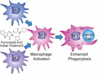
PBI1 can polarize undifferentiated M0 macrophages or re-program tumor-promoting M2 macrophages into the anti-tumor (M1)
phenotype and enhance the phagocytosis of cancer cells.You can also read