Maternal Serotonin Reuptake Inhibitor Antidepressants Have Acute Effects on Fetal Heart Rate Variability in Late Gestation
←
→
Page content transcription
If your browser does not render page correctly, please read the page content below
ORIGINAL RESEARCH
                                                                                                                                                published: 16 August 2021
                                                                                                                                          doi: 10.3389/fpsyt.2021.680177

                                                Maternal Serotonin Reuptake
                                                Inhibitor Antidepressants Have Acute
                                                Effects on Fetal Heart Rate Variability
                                                in Late Gestation
                                                Kayleigh S. J. Campbell 1,2 , Abby C. Collier 3 , Michael A. Irvine 1 , Ursula Brain 1,4 ,
                                                Dan W. Rurak 1,2 , Tim F. Oberlander 1,4* and Kenneth I. Lim 1,2
                                                1
                                                  BC Children’s Hospital Research Institute, Vancouver, BC, Canada, 2 Department of Obstetrics and Gynaecology, University
                                                of British Columbia, Vancouver, BC, Canada, 3 Faculty of Pharmaceutical Sciences, University of British Columbia,
                                                Vancouver, BC, Canada, 4 Department of Pediatrics, University of British Columbia, Vancouver, BC, Canada

                                                Background: Prenatal exposure to serotonin reuptake inhibitor (SRI) antidepressants
                                                increases risk for adverse neurodevelopmental outcomes, yet little is known about
                                                whether effects are present before birth. In relation to maternal SRI pharmacokinetics, this
                                                study investigated chronic and acute effects of prenatal SRI exposure on third-trimester
                                                fetal heart rate variability (HRV), while evaluating confounding effects of maternal
                           Edited by:           depressed mood.
                          Julie M. Zito,
     University of Maryland, Baltimore,         Methods: At 36-weeks’ gestation, cardiotocograph measures of fetal HR and HRV
                         United States
                                                were obtained from 148 pregnant women [four groups: SRI-Depressed (n = 31),
                       Reviewed by:
                                                SRI-Non-Depressed (n = 18), Depressed (unmedicated; n = 42), and Control (n = 57)]
               Jonathan L. Slaughter,
 The Research Institute at Nationwide           before, and ∼5-h after, typical SRI dose. Maternal plasma drug concentrations
    Children’s Hospital, United States          were quantified at baseline (pre-dose) and four time-points post-dose. Mixed effects
                     Hermona Soreq,
Hebrew University of Jerusalem, Israel
                                                modeling investigated group differences between baseline/pre-dose and post-dose fetal
                   *Correspondence:
                                                HR outcomes. Post hoc analyses investigated sex differences and dose-dependent
                     Tim F. Oberlander          SRI effects.
                 toberlander@bcchr.ca
                                                Results: Maternal SRI plasma concentrations were lowest during the baseline/pre-dose
                   Specialty section:           fetal assessment (trough) and increased to a peak at the post-dose assessment;
         This article was submitted to          concentration-time curves varied widely between individuals. No group differences in fetal
     Child and Adolescent Psychiatry,
               a section of the journal
                                                HR or HRV were observed at baseline/pre-dose; however, following maternal SRI dose,
                Frontiers in Psychiatry         short-term HRV decreased in both SRI-exposed fetal groups. In the SRI-Depressed
           Received: 13 March 2021              group, these post-dose decreases were displayed by male fetuses, but not females.
            Accepted: 14 July 2021
                                                Further, episodes of high HRV decreased post-dose relative to baseline, but only among
          Published: 16 August 2021
                                                SRI-Non-Depressed group fetuses. Higher maternal SRI doses also predicted a greater
                              Citation:
 Campbell KSJ, Collier AC, Irvine MA,           number of fetal HR decelerations. Fetuses exposed to unmedicated maternal depressed
   Brain U, Rurak DW, Oberlander TF             mood did not differ from Controls.
 and Lim KI (2021) Maternal Serotonin
   Reuptake Inhibitor Antidepressants           Conclusions: Prenatal SRI exposure had acute post-dose effects on fetal HRV
    Have Acute Effects on Fetal Heart           in late gestation, which differed depending on maternal mood response to SRI
     Rate Variability in Late Gestation.
         Front. Psychiatry 12:680177.           pharmacotherapy. Importantly, fetal SRI effects were sex-specific among mothers with
     doi: 10.3389/fpsyt.2021.680177             persistent depressive symptoms, as only male fetuses displayed acute HRV decreases.

Frontiers in Psychiatry | www.frontiersin.org                                         1                                          August 2021 | Volume 12 | Article 680177
Campbell et al.                                                                                                                           Acute Fetal SRI Effects

                                                At trough (pre-dose), chronic fetal SRI effects were not identified; however, concurrent
                                                changes in maternal SRI plasma levels suggest that fetal drug exposure is inconsistent.
                                                Acute SRI-related changes in fetal HRV may reflect a pharmacologic mechanism,
                                                a transient impairment in autonomic functioning, or an early adaption to altered
                                                serotonergic signaling, which may differ between males and females. Replication is
                                                needed to determine significance with postnatal development.

                                                Keywords: serotonin reuptake inhibitor antidepressants, prenatal exposure, fetal heart rate variability, sex
                                                differences, antidepressant pharmacokinetics, maternal depressed mood, pregnancy, third-trimester

INTRODUCTION                                                                          disrupted cardiovascular function (28–30) and increased fetal
                                                                                      motor activity (28, 31–33); though, findings are not consistent,
Up to 20% of women experience depressed mood during                                   primarily due to variations in methodology, gestational age
pregnancy (1, 2), and nearly one half of these women are                              and the ability to account for maternal mood. In SRI exposed
treated with a serotonin reuptake inhibitor (SRI) antidepressant                      fetuses, Mulder et al. report increased motor activity in
(3). Since their introduction nearly 30 years ago, the decision                       the second trimester and increased motor activity during
to start, continue or discontinue SRI antidepressant treatment                        quiet sleep state (i.e., stable fetal HR, low variability) in the
during pregnancy remains complex, as clinicians and women                             third-trimester; however, SRI-treated mothers had comparable
continue to weight risks of adverse outcomes against relapse (4,                      psychiatric symptoms to the unmedicated depressed group (28).
5). Prenatal SRI exposure has been associated with increased risks                    Gustafsson et al. also observed increased motor activity in
for preterm birth, lower birth weight and neonatal behavioral                         SRI-exposed fetuses, but only prior to 30-weeks’ gestation and
disturbances (6), as well as altered stress-regulation, social-                       found no SRI-related effect on fetal HR, HR variability, or HR-
emotional behaviors and other neurodevelopmental outcomes                             movement coupling (31). Conversely, lower fetal HR variability
from infancy-to-childhood (7–13). However, many of these long-                        at 36-weeks’ gestation and reduced cerebral blood flow resistance
term associations may be confounded by the underlying maternal                        was observed in SRI-exposed fetuses (30), as well as elevated
psychiatric disorder (14). Antenatal maternal mood disturbances                       pulmonary blood flow in SRI-exposed fetuses who experienced
are, similarly, associated with altered neurobehavioral outcomes                      transient respiratory difficulties at birth (29). Critically, outcomes
in infancy (15–17), stress-regulation in childhood (18) and a risk                    from previous fetal SRI studies remain confounded by maternal
for later psychopathology, emotional, or behavioral disturbances                      psychiatric symptoms. A case in point, altered fetal motor
(19). Whether the early origins of these outcomes are already                         activity, HR and HR variability have also been associated
evident before birth remains unclear. This study was undertaken                       with antenatal maternal depression (34–37) and anxiety [e.g.,
to investigate the effect of prenatal exposure to SRIs on fetal                       reviewed in (38)]. Thus, investigating fetal outcome related to
heart rate (HR) and relationships to maternal antidepressant                          prenatal SRI exposure requires appropriate control groups for
pharmacokinetics in late gestation, controlling for the effects of                    maternal mood.
depressed mood.                                                                           Fetal outcome may also be differentially sensitive to acute and
    Both maternal psychiatric distress and its treatment with SRI                     chronic drug effects, whereby outcomes vary depending on the
antidepressants are early exposures that may influence the in                         time of assessment relative to SRI exposure. In fetal sheep studies,
utero environment (20–22), possibly through the modulation                            acute and chronic SRI effects have been observed, with transient
of fetal, maternal, or placental serotonin (5-hydroxytryptamine;                      reductions in uterine blood flow and reduced fetal oxygenation
5-HT) signaling (23, 24). In particular, SRIs act by inhibiting                       status following acute SRI infusion (39), but a sustained decrease
the reuptake of the extracellular 5-HT leading to an increased                        in low-voltage electrocortical fetal brain activity with prolonged
duration and magnitude of serotonergic activity on pre- and                           SRI exposure (40). While it is presently unknown whether SRI
postsynaptic receptors. During development, 5-HT is present                           exposure has distinct acute and chronic effects on human fetuses,
from early gestation (25) and has been identified as a key                            a pharmacologic mechanism has been suggested (41, 42). SRI
neurotrophic factor regulating the construction and plasticity of                     dose-relationships with fetal, obstetric, and neonatal outcomes
neuronal circuits within its own and non-serotonergic systems                         have been reported (28, 43, 44), and there is high correspondence
(26). Across the lifespan, 5-HT also has extensive roles in                           between maternal and fetal plasma drug concentration ratios in
neuropsychological and other central, autonomic, and peripheral                       amniotic fluid (45) and cord blood (46, 47) that vary with SRI
nervous system processes (27). As SRIs are lipophilic compounds                       type. Importantly, fetal exposure to other psychoactive agents
with high placental permeability, it is conceivable that altered 5-                   have produced differential acute outcomes, such as an acute
HT signaling before birth may have broad neurodevelopmental                           suppressive effect of buprenorphine on fetal HR and movement
and physiologic implications.                                                         (48) and decreased fetal HR variability following acute nicotine
    To date, only few studies have investigated whether outcomes                      exposure (49). Together, these studies suggest that fetal HR may
of prenatal SRI exposure emerge before birth: during the period                       be sensitive in detecting differences between acute and chronic
of drug exposure. Fetal SRI exposure has been associated with                         psychotropic drug exposures.

Frontiers in Psychiatry | www.frontiersin.org                                     2                                     August 2021 | Volume 12 | Article 680177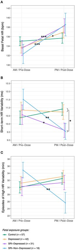
Campbell et al.                                                                                                             Acute Fetal SRI Effects

    Fetal HR and its variability are prenatal markers of                  March 2013–August 2017). Informed consent was obtained
cardiovascular regulation and can be studied non-invasively               from all participants. Both SRI-treated and non-SRI-treated
using Doppler ultrasound-based technologies, such as                      women were recruited who were experiencing a range of
cardiotocography. Fetal HR and HR variability are widely                  antenatal depressive symptoms, some meeting a diagnostic
described as indies of early autonomic functioning (50, 51),              threshold for a DSM-V mood disorder (65), while others
and the coalescence of fetal HR patterns and accelerations                were symptomatic at a subthreshold level or were relatively
with motor activity around 32-weeks’ gestation is viewed as               euthymic. Inclusion criteria for SRI-treated women required
organized neurobehavior (52–54). As the fetus matures, the well-          the initiation of pharmacotherapy before or during pregnancy
characterized decrease in fetal HR and increase in HR variability         for a minimum of 90 days prior to delivery (i.e., entire
(52, 55–58) are thought to reflect increasing sympathetic                 duration of the third-trimester). Demographic characteristics
responsiveness and an emerging influence of parasympathetic               were collected by clinician interviews and health records chart
(i.e., vagal) modulation (51). Fetal HR variability has been              review. Fetal gestational age was calculated using the first
described as a psychophysiological construct with behavioral              trimester dating scan, as per the Society of Obstetricians and
trait-like correspondence (50), reflecting an individual’s                Gynaecologists of Canada Clinical Practice Guidelines (66).
emerging capacities for adaptive flexibility and interaction              Exclusion criteria comprised of maternal psychiatric disorders
with environment, serving to prime the fetus for extrauterine             other than unipolar depression or anxiety, illicit substance use,
life (59). Fetal cardiac patterning demonstrates developmental            gestational hypertension or diabetes, placental insufficiency, or
stability into the postnatal period, as it’s highly correlated with       any other significant maternal or fetal medical condition. Fetuses
neonatal and infant HR (60) and predicts temperament and                  born prior to 36-weeks’ gestation were excluded.
neurodevelopmental outcomes in infancy (61–63), as well as                   Of the 188 recruited women, 153 were eligible for inclusion
behavioral regulation in childhood (64).                                  in the present study. Reasons for exclusion were as follows:
    The present study was undertaken to investigate acute and             cancelation for technical reasons (n = 12), preterm delivery (n
chronic effects of prenatal SRI antidepressant exposure on fetal          = 8), obstetrical complications (n = 8), emergent issues during
HR and HR variability in late gestation, while evaluating the             the study protocol necessitating clinical assessment (n = 4),
concurrent effects of prenatal maternal depressed mood. Chronic           voluntary withdrawal (n = 2), and development of an exclusion
effects of SRI exposure were determined by comparing fetal                criterion after recruitment (n = 1).
outcomes at a baseline period prior to typical morning oral                  Of note, the present study reports on two maternal-fetal
SRI dose (i.e., pre-dose; at pharmacologic trough). Acute SRI-            cohorts that underwent nearly identical data collection sequences
exposure effects were determined at peak drug levels (∼4–5 h              at 36-weeks’ gestation, with the exception of maternal blood
post-dose). Maternal SRI plasma drug concentrations across five           collection (detailed below) on the first cohort only. These
time-points were used to characterize pharmacokinetics and                cohorts did not differ in clinical or demographic characteristics.
assess drug level changes relative to periods of chronic and acute        Subsets of data from participants in the present study had
SRI exposure. To distinguish SRI-related effects from prenatal            been included in two prior reports investigating fetal outcomes
maternal depressed mood, we compared fetal HR outcomes                    in healthy, uncomplicated pregnancies (n = 68) (67), and
from a control group (non-SRI treated/non-depressed) with                 SRI-exposure effects on brain blood flow (n = 74) (30).
three prenatal exposure groups: fetuses of mothers who were               While primary study protocols were similar, the present
SRI-treated/depressed, SRI-treated/non-depressed, and non-SRI             study investigated acute and chronic effects of SRI exposure
treated/depressed. These groups captured how maternal response            in relation to fetal HR variability, maternal pharmacologic
to SRI pharmacotherapy, namely whether depressive symptoms                data and the potentially confounding effects of depressed
persisted or remitted, may differentially influence the fetus.            mood. These augmented data and outcomes have not been
We hypothesized that acute SRI exposure would be associated               previously reported.
with reduced fetal HR variability and that SRI-exposed fetuses
with concurrent exposure to maternal depressed mood would
have the greatest changes compared with outcomes in non-                  Maternal Depressed Mood and SRI
exposed fetuses.                                                          Antidepressants
                                                                          Maternal depressive symptoms were assessed with the Hamilton
MATERIALS AND METHODS                                                     Rating Scale for Depression (HAM-D) (68), a 17-item clinician-
                                                                          rated questionnaire administered by trained research staff,
Study Cohort                                                              blinded to SRI exposure-status. Mothers were considered to be
The study protocols were approved by the UBC Clinical                     symptomatically depressed with a total HAM-D score > 8 (69). In
Research Ethics Board and the BC Women’s Hospital Research                this study, SRI antidepressants included any selective serotonin
Review Committee (H05-70629 and H12-00733). During the late               reuptake inhibitor (SSRI) or serotonin-norepinephrine reuptake
second trimester, 188 women with singleton low-risk pregnancies           inhibitor (SNRI).
were recruited in two cohorts from the Reproductive Mental                   To detect SRI-related fetal effects and distinguish them from
Health Clinic at BC Women’s Hospital and Health Center,                   exposure to maternal depressed mood, mothers were then
community midwives, or family physicians in metropolitan                  grouped based on SRI treatment and the presence of depressive
Vancouver, Canada (from November 2006–January 2010 and                    symptoms at 36-weeks’ gestation, yielding four study groups:

Frontiers in Psychiatry | www.frontiersin.org                         3                                   August 2021 | Volume 12 | Article 680177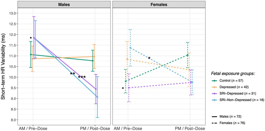
Campbell et al.                                                                                                                Acute Fetal SRI Effects

SRI-Depressed (SRI-treated + HAM-D > 8, i.e., depressive                     using a handheld event marker, which we assessed as an indirect
symptoms persisted), SRI-Non-Depressed (SRI-treated + HAM-                   measure of fetal motor activity. Refer to Pardey et al. for further
D ≤ 8, i.e., depressive symptoms remitted), Depressed (non-SRI-              details on reported measures (72).
treated + HAM-D > 8), and Control (non-SRI-treated + HAM-
D ≤ 8). Thus, fetal outcome was assessed as an exposure to one               Maternal SRI Plasma Levels
of these groups.                                                             Changes in maternal plasma drug concentration between fetal
                                                                             assessments were determined by analyzing blood samples
Study Protocol                                                               from SRI-treated mothers pre-dose (T0 , baseline levels;
Figure 1 outlines the fetal and maternal data collection sequence            ∼08h00) and at four time-points post-dose: T1 (∼10h30),
that occurred at 36-weeks’ gestation. On the day of the study, all           T2 (∼12h30), T3 (∼13h30), and T4 (∼14h30). Serum was
participants were instructed to eat and drink as per usual prior to          separated by centrifugation at 3,000×g for 10 min, transferred
arrival. Participants underwent two sequential fetal assessments             to polypropylene tubes and stored at −70◦ C until analysis. High
in a dedicated quiet room at the BC Women’s Hospital Center for              performance liquid chromatography tandem mass spectrometry,
Prenatal Diagnosis, first in the morning (AM/baseline; ∼09h30)               performed offsite (CANTEST Ltd.; Burnaby, Canada), was used
and again in the afternoon (PM; ∼13h30); methodological                      to determine levels of fluoxetine, norfluoxetine, paroxetine,
details are described below. Mothers were positioned in the left             sertraline, citalopram, escitalopram and venlafaxine. The
recumbent position to prevent aortocaval compression. Fetal                  calibration range was 0.1–100 ng/ml for analytes (except
assessments were separated by a 2-h controlled break, involving              sertraline, where the lower limit of quantification was
the administration of the HAM-D and time for participants to                 0.25 ng/ml). The intra- and inter-assay coefficients of variation
mobilize and have lunch (provided).                                          and relative errors were < 20% for all drugs and metabolites.
   To investigate chronic and acute SRI effects on the fetus, SRI-              Plasma drug concentrations were adjusted for maternal oral
treated women were asked to withhold their typical morning oral              dose (ng/ml·mg). To quantify the relative change in maternal
dose until ∼10h00, resulting in the AM/baseline and PM fetal                 SRI level between the pre- and post-dose fetal assessments,
assessments corresponding to pre-dose and post-dose periods,                 the difference in dose-adjusted plasma drug concentration
respectively. To characterize concurrent SRI pharmacokinetics                between T0 and T3 was determined. Plasma concentrations for
across the study protocol, plasma drug concentrations were                   metabolites were not reported as they reflect parent drugs.
quantified at baseline (pre-dose) and four time-points post-dose;
details on the drug level assay and pharmacologic variables                  SRI Pharmacokinetics and Standardized Dose
are described below. Timing for each component of this study                 Maternal SRI plasma levels were further characterized by
considered the need for a sufficient antidepressant baseline                 performing a non-compartmental pharmacokinetic analysis,
(pharmacologic trough), half-life, and time-to-peak plasma                   yielding estimates of maximum plasma drug concentration
levels, weighted against length of study in effort to minimize               (Cmax ), time-to-peak (Tpeak ) and area under the curve (AUClast ).
maternal discomfort/inconvenience and potential effects of                   Pharmacokinetic variables were calculated using the PKNCA R
diurnal variations in the fetal variables obtained.                          package (73).
                                                                                Further, we computed a standardized SRI dose variable to
Fetal Cardiotocography                                                       investigate whether dose-dependent relationships were present
Fetal cardiotocography (CTG) was used to investigate patterns of             among SRI-related fetal outcomes. As per methods described by
fetal HR and HR variability. Fetal HR was recorded continuously              Mulder et al. (28), standardized SRI dose was defined according
for 50-min using a Sonicaid Fetal Care computerized CTG                      to the World Health Organization Anatomical Therapeutic
system (Huntleigh Healthcare Ltd.; Cardiff, UK; software version             Chemical-Defined Daily Dose (ATC-DDD) Index (74). DDDs
2.2.3.0), a clinical tool widely used for antenatal fetal surveillance       were as follows: 10 mg for escitalopram; 20 mg for citalopram,
(70). Briefly, the software baseline-fits the continuous fetal HR            fluoxetine, and paroxetine; 50 mg for sertraline; 100 mg for
tracing then computes several variables based on its averaging               venlafaxine; and 300 mg for moclobemide. Mothers prescribed
algorithm (71, 72): basal fetal HR (i.e., average resting HR, in             their antidepressant’s DDD were set to 1; higher or lower doses
beats per minute; bpm), number of fetal HR accelerations and                 were expressed as a multiple of the DDD.
decelerations, as well as three measures of fetal HR variability:
short-term variation (STV), high variability and low variability.            Statistical Analyses
STV, a measure of micro-fluctuations in fetal HR, was computed               Statistical analyses were performed using R Statistical Computing
as the average epoch-to-epoch variation across the entire HR                 Environment version 3.6.1 (75); the significance level was set at
tracing in pulse intervals (i.e., time between consecutive heart             α = 0.05. Group differences in maternal and fetal characteristics
beats, in milliseconds; ms). Whereas high and low variability                were assessed using one-way analysis of variance (ANOVA) or
reflect specific HR patterns that occur during periods of fetal              Kruskal-Wallis rank sum test for continuous normal and ordinal
activity and quiescence, respectively. Episodes of high and low              data, respectively; significant between-group effects were further
variability were computed as the sum of all individual episodes              explored using post hoc Tukey’s HSD or the Dunn test. Chi Square
(in minutes) each HR pattern was displayed in the tracing,                   tests were used for group comparisons of categorical variables.
corrected to 50-min. Additionally, the number of maternally-                    Generalized linear mixed-effects models (GLMMs) were used
perceived fetal movements (FMs) during each CTG was recorded                 to investigate group differences in fetal HR and HR variability

Frontiers in Psychiatry | www.frontiersin.org                            4                                   August 2021 | Volume 12 | Article 680177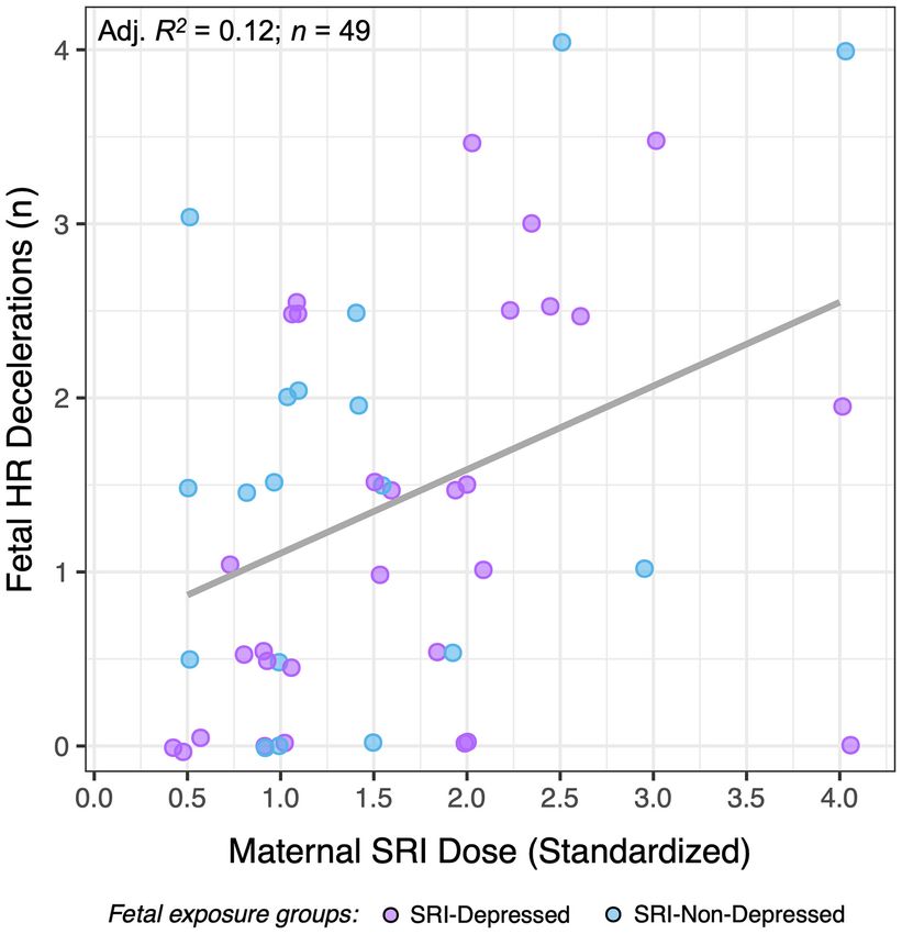
Campbell et al.                                                                                                                                 Acute Fetal SRI Effects

 FIGURE 1 | Data collection sequence at 36-weeks’ gestation assessing pre- and post-SRI dose effects on the fetus. Text in gray pertains to SRI-treated mothers only.
 Times are approximate and represent the median, rounded to the nearest 30-min.

outcomes across time. GLMMs describe each outcome as a linear                         characteristics did not differ between those included in the
combination of fixed and random effects; here, fixed effects                          analysis sample (n = 148) compared to those who did not
were an interaction between one between-factor (Group: Control,                       participate/were excluded (n = 40) (Supplementary Table 1),
Depressed, SRI-Depressed, SRI-Non-Depressed) and one within-                          other than in characteristics related to exclusion criteria
factor (Time: AM/pre-dose, PM/post-dose). Gestational age                             (i.e., preterm delivery).
at the time of assessment and fetal sex were also included
as fixed effects terms. Because pre- and post-dose outcomes                           Maternal and Fetal Characteristics
were not independent, random effects were specified to                                Maternal characteristics generally did not differ between groups
account for individual differences at baseline (AM/pre-dose; i.e.,                    (Table 1), apart from maternal weight at 36-weeks’ gestation,
random intercept for subjects) and the within-subject variability                     which was higher in both Depressed (padj = 0.05) and SRI-
explained by the repeated measures (i.e., random slope for                            Depressed (padj = 0.05) women compared to Controls. Maternal
subjects across Time) (76). Linear or Poisson (log) link functions                    depressed mood symptoms differed between groups, with
were specified according to the underlying distribution. Mixed                        significantly higher HAM-D scores in the Depressed and SRI-
modeling was conducted using the lme4 library in R (77)                               Depressed groups compared to women in both the Control and
and fit by restricted maximum likelihood. Type III Wald F-                            SRI-Non-Depressed groups (all: padj < 0.001). Mood symptoms
statistics (or X 2 -statistic, if Poisson model) and associated p-                    among SRI-Non-Depressed women did not differ from Controls
values are reported for significant interaction or main effects;                      (padj = 0.4).
effective degrees of freedom were estimated with the Kenward-                             SRI-treated women were taking a daily oral dose within
Roger approximation.                                                                  the typical therapeutic range and were prescribed their
    Post hoc tests explored significant Group × Time interactions                     antidepressant for the entire duration of pregnancy, except four
to detect group differences at AM/pre-dose and PM/post-                               mothers with third-trimester exposure only (i.e., n = 4 taking
dose assessments, as well as within-group changes across time.                        SRI for 137 ± 44 days prior to delivery). Neither standardized
Results are reported as the estimated difference between relevant                     SRI dose nor length of gestational SRI exposure differed between
factor contrasts, along with 95% confidence intervals (CI)                            SRI-Depressed and SRI-Non-Depressed mothers. Included in
and associated p-values, adjusted for multiple comparisons                            the SRI-Non-Depressed group was one mother treated with
with Tukey’s method. Further, given the previous reports of                           moclobemide, a reversible inhibitor of monoamine oxidase-A,
sex differences in fetal HR [e.g. (78)], we also investigated                         which also acts to increase serotonergic activity by inhibiting
whether any significant effect differed between male and                              5-HT deamination within neurons and synaptic vesicles (80).
female fetuses. Additional post hoc GLMMs examined Group                                  Fetuses were assessed at 35.9 ± 0.81 weeks’ gestation;
× Sex × Time (three-way) interactions, adjusted for gestational                       their characteristics are summarized in Table 2. All fetuses
age. Post hoc testing was performed using the emmeans                                 included in analysis were delivered at term and were clinically
R package (79).                                                                       healthy newborns discharged from hospital according to
                                                                                      routine schedules. Gestational age at birth was significantly
RESULTS                                                                               lower for fetuses in the SRI-Depressed group compared to
                                                                                      the Control (padj < 0.001) and Depressed (padj = 0.002)
Of the 153 mother-fetal participants, 148 were included in the                        groups. In the newborn period, the SRI-Depressed group
study sample: one SRI-treated mother was not compliant with                           also had lower birth weight, length and head circumference
study protocols, one fetus did not meet the Dawes/Redman                              compared the Control and Depressed groups; however, these
criteria for normality during CTG sessions (71, 72), one                              effects all diminished when adjusting for gestational age
fetus was found to have a cardiac abnormality, one fetus                              at birth.
had overall poor data, and one fetus was consistently an
outlier in analysis. The final study cohort comprised 57                              Maternal SRI Pharmacokinetics
Control, 42 Depressed, 31 SRI-Depressed, and 18 SRI-                                  Plasma drug concentrations were quantified for a minimum of
Non-Depressed mother-fetus pairs. Maternal and fetal                                  three of the five time-points in 24 of the 49 SRI-treated women.

Frontiers in Psychiatry | www.frontiersin.org                                     5                                          August 2021 | Volume 12 | Article 680177
Campbell et al. Acute Fetal SRI Effects
TABLE 1 | Maternal characteristics (n = 148).
Control Depressed SRI-Depressed SRI-Non-Depressed Test statistic
(n = 57) (n = 42) (n = 31) (n = 18) (p-value)
Maternal age (years) 32.9 ± 3.5 34.5 ± 4.5 33.9 ± 5.9 35.1 ± 5.1 F (3, 144) = 1.6 (0.2)
Maternal weight at 36-weeks’ (kg) 75.1 ± 9.7 81.9 ± 16.0 82.5 ± 15.0 79.6 ± 9.8 F (3,144) = 3.2 (0.02)*
Parity 0 (0, 1) 0 (0, 1) 0 (0, 1) 0 (0, 0) H(3) = 1.9 (0.6)
Education (total years) 18.7 ± 3.1 18.0 ± 3.9 17.3 ± 3.6 18.3 ± 3.7 F (3, 144) = 1.1 (0.3)
Alcohol during pregnancy (n total drinks)† 0 (0, 2) 0 (0, 2) 0 (0, 2.5) 1 (0, 4.75) H(3) = 2.6 (0.5)
Smoking during pregnancy (n smoker/n non-smoker) 0/57 1/41 1/30 1/17 (0.2)
HAM-D at 36-weeks’ 4.7 ± 2.3 12.7 ± 4.0 13.4 ± 3.1 6.0 ± 2.1 F (3, 144) = 88 (< 0.001)***
SRI antidepressants (n, [dose range])
Citalopram (n = 14) — — 10 [10–60 mg] 4 [10–50 mg] —
Escitalopram (n = 7) — — 3 [5–20 mg] 4 [10 mg] —
Fluoxetine (n = 5) — — 2 [20–80 mg] 3 [20–60 mg] —
Paroxetine (n = 4) — — 3 [20–40 mg] 1 [30 mg] —
Sertraline (n = 6) — — 4 [50–200 mg] 2 [75–200 mg] —
Venlafaxine (n = 12) — — 9 [75–262.5 mg] 3 [75–150 mg] —
Moclobemide‡ (n = 1) — — — 1 [150 mg] —
Standardized daily SRI dose — — 1.5 (1.0, 2.0) 1.0 (1.0, 1.5) t(47) = 0.9 (0.4)
Length of gestational SRI exposure (days) — — 264 ± 36 260 ± 48 t(47) = 0.31 (0.8)
Continuous variables reported as mean ± SD if normally distributed, or median (first, third quartile) if skewed. Categorical variable reported as total number (n). Test statistics, degrees
of freedom, and associated p-values are reported for between-group differences using: one-way ANOVA (F), Kruskal-Wallis test (H), Fisher’s Exact test, or two-sample t-test (t), where
appropriate. P-value significance levels: *p < 0.05, **p < 0.01, ***p < 0.001.
SRI, serotonin reuptake inhibitor; HAM-D, total score from Hamilton Rating Scale for Depression; kg, kilograms; mg, milligrams.
† Alcohol during pregnancy represents n total standard drinks consumed during the course of pregnancy (study sample range: 0–52 total drinks).
‡ Reversible monoamine oxidase inhibitors included in cohort as “SRI-exposed.”
TABLE 2 | Fetal characteristics (n = 148).
Control Depressed SRI-Depressed SRI-Non-Depressed Test statistic
(n = 57) (n = 42) (n = 31) (n = 18) (p-value)
Gestational age at fetal study (weeks) 36.0 ± 0.9 35.9 ± 0.8 35.9 ± 0.7 35.9 ± 0.8 F (3, 144) = 0.1 (> 0.9)
Gestational age at birth (weeks) 39.9 ± 1.1 39.8 ± 1.3 38.9 ± 1.2 39.6 ± 1.5 F (3, 144) = 5.5 (0.001)**
Sex (n male/n female) 26/31 26/16 14/17 6/12 χ 2 (3) = 5.0 (0.2)
Birth weight (g) 3532 ± 408 3588 ± 416 3312 ± 490 3514 ± 431 F (3, 144) = 2.7 (0.05)
Length at birth (cm) 52.0 ± 2.1 51.7 ± 2.3 50.3 ± 1.8 51.4 ± 2.7 F (3, 144) = 4.2 (0.007)**
Head circumference at birth (cm) 35.2 ± 1.3 35.1 ± 1.4 34.3 ± 1.4 34.9 ± 1.1 F (3, 144) = 3.6 (0.02)*
Apgar at 5 min 9 (9, 9) 9 (9, 9) 9 (9, 9) 9 (9, 9) H(3) = 2.2 (0.5)
Continuous variables reported as mean ± SD if normally distributed, or median (first, third quartile) if skewed. Categorical variable reported as total number (n). Test statistics, degrees
of freedom, and associated p-values are reported for between-group differences using: one-way ANOVA (F), Kruskal-Wallis test (H), or Chi Square test (χ2 ), where appropriate. P-value
significance levels: *p < 0.05, **p < 0.01, ***p < 0.001.
SRI, (fetal exposure to) serotonin reuptake inhibitor antidepressant; g, grams; cm, centimeters.
One mother’s plasma drug concentrations were below lower Figure 2 shows the inter-individual variability in
levels of quantification (Campbell et al. Acute Fetal SRI Effects
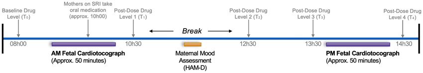
FIGURE 2 | Dose-adjusted plasma drug concentrations (ng/ml·mg) at baseline (T0 ) and four time-points post-dose across the study protocol for 23 SRI-treated
mothers, grouped by antidepressant type. Concentration-time curves demonstrate inter-individual variability in SRI pharmacokinetics and maternal drug levels relative
to the start of each fetal assessment; curves were fit to each individual’s data with local polynomial regression. SRI oral dosing occurred a median of 1.83 h after T0 .
Median blood collection times were: baseline (T0 ) at 08h06, post-dose 1 (T1 ) at 10h21, post-dose 2 (T2 ) at 12h50, post-dose 3 (T3 ) at 13h38, and post-dose 4 (T4 ) at
14h57. Median start times of the baseline/pre-dose and post-dose fetal assessments were at a 1.3 and 5.6 h after T0 , respectively (dotted vertical lines).
TABLE 3 | Maternal pharmacokinetic variables (mean ± SE) for each antidepressant type on a subset of SRI-treated women (n = 23).
Antidepressant N AUClast Cmax Tpeak 1 T3-T0
(ng/ml) (h) (ng/ml·mg)
Citalopram 5 906 ± 271 274 ± 92 4.9 ± 0.14 3.2 ± 1.0
Fluoxetine† 3 3221 ± 1919 613 ± 342 5.8 ± 1.0 4.3 ± 2.5
Paroxetine 2 125 ± 66 24 ± 5.2 4.6 ± 0.04 0.13 ± 0.21
Sertraline 2 728 ± 256 197 ± 105 5.3 ± 0.18 0.71 ± 0.53
Venlafaxine 11 621 ± 314 163 ± 74 5.7 ± 0.30 0.43 ± 0.13
AUClast , area under the curve from T0 (baseline) to the last measured plasma drug concentration (T4 ); Cmax , maximum (peak) plasma drug concentration (ng/ml); Tpeak , time (hours)
from baseline to reach Cmax ; 1 T3-T0, change in dose-adjusted plasma concentration (ng/ml·mg) from baseline (T0 ) to post-dose 3 (T3 ), representing the change in maternal drug
concentration between baseline/pre-dose and post-dose fetal assessments.
† Fluoxetine not yet reached maximum plasma concentration at time of last blood collection (i.e., T estimated as T
4 peak ). AUClast and Cmax for fluoxetine may be underestimations.
concentration (Cmax ) between 4.5–6 h (Tpeak ), followed by before T1 , which therefore occurred during the period of steady-
the initial elimination phase. Fluoxetine-treated mothers state pharmacologic trough (T0 -T1 ). At a median of 5.6 h (IQR:
appear to still be in the absorption phase when final drug 5.2−6.0) after T0 , the start of the post-dose fetal assessment
levels were collected (T4 ), consistent with a Tpeak of 6–8 h. corresponded to the late absorption phase or early elimination
AUClast , representing total observed maternal drug exposure phase, depending on SRI type. Dose-adjusted plasma drug
during our study protocol, was highest for fluoxetine and concentration significantly increased between baseline/pre-dose
lowest for paroxetine. Maternal pharmacokinetic responses are and post-dose fetal assessments [n = 23; paired t-test: t (21) =3.13,
summarized in Table 3. p = 0.005], with a mean (± SE) change from T0 -to-T3 of 1.54 ±
0.48 ng/ml·mg.
Validation of Study Design: Fetal Assessments at
Pharmacologic Trough and Peak Fetal HR and HR Variability
Figure 2 also illustrates the start times of each fetal assessment Fetal CTG measures (n = 148) are presented in Table 4, and were
relative maternal plasma SRI levels. For the antidepressants within clinically normative ranges for gestational age (56, 77).
studied, the baseline/pre-dose fetal assessment started a median AM/pre-dose and PM/post-dose fetal CTG sessions were 50.1 ±
1.3 h (IQR: 0.99−1.5) after T0 and 0.92 h (IQR: 0.87−1.00) 3.0 min with minimal HR tracing signal loss (3.5 ± 5.4 %); neither
Frontiers in Psychiatry | www.frontiersin.org 7 August 2021 | Volume 12 | Article 680177Campbell et al. Acute Fetal SRI Effects
the duration nor amount of signal loss differed between groups at
either fetal assessment.
Basal Fetal HR
There were no group differences in basal fetal HR at either fetal
assessment. However from AM/pre-dose, fetal HR increased to
be significantly higher at the PM/post-dose assessment in all fetal
groups, except the Controls (Group × Time interaction: F (3, 142.0)
= 3.4, p = 0.02) (Figure 3A). Between assessments, basal fetal
HR increased by 5 bpm (95% CI: 2.3, 7.7; padj < 0.001) in the
SRI-Depressed group, by 6 bpm (95% CI: 2.7, 9.8; padj < 0.001) in
the SRI-Non-Depressed group, and by 3 bpm (95% CI: 0.8, 5.4;
padj = 0.01) in the Depressed group. In contrast, fetal HR in the
Control group did not change between assessments (padj = 0.4).
There were no covariate effects on basal fetal HR.
Fetal HR Accelerations and Decelerations
Fetuses had a median of 14 HR accelerations (IQR: 11–18)
and 1 HR deceleration (IQR: 0–2) during each 50-min CTG
session (Table 4). Fetal HR accelerations did not differ between
groups at either assessment; however, averaged across groups,
the number of HR accelerations significantly increased relative
to AM/pre-dose assessment (main effect of Time: X 21 = 8.9, p
= 0.003). Whereas, fetal HR decelerations did not significantly
differ between groups nor across time, but were found to be
positively associated with gestational age (X 2(1) = 4.2, p = 0.04).
Short-Term HR Variability
STV, reflecting the average HR variation across each CTG tracing,
in pulse intervals (ms) (72), did not differ between groups at the
AM/pre-dose fetal assessment. Following SRI dose, a significant
decrease in STV was observed relative to baseline among fetuses
in both SRI-exposed groups (Group × Time interaction: F (3, 142.6)
= 5.1, p = 0.002) (Figure 3B): STV decreased by 1.0 ms (95%
CI: 0.05, 1.9; padj = 0.04) in SRI-Depressed group fetuses and by
2.0 ms (95% CI: 0.80, 3.2; padj = 0.001) in SRI-Non-Depressed
group fetuses. These post-dose decreases resulted in SRI-exposed
fetuses to have 1.1 ms (95% CI: 0.80, 3.2; padj = 0.04) lower
STV compared to non-exposed fetuses at the PM/post-dose
assessment, controlling for covariates.
High and Low Fetal HR Variability
Episodes of high fetal HR variability did not differ between
groups at the AM/pre-dose fetal assessment; however post-SRI
dose, a Group × Time interaction was identified (F (3, 142.9) = 3.7,
p = 0.01), whereby the time fetuses in the SRI-Non-Depressed
group spent displaying high HR variability decreased by 9.2 min
(95% CI: 3.0, 15.4; padj = 0.004), controlling for covariates
(Figure 3C). No between-group differences were found for
episodes of low HR variability.
Fetal Motor Activity
Fetal movements (FMs), which did not differ between groups,
occurred at a median frequency of 48 (IQR: 32–70) and 53
(IQR: 32–83) movements/hour during the AM/pre-dose and
FIGURE 3 | Fetal HR and HR variability (mean ± SE) for each exposure group
across AM/pre-dose and PM/post-dose fetal assessments for (A) basal fetal PM/post-dose assessments, respectively (Table 4). As expected,
HR, (B) short-term variability, and (C) episodes of high fetal HR variability (post the number of FMs per minute during episodes of high HR
hoc test significance levels: *p < 0.05, **p < 0.01, ***p < 0.001). variability was significantly higher than during low HR variability
Frontiers in Psychiatry | www.frontiersin.org 8 August 2021 | Volume 12 | Article 680177Campbell et al. Acute Fetal SRI Effects
TABLE 4 | Fetal HR, HR variability and movement (n = 148).
Control Depressed SRI-Depressed SRI-Non-Depressed
(n = 57) (n = 42) (n = 31) (n = 18)
Fetal variables AM PM AM PM AM/pre-dose PM/post-dose AM/pre-dose PM/post-dose
Basal HR (bpm)a 135 ± 7 136 ± 8 134 ± 7 137 ± 8 134 ± 9 138 ± 8 132 ± 9 139 ± 10
HR accelerations (n)b 14 (11, 18) 16 (13, 20) 16 (11, 20) 15.5 (12, 19) 12 (9, 15) 14 (12, 17) 14 (10, 18) 13.5 (10, 15)
HR decelerations (n) 1 (0, 1) 1 (0, 2) 0 (0, 2) 1 (0, 2) 1 (0, 2) 1 (0, 2) 1 (0, 2) 2 (0.25, 3)
STV (ms)a 10.4 ± 3.1 10.9 ± 2.9 10.9 ± 3.1 10.7 ± 3.0 10.6 ± 3.4 9.6 ± 2.4 11.5 ± 2.7 9.5 ± 2.1
High HR variability (min)a 30.0 ± 13.0 32.5 ± 11.9 31.2 ± 12.1 33.1 ± 11.2 30.3 ± 14.3 31.4 ± 11.0 34.9 ± 9.3 25.7 ± 12.4
Low HR variability (min) 0.97 ± 2.5 1.8 ± 4.1 1.7 ± 4.2 1.4 ± 4.0 1.5 ± 2.9 0.70 ± 2.2 0.50 ± 1.9 1.8 ± 4.4
Fetal movements/hour (n)† 50 (31, 66) 53 (35, 82) 53.5 (37, 77) 57 (38, 86) 46 (28, 62) 47 (30, 69) 43.5 (30, 83) 60 (32, 83)
Fetal variables for each group are summarized as mean ± SD if continuous and normally-distributed, or median (first, third quartile) if skewed or count data.
GLMM Statistics: significant fixed effects are identified as: a significant Group × Time interaction; b significant effect of Time; and c significant effect of Group. Refer to text for model
statistics and estimated marginal means between relevant factor contrasts.
AM, morning/baseline fetal assessment; PM, afternoon fetal assessment; HR, heart rate; STV, short-term variation; bpm, beats per minute; n, total number; ms, milliseconds; min, minutes.
† Maternally-perceived fetal movements per hour (adjusted from ∼50 min).
FIGURE 4 | Sex differences in fetal short-term HR variability (mean ± SE) between exposure groups (post hoc test significance levels: *p < 0.05, **p < 0.01, ***p <
0.001; *indicates within-sex group difference, indicates within-group sex difference).
(t = 6.8, p < 0.001); this also did not differ between groups, nor CI: 0.22, 3.0; padj = 0.02). Conversely, female fetuses in the
across assessments. There were no covariate effects on FMs. SRI-Depressed group did not undergo this post-dose decrease
in STV, but instead, were found to have 2.2 ms (95% CI: 0.13,
4.4; padj = 0.04) lower STV than SRI-Depressed males at the
Sex-Specific Fetal SRI Effects baseline/pre-dose assessment and remained unchanged post-
Post hoc analysis revealed sex-specific effects on group differences
dose. There were no other significant effects of fetal sex on group
in fetal STV (three-way interaction: F (3, 138.4) = 2.7, p =
differences reported.
0.04) (Figure 4). In the SRI-Depressed group, only male fetuses
underwent a significant post-dose decrease in STV: from pre- to
post-dose assessments, STV decreased in SRI-Depressed group SRI Dose-Dependent Fetal Effects
males by 2.4 ms (95% CI: 1.1, 3.7; padj < 0.001), in SRI-Non- Maternal SRI oral dose (standardized) was found to be
Depressed group males by 2.7 ms (95% CI: 0.75, 4.7; padj = significantly associated with the number of fetal HR decelerations
0.008), and in SRI-Non-Depressed group females by 1.6 ms (95% during 50-min CTG sessions (Figure 5). Higher SRI doses were
Frontiers in Psychiatry | www.frontiersin.org 9 August 2021 | Volume 12 | Article 680177Campbell et al. Acute Fetal SRI Effects
associated with a greater number of fetal HR decelerations (n
= 49; F (1, 47) = 7.6, p = 0.008). This did not differ between
SRI-exposure groups, and effects were evident at both pre-
and post-dose assessments. No other dose-dependent effects
were observed in SRI-related fetal HR outcomes we report, nor
were there differences related to antidepressant class (i.e., SSRIs
vs. SNRIs).
DISCUSSION
This study reports three key findings. In late gestation, fetal
SRI exposure was associated with: (1) post-dose decreases in
fetal HR variability, (2) sex-specific fetal HR outcomes, and
(3) concurrent changes in maternal drug levels that reflected
pharmacologic trough/peak (acute) periods. Fetal HR increased
while fetal HR variability decreased in SRI-exposed fetuses
relative to the AM/pre-dose assessment, reflecting an acute effect
of SRI exposure. Importantly, fetal outcomes varied depending
on maternal response to SRI pharmacotherapy; namely whether
the mothers’ depressed mood remitted or remained symptomatic
(i.e., SRI-Non-Depressed, SRI-Depressed). In particular, STV
acutely decreased among fetuses in both SRI-Depressed and
FIGURE 5 | The relationship between standardized maternal SRI oral dose
SRI-Non-Depressed groups, thus occurring independent of
and the number of fetal HR decelerations for SRI-exposed fetuses
concurrent maternal mood; whereas, high HR variability was (SRI-Depressed and SRI-Non-Depressed groups). Data presented represent
found to acutely decrease only among fetuses in the SRI- average HR decelerations across 50-min CTG sessions, which did not differ
Non-Depressed group. SRI-related sex differences in fetal across time.
HR variability also varied with maternal mood context, with
differences between male and female fetuses observed only in fetal assessment. In contrast, women taking venlafaxine and
the SRI-Depressed group. Further, higher maternal SRI doses fluoxetine were still in the absorption phase by the last blood
were associated with a greater number of fetal HR decelerations collection. Trough and peak levels are an accepted phenomenon
across both study periods. Since neither standardized dose for multiple oral dosing regimens, but the critical finding
nor length of gestational SRI exposure differed between SRI- from this study was that the extremes of trough/peak observed
Depressed and SRI-Non-Depressed women, fetal outcomes in translated to a variable fetal response.
these groups may be acute drug exposure-related effects. In Our findings demonstrate that the fetus may experience a
particular, we did not observe group differences pre-dose during chronic exposure to steady-state plasma SRI levels, but subject to
a period of pharmacologic trough, which would have reflected a continual fluctuations in such exposure with respect to maternal
chronic/sustained effect of SRI exposure. oral dose across a typical day in late gestation. Although this
Importantly, changes in fetal HR variability we report were does not directly indicate that equivalent drug changes in fetal
within normative ranges for healthy typically developing fetuses circulation occur, changes in maternal SRI plasma levels would
at 36-weeks’ gestation (56, 78), and thus, are likely not clinically have implications toward factors that may impact the extent of
significant. However, even within a normative range of fetal fetal SRI exposure. SRIs have high placental permeability (46, 82),
physiology, we observe group differences that may reflect adverse and in rodents, fetal citalopram exposure was found to exceed
developmental effects of SRI exposure before birth that vary with that of the mother 2-h after maternal drug administration (83).
respect to the timing of maternal oral dose. Beyond transplacental drug transfer, several other factors could
also influence SRI pharmacology in this setting, such as genetic
Fetal SRI Exposure at Pharmacologic variations in maternal metabolic enzymes (further discussed
Trough and Peak below), fetoplacental metabolism and clearance, or exposure to
Maternal SRI plasma concentrations increased following a other pharmacologic agents (41, 42, 84, 85). Hence, it is almost
typical daily oral SRI dose in the third-trimester, demonstrating certain that fetal SRI exposure is not consistent and the distinct
an expected concentration-time relationship. Women in this acute SRI-related outcomes we report suggest a differential fetal
study were on long-term SRI pharmacotherapy (most prior sensitivity may exist to varying maternal SRI plasma levels and/or
to conception) and appear to have trough plasma SRI levels acute physiologic changes secondary to SRI exposure.
consistent with a pharmacologic steady-state, which were similar
to third-trimester maternal dose-adjusted plasma trough levels Fetal HR Variability Decreases Following
previously reported (81). Although sample size was limited, Acute SRI Exposure
women taking citalopram, sertraline, and paroxetine reached Transient reductions in fetal HR variability with acute SRI
Cmax and were in the early elimination phase at the post-dose exposure may indicate impairments in autonomic functioning.
Frontiers in Psychiatry | www.frontiersin.org 10 August 2021 | Volume 12 | Article 680177Campbell et al. Acute Fetal SRI Effects
The Dawes-Redman parameter of STV, a standardized clinical of active states among fetuses in the SRI-Non-Depressed group,
marker of perinatal compromise (56, 72, 86), not only possibly reflecting acute impairments or delayed development.
summarizes overall HR variation, but may also be a surrogate Our findings highlight the need for future studies focused on how
for fetal sympathovagal regulation. In a comparative study by SRIs interact with maternal mood to influence fetal autonomic
Seliger et al., CTG-derived STV was highly correlated with the functioning and neurobehavior.
standard deviation of normal-to-normal beat intervals, as well
as HR in the low frequency power spectra (87). These indices Acute SRI Effects on Fetal HR Variability
are commonly obtained from fetal electrocardiography, which Are Sex-Specific
has higher temporal resolution than ultrasound-based CTG (i.e., Acute SRI-related outcomes in fetal HR variability were found
ability to detect QRS complexes in continuous cardiac signal) and to be moderated by fetal sex. Specifically, the post-dose
are among parameters widely used to examine sympathetic and decrease in STV was observed among SRI-Depressed group
integrative sympathovagal-mediated HR fluctuations (88). Thus, male fetuses, compared with the relative stability in STV
acute decreases in STV, accompanied by acute increases in basal between assessments in SRI-Depressed females. Indeed, sex
fetal HR we report in SRI-exposed fetuses, may be consistent with difference in fetal HR variability have been reported in low-
sympathetic activation and/or autonomic withdrawal leading to risk singleton pregnancies (98, 99), for example in a large
diminished HR variability. CTG study, males had lower baseline HR but higher STV
In adults, reduced HR variability is associated with major than females throughout gestation (78). Although basal HR
depressive disorder (89, 90); however, these effects appear to and STV in males and females in the Control group did not
be strongly mediated by antidepressants (91, 92). Additionally, differ significantly, SRI-Depressed males did have higher STV
higher SRI doses may have cardiac side effects in adults, such as than SRI-Depressed females at the baseline/pre-dose assessment,
QT interval prolongation (93). However, to our knowledge, only pointing to a chronic/sustained SRI-related sex difference
two other groups have assessed fetal HR variability in relation to that is evident when maternal depressive symptoms persist.
prenatal SRI exposure (28, 31), who each described fetal cardiac Sex differences were not observed in the SRI-Non-Depressed
patterning using differing methodology, consequently limiting group, further suggesting that sex-specific SRI effects vary
direct comparison with our findings. Critically, neither study with maternal mood. Several rodent studies report sex-specific
reported fetal outcome with respect to the timing of maternal neurodevelopmental outcomes following perinatal SRI exposure,
SRI oral dose, so it is unknown whether previous findings reflect with outcomes that vary with maternal stress/psychiatric context
fetal outcomes of chronic or acute SRI exposure and may be why (100). For example, hippocampal neurogenesis and plasticity
no SRI effect on fetal HR/variability was observed in Gustafsson appear to have a particular sex-specific sensitivity to SRIs
et al. (31). Despite these methodological differences, our findings and maternal stress (101); interestingly, hippocampal-brainstem
may have consistencies with disrupted neurobehavioral state connectivity has critical integrative roles in vagal modulation of
previously reported in the near-term fetus by Mulder et al. (28). cardiovascular function (102). In humans, studies reporting sex-
These effects may reflect altered fetal autonomic functioning, specific infant or child outcomes following prenatal SRI exposure
particularly given the roles of serotonin as a neuromodulator are extremely scarce; however, Erickson et al. report that male and
of autonomic pathways (94, 95). In the postnatal period, female infant temperament trajectories from 3–10 months are
altered cardiac autonomic function following an acute noxious differentially associated with prenatal SRI exposure and maternal
event (phenylketonuria heel lance) was observed in both internalizing symptoms (103), and recently, we identified sex-
2–3 day-old neonates (7) and infants at 2-months of age (8) specific alterations in brain microstructure in neonates with
with prenatal SRI exposure. Additional studies are needed prenatal SRI exposure (104). Moreover, sex differences may
to further characterize acute SRI-related changes in fetal HR influence pharmacologic factors contributing to the extent and
variability and determine to what extent such changes exert a effect of SRI exposure on the fetus, such placental functioning
fetal programming effect on long-term neurodevelopmental (23), metabolic enzyme activity and synaptic transmission (105).
outcome in stress-reactivity, emotion/affective processes, While our findings provide the first preliminary evidence that
and self-regulation. sex-specific SRI effects may emerge in the fetal period with
Importantly, our findings suggests that maternal mood outcomes varying with maternal mood, this topic warrants
response to SRI pharmacotherapy may be a key modifier of further investigation in a larger sample.
fetal outcome. However, it remains unknown as to why fetuses
of SRI-treated mothers whose depressive symptoms remitted Maternal SRI Pharmacology
would uniquely display acute reductions in high HR variability: High inter-individual variability in maternal SRI plasma
a cardiac pattern that, when coupled with HR accelerations concentrations was observed in this study, particularly in the
and movement, occurs during periods of active neurobehavioral concentration-time curves. These differences are indicative of
states (72). Although our study did not assess patterns of fetal the known population-level heterogeneity in pharmacokinetic
motor activity, previous studies have identified fetal state based factors, likely compounded by pregnancy-induced physiologic
on HR variability alone [e.g. (96, 97)]. Given the high incidence changes that influence drug disposition, such as increased
of concordance between fetal HR and motor activity by 32- gastrointestinal motility, plasma volume, cardiac output and
weeks’ gestation (52), it is conceivable that reduced episodes of renal function (106). In particular, hepatic cytochrome P450
high HR variability may indicate fewer and/or shorter periods (CYP) enzymes, which metabolize SRIs, have altered expression
Frontiers in Psychiatry | www.frontiersin.org 11 August 2021 | Volume 12 | Article 680177Campbell et al. Acute Fetal SRI Effects
and activity across gestation (107, 108). CYP450s are highly methodological constraint of “confounding by indication”. While
polymorphic with high-to-low activity allelic variants (109) this approach identified appropriate exposure groups, the impact
and have been associated with individual differences in of maternal depressive illness severity, or variations in symptoms
drug disposition and treatment outcome (110); thus without across pregnancy, could not be addressed. Moreover, women
genetic screening, antidepressant levels will vary widely and scoring close to the depressed/non-depressed cut-off may not
unpredictably (111). Indeed, variations in CYP2D6 genotype are differ in a clinically meaningful manner, even though a HAM-
reported to have divergent effects on maternal plasma levels and D score > 8 (as used here) has been clinically validated as
SRI efficacy during pregnancy (112), which may partially explain a cut-off between symptomatic and asymptomatic depression
why over 60% of our SRI-treated sample remained symptomatic. (69). However, our findings suggest a differential fetal sensitivity
Additional factors may also contribute to variable may exist in the context of maternal response to SRI treatment,
antidepressant efficacy, such as history and initial severity of highlighting the importance of making such distinctions in
mental illness, treatment compliance, and other neurobiological future studies.
factors associated with the pathophysiology of depression and/or Regarding study design limitations, it is possible diurnal
antidepressant mechanisms, such as individual differences in rhythms in fetal cardiovascular variables [e.g., (117, 118)] were an
synaptic transmission in multiple brain regions (105), genetic unmeasured source of variability. Even with effort to minimize
expression and endogenous signaling molecules [e.g., reviewed diurnal effects with the careful consideration of timing for
in (113)]. For example, polymorphisms in the serotonin each component of this study, such influences may be driving
transporter gene promoter (5-HTTLPR) are associated with the increase in basal fetal HR and HR accelerations observed
antidepressant efficacy (114). Emerging evidence also suggests between assessments. It is also possible maternal mood and/or
that microRNAs may have regulatory roles in psychological stress antidepressant treatment may impact maternal circadian cycles,
pathways, with potential to serve as biomarkers for monitoring to which the developing fetus may be sensitive (119, 120).
antidepressant treatment response (115, 116). In this study, the Further, with a cross-sectional approach at 36-weeks’ gestation,
extent to which each SRI-treated woman experienced symptom these findings are only relevant to the late gestation fetus
remission—or relapse, possibly due to increased maintenance and may not reflect changes across earlier periods of prenatal
dose requirements with advancing gestation (81)—remains development. Future studies should determine if other aspects
unknown. Future studies combining extended mental health of fetal physiology or neurodevelopment demonstrate varying
histories with genetic screening, use of novel biomarkers, etc. chronic/acute outcomes with respect to maternal SRI dosing.
are needed to elucidate why some women and not others benefit Lastly, key methodological limitations should also be
from prenatal SRI treatment, and by extension, how this impacts considered. Doppler-based detection of continuous fetal HR
fetal development. with CTG suffers from low temporal resolution compared
to more sophisticated tools, such as fetal electrocardiography
Limitations or magnetocardiography that can be used for complex HR
We note several key limitations pertaining to sample size, variability analyses and resolving fast vagal activity (121).
study design and methodology in this study. First, sample However, fetal CTG is widely accessible, cost-effective, and
sizes of fetal exposure groups were relatively small, especially does not require a specialist to administer, thereby aiding in
when assessing sex differences. Thus, our findings should reproducibly. Our findings may also have clinical implications,
be replicated to determine their generalizability. We were as fetal CTG measures are implemented in national guidelines
also unable to determine whether acute fetal SRI effects for antenatal fetal monitoring (70). Another methodological
were related to specific antidepressants, although we found limitation was the measure of fetal motor activity by maternal
that fetal outcomes did not differ between antidepressant perception. Although this provides a crude index of relative
classes (i.e., SSRIs vs. SNRIs). Further, maternal blood fetal activity during the assessment period, many factors can
was collected on a subsample of participating women, influence maternal perception of her fetus, such as BMI, levels of
resulting in particularly small numbers for the SRI maternal activity, and the size/growth rate of the fetus (50, 122).
pharmacokinetic analysis. Inherent differences in bioavailability As such, the lack of independently recorded fetal movement
and half-life between formulations, along with other factors data limited our ability to separately assess fetal neurobehavioral
influencing SRI pharmacokinetics (as discussed above), limited state from HR tracings. Although it is possible acute decreases
our ability to pool dose-adjusted concentrations for analysis. in HR variability may reflect variations in fetal state, future
Between a lack of pooling, small sample size and limited time- studies are needed to investigate fetal HR-movement coupling in
frame for sampling (7–8 h), relationships between maternal this context.
pharmacokinetic variables (i.e., AUClast , Cmax , Tpeak ) and
SRI-related fetal outcomes were not identified. Future work
should investigate whether pharmacokinetic variables, or other CONCLUSIONS
biomarkers, may be predictive of acute or chronic fetal outcome
as routine blood sampling is rapid and economical. Prenatal SRI antidepressant exposure had acute, but not chronic,
Our use of four prenatal exposure groups allowed for the effects on fetal HR and HR variability in late gestation,
distinction between SRI-related fetal HR outcomes from those which differed depending on maternal mood response to
related to maternal depressed mood, thereby addressing the key SRI pharmacotherapy. Maternal SRI pharmacokinetics had
Frontiers in Psychiatry | www.frontiersin.org 12 August 2021 | Volume 12 | Article 680177You can also read