Mga, a dual-specificity transcription factor that interacts with Max and contains a T-domain DNA-binding motif
←
→
Page content transcription
If your browser does not render page correctly, please read the page content below
The EMBO Journal Vol.18 No.24 pp.7019–7028, 1999

Mga, a dual-specificity transcription factor that
interacts with Max and contains a T-domain DNA-
binding motif

Peter J.Hurlin1,2,3, Eirı̀kur Steingrı̀msson4,5,                      DNA binding and transcriptional regulation at E-box
Neal G.Copeland4, Nancy A.Jenkins4 and                                sequences. In addition to Myc proteins, Max heterodimer-
Robert N.Eisenman1                                                    izes with Mad family proteins and the related protein Mnt/
                                                                      Rox (Ayer et al., 1993; Zervos et al., 1993; Hurlin et al.,
1Divisionof Basic Sciences, Fred Hutchinson Cancer Research Center,   1995, 1997; Meroni et al., 1997). Transcription assays
1100 Fairview Avenue N., PO Box 19024, Seattle, WA 98109-1024         using reporter genes containing promoter-proximal E-box
and 4Mammalian Genetics Laboratory, ABL-Basic Research Program,
NCI-Frederick Cancer Research and Development Center, Frederick,      sites show that Myc–Max complexes activate transcription
MD 21702, USA                                                         (Amati et al., 1992; Kretzner et al., 1992) and Mad–Max
                                                                      and Mnt/Rox–Max complexes repress transcription (Ayer
2Present address: Shriners Hospital for Children, Rm 610, 3101 SW     et al., 1993; Hurlin et al., 1995, 1997, Meroni et al.,
Sam Jackson Park Road, Portland, OR 97201, USA
5Present address: Department of Biochemistry and Molecular Biology,   1997). For both Mad and Mnt/Rox proteins, transcriptional
University of Iceland, 101 Reykavik, Iceland                          repression is dependent on a small N-terminal motif that
3Corresponding                                                        interacts with corepressor proteins mSin3A and mSin3B
               author
e-mail: pjh@shcc.org                                                  related to the yeast protein Sin3p (Ayer et al., 1995;
                                                                      Hurlin et al., 1995, 1997; Schreiber-Agus et al., 1995;
The basic-helix-loop-helix-leucine zipper (bHLHZip)                   Meroni et al., 1997). The mSin3-interaction domain (SID)
proteins Myc, Mad and Mnt are part of a transcription                 in the Mad and Mnt/Rox proteins and its associated
activation/repression system involved in the regulation               transcription repression function are required for biological
of cell proliferation. The function of these proteins as              activity of these proteins, as measured by their ability to
transcription factors is mediated by heterodimerization               suppress Myc-dependent cell transformation and prolifer-
with the small bHLHZip protein Max, which is                          ation (Koskinen et al., 1995; Schreiber-Agus et al., 1995;
required for their specific DNA binding to E-box                      Roussel et al., 1996; Hurlin et al., 1997). Taken together,
sequences. We have identified a novel Max-interacting                 these results suggest that Max-interacting proteins
protein, Mga, which contains a Myc-like bHLHZip                       function as a transcription activation/repression system
motif, but otherwise shows no relationship with Myc                   that regulates a common or overlapping set of target genes
or other Max-interacting proteins. Like Myc, Mad and                  involved in controlling cell proliferation.
Mnt proteins, Mga requires heterodimerization with                       Like the bHLHZip domain, the T-domain is a highly
Max for binding to the preferred Myc–Max-binding                      conserved DNA-binding and dimerization motif (Kispert
site CACGTG. In addition to the bHLHZip domain,                       and Herrmann, 1993; Müller and Herrmann, 1997). The
Mga contains a second DNA-binding domain: the                         T-domain was first identified in the protein Brachyury, a
T-box or T-domain. The T-domain is a highly conserved                 transcription factor that plays an essential role in posterior
DNA-binding motif originally defined in Brachyury                     mesoderm induction and is mutated in the mouse mutant,
and characteristic of the Tbx family of transcription                 tailless (Herrmann and Kispert, 1994). A family of
factors. Mga binds the preferred Brachyury-binding                    proteins has now been identified that contain the T-domain,
sequence and represses transcription of reporter genes                and is known collectively as the Tbx family. Several
containing promoter-proximal Brachyury-binding                        members of the Tbx family, like Brachyury, play important
sites. Surprisingly, Mga is converted to a transcription              roles in the specification, induction and differentiation of
activator of both Myc–Max and Brachyury site-con-                     mesoderm during embryonic development (Papaioannou
taining reporters in a Max-dependent manner. Our                      and Silver, 1998). For example, inactivation of Tbx6 by
results suggest that Mga functions as a dual-specificity              gene targeting in the mouse revealed an essential role in
transcription factor that regulates the expression of                 paraxial mesoderm induction during development (Chap-
both Max-network and T-box family target genes.                       man and Papaioannou, 1998) and mutations in Tbx3 and
Keywords: Brachyury/Max/mesoderm/Mga/Tbx                              Tbx5 are responsible for human disease syndromes that
                                                                      affect limb and other organ systems in which mesoderm
                                                                      induction and/or mesoderm–epithelial interactions are
                                                                      impaired (Bamshad et al., 1997; Basson et al., 1997; Li
Introduction                                                          et al., 1997; Horb and Thomsen, 1999). It has also been
The Myc family of transcription factors plays an important            recently demonstrated that the T-domain protein VegT is
role in the regulation of cell proliferation and differ-              required to establish the earliest stages of germ layer
entiation, apoptosis, and embryonic development                       specification in Xenopus (Zhang et al., 1998). Thus, T-
(Henrikson and Lüscher, 1996). Myc proteins are members              domain-containing proteins are thought to act as master
of a group of basic-helix-loop-helix-leucine zipper                   regulators, directing the transcription of genes essential
(bHLHZip) transcription factors that require heterodimeri-            for a variety of embryonic inductive events.
zation with another bHLHZip protein, Max, for specific                   In this study, we report the identification and charac-

© European Molecular Biology Organization                                                                                     7019
P.J.Hurlin et al. Fig. 1. (A) Schematic diagram of Mga. (B) Comparison of the murine Mga bHLHZip domain (aa 2365–2443) with those of Max and Max- interacting proteins. (C) Comparison of the Mga T-domain (aa 84–259) with several T-domain-containing proteins. Amino acids outlined in black indicate identical positions and serve to highlight conserved positions in distinct murine T-domain proteins. (D) Northern Blot analysis of duplicate blots of total RNA prepared from PC12 cells, using probes from either the Mga T-box region or the bHLHZip region as indicated. Lanes 1 and 2 were loaded with 40 and 20 µg, respectively. The T-box and bHLHZip probes hybridize to an ~14 kb RNA (arrows). terization of a novel Max-interacting protein, Mga, which Results contains not only a Myc-like bHLHZip domain, but also Identification of Mga a T-domain DNA-binding motif. Our data suggest that To identify proteins that interact with Max, a yeast two- Mga functions as a dual-specificity transcription factor hybrid assay was used to screen a mouse embryonic day capable of regulating the expression of both Max-network (e)9.5 and e10.5 cDNA library. Four proteins that contained and T-domain target genes. a bHLHZip region related to the bHLHZip domain of 7020
Dual-specificity transcription factor
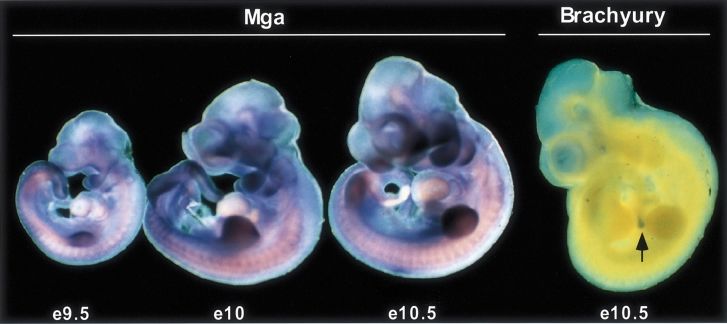
Fig. 2. Whole-mount in situ hybridization analysis of Mga expression in the mouse at embryonic day (e) 9.5, e10 and e10.5. Brachyury expression
at e10.5, which is limited to the distal tip of the tail (arrow) at this stage, was used as a control. Mga expression is widespread, but with highest
levels in the limb buds and branchial arches.
Max, Myc or Mad were identified in this screen. The ~14 kb. Using duplicate Northern blots prepared from
cloning and characterization of three of these proteins PC12 RNA, we found that separate probes specific for
have been described previously (Hurlin et al., 1995, 1997). either the Mga T-box region or the bHLHZip region gave
The characterization of the fourth of these proteins, Mga, a hybridization signal at apparently identical positions
is presented here. From the Max yeast two-hybrid screen, (Figure 1D). The size of the mRNA detected by both
several interacting clones were identified that contained probes is consistent with the ~14 kb Mga cDNA obtained
cDNAs coding for the same novel bHLHZip DNA- in library screens (not shown). In addition to these data,
binding, dimerization domain. To isolate a full-length we found that the T-box and bHLHZip regions map to
cDNA, the region obtained in the two-hybrid screen was the same chromosomal position (Figure 6), providing
initially used to screen a variety of murine and human further evidence that both domains are encoded within a
cDNA libraries. cDNAs containing an identical bHLHZip single ORF.
region were found using a murine e14.5 kidney library (a The embryonic expression pattern of Mga in the mouse
generous gift of V.Dixit). An apparent full-length sequence was examined by whole-mount in situ hybridization at
was constructed using several overlapping cDNA clones e9.5, e10 and e10.5 using probes specific for the bHLHZip
obtained from sequential screening of the embryonic (Figure 2) and T-box regions (not shown). Apparently
kidney library. This cDNA, containing an open reading identical patterns of expression were obtained using these
frame (ORF) of 9018 nucleotides and coding for a two probes (not shown). The general pattern of Mga
protein of 3006 amino acids (aa), was named Mga (DDBJ/ expression was similar during the embryonic stages exam-
EMBL/GenBank accession No. AF205935). The cDNA ined with highest expression levels in the limb buds,
contained a start codon in good context, preceded by branchial arches and the tail region (Figure 2). In contrast
numerous stop codons in all three reading frames. A stop to Mga, Brachyury expression at e10.5 was limited to the
codon followed by a polyadenylation signal and sequence distal tip of the tail (Figure 2) as demonstrated previously
indicated the 3⬘ end of the cDNA. Database comparisons (Kispert et al., 1995). Notably, the expression pattern of
of the full- length Mga sequence revealed not only a Mga appears to overlap with many other T-box-containing
bHLHZip region most closely related to c-Myc (Figure 1B; genes, including Brachyury, Tbx2, 3, 4 and 5 (Papaionnou
39% identical to c-Myc), but also strong homology and Silver, 1998). These results raise the possibility that
between an N-terminal region in the Mga protein and the Mga may participate in regulatory pathways controlling
T-box DNA-binding domain of the Tbx family of proteins mesoderm induction, possibly in concert with other
(Figure 1C). An alignment of the Mga T-domain with T-domain proteins.
several Tbx family members, including Tbx6, which it is
most similar to (55% identical in the T-domain), is shown Interaction with Max
in Figure 1C. Mga showed little or no homology with To initially confirm the yeast two-hybrid results, in vitro
Tbx proteins outside the T-box or with Myc proteins (and co-immunoprecipitation experiments were performed. The
related bHLHZip proteins) outside the bHLHZip domain. yeast two-hybrid library plasmid containing the Mga
After surveying RNA from a number of different cell bHLHZip region was used to produce in vitro translated,
lines and adult tissues by Northern blot analysis using a [32S]methionine-labeled VP16–Mga fusion protein. This
Mga bHLHZip probe, the only positive samples found protein was incubated with baculovirus-produced Max
were the rat pheochromocytoma cell line PC12 (Figure 1D) and immunoprecipitated with Max antisera. The Max
and C2C12 myoblasts (H.Carlson and P.J.Hurlin, antisera efficiently co-immunoprecipitated the VP16–Mga
unpublished) with each showing a hybridizing signal at fusion protein in the presence but not the absence of added
7021P.J.Hurlin et al.
(Hurlin et al., 1997). The low stringency immuno-
precipitations were washed three times with low stringency
buffer and co-immunoprecipitated proteins eluted by
incubation in high stringency buffer. The eluted proteins
were subjected to a second immunoprecipitation with an
anti-Flag antibody and analyzed by PAGE (Figure 3B). A
high stringency immunoprecipitation of Flag-Mga trans-
fected cell extracts with anti-Flag antibody or immunogen
blocked anti-Flag antibody was run on the same gel
(Figure 3B). A large protein was detected in the low
stringency Max immunoprecipitate that co-migrated with
the Flag-Mga protein immunoprecipitated under high
stringency conditions (Figure 3B). These results show
that overexpressed Mga interacts with Max and strongly
suggest that Max and Mga associate in vivo.
DNA-binding and transcriptional activities of Mga
The DNA-binding properties of the bHLHZip and
T-domain regions of Mga were investigated by electro-
phoretic mobility shift assays (EMSA) employing either
oligonucleotides containing the preferred Myc–Max
bHLHZip-binding sequence CACGTG or the preferred
Brachyury recognition sequence AATTTCACACCTA-
GGTGTGAAATT (Kispert and Herrmann, 1993). For
binding to CACGTG, a glutathione S-transferase (GST)
fusion protein containing the Mga bHLHZip domain (aa
2347–2479) or a control GST fusion protein containing
a Mga C-terminal region (aa 2771–1940) showing no
homology to any known proteins was used. As shown in
Figure 4A, the Mga bHLHZip domain alone failed to
bind CACGTG, but produced a strong, concentration-
dependent band shift when pre-incubated with baculovirus-
produced Max.
To test Mga T-domain binding to the preferred
Brachyury-binding sequence, the plasmid pCDNAMgaX1-
Pst was used to produce an in vitro translated N-terminal
fragment of Mga containing the T-domain region
(N-terminal 568 aa). Mga binding was compared with that
Fig. 3. Mga interacts with Max in vitro and in vivo. (A) The Mga of in vitro translated Brachyury containing an HA tag
bHLHZip region fused to VP16 was in vitro translated (ivt Mga) and (made from plasmid pCS2-Bra5⬘Ha). Mga bound the
interaction with baculovirus-produced Max was tested by performing
immunoprecipitations with either Max immune or Max preimmune Brachyury site (Figure 4B) and both Mga and Brachyury
serum as indicated. (B) HEK 293T cells were transfected with band shifts were supershifted using antisera raised against
pMEFLAG-Mga expressing a FLAG-tagged Mga protein. To identify the N-terminus of Mga or anti-HA antibody, respectively,
the expressed FLAG-Mga protein, transfected cells were but not by control antibodies (anti-Mga C-terminus and
immunoprecipitated under high stringency (HS) conditions with a
FLAG monoclonal antibody or the same antibody blocked (b) by pre-
anti-FLAG, respectively).
incubation with a FLAG peptide. To test for co-immunoprecipitation Based on these DNA-binding properties, the transcrip-
between Mga and Max, endogenous Max was immunoprecipitated tional activities of Mga were tested using either an
under low stringency (LS) conditions, the immunoprecipitates were empty reporter plasmid [pGL2pro (Promega)] or ones
washed in the same LS buffer and then incubated in high stringency containing either four promoter-proximal Myc–Max
buffer to release proteins bound to Max. High stringency eluates were
immunoprecipitated with anti-FLAG antibody or immunogen blocked (CACGTG)-binding sites (pGL2M4; Ayer et al., 1995),
(b) anti-FLAG antibody. The location of FLAG-Mga is indicated with two Brachyury (T)-binding sites (pGL2T2) or both
an arrow. Brachyury and Myc–Max sites (pGL2T2M4). When trans-
fected into HEK 293 cells, the activity of the empty
Max (Figure 3A). Additional in vitro studies investigating reporter (pGL2pro) was very low relative to the other
potential interaction between Mga and Myc, Mad and Mnt reporter plasmids and was not affected by co-transfection
proteins were negative (data not shown). of any of the expression plasmids used in this study
To investigate the interaction between Mga and Max (data not shown), which include ones expressing Mga,
in vivo, co-immunoprecipitation experiments were carried Brachyury, Max and ∆BRMax. ∆BRMax lacks its DNA-
out in HEK 293 cells transfected with a Flag-tagged binding basic region and functions as a dominant negative
version of the full-length Mga cDNA. Transfected cells by forming heterodimers incapable of binding DNA Myc–
were metabolically labeled with [35S]methionine and cell Max target sites (Ayer et al., 1993). Consistent with a
extracts were immunoprecipitated with Max antisera or previous study (Kispert et al., 1995), Brachyury activated
Max preimmune sera under low stringency conditions transcription of reporters containing Brachyury sites, but
7022Dual-specificity transcription factor
Fig. 4. DNA binding by the Mga bHLHZip and T-domain regions. (A) DNA binding by the Mga bHLHZip region to an oligonucleotide containing
the preferred Myc–Max-binding site CACGTG was tested using a purified GST fusion protein containing the Mga bHLHZip domain. The Mga
bHLHZip fusion protein was incubated with the oligonucleotide in the presence and absence of baculovirus-produced Max and with increasing
amounts of Mga fusion protein in the presence of a constant amount of Max (sloped line). A GST fusion protein containing the C-terminal 96 aa of
the c-MYC protein (GST–cMyc) was used as a positive control. (B) Binding of the Mga T-domain to the preferred Brachyury-binding site
AATTTCACACCTAGGTGTGAAATT was tested using an in vitro transcribed and translated N-terminal fragment (568 aa) of Mga containing the
entire T-domain. For a positive control, in vitro transcribed and translated Brachyury containing an HA tag was used. Supershift analysis of Mga
complexes was performed using antiserum raised against the N-terminal 268 aa of Mga (αNt) or a C-terminal portion of Mga (αCt). For supershift
analysis of Brachyury, anti-HA or anti-FLAG (negative control) antibodies were used.
no activity was observed when it was co-transfected with (Figure 6). A dose-dependent inhibition of Mga repression
the pGLM4 reporter containing only proximal CACGTG and a switch to activation were observed. Since neither
sites (data not shown). In contrast, transfection of our Max nor ∆BRMax alone had little effect on this reporter,
full-length Mga cDNA repressed reporter transcription we conclude that Max binding to Mga is necessary
from the pGLT2 and pGLT2M4 plasmids, but showed for the transcription activation observed. Therefore, we
little activity on the pGL2M4 reporter (Figure 5). Thus, conclude that Max binding to Mga confers a DNA-
repression by Mga was enhanced or dependent on the binding-dependent function (on promoters containing E-
presence of T-domain-binding sites. Surprisingly, co-trans- box sites) and a DNA-binding-independent function (on
fection of Max with Mga resulted in transcriptional activa- promoters containing Brachyury-binding sites). Although
tion of each of the reporters. To determine whether the complicated, our results indicate that Mga is capable of
observed activation was dependent on E-box DNA binding regulating transcription from promoters containing either
by Mga, ∆BRMax was co-transfected with Mga. ∆BRMax Brachyury or Myc–Max-binding sites and suggest that
relieved activation of the pGL2M4 promoter (Figure 5A) cellular Max levels dictate whether it functions as a
and partially relieved activation of the pGL2M4T2 reporter repressor or an activator.
(Figure 5C), consistent with the idea that Max-mediated
E-box binding by Mga contributes to its activation function Mga inhibits Myc-dependent cell transformation
on promoters containing Myc–Max sites. However, co- The DNA-binding and transcriptional activities of Mga
transfection of ∆BRMax functionally substituted for Max suggest that it may impinge on the function of other
in mediating activation of the pGL2T2 reporter Max-interacting proteins. We used the Myc⫹Ras co-
(Figure 5B), indicating that Max binding to Mga also has transformation assay (Land et al., 1983) as a system to
a function independent of its role in DNA binding. determine whether Mga expression influences the onco-
To investigate more rigorously the unexpected ability genic activities of Myc. Transfections of primary rat
of Max and ∆BRMax to mediate transcriptional activation fibroblasts with Myc and Ras expression plasmids were
by Mga on the pGL2T2 reporter, a series of titration performed with or without the addition of Mga expression
experiments was performed. Using constant amounts of plasmid and foci of transformed cells were counted after
transfected Mga, increasing amounts of Max or ∆BRMax 2 weeks in cell culture (Figure 7A). In the absence of
were co-transfected along with the pGL2T2 reporter Mga, transfection of Myc⫹Ras resulted in the development
7023P.J.Hurlin et al.
Fig. 5. Transcriptional activities of Mga. The indicated cDNAs in the vector pCS2 were transfected into HEK 293 cells together with the reporter
pGLM4 (A) containing four promoter-proximal Myc–Max-binding sites, the reporter pGLT2 (B) containing two promoter-proximal Brachyury-
binding sites (Kispert and Herrmann, 1993), or the reporter pGLM4T2 (C) containing four Myc–Max and two Brachyury sites at the same positions
as in the pGLM4 and pGLT2 reporters, respectively. The total amount of DNA transfected in each assay was equalized using empty vector (pCS2).
Luciferase activity was normalized to β-galactosidase activity produced by a co-transfected plasmid, pCMVβGAL, and measured using the
luminescent substrate Galacton Plus (Tropix). Transfections were performed in triplicate and repeated at least two times. Results are represented as
mean ⫾ SEM.
of numerous foci. In the presence of transfected Mga, a chromosome 2 (Figure 8). This position did not recombine
dramatic reduction in the number of foci in each of three with Epb4.2 in 161 animals typed in common, suggesting
separate experiments was seen. An expression plasmid that these loci are within 1.9 cM of each other (upper
containing the N-terminal 758 aa of Mga (pCDNA- 95% confidence limit).
MgaA2) and thus containing the T-domain and lacking The central region of chromosome 2 shares a region of
the bHLHZip, failed to reduce the number of foci produced homology with human chromosome 15q. In particular,
by Myc and Ras. Likewise, co-transfection of Brachyury the tight linkage between Mga and Epb4.2 suggests
also failed to reduce the number of foci produced by Myc that human MGA lies on human chromosome 15q15. The
and Ras. Therefore, these results suggest that the T-domain location of Mga at human chromosome 15q15
region of Mga is insufficient for inhibition of Myc/Ras was confirmed using fluorescent in situ hybridization
co-transformation and that regions C-terminal (including (K.Sushia and C.Disteche, unpublished). Although there
the bHLHZip region) are required. have been several reports of chromosomal translocations
The results of our cell transformation assays suggested or deletions occurring at human 15q15 (Hunger et al.,
that the mechanism of suppression by Mga might involve 1993; D’Alessandro et al., 1994), no common disease
interference with the transcription activation of Myc target syndromes linked to unidentified genes appear to map to
genes. We tested this posibility by performing transcription this position.
assays in which increasing amounts of Mga expression
plasmid were transfected in the presence of a constant
Discussion
amount of transfected c-Myc expression plasmid
(Figure 7B). Whereas Myc alone activated transcription The Myc, Mad and Mnt bHLHZip proteins possess
6-fold, co-transfection of Mga resulted in a dose-dependent intrinsic transcriptional activities, but require heterodimer-
suppression of activation. We note that suppression of ization with the small bHLHZip protein Max for specific
Myc activation by Mga appears less potent than that DNA binding to E-box sites. Thus, Max functions as an
caused by Mad proteins and Mnt, which also both effi- obligate partner for members of a transcription factor
ciently suppress Myc-dependent transformation (Hurlin network that participate in regulating cell proliferation.
et al., 1995, 1997). We show here that Max interacts with another bHLHZip
protein, Mga, which also requires heterodimerization with
Chromosomal location of Mga Max for E-box binding. The Mga bHLHZip region falls
The mouse chromosomal location of Mga was determined within a subclass of bHLHZip proteins that includes Max,
using separate cDNA probes specific for either the Myc, Mad and Mnt proteins. However, Mga is distinctly
bHLHZip region or the T-domain of Mga. Interspecies different from these proteins in that it contains a second
backcross analysis was performed using progeny derived highly conserved DNA-binding domain, the T-domain.
from matings of [(C57BL/6J ⫻ M.spretus)F1 ⫻ C57BL/ Although the presence of two DNA-binding domains in
6J] mice (see Methods in Copeland and Jenkins, 1991). the same molecule is not unprecedented [for example
The Mga bHLHZip and T-domain probes localized to several members of the Pax family of transcription factors
the same chromosomal position at the central region of contain both homeobox and paired-box DNA-binding
7024Dual-specificity transcription factor
Fig. 6. E-box-independent activities of Max on Mga function. (A and C) Titrations of increasing amounts (µg shown in parentheses) of transfected
Max or ∆BRMax plasmids were performed in the presence of a constant amount of transfected Mga plasmid (5 µg). (B and D) Titrations of
increasing amounts of transfected Max or ∆BRMax plasmids were also carried out in the absence of transfected Mga to measure their Mga-
independent activities. Experiments were carried out in triplicate and performed twice. Results are represented as mean ⫾ SEM.
domains (Stuart et al., 1994)], it is nonetheless highly Max- and Brachyury-binding sites (Figure 4) raises the
unusual. In an attempt to determine whether the Mga possibility that Mga may regulate the same, or an over-
cDNA was an artifact of cloning, both the T-domain and lapping set of genes regulated by Myc–Max heterodimers,
the bHLHZip regions were used as probes to identify Brachyury and their related proteins. Although we find
chromosomal position (Figure 6), mRNA transcript size, that Mga exhibits very little net transcriptional activity
and tissue and embryonic expression patterns (Figures 1 alone on a reporter plasmid containing only promoter-
and 2; data not shown). In each situation, apparently proximal CACGTG-binding sites (Figure 5A), our results
identical results were achieved with the separate probes, demonstrating that Mga suppresses transcription activation
strongly suggesting that the cloned Mga cDNA represents by c-Myc and inhibits Myc-dependent cell transformation
a naturally occurring gene product. Interestingly, sequen- (Figure 7) are consistent with the theory that Mga regulates
cing of the mouse Mga genomic T-box region revealed Myc–Max target genes in vivo. Our studies of the transcrip-
that Mga does not contain the highly conserved intron– tional activities of Mga using promoters engineered to
exon structure characteristic of T-box-containing genes contain proximal Brachyury sites and/or Myc–Max sites
(data not shown). Instead, the Mga T-box is contained on indicate that Mga has similar and distinct binding site-
a single exon, raising the possibility that the Mga gene dependent activities and that it can function as both a
evolved from the insertion of a reverse transcribed mRNA transcriptional repressor and activator (Figures 5 and 6).
of a T-box gene into a gene coding for a Max-interacting Whereas the transcriptional repression function of Mga
bHLHZip protein. appears to be dependent on the presence of Brachyury-
The presence of both a T-domain and a bHLHZip binding sites, transcriptional activation was seen with
in Mga suggests that its transcriptional and biological reporters containing either Brachyury- or Myc–Max-
activities will be more complex than other members of binding sites and was dependent on transfected Max
the bHLHZip and T-domain families. Indeed, the ability (Figures 5 and 6). Therefore, our results indicate that
of Mga–Max heterodimers to bind to the preferred Myc– Mga–Max heterodimers mediate the activation function
7025P.J.Hurlin et al.
Fig. 8. The Mga murine chromosomal position. A partial chromosome
linkage map is shown indicating the location of Mga in relation to
several linked genes. The number of recombinant F2 animals over the
total number of F2 animals typed plus the recombination frequencies,
expressed as genetic distance in centimorgans (⫾ SE), is shown for
each pair of loci on the left of the chromosome map. Where no
recombinants were found between loci, the upper 95% confidence
limit of the recombination distance is given in parentheses. The human
chromosomal positions for the indicated loci are shown to the right.
some cells due to competition with additional Max-
interacting proteins for binding to Max. Our results predict
that the transcriptional and biological activities of Mga
would be modulated under these conditions.
Although several studies have investigated the transcrip-
tional activities of T-domain proteins including Brachyury
(Kispert et al., 1995; Casey et al., 1998) and Tbx2 (Carreira
et al., 1998), the major focus of studies investigating the
function of Tbx family proteins has been their role in
embryonic development. The gene encoding the proto-
Fig. 7. Suppression of Myc transcription activation and inhibition of
Myc-dependent cell transformation by Mga. (A) Primary rat embryo typical T-domain protein Brachyury was cloned by virtue
fibroblasts were transfected with expression plasmids containing the of it being responsible for the classic mouse mutant,
indicated cDNAs and maintained in medium containing 5% fetal tailless, in which posterior mesoderm induction is abrog-
bovine serum for 2 weeks. The total number of foci produced is ated (for review see Herrmann and Kispert, 1994). The
indicated for each of three independent experiments. MgaA2* is a
deletion mutant of Mga containing the N-terminal 758 aa of Mga,
mouse Brachyury phenotype corresponds well to Brach-
which includes the T-domain. (B) Transcription assays using the yury expression in posterior mesoderm precursors and
pGL2M4 reporter plasmid co-transfected with a constant amount of c- ectopic expression of Brachyury induces the expression of
Myc plasmid and with increasing amounts of Mga (µg shown in mesodermal markers (Cunliffe and Smith, 1992; O’Reilly
parentheses). Results are represented as mean ⫾ SEM. et al., 1995). Thus, Brachyury appears to fulfill a role
as a primary regulator of mesoderm induction during
of Mga. However, it is difficult to explain why Max, as gastrulation. Like Brachyury, several other T-box genes
well as ∆BRMax, a dominant-negative form of Max play essential roles in the regional induction and specifica-
predicted to extinguish E-box binding by Mga, is also tion of mesoderm during development (Papaioannou and
capable of mediating Mga-dependent activation of a Silver, 1998). Although Mga expression is widespread,
reporter containing only Brachyury sites (Figures 5B and highest levels are found in limb buds, branchial arches
6C). One possiblity is that Max binding to Mga serves and tail, regions patterned by mesoderm and mesodermal–
the dual function of both generating an E-box-binding epithelial interactions (Figure 2). This expression pattern
heterodimer and simultaneously blocking interaction of a raises the possibility that Mga may participate in regulating
corepressor that also interacts with the bHLHZip region mesoderm induction or differentiation at these sites.
of Mga. Thus, Max binding may provide a mechanism to Furthermore, since Mga expression overlaps with the
regulate both target gene specificity and transcriptional expression of several other Tbx genes, complex regulatory
activity (whether it functions as a repressor or activator) mechanisms governing mesoderm induction may exist that
of Mga. Although Max protein levels are generally thought utilize multiple T-domain proteins. Indeed, in Xenopus
to be in excess of Myc, there are reports of changes in regulatory pathways involving Xenopus Brachyury (Xbra),
Max levels during differentiation (Delgado et al., 1995) and the T-domain proteins VegT and Eomes have been
and apoptosis (Shichiri et al., 1999), and indeed PC12 identified that are thought to coordinate the transcriptional
cells have been found to lack Max entirely (Hopewell and regulation of mesoderm specific genes in response to
Ziff, 1995). Furthermore, Max levels may be limiting in mesoderm signaling proteins such as activin and fibroblast
7026Dual-specificity transcription factor
growth factor family members (Lustig et al., 1996; Ryan Luciferase assays
HEK 293 cells were transfected using the calcium phosphate precipitation
et al., 1996; Stennard et al., 1996; Zhang and King, 1996; method (Graham and van der Eb, 1973). The T2 reporter plasmid was
Horb and Thomsen, 1997). constructed by inserting tandem consensus Brachyury-binding sites
Although the biological function of Mga has yet to be derived from the plasmid pG.Cat.BS.p2 (Kispert et al., 1995) into the
determined, the presence in Mga of both a T-domain and proximal promoter XhoI site of pGL2pro (Promega). The T2M4 reporter
a Max-interacting bHLHZip domain suggests a role in the was constructed by inserting the same Brachyury sites into the XhoI site
of pGL2M4 (Ayer et al., 1993), containing a 4-fold reiteration of the
coordinate regulation of target genes recognized by both consensus Myc–Max-binding site CACGTG. The plasmid pCS2 (CMV
Max-network and T-domain proteins. Thus, Mga may promoter; D.Turner and R.Rupp, unpublished) was used to express Mga,
function to integrate regulation of cell proliferation with Max, ∆BRMax and c-Myc. The total amount of plasmid DNA in
mesoderm specification/induction during embryonic transfections was adjusted with empty pCS2 plasmid to maintain uniform
levels. β-galactosidase and luciferase assays were performed using
development. Galacton Plus (Tropix) and luciferin luminescent substrates and a Tropix
TR717 luminometer.
Materials and methods Myc⍣Ras transformation assay
Transformation assays were performed as described previously (Hurlin
Cloning Mga et al., 1997) with the exception that 5 µg of pCS2-based Mga and
A yeast two-hybrid screen was carried out using Max as bait as described Brachyury expression plasmids were used in co-transfections with Myc
previously (Hurlin et al., 1995) using a cDNA library derived from 9.5 and Ras cDNAs.
and 10.5-day-old mouse embryos (Wilkinson and Nieto, 1993). The
cDNA fragment recovered in the two-hybrid screen coding for a Northern blotting and in situ hybridization
bHLHZip motif was used to screen a mouse e14.5 kidney cDNA library Northern blots were prepared from total RNA purified from PC12 cells
(a kind gift of V.Dixit). Sequential library screens were performed using using Trizol (Gibco-BRL). RNA was separated by electrophoresis in a
cDNA fragments obtained until consensus start and stop sites were 1% agarose, 2.2 M formaldehyde gel and transferred onto Hybond N⫹
identified. Because of the surprising finding of a T-domain DNA-binding (Amersham). 32P-labeled probes specific to the Mga T-box and bHLHZip
domain within the putative full-length ORF, additional library screens were made from BamHI–EcoRI restriction enzyme (New England
were performed to generate multiple independent overlapping cDNA Biolabs) released inserts of plasmids GST–MgaNT and GST–bHLHZip,
clones covering the entire putative full-length cDNA. The extensive set respectively. Whole-mount in situ hybridization using digoxygenin-
of independent cDNA fragments was used to confirm the Mga cDNA labeled probes was carried out as described by Wilkinson and Nieto
structure. The putative full-length Mga cDNA was constructed from (1993). Digoxygenin 11 UTP (Boehringer) incorporated antisense probes
three separate, overlapping cDNAs (designated X1, N6 and I28) in the specific for the T-box region and the bHLHZip region of Mga were
plasmid pBC SK⫹ (Stratagene). The Mga sequence was determined by produced from plasmids pCDNA1MgaX1 and pCDNA1MgaK32,
sequencing the ends of individual cDNAs obtained in library screens respectively, using T7 RNA polymerase. A Brachyury probe was made
and by sequencing nested exonuclease III deletions (Henikoff, 1987) of from plasmid pBT1 (a gift from P.Soriano) using T3 RNA polymerase.
the putative full-length cDNA. The putative full-length Mga cDNA
contained a 9018 bp ORF coding for a protein of 3006 aa. The full- Chromosomal localization
length cDNA was subcloned into the mammalian expression vectors Interspecies backcross mapping was performed as described previously
pRCCMV (Invitrogen) and pME (a gift of Y.Shio). A FLAG epitope (Copeland and Jenkins, 1991). Two separate probes were used: one
tag sequence was introduced into the 5⬘ end of the Mga cDNA by PCR specific for the Mga bHLHZip region (0.5 kb bHLHZip clone10 fragment
prior to cloning into the pME vector. GST fusion proteins containing from the library plasmid obtained in the yeast two-hybrid screen), and
the Mga T-domain (pGEX-MgaNT, aa 1–333), the Mga bHLHZip one specific for the Mga T-domain (2 kb fragment from the kidney
domain (pGEXMgabHLHZip, aa 2347–2479) and a portion of the cDNA library plasmid pCDNA1-A2). A description of the probes and
C-terminus (pGEXMgaCT, aa 2771–1940) were constructed in the vector RFLPs for loci linked to Mga including thrombospondin (Thbs1),
pGEX2T (Pharmacia). adenosine deaminase (Ada), β2-microglobulin (β2m), and α2-adrenergic
receptor subtype b (Adra2b) has been reported previously (Lawler et al.,
1991; Chang et al., 1994). The erythrocyte protein 4.2 (Ebp4.2) locus
Interaction and DNA-binding assays has not been reported previously for this interspecies backcross. Recom-
The cDNA fragment of Mga obtained from the two-hybrid screen was bination distances and gene order were determined using Map Manager
in vitro transcribed and translated from the library plasmid BTM116 version 2.6.5 (Manley, 1993) by minimizing the number of recombination
(Hollenberg et al., 1995) and used in co-immunoprecipitation assays events required to explain the allele distribution patterns.
with 20 ng of baculovirus-produced Max (Ayer et al., 1993). Anti-Max
antisera 8711 was used to immunoprecipitate Max under low stringency
conditions (L-buffer: PBS⫹0.2% NP-40) in the presence of the in vitro Acknowledgements
translated, [35S]methionine-labeled Mga fragment. Immunoprecipitates
were washed 4⫻ with L-buffer and analyzed by SDS–PAGE and The authors would like to thank B.Herrmann, A.Kispert, V.Dixit and
autoradiography. To test for in vivo association between Mga and Max, Y.Shio for reagents. Thanks also to P.Soriano for help with in situ
a FLAG-tagged version of Mga in the vector pME was transfected into hybridizations and to S.Willis, P.-F.Cheng, A.Bush, Z.Luo and D.Gilbert
HEK 293T cells. Co-immunoprecipitation experiments were carried out for technical help. E.S., N.G.C. and N.A.J. were supported in part by
as described above using Max antiserum 8711 and anti-FLAG monoclonal the National Cancer Institute, DHHS. R.N.E. was supported by NIH
antibodies (Sigma). grant CA20525. R.N.E. is a Research Professor of the American
EMSAs employing an oligonucleotide containing the preferred Myc– Cancer Society.
Max-binding sequence CACGTG were performed as described previ-
ously (Hurlin et al., 1997) using baculovirus Max, and the GST References
fusion proteins GST–MgabHLHZip and GST–MgaCt. GST–Myc(c96)
(Blackwood et al., 1991) fusion protein was used as a positive control. Amati,B., Dalton,S., Brooks,M.W., Littlewood,T.D., Evan,G.I. and
For EMSA of the Mga T-domain and for Brachyury, the preferred Land,H. (1992) Transcriptional activation by the human c-Myc
Brachyury-binding sequence AATTTCACACCTAGGTGTGAAATT oncoprotein in yeast requires interaction with Max. Nature, 359,
(Kispert and Herrmann, 1993) was used. The Mga T-domain region was 423–425.
in vitro transcribed and translated from the plasmid pCDNAMgaX1-Pst. Ayer,D.E., Kretzner,L. and Eisenman,R.N. (1993) Mad: a heterodimeric
Brachyury was in vitro transcribed and translated from the plasmid partner for Max that antagonizes Myc transcriptional activity. Cell,
pCS2-Bra5⬘HA, containing an introduced HA tag at the 5⬘ end of 72, 211–222.
Brachyury. Supershift analysis of Mga DNA-binding complexes was Ayer,D.E., Lawrence,Q.A. and Eisenman,R.N. (1995) Mad–Max
performed using polyclonal antisera raised against GST fusion proteins transcriptional repression is mediated by ternary complex formation
produced from plasmids pGEXMgaNT and pGEXMgaCT (see above). with mammalian homologs of yeast repressor Sin3. Cell, 80, 767–776.
Anti-HA antibodies (BMB) were used to supershift Brachyury complexes. Bamshad,M. et al. (1997) Mutations in human TBX3 alter limb, apocrine
7027P.J.Hurlin et al.
and genital development in ulnar-mammary syndrome. Nature Genet., Ras co-transformation by Mad is mediated by multiple protein–protein
16, 311–315. interactions. Cell Growth Differ., 6, 623–629.
Basson,C.T. et al. (1997) Mutations in human TBX5 cause limb and Kretzner,L., Blackwood,E.M. and Eisenman,R.N. (1992) The Myc and
cardiac malformation in Holt-Oram syndrome. Nature Genet., 15, Max proteins possess distinct transcriptional activities. Nature, 359,
30–35. 426–428.
Blackwood,E.M and Eisenman,R.N. (1991) Max: A helix–loop–helix Land,H., Parada,L.P and Weinberg, RA. (1983) Tumorigenic conversion
zipper protein that forms a sequence-specific DNA binding complex of primary embryo fibroblasts requires at least two cooperating
with Myc. Science, 251, 1211–1217. oncogenes. Nature, 304, 596–601.
Carreira,S., Dexter,T.J., Yavuzer,U., Easty,D.J. and Goding,C.R. (1998) Lawler,J., Duquette,M., Ferro,P., Copeland,N.G., Gilbert,D.J. and
Brachyury-related transcription factor Tbx2 and repression of the Jenkins,N.A. (1991) Characterization of the murine thrombospondin
melanocyte-specific TRP-1 promoter. Mol. Cell. Biol., 18, 5099–5108. gene. Genomics, 11, 587–600.
Casey,E.S., O’Reilly,M.A., Conlon,F.L. and Smith,J.C. (1998) The Li,Q.Y. et al. (1997) Holt-Oram syndrome is caused by mutations in
T-box transcription factor Brachyury regulates expression of eFGF TBX5, a member of the Brachyury (T) gene family. Nature Genet.,
through binding to a non-palindromic response element. Development, 15, 21–29.
125, 3887–3894. Lustig,K.D., Kroll,K.L., Sun,E.E. and Kirschner,M.W. (1996) Expression
Chang,N.C., Jenkins,N.A., Gilbert,D.J., Copeland,N.G., Chang,Y.-H., cloning of a Xenopus T-related gene (Xombi) involved in mesodermal
Chen,W.-M. and Chang,A.C. (1994) Assignment of two α2-adreno- patterning and blastopore lip formation. Development, 122, 4001–4012.
ceptor subtype genes to murine chromosomes. Neurosci. Lett., 167, Manley,K.F. (1993) A Machintosh program for storage and analysis of
105–108. experimental genetic mapping. Mamm. Genome, 4, 303–313.
Chapman,D.L. and Papaioannou,V.E. (1998) Three neural tubes in mouse Meroni,G. et al. (1997) Rox, a novel bHLHZip protein expressed in
embryos with mutations in the T-box gene Tbx6. Nature, 391, 695–697. quiescent cells that heterodimerizes with Max, binds a non-canonical
Copeland,N.G. and Jenkins,N.A. (1991) Development and applications E box and acts as a transcriptional repressor. EMBO J., 16, 2892–2906.
of a molecular genetic linkage map of the mouse genome. Trends Müller,C.W. and Herrmann,B.G. (1997) Crystallographic structure of
Genet., 7, 113–118. the T-domain–DNA complex of the Brachyury transcription factor.
Cunliffe,V. and Smith,J.C. (1992) Ectopic mesoderm formation in Nature, 389, 884–888.
Xenopus embryos caused by widespread expression of a Brachyury O’Reilly,M.A., Smith,J.C. and Cunliffe,V. (1995) Patterning of the
homologue. Nature, 358, 427–430. mesoderm in Xenopus: dose-dependent and synergistic effects of
D’Alessandro,E., Ligas C., Lo Re,M.L., Marcanio,M.P., Gentile,T. and Brachyury and Pintallavis. Development, 121, 1351–1359.
Del Porto,G. (1994) Partial monosomy of 7q32 in a case of de novo Papaioannou,V.E. and Silver,L.M. (1998) The T-box gene family.
rcp (7;15) (q32;q15). J. Med. Genet., 31, 413–415. BioEssays, 20, 9–19.
Delgado,M.D., Lerga,A., Canelles,M., Gomez-Casares,M.T. and Leon,J. Roussel,M.F., Ashmun,R.A., Sherr,C.J., Eisenman,R.N. and Ayer,D.E.
(1995) Differential regulation of Max and role of c-Myc during (1996) Inhibition of cell proliferation by the Mad1 transcriptional
erythroid and myelomonocytic differentiation of K562 cells. repressor. Mol. Cell. Biol., 16, 2796–2801.
Oncogene, 10, 1659–1665. Ryan,K., Garnett,N., Mitchell,A. and Gurdon,J.B. (1996) Eomesodermin,
Graham,F. and van der Eb,A. (1973) A new technique for the assay of a key early gene in Xenopus mesoderm differentiation. Cell, 87,
infectivity of human adenovirus 5 DNA. Virology, 52, 456–457. 989–1000.
Henikoff,S. (1987) Unidirectional digestion with exonuclease III in DNA Schreiber-Agus,N., Chin,L., Chen,K., Torres,R., Rao,G., Guida,P.,
sequence analysis. Methods Enzymol., 155, 156–165. Skoultchi,A.I. and DePinho,R.A. (1995) An amino-terminal domain
Henriksson,M. and Lüscher,B. (1996) Proteins of the Myc network: of Mxi1 mediates anti-Myc oncogenic activity and interacts with a
essential regulators of cell growth and differentiation. Adv. Cancer homolog of the yeast repressor SIN3. Cell, 80, 777–786.
Shichiri,M., Kato,H., Doi,M., Marumo,F. and Hirata,Y. (1999) Induction
Res., 68, 109–182.
of max by adrenomedullin and calcitonin gene-related peptide
Herrmann,B.G. and Kispert,A. (1994) The T genes in embryogenesis.
antagonizes endothelial apoptosis. Mol. Endocrinol., 13, 1353–1363.
Trends Genet., 10, 280–286.
Stennard,F., Carnac,G. and Gurdon,J.B. (1996) The Xenopus T-box gene,
Hollenberg,S.M., Sternglanz,R., Cheng,P.-F. and Weintraub,H. (1995)
Antipodean, encodes a vegetally localised maternal mRNA and can
Identification of a new family of tissue-specific basic helix–loop–helix
trigger mesoderm formation. Development, 122, 4179–4188.
proteins with a two-hybrid system. Mol. Cell. Biol., 15, 3813–3822.
Stuart,E.T., Kioussi,C. and Gruss,P. (1994) Mammalian Pax genes. Annu.
Hopewell,R. and Ziff E.B. (1995) The nerve growth factor-responsive
Rev. Genet., 28, 219–236.
PC12 cell line does not express the Myc dimerization partner Max.
Wilkinson,D.G. and Nieto,M.A. (1993) Detection of messenger RNA
Mol. Cell. Biol., 15, 3470–3478.
by in situ hybridization to tissue sections and whole mounts. Methods
Horb,M.E. and Thomsen,G.H. (1997) A vegetally localized T-box Enzymol., 225, 361–373.
transcription factor in Xenopus eggs specifies mesoderm and endoderm Zervos,A.S., Gyuris,J. and Brent,R. (1993) Mxi1, a protein that
and is essential for embryonic mesoderm formation. Development, specifically interacts with Max to bind Myc-Max recognition sites.
124, 1689–1698. Cell, 72, 223–232.
Horb,M.E. and Thomsen,G.H. (1999) Tbx5 is essential for heart Zhang,J. and King,M.L. (1996) Xenopus VegT RNA is localized to the
development. Development, 126, 1739–1751. vegetal cortex during oogenesis and encodes a novel T-box
Hunger,S.P., Tkachuk,D.C., Amylon,M.D., Link,M.P., Carroll,A.J., transcription factor involved in mesodermal patterning. Development,
Welborn,J.L., Willman,C.L. and Cleary,M.L. (1993) HRX involvement 122, 4119–4129.
in de novo and secondary leukemias with diverse chromosome 11q23 Zhang,J., Houston,D.W., King,M.L., Payne,C., Wylie,C. and Heasman,J.
abnormalities. Blood, 81, 197–203. (1998) The role of maternal VegT in establishing the primary germ
Hurlin,P.J., Quéva,C., Koskinen,P.J., Steingrı́msson,E., Ayer,D.E., layers in Xenopus embryos. Cell, 94, 515–524.
Copeland,N.G. Jenkins,N.A. and Eisenman,R.N. (1995) Mad3 and
Mad4: novel Max-interacting transcriptional repressors that suppress Received July 12, 1999; revised and accepted October 28, 1999
Myc-dependent transformation and are expressed during neural and
epidermal differentiation. EMBO J., 14, 5646–5659.
Hurlin,P.J., Queva,C. and Eisenman,R.N. (1997) Mnt, a novel Max-
interacting protein is coexpressed with Myc in proliferating cells and
mediates repression at Myc binding sites. Genes Dev., 11, 44–58.
Kispert,A. and Herrmann,B.G. (1993) The Brachyury gene encodes a
novel DNA binding protein. EMBO J., 12, 3211–3220.
Kispert,A. and Herrmann,B.G. (1994) Immunohistochemical analysis of
the Brachyury protein in wild-type and mutant mouse embryos. Dev.
Biol., 161, 179–193.
Kispert,A., Koschorz,B. and Herrmann,B.G. (1995) The T protein
encoded by Brachyury is a tissue-specific transcription factor. EMBO
J., 14, 4763–4772.
Koskinen,P.J., Ayer,D.E. and Eisenman,R.N. (1995) Repression of Myc-
7028You can also read