Micro-computed tomography - 3D X-ray imaging - Brittle stars in the Bokkeveld - Research at ASSAf
←
→
Page content transcription
If your browser does not render page correctly, please read the page content below
VOLUME 15 | NUMBER 2 | 2019
                                                                ISSN 1729-830X

 Micro-computed tomography
                 3D X-ray imaging

Brittle stars in the MicroCT for metal            Forecasting sea
Bokkeveld			3D printing                           conditions

                  ACADEMY OF SCIENCE OF SOUTH AFRICA
The Universe is right next door
The South African Astronomical Observatory (SAAO) brings the Universe closer to home.

                                                    We work with leading astronomers and
                                                    scientists to unwrap the mysteries of the
                                                    cosmos and continue to play an important role
                                                    in new discoveries.

                                                    We also operate some amazing technologies
                                                    such as SALT, the biggest optical telescope in
                                                    the Southern Hemisphere.

If you are thinking of a career in astronomy, the technology behind it or just interested
in what goes on in the Universe, why not visit us?
   Tours and visits to SALT and the Cape Town Observatory
   Astronomical information
   Science resources and activities for schools
   Astronomy career information
   In-service technical training
   Scholarships
   National Astrophysics and Space Science Programme

Website: www.saao.ac.za
Telephone: 021 447 0025
Email: enquiries@saao.ac.za
Fax: 021 447 3639
Address: P.O. Box 9, Observatory, 7935
         Observatory Road, Observatory, Cape Town
CONTENTS                          Volume 15 | Number 2 | 2019

THEME                                   FEATURES
6     MicroCT                           20   Scarred Earth

                                                                                    10
      An introduction to micro-              The SALDi project aims to
      computed tomography                    develop tools for monitoring
                                             land degradation
8     Digital avatars new in
      taxonomers’ toolkit               24   ZACube-2

                                                                               20
      Helen Swingler explains                South Africa’s latest
      how microCT can be used                nanosatellite will help protect
      in taxonomy                            the marine environment

10    Looking inside a brooding         26   Marine forecasting
      brittle star                           The role of the South African
      Jannes Landschoff used                 Weather Service’s Marine Unit
      microCT to research
      reproduction of a marine          30   Young scientists meet
      invertebrate                           Nobel Prize winners
                                             The annual Lindau Nobel
11    Brittle stars in the                   Laureate Meeting
      Bokkeveld
      Studying fossil echinoderms
      with microCT
                                        NEWS
14    Little Foot’s inner ear
      MicroCT scans of the hominin      33   Coding and robotics
      fossil shed light on how she
      lived and moved                   34   Beating the Russians at
                                             their own game
15    ‘Bakeng se Afrika’
      Forensic applications of          36   The pen is mightier
      microCT
                                        39   Indigenous knowledge

                                                                                    33
16    MicroCT for metal 3D
      printing
      Anton du Plessis tells us how
      additive manufacturing benefits   REGULARS
      from microCT
                                        40   Back page science

                                        41   Books

                                        42   Puzzles

                                        43   Subscription

                        40
WWW.ASSAF.ORG.ZA
EDITOR’S NOTE

                        Editor
                     Sue Matthews

                Editorial Board
         Himla Soodyall (EO: ASSAf) (Chair)

                                                         The microCT – Nobel Laureates link
                 John Butler-Adam
         (South African Journal of Science)
      Debra Meyer (University of Johannesburg)
              Kevin Govender (SAAO)
              Caradee Wright (MRC)

              Correspondence                             In 1979 Allan Cormack became the                     develop a prototype machine. The
               and enquiries                             third South African to be awarded                    ‘EMI brain scanner’ was installed
                      The Editor                         a Nobel Prize after Max Theiler,                     in a hospital and used for its first
          e-mail: questeditor@iafrica.com
     e-copies: https://www.assaf.org.za/index.           who received the Nobel Prize for                     patient – a woman with a brain
        php/publications/quest-magazine                  Physiology or Medicine in 1951 for                   tumour – on 1 October 1971. The
                 @questSa1 - Twitter
     Quest: Science for South Africa - Facebook          developing a yellow fever vaccine,                   process subsequently became
                                                         and former ANC leader Albert Lutuli,                 known as computed tomography,
           Advertising enquiries                         who won the Nobel Peace Prize in                     computer-assisted tomography or
                   Barbara Spence
                Avenue Advertising                       1960 for his non-violent resistance                  computerised axial tomography,
                     PO Box 71308                        to the apartheid regime. Cormack                     and ‘CAT scans’ soon became
                    Bryanston 2021
                  Tel: (011) 463 7940                    was awarded the Physiology or                        standard medical procedures.
                  Cell: 082 881 3454                     Medicine prize jointly with Godfrey
           e-mail: barbara@avenue.co.za
                                                         Hounsfield for the development                       Later, machines able to scan fine
          Subscription enquiries                         of computer-assisted tomography                      detail at massively increased
            and back issues                              (CAT), although they had come up                     resolution were developed for non-
                    Tsepo Majake                         with the idea quite separately in                    medical applications. The so-called
                 Tel: (012) 349 6645
              e-mail: tsepo@assaf.org.za                 different parts of the world.                        micro-computed tomography is our
                                                                                                              theme for this issue of Quest, and
                    Copyright                            Cormack was a physicist who                          we showcase a number of different
             ©2019 Academy of Science
                   of South Africa
                                                         worked at the University of Cape                     applications of the technology.
                                                         Town before moving to the United
                                                         States with his American wife in                     Interestingly, the discovery of X-rays
                                                         1957 and taking up a professorship                   in 1895 by Wilhelm Röntgen was
                                                         at Tufts University. Before leaving                  recognised with a Nobel Prize in
                Published by the
                                                         South Africa he had begun                            1901, the very first to be awarded
  Academy of Science of South Africa (ASSAf)             developing the theory behind CAT                     for physics. This year’s Lindau Nobel
PO Box 72135, Lynnwood Ridge 0040, South Africa          scans, and published this in the                     Laureate Meeting – a gathering
              Subscription rates                         early 1960s in two scientific papers.                of previous winners of the Nobel
                 (4 issues and postage)                  He proposed that X-rays taken from                   Prize and up to 600 young scientists
      (for other countries, see subscription form)       different angles around a body or                    selected from around the world – is
            Individuals/Institutions - R130.00
             Students/schoolgoers - R65.00               organ could be reconstructed into                    dedicated to physics, and South
                                                         a three-dimensional representation                   Africa will be the ‘host country’ of
             Design and layout                           by computer, but he never put                        the International Day. A larger than
                   LedCool Graphics
                                                         the concept to the test, probably                    normal contingent of
                                                         because the computers of the day                     20 young South
                                                         were not up to the task.                             Africans will be
                                                                                                              in attendance,
                                                         Hounsfield was an electrical                         and we trust
                                                         engineer working at the Beatles’                     they will benefit
                                                         record label EMI (Electrical and                     from the
                                                         Musical Instruments) in England.                     experience as
                                                         He was completely unaware of                         much as the
                                                         Cormack’s work, but his experience                   two Lindau
                                                         with radar systems gave him the                      alumni profiled
                                                         idea of reversing the process, and                   elsewhere in
                                                         sending radiation beams into an                      this issue.
                                                         object from all angles to obtain
                                                         cross-sectional slices that could be
                                                         reconstructed in 3D. He proposed
                                                         X-rays as the form of radiation, and
                                                         started working with a radiologist to
      Every care is taken in compiling the contents of
     this publication, but we assume no responsibility
    for effects arising therefrom. The views expressed
     in this magazine are not necessarily those of the
    publisher. Content may be copied or shared with
                                                         Our cover image, courtesy of Prof. Anton du Plessis of Stellenbosch University’s CT Scanner Facility,
    acknowledgement to Quest as well as the author       shows five paper wasps in their hive. The artificial colour of the wasps, which had been dead for some
     where stated. Copyright of republished material     months, was digitally applied to the image for visual appeal. More images and a 3D video can be viewed
       and images remains with the original source.      at: http://blogs.sun.ac.za/ctscanner/3d-imaging-of-a-wasp-hive/

4    Quest Vol. 15 No. 2 | 2019                                                                                                             WWW.ASSAF.ORG.ZA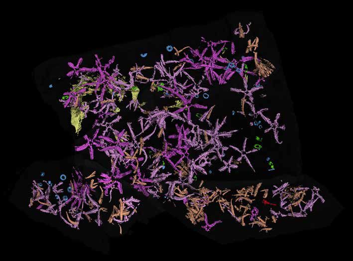
THEME | MicroCT

                                            MicroCT
                 Our theme for this issue of Quest is micro-computed tomography,
                                    known as microCT or just µCT

What is microCT?
MicroCT is a 3D imaging technique utilising X-rays to
see inside an object, slice by slice. MicroCT, also called
microtomography or micro-computed tomography, is
similar to hospital CT or ‘CAT’ scan imaging but on a small
scale with greatly increased resolution. Samples can be
imaged with pixel sizes as small as 100 nanometres and
objects can be scanned as large as 200 millimetres in
diameter.

MicroCT scanners capture a series of 2D planar X-ray
images and reconstruct the data into 2D cross-sectional
slices. These slices can be further processed into 3D
models and even printed as 3D physical objects for              Following microCT scanning, the image data can be processed
                                                                for detailed analysis. © Dragonfly ORS, Wikimedia
analysis. With 2D X-ray systems you can see through
an object, but with the power of 3D microCT systems
you can see inside the object and reveal its internal           samples to be imaged in their native states, including
features. It provides volumetric information about the          light microscopy, laser scanning, visible and other
microstructure, non-destructively.                              spectrum photography. MicroCT is one such technique
                                                                where most of the samples studied are scanned in an
How does a microCT scanner work?                                unaltered state.
X-rays are generated in an X-ray source, transmitted
through the sample, and recorded by the X-ray detector          What are the advantages of microCT scanning?
as a 2D projection image. The sample is then rotated a          MicroCT provides high-resolution 3D imaging information
fraction of a degree on the rotational stage, and another       that can’t be obtained by any other non-destructive
X-ray projection image is taken. This step is repeated          technology. It can be used to study the interior structure
through a 180° turn (or sometimes 360°, depending on            of both material and biological samples without having
sample type). The series of X-ray projection images is          to cut the samples, preserving the samples or specimens
then computed into cross-sectional images through the           for future studies. The quantitative information obtained
computational process called ‘reconstruction’. These            from microCT scanning can only be obtained from 3D
slices can be analysed, further processed into 3D models,       images, and 3D digital models created from microCT
made into movies, printed into 3D physical objects, etc.        virtual slices allow scientists to measure any parameters
                                                                for comparison in before-and-after studies.
What does non-destructive testing mean?
Non-destructive testing means that the sample or                These unique features of microCT scanning allow
specimen being scanned isn’t altered or destroyed               scientists to look at the morphology of a sample and
during testing, or in the preparation for testing. This         study features such as porosity, structure / bone
allows the sample to be preserved for historical record,        thickness, volume fraction, defect analysis, density,
tested again at a later date, used in another test or put       particle size, voids and fibre orientation. Researchers use
into final production. Some other techniques require            microCT to study bone, teeth, tissue / organs, composite
staining, cutting or coating of samples, which can affect       materials, medical devices, batteries, etc.
the sample’s structure, its ongoing usefulness, or its use
in later studies. There are several techniques that allow       What is the difference between medical CT and
                                                                microCT scans?
                                                                MicroCT scanning is X-ray imaging in 3D, using the same
    Indaba yethu kulesisqephu igxile kwi computer               method as medical CT (or ‘CAT’) scans, but microCT is on
    encane yokuthatha ezithombe emzimbeni, ibuye                a much smaller scale with greatly increased resolution.
    yaziwe ngokuthi I microCT noma μCT. Lona ngumshini          Scans from medical CT – introduced as a tool for medical
    okwazi ukukhipha izithombe zamathambo noma                  imaging in the 1970s – are limited to a resolution of one
    izicubu zomzimba, usebenzisa imisebe evela macala           millimetre, which provides sufficient detail for clinical use.
    onke ento ayishuthayo ukuze kuphume izithombe ezi           For materials science and small animal imaging, much
    two-dimensional (2D), bese zihlanganiswa ukukhanda          higher resolution was needed, so microCT scanning was
    I three-dimensional (3D).                                   introduced in the 1980s. MicroCT scanners can work
                                                                at the level of one micrometre (or micron), which is a
                           Translation by Zamantimande Kunene   thousandth of a millimetre, and smaller.

6    Quest Vol. 15 No. 2 | 2019                                                                               WWW.ASSAF.ORG.ZA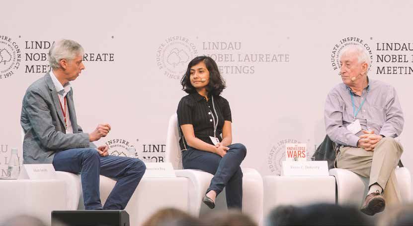
MicroCT | THEME

What is the difference between in vivo and ex
vivo microCT scanning?
Simply put, in vivo (Latin for within life) is the scanning of
live specimens and ex vivo (Latin for out of living) typically
refers to things that used to be alive or samples excised
from something that had been alive. For microCT, in vivo
typically refers to systems that scan mice and rats and in
some cases rabbits, while ex vivo systems typically handle
the remainder of the applications.

With in vivo microCT instruments, since the animal
remains alive, longitudinal studies can be performed to
measure the effects of drug, diet, hormonal and other
treatments on tumours; bone growth and quality; body
mass and other applications on the same subject. This

                                                                                                                                                                    https://doi.org/10.1163/15685381-20181102
can reduce the number of animals needed for a study.

Ex vivo microCT instruments typically handle the
remaining applications, which include end-point studies
of specific regions of an animal that get excised (lungs,
bone, tumours, implants, grafts, etc.), biomaterial
studies, implants in large animals, materials studies,
compression studies, and more. Ex vivo microCT
instruments allow for higher spatial resolution, longer
scan times (since dose to the sample isn’t of concern),
better signal to noise ratios, and therefore better images.
                                                                 Above: Schematic of the microCT scanning and data-
Ex vivo systems have typically been used for most
                                                                 processing technique. © Broekhoven and Du Plessis
applications outside of a living animal.
                                                                 Below: This live armadillo lizard entered defensive mode,
© 2019 Micro Photonics Inc. (www.microphotonics.com)             with typical tail-biting behaviour, prior to the cooling stage
Reproduced with permission. The website above includes a         and maintained this ‘pose’ throughout the scanning period.
blog showcasing numerous examples of microCT applications.       The 3D-rendered image shows the arrangement of the
                                                                 bony plates, called osteoderms, embedded in the skin after
                                                                 digitally removing the scales. © Broekhoven and Du Plessis
       The word tomography is derived
    from the Greek word ‘tomos’, meaning
         slice or section, and ‘graphe’
               meaning drawing.
                                                                                                                      https://doi.org/10.1111/2041-210X.12661

Close Encounters of the 3D Kind
Live animals usually need to be anaesthetised, either
via injection or inhalation, prior to microCT scanning
to ensure they keep perfectly still during the process,
because any movement would result in blurry images.
Chris Broeckhoven and co-researchers at Stellenbosch
University recently described a protocol to immobilise
and restrain live reptiles and amphibians without
administering anaesthetic. These ‘cold blooded’ animals          image quality and scan time. Apart from some blurriness
– more correctly known as ectotherms – cannot produce            in images of the sides of the thorax due to the lizards’
internal heat to maintain their body temperature, so if          breathing, the results were comparable with microCT
they become cold they quickly enter a state of torpor            scans of dead lizards. After the scanning procedure,
(chill coma).                                                    which took up to 16 minutes per individual, the lizards
                                                                 were monitored for a month in the laboratory, but no
The researchers used live lizards as their example to            ill effects of the irradiation were apparent. The lizards
explain the protocol. Each lizard was restrained between         were then released back into their natural habitat, and
two taped-together Styrofoam blocks, and then placed in          monitored for another three months using remote
a holder with crushed ice in a way that ensured no part          camera traps. Most of the lizards were recovered at the
of the animal came into contact with ice or meltwater.           end of this period, and the researchers felt it likely that
This cooled the specimen to a chilly 8°C before scanning         all the lizards were still alive at that point. But what an
began. Experiments were conducted with various scan              experience they had had – rather like the classic alien-
settings to determine the effect on radiation dose rate,         abduction movie Close Encounters of the Third Kind!

WWW.ASSAF.ORG.ZA                                                                                   Quest Vol. 15 No. 2 | 2019                                   7
THEME | TAXONOMY

                            Digital avatars
          new in taxonomers’ toolkit
                 Helen Swingler explains how microCT can be used in taxonomy –
                   the science of naming, describing and classifying organisms

Digital avatars – specimens captured via X-ray 3D micro-            crabs (Crustacea: Decapoda: Anomura: Paguroidea)’.
computed tomography (microCT) scanning technology                   His co-authors are his supervisor in the Department of
– give taxonomers a new tool to ‘dissect’ and study rare            Biological Sciences, Emeritus Professor Charles Griffiths,
or delicate specimens on their computer screens, without            and Associate Professor Anton du Plessis of Stellenbosch
(or in addition to) museum or natural history collections           University.
and artists’ replicas.
                                                                    These specimens are held at the Iziko Museum in Cape
Marine taxonomer Dr Jannes Landschoff says although                 Town and at the Smithsonian National Museum of
the tool will never fully replace physical specimens, it            Natural History in Washington, DC. But scientists, or even
does allow scientists to download interactive, printable            amateurs, who’re also interested in having a look can
scans – or digital avatars – for 3D examination of                  now do so online. The 3D scans and 3D printer files are
specimens.                                                          available from the GigaDB repository.

This is an important development for the description                Until now, scientists have relied heavily on meticulously
of new species, and will aid scientific collaboration as            drawn illustrations of specimens and their sections. But
researchers can examine the digital sample of the same              this is not ideal, says Landschoff.
specimen at different places and times. Actual specimens,
many rare and valuable, are difficult to ship around and            “Taxonomic biodiversity research is a niche market and
can be examined by only one person at a time.                       a difficult field, and the new technology is important as
                                                                    there are several limitations to hand-drawing specimens,
Marine biologists at the University of Cape Town                    especially complex specimens such as hermit crabs.”
and Stellenbosch University used this technology to
scan seven hermit crab species. These include one                   “A great artist will produce marvellous illustrations. The
undescribed species as well as two scans of rare species,           main limitation, however, is the artist’s skill and the time
one from a deep-sea habitat more than 500 metres                    it takes to produce complicated drawings. When I draw
below the surface. Three are species that are new to                complex structures, for example, of the crab’s pincers, a
science and are described in Landschoff’s PhD thesis.               single illustration can take me a week.”

A niche market                                                      Hand-drawn illustrations are also subjective, he adds.
Landschoff is the lead author of a paper on the
technology, featured in GigaScience, titled ‘A micro X-ray          “On the one hand this is a good thing because the artist
computed tomography dataset of South African hermit                 can highlight certain characteristics they think are

A 3D microCT image, or ‘digital avatar’ (left), of the hermit crab Paragiopagurus atkinsonae (right). The radiating patterns on the
shell are colonial sea anemones, called zoanthids, living on the crab in a symbiotic relationship. Sand and organic material
adheres to their mucous coat, building up to form a soft shell around the crab. In other families of hermit crabs, the shell of a
dead sea snail is used as the crab’s ‘home’.

8   Quest Vol. 15 No. 2 | 2019                                                                                       WWW.ASSAF.ORG.ZA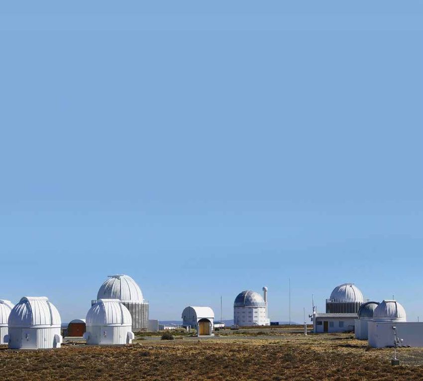
TAXONOMY | THEME

The male hermit crab, here shown without its soft ‘shell’ covering, has one enlarged pincer, or cheliped. It is not known whether
this is used to fight off competing males during courtship, grasp the female for mating, or simply attract females that may be
impressed by large pincers!

important. On the other hand, this might over-emphasise           The new technology does need a lot of computing power
some things. So, it can result in mistakes. There is also         and special software, which can make it expensive. While
the issue of reproducibility. Each scientific experiment          the scanning process is relatively quick and easy, the
should be reproducible.”                                          analysis of the 3D data is time-consuming, as is working
                                                                  on the scan data on the computer, cleaning unwanted
Machine-derived images are less vulnerable to bias.               scanning artefacts, cutting the specimens open virtually,
                                                                  and so on.
Computing power
In an interview for the GigaScience blog, Landschoff says         As Landschoff says, drawing is cheap and doesn’t come
that the power and magnification of X-ray microCT also            with these challenges.
gives a precise picture of surface textures, which are
often missing in photographs or manual drawings.                  “I require only a pencil, eraser and paper.”

Ironically, hermit crabs are among the most challenging           A crab in hand
model organisms within Decapoda to use CT scanning on.            On the other hand, the CT images are very powerful,
Many are small, and half of their body – the pleon, which         which makes this a useful tool for education and training.
is encased by the scavenged sea shell it lives in – is soft.      The scans provide a powerful connection to the real
                                                                  thing.
“The pleon has many fine-scaled, soft tissue identification
characteristics,” he adds. “Because CT scanning is based          “When I can show a high-resolution rotation movie to an
on X-ray detection, these soft structures are difficult to        audience instead of a picture of a colourless specimen
detect. If hermit crabs can be scanned, other groups like         in a jar of alcohol, that’s a huge advantage in making
Brachyura [normal crabs], which are entirely calcified,           taxonomy more interesting in general,” Landschoff
would be very visible for scanning.”                              explains. “I still believe that having a living specimen
                                                                  at hand is the most fascinating thing for students of
                                                                  biology.”

                                                                  With over 1 100 species and 120 genera of hermit
                                                                  crabs described to date, and with very many species
                                                                  undescribed on museum and laboratory shelves, this
                                                                  digital advance should broaden options and possibilities
                                                                  for greater coverage of specimens.

                                                                  Helen Swingler is a senior writer at the University of Cape Town.
                                                                  Article republished from UCT News.

                                                                  All images courtesy of Jannes Landschoff.
                                                                  A short animation entitled ‘3D video microCT scans of new and
                                                                  rare hermit crab species’ is available on the GigaScience Journal’s
                                                                  YouTube channel.
Hand-drawing a hermit crab’s claw could take as long as a
week, while microCT scanning from all angles can be done in
an hour or two, although the subsequent data processing and
analysis is very time-consuming.

WWW.ASSAF.ORG.ZA                                                                                       Quest Vol. 15 No. 2 | 2019       9
THEME | BIOLOGY

            Looking inside
         a brooding brittle star
                    Jannes Landschoff tells us how he used microCT to research
                              reproduction of a marine invertebrate

Brittle stars are echinoderms in the class Ophiuroidea.
They consist of a central disc and five long, thin arms
that can break quite easily – hence the name! Although
not as well known as their cousins the starfishes (class
Asteroidea), brittle stars are widespread in the marine
environment, occurring from very shallow waters down
to the deepest depths of the ocean. More than 2 200
species are recognised in the world, of which about 140
occur in South Africa.

Normally, brittle stars release their eggs and sperm
into the water, where they are externally fertilised and
then develop as tiny larvae in the plankton. This method
is called broadcast spawning. However, about 5% of
all brittle star species actively brood their young in         A brittle star with seven young, artificially coloured to show
                                                               their size and position. © Jannes Landschoff
specialised pouches called ‘bursae’. This behaviour has
long fascinated biologists. It is remarkable that an animal
without a centralised brain, and with one opening to the       MicroCT scans have now shown that most young would
stomach that serves both as the mouth and the anus,            indeed be positioned with their mouth against the wall,
is capable of such a complex reproductive behaviour,           but not all. Especially when they occur with other siblings
similar to that of humans.                                     in the same brood pouch, they are often stacked on
                                                               top of each other. The siblings inside may well fight for
The young brittle stars stay inside the mother for an          the best positions and compete for the available food
extended period, potentially up to a year. During this         sources. What remains unknown is how much the young
time they need to be nurtured and fed by the mother.           actually move around inside. Perhaps by scanning living
Previously it was not entirely clear how the young brittle     organisms, microCT imaging could help to solve this
stars lie within the mother’s bursae – to see them inside,     mystery in the future.
biologists would have to cut the specimen open. This
would destroy the brood pouch, changing the position           A two-minute
and orientation of the young in the process.                   video animation of
                                                               microCT images of
                                                               a brooding brittle
MicroCT scanning has helped to visualise the internal          star is available on
brooding of brittle stars, as this non-invasive technique      YouTube. Search for
can produce in situ images of the young in their natural       Brittle Star [2014].
position. There are usually 10 brood pouches in a
brooding brittle star. The serpent-skinned brittle star
Ophioderma wahlbergii, which is common in the coastal
waters of Cape Town, was found to brood up to 33 young
at a time, and different developmental stages could be
seen in different brood pouches.

It was also previously hypothesised that the young are
                                                               Young brittle
fed by the mother secreting nutrients through the bursal       stars poke their
body wall. This would mean that these animals are ‘truly       arms through
viviparous’, giving birth to live young that were nourished    their mother’s
and developed inside the parent’s body. Some research          bursal slits,
                                                               getting a taste of
suggests that bacterial activity at the surface of this body
                                                               the underwater
wall helps the young gain sufficient food. The young           world that awaits
would therefore be expected to press their mouths              them.
against that surface, but this could never be verified.        © Jannes Landschoff

10   Quest Vol. 15 No. 2 | 2019                                                                                WWW.ASSAF.ORG.ZA
FOSSILS | THEME
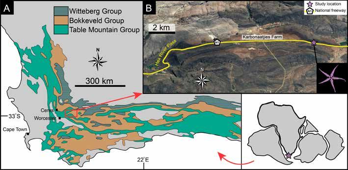
Brittle stars
in the
Bokkeveld
The next time you travel along the N1 national with Honorary Research Associate Dr Wendy Taylor, an
road from Johannesburg to Cape Town, look expert on echinoderm fossils. Mhairi was searching for
around at the arid landscape before you a suitable subject for her Honours project, which she
descend the pass into the Hex River Valley. specifically wanted to do on fossils.
Consider the fact that you are 125 km from
“We went out for a week, driving around and looking at
the nearest coastline, and some 950 m above outcrops, and we stumbled across it by pure chance,”
present-day sea level – and then picture the says Mhairi.
marine brittle stars that were found here, as
ancient fossils! What they discovered, at a road-cutting next to the N1,
was an entire bed of fossil brittle stars – or ophiuroids.
In the Devonian Period some 400 million years ago, when The bed was lying about 120 cm below the surface of the
the land that is now the African continent was part of the 2 m high rocky embankment, which had a sedimentary
giant supercontinent Gondwana, the southern Cape was profile representative of the Voorstehoek formation.
underwater, part of a basin covered by the Agulhas Sea.
It was during this time that the fine-grained sediments “It’s quite rare to find echinoderms, just because they’re
of the Bokkeveld Group were deposited on the seafloor, so delicate,” says Mhairi. “They need exceptional events
which was home to a thriving community of bottom- to be preserved.”
dwelling invertebrates.
These events, known as obrution events, involve rapid
The Bokkeveld Group was laid down on top of the Table burial or smothering by an underwater ‘landslide’ or a
Mountain Group, and was subsequently overlain by the storm-induced current that sweeps sediment down a
Witteberg Group. These three groups make up the Cape slope. Sedentary bottom-dwelling (benthic) invertebrates
Supergroup, which was later buckled and thrust up by that cannot move fast enough to escape are particularly
tectonic activity to form the Cape Fold Belt mountains. prone to being entombed in this way. Over time their soft
The Bokkeveld is known to be rich in fossils, as the tissues and eventually their calcareous shells – or ossicles
marine organisms of the time were better preserved in in the case of echinoderms – dissolve away, but by then
the fine sediments that ultimately hardened to mudstone
and shale, compared to the coarser sandstones of
the other two groups. Two of the oldest Bokkeveld
layers, the Voorstehoek and deeper Gydo formations,
have particularly well-preserved ‘shelly’ fossils such
as trilobites (now extinct), brachiopods (lamp shells),
bivalves (mussels and clams), gastropods (sea snails) and
even echinoderms (including starfish, feather stars and The presence of the
brittle stars). brittle star bed was
given away by orange,
star-shaped patterns on
With this in mind, Mhairi Reid – then a BSc Honours the surface of the rock,
student in the University of Cape Town’s geology caused by oxidation of iron
department – went on a fossil-finding road trip in 2014 in the mould fossils.
WWW.ASSAF.ORG.ZA Quest Vol. 15 No. 2 | 2019 11THEME | FOSSILS
their bodies have left impressions in the rock. The fossil What was even more special about the Karbonaatjies
brittle stars were examples of these mould fossils, rather obrution bed, named after the local farm, is that more
than a mummified version of the animal itself. than half of the brittle star fossils were ‘intact’ – in other
words, all five arms remained joined to the central disk.
Initially, for Mhairi’s Honours project, some rock samples Brittle stars have arms that easily detach, which is an
containing the fossils were collected, but for her MSc adaptation to escape predators. When grabbed by an
project an entire section – about 2 m long, 1 m wide and arm, they can simply shed it and grow a new one later,
4 cm thick – was excavated using a pickaxe, flat brick but they also tend to fall apart quickly after death, as the
chisel and geological hammer. The weathered nature soft tissue and ligaments attaching the arms to the disc
of the rock meant that it was impossible to remove the soon decay. The number of intact specimens lend proof
section as a single slab though. It broke up into 55 pieces, to the theory that the animals were buried alive.
which were carefully labelled and then put back together
again in the laboratory to be photographed. Mhairi also counted more than 600 detached arms in
the microCT scans, and noted that many of the brittle
While photos were fine for capturing the fossils on stars were lying upside down. Since there are a number
the surface of the rock pieces, they were hopelessly of records of mass amputation in modern-day brittle
inadequate for showing what lay inside, so the pieces stars after storms and hurricanes, she interprets her
were taken to Stellenbosch University’s microCT facility observations as being consistent with a strong, storm-
for scanning. This allowed the internal contents of the induced current that tumbled the ancient brittle stars
rocks to be ‘exposed’ as cross-sectional images from all over as they were covered in sediment. Some even had
angles, without any risk of damaging the fossils. All the one or more arms raised, which is exactly the posture
images were then digitally stitched together to create a modern brittle stars adopt when crawling up out of a
virtual 3D model, which revealed that there were more thin layer of sediment, but the ancient brittle stars
than 700 brittle stars in the excavated section, along were clearly buried too fast and too deep to escape.
with trilobites, brachiopods, bivalves, gastropods and Their muddy grave was beyond the reach of scavenging
other echinoderms. The microCT images essentially or burrowing animals, which would either have eaten
represented a snapshot of a benthic marine community their remains or facilitated aerobic decomposition
that lived together 400 million years ago. through bioturbation.
A virtual reconstruction of a portion of the fossil bed showing about 80 complete brittle stars (light pink = right side up, dark
pink = upside down), as well as fragmented arms (pale orange) and parts of other animals.
12 Quest Vol. 15 No. 2 | 2019 WWW.ASSAF.ORG.ZAFOSSILS | THEME
A simplified geological map of the Cape Supergroup showing the fossil bed’s location beside the N1 national road, near the Hex
River Pass. The small inset depicts Africa’s position within Gondwana.
Dr Emese Bordy, formally describing the new species. She
named it Gamiroaster tempestatis.
“For the genus name, it had to end in ‘aster’ [Greek for
star], but I wanted to name it something African, so I
went with ‘Gamiro’, which is the Khoikhoi word for a star
in the sky – although I had to take out the click syllable!”
explains Mhairi. “The species name, ‘tempestatis’, refers to
the storm that smothered the animals.”
The fossil-bearing rocks have now been curated in the
Iziko South African Museum, and it is hoped that the 3D
microCT images will form part of a future display.
• Fossils are protected by the National Heritage
Resources Act (No. 25 of 1999) and may not be
destroyed, damaged, altered, excavated or removed
from their original site, without a permit from the
relevant heritage resources authority. The necessary
permits were obtained in order to conduct this
research.
Article by Sue Matthews, images courtesy of Mhairi Reid.
For more detail, and access to the 3D model of the fossil bed, refer
to the open-access paper: Reid et al. (2019) in GigaScience, Vol. 8 (3).
https://doi.org/10.1093/gigascience/giy156
A short animation entitled ‘A 3D visualisation of a microCT
dataset of fossil starfish in an ancient sea bed’ is available on
the GigaScience Journal’s YouTube channel.
A 2D microCT scan representing a cross-section of rock with
brittle star fossils.
CURRICULUM CORNER
The fossil brittle stars were recognised as members of LIFE SCIENCES GRADE 10
the Family Protasteridae, but were a genus and species Support systems in animals; Biodiversity and
new to science – although long extinct and only distantly classification; History of life on Earth
related to modern-day brittle stars. Mhairi has recently
co-authored a paper with British fossil echinoderm GEOGRAPHY GRADE 10
expert Dr Aaron Hunter, Dr Taylor and her MSc supervisor Geomorphology
WWW.ASSAF.ORG.ZA Quest Vol. 15 No. 2 | 2019 13THEME | PALEO-ANTHROPOLOGY
Little Foot’s inner ear
MicroCT scans of the hominin fossil known as Little Foot shed light on
how she lived and moved, more than three million years ago
The inner ear of hominin fossils has the potential to could suggest a high degree of variation in locomotor
provide valuable information about how an individual behaviour in this group.
moved, what its hearing capacities were, and how the
evolution of the species relates to others. “Our analysis of the inner ear might be compatible with
the hypothesis that Little Foot and the Australopithecus
Based on microCT scans performed at the Evolutionary specimens in general were walking on two legs on the
Studies Institute at the University of the Witwatersrand, ground, but also spent some time in the trees,” says Dr
a Wits scientist and colleagues have been able to Beaudet. Little Foot’s cochlea, though, is quite similar
‘virtually extract’ the inner ear of Little Foot, the to other Australopithecus specimens in the study and to
Australopithecus fossil dated at 3.67 million years old. Dr Paranthropus, but it differs with fossil Homo specimens.
Amélie Beaudet, Professor Ronald Clarke and their team “This organ is related to sound perception and to
published a description of the inner ear in the Journal of ecological factors such as diet, habitat or communication,
Human Evolution in January. They also compared it with which means that Little Foot differed in this regard
17 hominin specimens from Sterkfontein, Swartkrans and with early members of our own genus, implying some
Makapansgat belonging to the genera Australopithecus, difference in behaviour,” says Dr Beaudet.
Paranthropus and Homo and dating between three and
1.8 million years ago, as well as with 10 chimpanzees and The dimension and shape of the cochlea are related
10 modern humans. to the range of frequencies that can be detected by
a species. The shape of the cochlea of fossil Homo
Overall, Little Foot’s inner ear has both ape-like and specimens is compatible with an extended low-frequency
human-like features, because the inner ear canals and hearing limit. This was not the case for Australopithecus,
the cochlea provided different results. The semicircular including Little Foot, nor for Paranthropus.
canals in Little Foot’s inner ear are different from both
modern humans and from Paranthropus – a genus of “At the moment, we are not yet sure what this means.
extinct hominins that lived at the same time as the first It may be that the early Homo species had to extend
humans. The Paranthropus canals have a very specific their range of frequencies for adapting to a different
shape that is not shared with any of the fossil specimens. environment or perhaps even to communicate to each
other. We don’t really know.”
“By contrast, we found that the Little Foot inner ear
canals are close to those of chimpanzees,” says Dr Of all the comparisons of Little Foot to other specimens,
Beaudet, lead researcher of the study. “They differ from the greatest similarity in the overall inner ear pattern was
modern human inner ear canals in that modern humans’ with a specimen of similar age from the Jacovec Cavern in
canals evolved for unique activities such as running.” the Sterkfontein Caves.
The study also shows a large diversity in the shape of the “Having a reference point, such as comparing Little
inner ear canals among Australopithecus species, which Foot to the Jacovec specimen, is important in detecting
which traits are specific to us – humans – and whether
humans evolved more distinct characteristics. With
this finding we now would be able to know what
is specific to Homo and Paranthropus, and when
these features emerged in the fossil record,”
says Dr Beaudet.
Blausen Medical: Wikimedia
Issued by Wits University. Read about Little Foot’s
discovery in Quest Vol. 14 No. 1 (2018).
A longer article by Dr Amélie Beaudet was published
online in January: https://theconversation.com/
virtual-images-reveal-secrets-of-an-ancient-fossils-
brain-and-inner-ear-108349
It includes a 36-second video animation of the
virtual reconstruction of Little Foot’s brain and
inner ear, which can also be viewed at:
https://vimeo.com/306999133
14 Quest Vol. 15 No. 2 | 2019 WWW.ASSAF.ORG.ZAFORENSICS | THEME
‘Bakeng se Afrika’
Forensics ‘For Africa’
MicroCT will play a central role in a new project to
develop a digital repository of skeletal images for
research and teaching purposes.
The three-year project, titled Bakeng se Afrika (For
Africa), will be coordinated by the University of Pretoria’s
Forensic Anthropology Research Centre (FARC), but two
other South African universities – Stellenbosch University
and Sefako Makgatho Health Sciences University – as well
as three European universities and the National Energy
Corporation of South Africa (Necsa) will also be involved.
The project has been made possible by a €1 million
(R15.9 million) grant from the Erasmus+ Programme of
the European Union.
A microCT facility was installed at Necsa in 2011, and has
been used by researchers and postgraduate students
to acquire a large quantity of three-dimensional data
on human bones, especially skulls. This data is useful to
other researchers – both nationally and internationally
– from the anatomical science, bio-engineering and
dentistry fields. Together with outputs from other
methods, such as Lodox Statscans and CBCT scans, it
will be made available in a digital archive. Strict ethical
guidelines will apply, however, with research projects
requiring ethical approval from institutional committees
before access is granted to the repository. Marine Cazenave, SMU funded by EU Erasmus+
MicroCT meets ...
Forensic Files
MicroCT may also be used by forensic scientists in
analysing evidence that might assist in prosecution.
For example, toolmarks on bone can provide
information about a victim’s murder or subsequent
dismemberment, with differences between blunt-force
trauma and straight-edged or serrated blades clearly
apparent.
Brandon Anderson, Flickr
Research studies have used microCT to analyse both
What is Forensic Anthropology? gunshot residue in wounds and ballistic impacts on
Forensic anthropology is the examination of human bone to estimate firing distance, as well as blood
skeletal remains to assist law enforcement in spatter on textiles. And detailed microCT imaging of
identifying the deceased. A biological profile that the metamorphic development of blowfly larvae may
includes sex, approximate age, height and ancestry ultimately help to refine estimates of post-mortem
can be created, based upon physical characteristics interval.
that have been determined from the study of
human skeletal differences. The time since death The limited availability of microCT – together with a lack
can be estimated, while evidence of bone injuries, of replicated scientific studies to determine the validity
medical procedures and diseases can assist in of such research findings – means that it is still an
identification, and sometimes even indicate the emerging technology in forensic science, but it promises
likely cause of death. to be a powerful tool in future investigations.
WWW.ASSAF.ORG.ZA Quest Vol. 15 No. 2 | 2019 15THEME | METAL 3D PRINTING
MicroCT
for metal 3D printing
By Anton du Plessis. Images courtesy of the CT Scanner Facility,
Stellenbosch University
Most people have heard of 3D printing by now, but not The process works by using a focussed high-power
everyone knows it can be done with metals. It is actually laser that is scanned across a surface to melt fine metal
possible to produce custom and complex designs, similar powder in a single layer, according to the 3D model. This
to plastic 3D print models, in solid metal. This is known is followed by lowering the solidified layer, and then
as additive manufacturing, and while it is not something adding the next layer of powder and melting this layer on
for your hobby room or garage, it has been introduced top of the previous one and so on, until the entire model
globally in many big companies and is starting to be used is created in solid metal, surrounded by powder. The part
in real applications – for example, in Formula 1 racing is removed from the powder and processed further to
cars and fighter jets! ensure it has acceptable mechanical properties.
© Siemens
16 Quest Vol. 15 No. 2 | 2019 WWW.ASSAF.ORG.ZAMETAL 3D PRINTING | THEME
These titanium brackets were ‘additively manufactured’ using metal 3D printing and then tested using microCT. The small
coloured dots represent porosity of different sizes. These are gas bubbles inside the metal that need to be minimised for
optimal quality of the part.
Obviously this is a complex process, with expensive various parameters in the additive manufacturing system
equipment and powder, and many things that can go before starting to build the parts – in other words,
wrong. In traditional manufacturing, various well-known fine-tuning the machine. It allows for easy checking of
quality-control tools are used to ensure structural which parameters result in the minimum porosity and
integrity and quality of the final parts. These include crack formation, with 3D images indicating what could
dimensional measurements, density measurements, be wrong in the process. During the laser melting of the
surface-quality measurement, 2D X-ray inspection (digital powder particles, gas bubbles can form in the molten
radiography) and sometimes even 3D X-ray inspection metal region under the laser spot, and then get trapped
(microCT). By contrast, additive manufacturing relies in the solid metal due to the rapid cooling as the laser
heavily on microCT, because many of the traditional spot moves. It can also happen that the laser moves too
quality-control tools are difficult to use for these parts. fast and does not melt enough metal powder, so layers
In additive manufacturing the parts are more intricate, on top of one another do not weld together sufficiently,
so some areas are not accessible, and the internal pores, and even have spaces called ‘lack of fusion’. Obviously
cracks and other flaws that can occur are smaller. The these two errors have different resulting ‘pore shapes’,
high-detail imaging capabilities of microCT make it very which can be easily seen in microCT images.
useful for checking these parts quickly for major flaws.
In general, no production method is perfect, and gas
Besides final quality checks for parts, microCT is also bubbles are also present in other traditional parts such
very useful and cost-effective for optimising all the as metal castings. In the case of additive manufacturing,
MicroCT of a 3D-printed titanium cube of 10 mm, shown in 3D with colour coding and in a virtual cross-section to visualise
porosity. The pores, represented by black dots, occur mainly near the top surface.
WWW.ASSAF.ORG.ZA Quest Vol. 15 No. 2 | 2019 17THEME | METAL 3D PRINTING
Colour mapping of surface roughness of a 10 mm cube, Colour coding shows deviation of up to 1.1 mm on the right
showing side surfaces much rougher than the top surface. side of the part compared to the CAD design file.
however, these flaws may be extremely small, while still In South Africa there are various facilities with metal
affecting the properties of the bulk material. MicroCT 3D printers doing research into new processes and
is therefore crucial in helping to minimise this porosity developing new applications, much of this R&D work
by adjusting the laser power, scan speed, and other being supported by the Department of Science and
parameters of the additive manufacturing system. Technology through the Collaborative Programme in
By fine-tuning the settings, highly dense parts can be Additive Manufacturing (CPAM). These facilities are also
produced with excellent properties. making additive manufacturing available to local industry
players that recognise the advantage of being able to
Besides porosity, the surface quality varies from machine produce extremely complex parts with exactly the shape
to machine, and even between vertical and horizontal needed for the application. These metal parts can have
sections on the parts. This can also be measured by the same strength as a traditionally designed one, but
microCT and can be used to assess requirements for with a fraction of the mass, which is very important for
post-processing, such as smoothing rough surfaces. vehicle and airplane efficiency. MicroCT obviously assists
in obtaining the best quality for such parts, but it can also
More importantly, microCT can be used to detect help in designing them.
whether a part has been produced inaccurately
compared to the design specification. The example The parts are often visually impressive, with designs
shown here is a bracket that warped inwards by 1 mm resembling natural structures, such as branch-like or
on both sides. This happens during the layer-by-layer spider-web connections. This is where microCT has
process if the heat input is not removed fast enough. another role to play – in the design of biomimetic parts.
Irregular thermal distribution results in stress in the
material, which causes it to distort or warp and even Biomimicry is the practice of ‘learning from nature’ to
crack in extreme cases. Although a 1 mm warping seems design and produce materials, objects and systems.
insignificant, it can be critical for a fighter jet! By allowing nature’s designs to be visualised and
analysed in full 3D, microCT can assist in the creation of
engineering blueprints for new biomimicry applications,
many of which could be implemented through additive
manufacturing. The protective bony plates in reptiles or
overlapping scales in fish, for example, might inspire the
development of new kinds of body armour, from bullet-
proof vests to shark-proof wetsuits!
• A thorough overview of microCT applications is
available online in the open-access publication:
Du Plessis et al. (2019) Advancing X-ray micro-
computed tomography in Africa: Going far,
together. Scientific African Vol. 3.
https://doi.org/10.1016/j.sciaf.2019.e00061
Prof. Anton du Plessis is an Associate Professor in the Physics
Department at Stellenbosch University and heads the university’s
CT scanner facility (http://blogs.sun.ac.za/duplessis/).
The internal structure of a cuttlefish bone may be a design For more information, including a variety of microCT case studies,
inspiration for additively manufactured lightweight materials. see: http://blogs.sun.ac.za/ctscanner/.
18 Quest Vol. 15 No. 2 | 2019 WWW.ASSAF.ORG.ZAFEATURE | LAND DEGRADATION
Scarred Earth
Monitoring land degradation
The German-funded research project SALDi aims to develop new tools for assessing
land degradation in South Africa so that the problem can be addressed
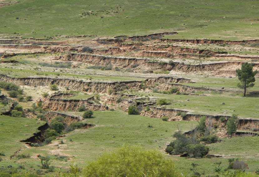
Large areas of South Africa have experienced a decline in observation data that is now available from the European
soil productivity, reduced vegetation cover and increased Sentinel satellites, as well as recent advances in modelling
soil erosion – sometimes evident as deep gullies that scar approaches.
the landscape. But to what extent is this land degradation
because of human impacts, such as overstocking cattle, “For instance, the USLE [Universal Soil Loss Equation]
clearing bush to plant crops or incorrectly channelling model that has been used in South Africa up to now
stormwater run-off from roads, rather than climate- works well for long-term averages, but not the kind
induced changes, particularly due to periodic droughts? of erosion damage that a farmer might see after one
storm,” explains project coordinator Dr Jussi Baade, from
A new German-South African collaboration aims to Germany’s Friedrich Schiller University Jena. “So with the
increase understanding of this conundrum in the long input of local partners we will adapt a physically based
term by implementing novel tools for assessing land soil erosion model from Germany to apply in South
degradation. The three-year project – known as the Africa.”
South African Land Degradation Monitor, or simply
SALDi – will build upon earlier research on the topic by a Six study sites, each covering an area of 100 km by
variety of local scientists. Much of this was collated by the 100 km, have been identified countrywide to represent
Agricultural Research Council’s Institute for Soil, Climate a range of climatic conditions, geological substrates,
and Water (ARC-ISCW) in developing a Land Degradation vegetation cover and land-use practices. In addition, a
Index, which takes into account soil erosion by water and modelling exercise will apply the WRF-Hydro (Weather
wind, soil salinisation and acidification, hydro-climatic Research and Forecasting Hydrological) Regional Earth
parameters, land cover and the loss of biodiversity. System Model to the whole of southern Africa. The
model essentially combines recorded precipitation
The SALDi project can improve interpretation of such data and predicted weather forecast data with a land
information and better assess the land degradation surface model for a variety of uses, including flash flood
problem by taking advantage of the high-resolution Earth prediction, river flow forecasts, seasonal water resource
20 Quest Vol. 15 No. 2 | 2019 WWW.ASSAF.ORG.ZALAND DEGRADATION |FEATURE
“We are collaborating to learn and assist each other
where possible,” says Dr Le Roux, who had suggested this
as a SALDi site based on previous research he had done
in the area. “Networking with the SALDi team will assist
our development of the required capacities in terms
of sediment measurement and mapping techniques.
We also plan to apply for funding from the SPACES
DAAD scholarship programme ‘Capacity Building /
Development’.”
If successful, this would allow Mrs Stander to spend
time at a German university as an exchange student,
increasing her exposure to cutting-edge erosion science.
The six SALDi study sites. On a future field trip, Dr Baade intends surveying about
15 dams in three of the SALDi study sites to compare
forecasts and land-atmosphere coupling studies. In this the siltation estimates with those of Rooseboom,
case, the model’s output will be used as input data for published in a Water Research Commission report in
soil erosion modelling. the early 1990s. These surveys will be done from a boat,
using an echosounder and GPS, to calculate the volume
By the end of the project, it is anticipated that an of accumulated sediment in the dam and hence the
automated observation system to monitor aspects such reduction in storage capacity.
as soil moisture, vegetation cover and potential land
degradation will have been developed. This would be a “We’re really looking forward to seeing whether siltation
freely accessible, online system allowing anyone – from rates went up or down, because in certain areas where
school learners to farmers to local and international there are good soil conservation measures in place
scientists – to zoom into a map, down to a resolution of you could maybe expect that siltation in reservoirs has
10–30 m, and view information derived from bi-weekly decreased,” says Dr Baade. “This would be useful for
satellite data. showing farmers that they can do things to maintain their
water storage capacity, because when that is lost they will
A kick-off meeting for the project was held in Pretoria be worse off in a drought.”
at ARC-ISCW in March, attended by collaborating
scientists, other potential partners and representatives Another component of the fieldwork is the surveying of
of stakeholder government agencies. After the meeting, erosion gullies, and this work began during the recent
the research team headed out on their first fieldwork trip. Dr Baade explains that a South African company was
campaign. One of the first stops was the Welbedacht contracted to do drone-based surveys, providing data
Dam, which was built in the early 1970s as part of used to construct highly detailed digital terrain models.
Bloemfontein’s water supply but has silted up to such
a degree that it now has only a fraction of its original “At the end of the project we’ll fly these areas again to
storage capacity. see how things have changed,” he says. “We’re doing
that mainly in the national parks because – given that
The visiting German scientists were joined here by Dr Jay they are directly protected by park management – it’s
Le Roux, a soil erosion scientist from the University of the basically just the rainfall or run-off signal that creates the
Free State, and his PhD student Mrs Marike Stander, who dynamics of the gullies. In certain areas they are not a
will be conducting research on erodible soils in the dam’s sign of land degradation, but we want to learn how they
catchment. evolve naturally.”
The project’s kick-off meeting was held at the Agricultural Some erosion gullies in the study sites have been surveyed
Research Council’s Institute for Soil, Climate and Water in using drones, and the resulting digital terrain models will
Pretoria in March. allow changes to be monitored over time.
WWW.ASSAF.ORG.ZA Quest Vol. 15 No. 2 | 2019 21FEATURE | LAND DEGRADATION
A moisture probe network consisting of eight sensors
attached to a central unit has been installed at each site so
that satellite-derived soil moisture data can be validated.
During the recent field campaign, a moisture probe degraded sites outside our parks for comparison
network was also installed in each of the six study sites purposes for the countrywide project. And secondly,
so that the soil moisture data derived from Sentinel-1 with greater relevance for SANParks, many of our parks
radar imagery can be validated. Each network consists include previously utilised land that was incorporated
of eight sensors placed in a 20 x 20 m area and attached into our parks in order to increase the area under
to a central unit. Since these need to be protected from conservation. Often, this recently acquired land has a
vandalism or theft, and the data downloaded every history of farming and therefore has some degree of
three months, they have been installed either in national degradation. Since soils are critical in supporting a range
parks or on the property of willing landowners. More of ecosystem processes, it is of utmost importance that
specifically, one is at the Kruger National Park’s sampling degraded soils are rehabilitated and restored.”
station near Lower Sabie, where there are a number
of other monitoring instruments, another in Mokala “SANParks invests a lot of time, money and effort into
National Park and a third in Augrabies National Park, various types of rehabilitation techniques aimed at
while the others are on private farms near Pilanesberg, restoring some newly acquired land into fully functioning
Ladybrand and Elim near Cape Agulhas. ecosystems. It is for this reason that this SALDi project
will be very useful for park management – it will help
This equipment will remain on site after the completion monitor land degradation and evaluate whether our
of the project, so that it can continue to be of use to rehabilitation and restoration programmes are working.”
South African scientists, together with all the processors,
algorithms and models developed during the project. Article by Sue Matthews; images courtesy of the SALDi project team.
The German research team hope that the project outputs
will ultimately assist in improving land degradation The project is funded by the German Federal
management in South Africa, but they point out that the Ministry of Education (BMBF) and forms part of
scientific exchange with local scientists is also of benefit the programme known as SPACES, an acronym
in addressing research questions they are working on in for Science Partnerships for the Assessment of
their own country. Complex Earth System Processes. The SPACES
programme has its roots in the 2012/2013
Some of the relationships supporting such exchange go German-South African Year of Science, which had
back many years, and there is an especially long history the theme ‘Strengthen research partnerships
of cooperation with SANParks scientists. For example, Dr for innovation and sustainable development’.
Baade did a reconnaissance survey of 15 reservoirs in the Climate research was one of seven subject areas
Kruger National Park a decade ago to establish whether identified for collaboration, with the main priority
their siltation could be used to assess spatial variation being to investigate the causes and consequences
of soil loss. He has published a number of papers on this of climate change. The SPACES programme was
and related land management research in the park in the subsequently developed to implement scientific
intervening years. cooperation projects in the broader southern
Africa region. The four projects that were funded
“One might be wondering why a land degradation in the first phase, starting in July 2013, have been
monitoring project is being conducted inside a national completed, and in July 2018 the second phase
park,” says SANParks abiotic scientist, Tercia Strydom. began, with nine projects funded – among them
“Well, there are two main reasons. Firstly, fully functional the SALDi project.
soils inside our parks will act as reference points for
22 Quest Vol. 15 No. 2 | 2019 WWW.ASSAF.ORG.ZAYou can also read