MicroRNA 101 inhibits renal tubular epithelial to mesenchymal transition by targeting TGF β1 type I receptor
←
→
Page content transcription
If your browser does not render page correctly, please read the page content below
INTERNATIONAL JOURNAL OF MOlecular medicine 47: 119, 2021

  MicroRNA‑101 inhibits renal tubular epithelial‑to‑mesenchymal
         transition by targeting TGF‑β1 type I receptor
              QINGLAN WANG1‑3, YANYAN TAO1, HONGDONG XIE1, CHENGHAI LIU1,2 and PING LIU1,2

      1
          Institute of Liver Diseases, Shuguang Hospital Affiliated to Shanghai University of Traditional Chinese Medicine;
                     2
                       E‑Institute of Shanghai Municipal Education Committee, 3College of Basic Medical Science,
                           Shanghai University of Traditional Chinese Medicine, Shanghai 201203, P.R. China

                                          Received July 11, 2020; Accepted March 30, 2021

                                                     DOI: 10.3892/ijmm.2021.4952

Abstract. MicroRNAs (miRNAs/miRs) are key regulators                   TGF‑β1/Smad3 signaling, while the opposite was observed
of renal interstitial fibrosis (RIF). The present study was            upon miR‑101 inhibition. To further confirm the ability of
designed to identify miRNAs associated with the development            miR‑101 to modulate EMT, the HK‑2 cells were treated with
of RIF, and to explore the ability of these identified miRNAs          the TβR‑I inhibitor, SB‑431542, which significantly suppressed
to modulate the renal tubular epithelial‑to‑mesenchymal                TGF‑β1‑induced EMT in these cells. Notably, miR‑101 inhi‑
transition (EMT) process. To this end, miRNAs that were                bition exerted a less pronounced effect upon EMT‑related
differentially expressed between normal and fibrotic kidneys           phenotypes in these T βR‑I inhibitor‑treated HK‑2 cells,
in a rat model of mercury chloride (HgCl2)‑induced RIF were            supporting a model wherein miR‑101 inhibits TGF‑β1‑induced
detected via an array‑based approach. Bioinformatics analyses          EMT by suppressing T β R‑I expression. On the whole,
revealed that miR‑101 was the miRNA that was most signifi‑             the present study demonstrates that miR‑101 is capable of
cantly downregulated in the fibrotic renal tissue samples, and         inhibiting TGF‑β1‑induced tubular EMT by targeting TβR‑I,
this was confirmed by RT‑qPCR, which also demonstrated that            suggesting that it may be an important regulator of RIF.
this miRNA was downregulated in transforming growth factor
(TGF)‑β1‑treated human proximal tubular epithelial (HK‑2)              Introduction
cells. When miR‑101 was overexpressed, this was sufficient
to reverse TGF‑β1‑induced EMT in HK‑2 cells, leading to                Renal interstitial fibrosis (RIF) is a pathological process that is
the upregulation of the epithelial marker, E‑cadherin, and the         common to the majority of chronic kidney diseases, resulting
downregulation of the mesenchymal marker, α‑smooth muscle              in functional deterioration which is largely independent of
actin. By contrast, the downregulation of miR‑101 using an             the initial renal injury (1). RIF is characterized by abnormal
inhibitor exerted the opposite effect. The overexpression of           interstitial extracellular matrix (ECM) deposition (2). While
miR‑101 also suppressed the expression of the miR‑101 target           a number of detailed studies on the mechanistic basis for RIF
gene, TGF‑β1 type I receptor (TβR‑I), and thereby impaired             have been published in recent years, the etiology of this condi‑
                                                                       tion has yet to be fully clarified.
                                                                           MicroRNAs (miRNAs/miRs) are short non‑coding RNAs,
                                                                       which can modulate gene expression by altering target mRNA
Correspondence to: Dr Chenghai Liu or Dr Ping Liu, Institute           stability and translation to control key physiological and
of Liver Diseases, Shuguang Hospital Affiliated to Shanghai            pathological processes (3,4). Indeed, miRNA dysregulation
University of Traditional Chinese Medicine, 528 Zhangheng Road,        has been linked to an array of different disease processes (5).
Pudong New Area, Shanghai 201203, P.R. China                           Within the kidneys, specific miRNAs regulate development,
E‑mail: chenghailiu@hotmail.com                                        homeostasis and normal physiology, while also regulating the
E‑mail: liuliver@vip.sina.com                                          onset of a range of renal diseases (6). As such, further studies
                                                                       elucidating the miRNAs that regulate the incidence of RIF
Abbreviations: α‑SMA, α‑smooth muscle actin; ANOVA, one‑way            have the potential to highlight novel approaches with which to
analysis of variance; DMEM, Dulbecco's modified Eagle's medium;        prevent or treat this debilitating condition.
ECM, extracellular matrix; EMT, epithelial‑to‑mesenchymal
                                                                           In the present study, a miRNA array‑based approach was
transition; FBS, fetal bovine serum; FDR, false discovery rate;
HgCl2, mercury chloride; miRNAs/miRs, microRNAs; RIF, renal
                                                                       utilized to identify miRNAs that were differentially expressed
interstitial fibrosis; TβR‑I, TGF‑β1 type I receptor; TβR‑II, TGF‑β1   between fibrotic and normal kidney tissues using a rat model
type II receptor; TGF‑β1, transforming growth factor‑β                 of mercury chloride (HgCl2)‑induced RIF (7,8). This analysis
                                                                       identified miR‑101 (also termed miR‑101a, miR‑101a‑3p) as
Key words: microRNA‑101, tubular epithelial‑to‑mesenchymal             the miRNA that was the most significantly downregulated
transition, renal interstitial fibrosis, TGF‑β1 type I receptor        in fibrotic renal tissue. In previous studies, miR‑101 has also
                                                                       been shown to be involved in fibrotic processes, such as liver
                                                                       fibrosis (9,10), pulmonary fibrosis (11), cardiac fibrosis (12,13),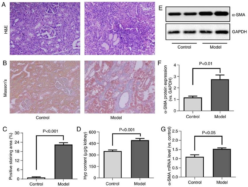
2 WANG et al: miR-101 INHIBITS RENAL TUBULAR EMT THROUGH TβR-I
bladder fibrosis (14) and cystic fibrosis (15). The role of this onset of rigor mortis (22). The body weight was measured at
miRNA in the context of RIF, however, remains to be clarified, the time of sacrifice, and body weight loss was not observed
as do the molecular mechanisms underlying such a role. As in any of the rats, with the body weight ranging from 331 to
such, the present study further evaluated the ability of miR‑101 492 g.
to regulate RIF.
One of the key steps in the development of RIF is renal Cells, cell culture and treatment. HK‑2 cells were obtained
tubular epithelial‑to‑mesenchymal transition (EMT), wherein from the Institute of Basic Medical Sciences, Chinese
tubular epithelial cells adopt a mesenchymal‑like phenotype Academy of Medical Sciences, and were grown in DMEM
and lose their epithelial‑like traits (16). The disruption of (Gibco; Thermo Fisher Scientific, Inc.) containing 10% fetal
tubular EMT is thus a viable approach for the treatment of bovine serum (FBS, Gibco; Thermo Fisher Scientific, Inc.)
RIF (17,18). Transforming growth factor‑β1 (TGF‑β1) is an for 18 h. Cells were then transferred to serum‑free medium
essential regulator of this renal tubular EMT process (19,20), and were treated for 48 h with 5 ng/ml TGF‑ β1 to achieve
signaling through its cognate cell‑surface type I and II recep‑ EMT induction. In appropriate experiments, cells were trans‑
tors (TβR‑I and TβR‑II, respectively) to induce appropriate fected with 20 nM of miR‑101 mimic (Guangzhou RiboBio
downstream signaling pathway activation. There is prior Co., Ltd.; the mature sequence of hsa‑miR‑101 mimic was
evidence to indicate that TβR‑I is a direct miR‑101 target (21), UACAGUACUGUGAUA ACUGAA) or 50 nM of miR‑101
and as such, it was hypothesized that miR‑101 may be able to inhibitor (Guangzhou RiboBio Co., Ltd.) or appropriate
regulate renal tubular EMT by targeting TβR‑I and thereby controls using a ribioFECT CP Transfection kit (Guangzhou
suppressing TGF‑β1 signaling. RiboBio Co., Ltd.) along with 5 ng/ml TGF‑β1 for 48 h. In
The present study thus examined the ability of miR‑101 order to assess how TβR‑I affects these results, in appropriate
to reverse TGF‑β1‑induced tubular EMT in human proximal experiments, cells were treated with 10 µM of SB‑431542
tubular epithelial (HK‑2) cells. Through these analyses, it was (Tocris Bioscience) to specifically inhibit T βR‑I for 12 h,
determined that TGF‑β1 treatment led to the downregulation and the cells were then transfected with miR‑101 inhibitor or
of miR‑101. In addition, miR‑101 overexpression suppressed appropriate controls using a ribioFECT CP Transfection kit
TGF‑β1‑induced EMT, whereas miR‑101 knockdown exerted (Guangzhou RiboBio Co., Ltd.) along with 5 ng/ml TGF‑β1
the opposite effect. At a mechanistic level, miR‑101 suppressed or SB‑431542 for a further 48 h.
TGF‑β1 signaling by inhibiting TβR‑I expression, and the
blocking of TβR‑I signaling ablated the effects of miR‑101 Hydroxyproline measurements. Renal hydroxyproline levels
inhibition on TGF‑β1‑induced EMT. Overall, the results thus were measured based upon HCl hydrolysis as per the method
demonstrated that miR‑101 inhibited tubular EMT, at least in described in the study by Jamall et al (23). Briefly, 100 mg of
part by suppressing TβR‑I expression. renal tissue was homogenized in 2.5 ml of ice‑cold ddH2O,
after which a BCA kit (Thermo Fisher Scientific, Inc.) was used
Materials and methods to quantify the protein levels in these samples. Subsequently,
6 M HCl were used to hydrolyze 2 ml of these homogenates
Rat model of HgCl 2 ‑induced RIF. In total, 20 male for 18 h at 105˚C, and the resultant hydrolysates were filtered
Sprague‑Dawley rats (4‑5 weeks old, weighing 120±10 g) using 3‑mm filter paper prior to drying at 40˚C. Samples
were obtained from the Shanghai Laboratory Animal were then incubated with Ehrlich's solution [25% (w/v)
Center, Chinese Academy of Sciences. All rats were housed p‑dimethylaminobenzaldehyde and 27.3% (v/v) perchloric
in a specific pathogen‑free environment that was maintained acid in isopropanol] at 50˚C for 1.5 h, followed by assessment
at 22‑24˚C with a relative humidity of 50‑60% and a 12‑h at 558 nm using a Tecan Infinite M200 Pro plate reader (Tecan
light/dark cycle. All rats were provided with ad libitum Life Sciences), with protein concentrations being used to
access to food and water. These animals were then randomly normalize the resultant values.
divided into the control (n=8) and model (n=12) groups, with
the animals in the latter group being orally administered Histological analysis. The kidney samples were fixed using
with HgCl 2 (8 mg/kg; Shanghai Tongren Pharmaceutical 10% formalin, paraffin‑embedded, and cut into 5‑µM‑thick
Co., Ltd.) once daily for nine weeks. These experimental sections. The sections were then subjected to Masson's
protocols were conducted in accordance with internation‑ trichrome staining using a modified Masson's Trichrome stain
ally accepted laboratory principles and all animals received kit (Solarbio Science & Technology Co., Ltd.) according to the
humane care as well as free access to food and water. The manufacturer's instructions. Briefly, the sections were stained
present study was approved by The Animal Research Ethics in Harris hematoxylin for 5 min, in Ponceau acid fuchsin
Committee of Shanghai University of Traditional Chinese staining solution for 10 min, and in Aniline Blue solution for
Medicine, Shanghai, China. The humane endpoint for this 5 min at room temperature. Hematoxylin and eosin (H&E)
study was a loss of 15% of the starting body weight. The staining was performed using a H&E staining kit (Yeasen
animals were anesthetized using 1% pentobarbital sodium Biotech Co., Ltd.) according to the manufacturer's instructions.
(50 mg/kg) by intraperitoneal (i.p.) injection. The samples Briefly, the sections were stained in hematoxylin for 5 min
of blood (0.8 ml) were collected from the vena cava. After and in eosin solution for 1 min at room temperature. Images
collecting the blood and kidney, at the end of the experi‑ were obtained using an Olympus IX73 microscope (Olympus
mental procedure, the abdominal vasculature, including Corporation). ImageJ software for Windows V 1.52v (NIH)
the vena cava was cut to cause exsanguination under deep was used to quantify the Masson's trichrome positive staining
anesthesia. Death was further confirmed by checking for the area.
INTERNATIONAL JOURNAL OF MOlecular medicine 47: 119, 2021                                           3

miRNA microarray. Renal miRNA profiles in these animals              spun for 15 min at 13,000 x g at 4˚C. A BCA assay (Thermo
(two samples in each group) were evaluated using an Agilent          Fisher Scientific, Inc.) was then used to assess the protein
Rat microRNA Microarray 16.0 (Agilent Technologies. Inc.).           quantities in the collected supernatants, and equal protein
Total renal tissue RNA was extracted using a mirVana™                amounts (30 µg) were separated via 10% SDS‑PAGE prior
miRNA Isolation kit (Ambion; Thermo Fisher Scientific, Inc.),        to transfer onto nitrocellulose membranes. Following a 1‑h
after which an Agilent Bioanalyzer 2100 was used to assess           blocking step using 5% non‑fat milk for 1 h at room tempera‑
the RNA quality based upon the RNA integrity number (RIN)            ture, these blots were probed overnight at 4˚C with primary
statistic. The miRNAs in these samples were then labeled and         anti‑ α ‑SMA (1:1,000, ab7817), anti‑E‑cadherin (1:1,000
hybridized with a miRNA Complete Labeling and Hyb kit                ab40772), anti‑Smad3 (1:1,000 ab40854), anti‑p‑Smad3 (1:500
(Agilent Technologies, Inc.) based on the provided directions,       ab52903), anti‑TβR‑I (1:1,000 ab31013) and anti‑GAPDH anti‑
and the Agilent Microarray Scanner and Feature Extraction            bodies (1:5,000 ab8245) (Abcam). The blots were then washed,
software (v10.7) was used to analyze these slides under default      incubated with secondary HRP‑conjugated goat anti‑mouse
settings. The Quantile algorithm was used to normalize the           antibody (1:5,000, ab97023, Abcam) or goat anti‑rabbit anti‑
resultant raw data with the Gene Spring Software v11.0 (Agilent      body (1:5,000, ab205718, Abcam) for 1 h at room temperature,
Technologies, Inc.). Shanghai Biotechnology Corporation              and protein bands were then visualized using an ECL reagent
conducted these microarray analyses.                                 (Thermo Fisher Scientific, Inc.), with a Tanon 5200 detection
                                                                     system (Tanon Science & Technology Co., Ltd.) being used
miRNA microarray analysis. Linear models and empirical               for imaging. Protein band densitometry was quantified using
Bayes methods were used to detect differentially expressed           ImageJ software for Windows V 1.52v (NIH) and normalized
miRNAs (24), with the thresholds for differential expression         to GAPDH.
being the following: P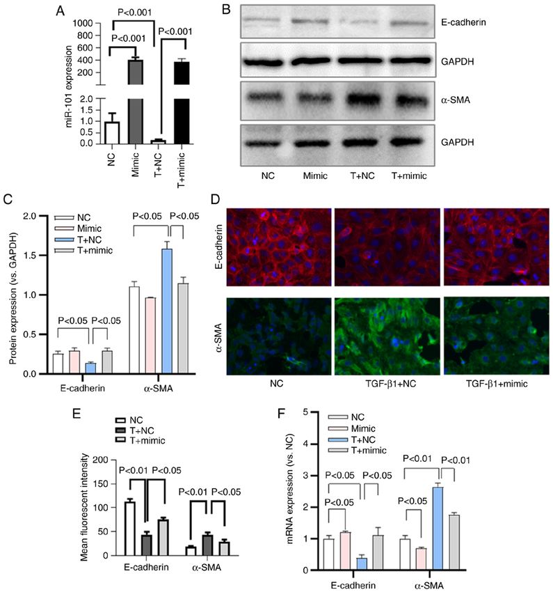
4 WANG et al: miR-101 INHIBITS RENAL TUBULAR EMT THROUGH TβR-I
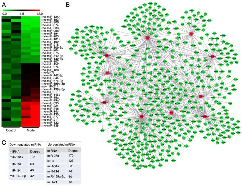
Figure 1. HgCl 2 induces renal inflammation and interstitial fibrosis in rats. (A) Representative H&E‑stained renal tissues. (B) Representative Masson's
trichrome‑stained tissues used to analyze RIF. (C) Quantification of Masson's trichrome positive staining area in renal tissue samples. (D) Renal hydroxypro‑
line levels were measured using the method by Jamall et al (23). (E) Renal α‑SMA levels were assessed via western blot analysis. (F) α‑SMA levels in western
blots were quantified via densitometry and were normalized to GAPDH. (G) Renal α‑SMA mRNA expression was assessed by RT‑qPCR. HgCl2, mercury
chloride; RIF, renal interstitial fibrosis; α‑SMA, α‑smooth muscle actin; Hyp, hydroxyproline.
deposition (Fig. 1B and C). The renal hydroxyproline content upregulated miRNAs (miR‑27a, let‑7i, miR‑34a, miR‑214,
was similarly elevated in the HgCl 2‑treated rats compared miR‑199a‑3p and miR‑21, with respective degree values of
with the controls (Fig. 1D). 170, 126, 93, 76, 60 and 42) (Fig. 2C).
α‑SMA is a marker of ECM‑secreting myofibroblasts,
and it was found that HgCl2 treatment significantly enhanced miR‑101 is downregulated in fibrotic kidney tissue and
α‑SMA expression at the protein and mRNA level (Fig. 1E‑G), TGF‑ β1‑treated HK‑2 cells. Given that miR‑101 was among
thus confirming that this treatment was linked to an enhanced the most significantly downregulated miRNAs in the
renal myofibroblast activation. miRNA‑mRNA network (Fig. 2C), the present study then
explored the role of this miRNA in the context of RIF. First
Identification of key differentially expressed miRNAs associ‑ it was confirmed that miR‑101 downregulation was evident in
ated with HgCl2‑induced RIF. Using an array‑based approach, the HgCl2‑treated fibrotic kidney tissue by RT‑qPCR, in line
40 miRNAs that were differentially expressed between the with the microarray findings (Fig. 3A).
control and RIF model animals were identified, of which 17 Renal tubular EMT is a key driver of the development of
and 23 were downregulated and upregulated, respectively RIF (27). The present study therefore explored this process
(Fig. 2A). TargetScan was used to predict target genes for in vitro by treating HK‑2 cells with TGF‑β1. Consistent with the
these miRNAs and a miRNA‑gene network based upon successful EMT induction, TGF‑β1 treatment resulted in both
predicted interactions among these miRNAs and genes was the loss of epithelial E‑cadherin expression and the upregu‑
then constructed using the Sanger miRNA database (Fig. 2B). lation of mesenchymal α‑SMA expression in HK‑2 cells, as
The degree metric was used to determine centrality within confirmed by western blot analysis (Fig. 3C‑E). Additionally,
this network, reflecting the degree to which a given miRNA miR‑101 expression was assessed in these cells by RT‑qPCR,
contributes to the regulation of the surrounding genes, with key and it was confirmed that this miRNA was downregulated in
miRNAs having a larger degree value (25). The top 10 most the context of TGF‑β1‑induced EMT (Fig. 3B).
central miRNAs within this network included four downregu‑
lated miRNAs (miR‑101a, miR‑107, miR‑194 and miR‑142‑3p, miR‑101 inhibits TGF‑ β1‑induced EMT in HK‑2 cells.
with respective degree values of 102, 63, 48 and 43) and six HK‑2 cells were then transfected with a miR‑101 mimic or a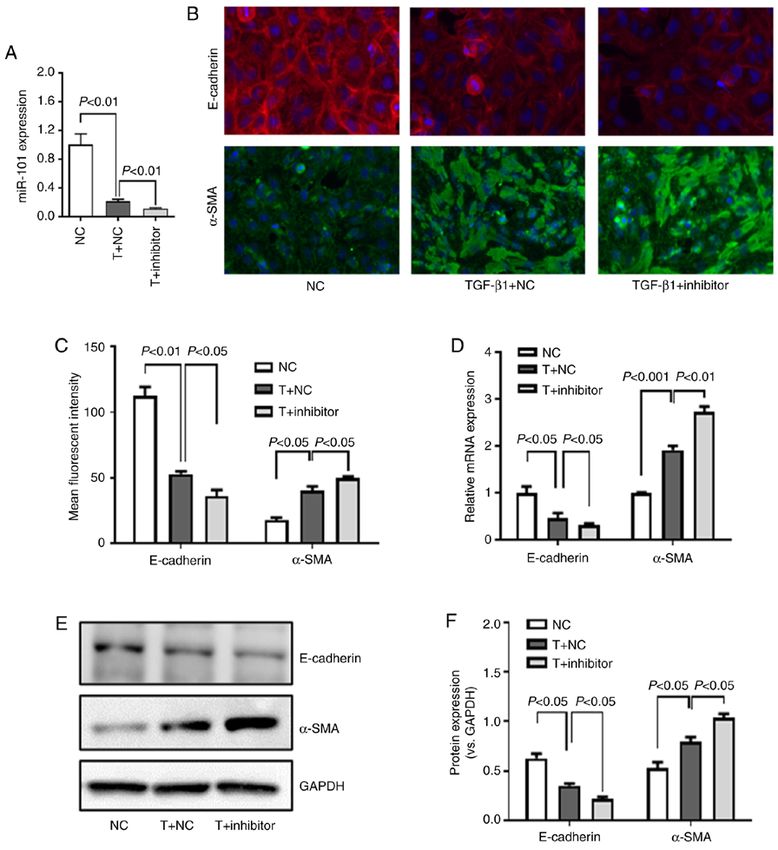
INTERNATIONAL JOURNAL OF MOlecular medicine 47: 119, 2021                                                            5

Figure 2. Identification of key differentially expressed miRNAs associated with HgCl2‑induced renal interstitial fibrosis. (A) Data from a miRNA microarray
comparing expression levels between control and RIF model rats were arranged in a heatmap. The color scale indicates the expression level of the miRNA, with
green and red colors indicating low and high expression, respectively. (B) A miRNA‑gene network was constructed, with squares and circles corresponding
to miRNAs and genes, respectively. Associations between these network elements are represented by edges, with network centrality corresponding to degree
values. (C) The degree values for the 10 top miRNAs within this network, with miR‑101 being identified as the most significant downregulated miRNA in this
disease context. The red square represents miRNA and the green circle represents mRNA. HgCl2, mercury chloride.

corresponding control construct, after which these cells were                   (Fig. 5B, C, E and F) and mRNA (Fig. 5D) expression, while
treated with TGF‑β1. As was expected, miR‑101 mimic trans‑                      α‑SMA protein (Fig. 5, C, E and F) and mRNA (Fig. 5D)
fection was associated with a significant increase in miR‑101                   expression increased significantly in these cells. These
expression, whereas TGF‑β1 treatment decreased miR‑101                          results thus revealed that miR‑101 downregulation enhanced
expression (Fig. 4A).                                                           TGF‑β1‑induced EMT in these HK‑2 cells.
    When the effects of miR‑101 on TGF‑β1‑induced EMT
were evaluated in these HK‑2 cells, it was found that miR‑101                   Effect of miR‑101 on TβR‑I and Smad3 expression in HK‑2
mimic transfection resulted in an increased E‑cadherin                          cells. Previous studies have identified TβR‑I as a miR‑101
expression at the protein (Fig. 4B‑E) and mRNA (Fig. 4F)                        target gene (21,28). The present study thus assessed the effects
level, along with a decreased α‑SMA protein (Fig. 4B‑E) and                     of miR‑101 on TβR‑I expression in the TGF‑β1‑treated HK‑2
mRNA (Fig. 4F) expression, consistent with the suppression of                   cells. This approach revealed that TGF‑β1 treatment enhanced
EMT in these cells.                                                             TβR‑I expression, whereas miR‑101 mimic transfection inhib‑
                                                                                ited its expression (Fig. 6A and B). By contrast, transfection
miR‑101 knockdown promotes TGF‑ β1‑induced EMT in                               with miR‑101 inhibitor was associated with an increased TβR‑I
HK‑2 cells. The present study then assessed the effects of                      expression in the TGF‑β1‑treated HK‑2 cells (Fig. 6D and E).
miR‑101 downregulation on TGF‑β1‑induced EMT in HK‑2                                The TGF‑β1/Smad pathway serves to facilitate canonical
cells by transfecting the cells with a miR‑101 inhibitor prior                  TGF‑ β1 signaling within cells, with Smad3 phosphoryla‑
to TGF‑β1 treatment, resulting in a significant knockdown of                    tion being a key step in this pathway (29). The present study
miR‑101 expression (Fig. 5A). This inhibition of miR‑101 was                    confirmed, by western blot analysis, that miR‑101 mimic
associated with a significant decrease in E‑cadherin protein                    transfection was associated with a decreased Smad3
6 WANG et al: miR-101 INHIBITS RENAL TUBULAR EMT THROUGH TβR-I
transfection on EMT‑related phenotypes (Fig. 7A and B), indi‑
cating that miR‑101 targets TβR‑I to inhibit TGF‑β1‑induced
EMT. When TβR‑I protein expression was assessed, it was
confirmed that SB‑431542 treatment markedly decreased
TβR‑I protein expression, whereas miR‑101 inhibitor treat‑
ment did not further affect the TβR‑I protein levels in these
SB‑431542‑treated HK‑2 cells (Fig. 7C and D).
Discussion
A number of studies to date have highlighted the roles played by
miRNAs in the context of both pathological and physiological
processes (5). Specific miRNAs have been found to regulate
renal development, homeostasis and the pathology of RIF as
well as other diseases (6,31). While the general etiology of RIF
has been thoroughly studied, the specific regulatory roles of
individual miRNAs in this disease context remain to be fully
elucidated. By further studying the ability of these miRNAs to
influence RIF progression, it may be possible to identify novel
approaches to preventing or treating this condition.
Herein, a previously described HgCl2‑induced rat model
of RIF was employed (8,32). By analyzing renal tissues from
these animals, 17 and 23 miRNAs were identified that were
downregulated and upregulated, respectively, in fibrotic kidney
tissue samples relative to the control kidney samples. Through
bioinformatics analyses, the 10 most critical of these miRNAs
were then identified, including four that were downregulated
(miR‑101, miR‑107, miR‑194 and miR‑142‑3p) and six that were
upregulated (miR‑27a, let‑7i, miR‑34a, miR‑214, miR‑199a‑3p
and miR‑21) in fibrotic renal tissue. The majority of these
miRNAs have previously been linked to renal diseases, such
as RIF. For example, Hou et al (33) found miR‑27a to suppress
peroxisome proliferator‑activated receptor‑γ signaling and to
thereby promote RIF, whereas miR‑34a has been shown to
regulate Klotho expression in tubular epithelial cells, thereby
Figure 3. miR‑101 downregulation is evident in both the RIF model renal
controlling RIF (34), while also inducing the apoptotic death
tissue and TGF‑ β1 treated HK‑2 cells. (A) RT‑qPCR was used to assess of these cells (35). There is also evidence to indicate that
miR‑101 expression in renal tissues. (B) RT‑qPCR was used to assess miR‑214 is upregulated in the context of renal injury, and the
miR‑101 expression in TGF‑β1‑treated HK‑2 cells. (C) RT‑qPCR was used knockdown of this miRNA is sufficient to attenuate unilateral
to assess E‑cadherin and α‑SMA expression in TGF‑β1‑treated HK‑2 cells.
(D) Western blot analysis was used to assess E‑cadherin and α‑SMA protein.
ureteral obstruction (UUO)‑induced RIF (36). miR‑21 is
(E) Densitometric quantification of the results in (D), with GAPDH used among the most well‑characterized miRNAs associated with
for normalization. RIF, renal interstitial fibrosis; α‑SMA, α‑smooth muscle fibrosis in a range of tissue types (37‑39). In healthy renal
actin; TGF‑β1, transforming growth factor‑β1. tissue, minimal miR‑21 expression is observed, whereas it is
significantly upregulated in the context of RIF, wherein it can
target PTEN and peroxisome proliferator‑activated receptor‑α
phosphorylation (Fig. 6A and C), whereas the inhibition of to promote fibrotic progression (38). These findings thus
miR‑101 exerted the opposite effect in TGF‑β1‑treated HK‑2 confirm that HgCl2 induces a model of RIF similar to that
cells (Fig. 6D and F). induced by a UUO‑based approach, while also confirming the
reliability of our miRNA array findings.
TβR‑I inhibition impairs TGF‑β1‑induced EMT and the effect Using bioinformatics analyses, the present study identified
of miR‑101 inhibition on HK‑2 cells. In an effort to more miR‑101 as the most downregulated miRNA in the rat model
fully assess whether miR‑101 targets TβR‑I to suppress the of HgCl 2‑induced RIF. This miRNA has previously been
EMT process in HK‑2 cells, the present study then assessed reported to play roles in other fibrotic processes, including liver
whether the effect of miR‑101 inhibitor transfection on EMT fibrosis (9,10), pulmonary fibrosis (11), cardiac fibrosis (12,13),
was disrupted when the cells were treated with the potent bladder fibrosis (14) and cystic fibrosis (15); however, its impor‑
TβR‑I inhibitor, SB‑431542 (30). As was expected, SB‑431542 tance in the context of RIF has not been well‑characterized.
markedly impaired TGF‑β1‑induced EMT in the HK‑2 cells, As such, the present study explored its role in this pathological
enhancing E‑cadherin expression and suppressing α‑SMA setting. Renal tubular EMT is a key step in RIF progression (40),
expression (Fig. 7A and B). When the cells were treated with and is characterized by tubular cells undergoing a shift from an
SB‑431542, this ablated the effects of miR‑101 inhibitor epithelial‑like to a mesenchymal‑like phenotype, whereuponINTERNATIONAL JOURNAL OF MOlecular medicine 47: 119, 2021 7
Figure 4. miR‑101 inhibits TGF‑β1‑induced EMT in HK‑2 cells. (A) miR‑101 expression was quantified in HK‑2 cells. (B) Levels of E‑cadherin and α‑SMA in
HK‑2 cells were measured by western blot analysis. (C) Densitometric quantification of data in (B), with GAPDH used for normalization. (D) E‑cadherin and
α‑SMA expression as assessed by immunofluorescence. (E) E‑cadherin and α‑SMA staining intensity was quantified using the Thermo HCS StudioTM 2.0
Cell Analysis Program. (F) RT‑qPCR was used to assess E‑cadherin and α‑SMA mRNA expression. NC, negative control; T, TGF‑β1; mimic, miR‑101 mimic;
EMT, epithelial‑to‑mesenchymal transition; α‑SMA, α‑smooth muscle actin; TGF‑β1, transforming growth factor‑β1.
these cells are able to produce high levels of ECM components TGF‑β1 to the cell surface TβR‑II molecule, in turn resulting
and to thereby drive RIF pathogenesis (8). TGF‑β1 is the most in TβR‑I activation, Smad2/3 phosphorylation and nuclear
well‑studied inducer of EMT (41), which is also downregulated translocation and altered gene expression. Previous research
in HgCl2‑induced RIF (8,32), and as such, this was utilized has demonstrated that miR‑101 can suppress fibrosis owing
in the present study to induce this process in HK‑2 cells. In to its ability to target TβR‑I and to thereby suppress TGF‑β1
line with the findings in vivo, a reduced miR‑101 expression signaling (21). In line with this finding, the present study deter‑
was observed in the cells following TGF‑β1 treatment. When mined that miR‑101 was able to inhibit EMT progression via
miR‑101 was overexpressed in these same cells, it was found downregulating TβR‑I, as miR‑101 overexpression disrupted
that this reversed TGF‑β1‑induced EMT, whereas miR‑101 TGF‑β1‑induced TβR‑I mRNA and protein expression. The
inhibition exerted the opposite effect. Taken together, these TGF‑β1/Smad pathway is an essential mediator of canonical
findings highlight miR‑101 as an inhibitor of tubular EMT. TGF‑β1 signaling (29), with Smad3 phosphorylation being a
The TGF‑ β1/Smad pathway is an essential mediator key component of this process. Consistently, it was determined
of EMT progression that is initiated upon the binding of that miR‑101 was able to inhibit TGF‑ β1‑induced Smad38 WANG et al: miR-101 INHIBITS RENAL TUBULAR EMT THROUGH TβR-I
Figure 5. miR‑101 inhibition enhances TGF‑β1‑induced EMT in HK‑2 cells. A miR‑101 inhibitor was transfected into HK‑2 cells, which were then treated
with TGF‑β1, (A) and miR‑101 expression was quantified. (B) E‑cadherin and α‑SMA expression as assessed by immunofluorescence. (C) E‑cadherin and
α‑SMA staining intensity as quantified using the Thermo HCS StudioTM 2.0 Cell Analysis Program. (D) RT‑qPCR was used to assess E‑cadherin and
α‑SMA mRNA expression. (E) E‑cadherin, and α‑SMA levels in HK‑2 cells were assessed by western blot analysis. (F) Densitometric quantification of the
data in (D), with GAPDH used for normalization; NC, negative control; T, TGF‑β1; inhibitor, miR‑101 inhibitor; α‑SMA, α‑smooth muscle actin; TGF‑β1,
transforming growth factor‑β1.
phosphorylation in HK‑2 cells, suggesting that this miRNA of the present study revealed that miR‑101 expression was
can suppress TGF‑β1/Smad3 signaling. Taken together, these downregulated in a rat renal fibrosis model, confirming that
findings demonstrate that miR‑101 can control TβR‑I expres‑ miR‑101 may be important in different rodent renal diseases
sion so as to suppress TGF‑β1 signaling. model. In an in vitro study, Zhao et al (42) found that miR‑101
To confirm these results, the cells were additionally treated overexpression using miR‑101 mimic inhibited EMT. Apart
with the potent TβR‑I inhibitor, SB‑431542. As was expected, from the gain‑of function experiment, in the present study, it
SB‑431542 suppressed TGF‑β1 induced EMT in HK‑2 cells. was further confirmed that the downregulation of miR‑101
When cells were treated with this TβR‑I inhibitor, the ability using miR‑101 inhibitor promoted the process of EMT. Taken
of miR‑101 inhibition to impact EMT phenotypes was ablated, together, the results further verified the key role of miR‑101 in
thus confirming that miR‑101 ablates TGF‑β1‑induced EMT by kidney diseases, and suggested that it may be a potential target
directly targeting TβR‑I. Recently, Zhao et al (42) also found for the treatment of renal fibrosis.
that miR‑101 inhibited acute kidney injury‑chronic kidney In conclusion, the findings of the present study highlight
disease transition by inhibiting the EMT process. The results miR‑101 as a key miRNA associated with HgCl2‑induced RIF.INTERNATIONAL JOURNAL OF MOlecular medicine 47: 119, 2021 9 Figure 6. Effect of miR‑101 on TβR‑I and Smad3 in HK‑2 cells. (A) TβR‑I, Smad3 and p‑Smad3 levels were assessed by western blot analysis in HK‑2 cells following miR‑101 mimic transfection. (B) Densitometric quantification of TβR‑I and Smad3, with GAPDH being used for normalization. (C) Ratio of p‑Smad3 vs. Smad3. (D) TβR‑I, Smad3 and p‑Smad3 levels were assessed by western blot analysis in HK‑2 cells following miR‑101 inhibitor transfection. (E) Densitometric quantification of TβR‑I and Smad3, with GAPDH being used for normalization. (F) Ratio of p‑Smad3 vs. Smad3. NC, negative control; T, TGF‑β1; mimic, miR‑101 mimic; inhibitor, miR‑101 inhibitor; α‑SMA, α‑smooth muscle actin; TGF‑β1, transforming growth factor‑β1. Figure 7. Effect of miR‑101 inhibition on TβR‑I inhibitor‑treated HK‑2 cells. (A) E‑cadherin and α‑SMA expression were assessed by immunofluorescence staining. (B) data in (A) were quantified using the Thermo HCS StudioTM 2.0 Cell Analysis Program. (C) TβR‑I expression in HK‑2 cells was assessed by western blot analysis. (D) Densitometric quantification of the data in (C), with GAPDH used for normalization. NC, negative control; T, TGF‑β1; inhibitor, miR‑101 inhibitor; α‑SMA, α‑smooth muscle actin; TGF‑β1, transforming growth factor‑β1.
10                               WANG et al: miR-101 INHIBITS RENAL TUBULAR EMT THROUGH TβR-I

It was found that this miRNA can suppress renal tubular EMT             7. Yuan JL, Tao YY, Wang QL, Shen L and Liu CH: Fuzheng
                                                                           Huayu Formula () prevents rat renal interstitial fibrosis induced
by targeting TβR‑I and thereby inhibiting TGF‑β1 signaling,                by HgCl2 via antioxidative stress and down‑regulation of nuclear
thus indicating that miR‑101 may be a viable target for treating           factor‑kappa B activity. Chin J Integr Med 23: 598‑604, 2017.
RIF, although further research is required needed to confirm            8. Wang QL, Tao YY, Yuan JL, Shen L and Liu CH: Salvianolic
                                                                           acid B prevents epithelial‑to‑mesenchymal transition through
this hypothesis.                                                           the TGF‑beta1 signal transduction pathway in vivo and in vitro.
                                                                           BMC Cell Biol 11: 31, 2010.
Acknowledgements                                                        9. Lei Y, Wang QL, Shen L, Tao YY and Liu CH: MicroRNA‑101
                                                                           suppresses liver fibrosis by downregulating PI3K/Akt/mTOR
                                                                           signaling pathway. Clin Res Hepatol Gastroenterol 43: 575‑584,
Not applicable.                                                            2019.
                                                                       10. Wang B, Yan Z, Li L, Wang Z and Liu H: Effect of MiR‑101
                                                                           on rats with CCl4‑induced liver fibrosis through regulating
Funding                                                                    Nrf2‑ARE pathway. Panminerva Med: Jul 30, 2019 (Epub ahead
                                                                           of print).
The present study was supported by the National Natural                11. Huang C, Xiao X, Yang Y, Mishra A, Liang Y, Zeng X, Yang X,
                                                                           Xu D, Blackburn MR, Henke CA and Liu L: MicroRNA‑101
Science Foundation of China (grant nos. 81573810 and                       attenuates pulmonary fibrosis by inhibiting fibroblast prolifera‑
30901943), the China Postdoctoral Science Foundation (grant                tion and activation. J Biol Chem 292: 16420‑16439, 2017.
no. 2015T80445) and the National Science and Technology                12. Pan Z, Sun X, Shan H, Wang N, Wang J, Ren J, Feng S, Xie L,
                                                                           Lu C, Yuan Y, et al: MicroRNA‑101 inhibited postinfarct cardiac
Major Project ‘Key New Drug Creation and Manufacturing                     fibrosis and improved left ventricular compliance via the FBJ
Program’ of China (grant no. 2019ZX09201001).                              osteosarcoma oncogene/transforming growth factor‑β1 pathway.
                                                                           Circulation 126: 840‑850, 2012.
                                                                       13. Li X, Zhang S, Wa M, Liu Z and Hu S: MicroRNA‑101 protects
Availability of data and materials                                         against cardiac remodeling following myocardial infarction via
                                                                           downregulation of runt‑related transcription factor 1. J Am Heart
The datasets used and/or analyzed during the current study                 Assoc 8: e013112, 2019.
                                                                       14. Wang N, Duan L, Ding J, Cao Q, Qian S, Shen H and Qi J:
are available from the corresponding author on reasonable                  MicroRNA‑101 protects bladder of BOO from hypoxia‑induced
request.                                                                   fibrosis by attenuating TGF‑ β ‑smad2/3 signaling. IUBMB
                                                                           Life 71: 235‑243, 2019.
                                                                       15. Viart V, Bergougnoux A, Bonini J, Varilh J, Chiron R, Tabary O,
Authors' contributions                                                     Molinari N, Claustres M and Taulan‑Cadars M: Transcription
                                                                           factors and miRNAs that regulate fetal to adult CFTR expres‑
PL, CL and QW conceived the study and established the initial              sion change are new targets for cystic fibrosis. Eur Respir J 45:
                                                                           116‑128, 2015.
design of the study. QW, YT and HX performed the experi‑               16. Liu Y: Cellular and molecular mechanisms of renal fibrosis. Nat
ments and analyzed the data. QW prepared the manuscript.                   Rev Nephrol 7: 684‑696, 2011.
All authors read and approved the final manuscript. QW and             17. Yang J and Liu Y: Blockage of tubular epithelial to myofibroblast
                                                                           transition by hepatocyte growth factor prevents renal interstitial
PL confirm the authenticity of all the raw data.                           fibrosis. J Am Soc Nephrol 13: 96‑107, 2002.
                                                                       18. Allison SJ: Fibrosis: Targeting EMT to reverse renal fibrosis. Nat
Ethics approval and consent to participate                                 Rev Nephrol 11: 565, 2015.
                                                                       19. Loeffler I and Wolf G: Transforming growth factor‑beta and
                                                                           the progression of renal disease. Nephrol Dial Transplant 29
The present study was approved by the Animal Research                      (Suppl 1): i37‑i45, 2014.
Ethics Committee of Shanghai University of Traditional                 20. Iwano M: EMT and TGF‑beta in renal fibrosis. Front Biosci
                                                                           (Schol Ed) 2: 229‑238, 2010.
Chinese Medicine, Shanghai, China.                                     21. Zhao X, Wang K, Liao Y, Zeng Q, Li Y, Hu F, Liu Y, Meng K,
                                                                           Qian C, Zhang Q, et al: MicroRNA‑101a inhibits cardiac fibrosis
Patient consent for publication                                            induced by hypoxia via targeting TGFβRI on cardiac fibroblasts.
                                                                           Cell Physiol Biochem 35: 213‑226, 2015.
                                                                       22. Close B, Banister K, Baumans V, Bernoth EM, Bromage N,
Not applicable.                                                            Bunyan J, Erhardt W, Flecknell P, Gregory N, Hackbarth H, et al:
                                                                           Recommendations for euthanasia of experimental animals: Part 1.
                                                                           DGXI of the European commission. Lab Anim 30: 293‑316, 1996.
Competing interests                                                    23. Jamall IS, Finelli VN and Que Hee SS: A simple method to
                                                                           determine nanogram levels of 4‑hydroxyproline in biological
The authors declare that they have no competing interests.                 tissues. Anal Biochem 112: 70‑75, 1981.
                                                                       24. Smyth GK: Linear models and empirical bayes methods for
                                                                           assessing differential expression in microarray experiments. Stat
References                                                                 Appl Genet Mol Biol 3: Article3, 2004.
                                                                       25. Joung JG, Hwang KB, Nam JW, Kim SJ and Zhang BT:
                                                                           Discovery of microRNA‑mRNA modules via population‑based
 1. Farris AB and Colvin RB: Renal interstitial fibrosis: Mechanisms       probabilistic learning. Bioinformatics 23: 1141‑1147, 2007.
    and evaluation. Curr Opin Nephrol Hypertens 21: 289‑300, 2012.     26. Livak KJ and Schmittgen TD: Analysis of relative gene expres‑
 2. Strutz F and Zeisberg M: Renal fibroblasts and myofibroblasts in       sion data using real‑time quantitative PCR and the 2(‑Delta Delta
    chronic kidney disease. J Am Soc Nephrol 17: 2992‑2998, 2006.          C(T)) method. Methods 25: 402‑408, 2001.
 3. He L and Hannon GJ: MicroRNAs: Small RNAs with a big role          27. Iwano M, Plieth D, Danoff TM, Xue C, Okada H and Neilson EG:
    in gene regulation. Nat Rev Genet 5: 522‑531, 2004.                    Evidence that fibroblasts derive from epithelium during tissue
 4. Fabian MR, Sonenberg N and Filipowicz W: Regulation of                 fibrosis. J Clin Invest 110: 341‑350, 2002.
    mRNA translation and stability by microRNAs. Annu Rev              28. Tu X, Zhang H, Zhang J, Zhao S, Zheng X, Zhang Z, Zhu J, Chen J,
    Biochem 79: 351‑379, 2010.                                             Dong L, Zang Y and Zhang J: MicroRNA‑101 suppresses liver
 5. Kloosterman WP and Plasterk RH: The diverse functions of               fibrosis by targeting the TGFβ signalling pathway. J Pathol 234:
    microRNAs in animal development and disease. Dev Cell 11:              46‑59, 2014.
    441‑450, 2006.                                                     29. Meng XM, Chung AC and Lan HY: Role of the TGF‑β/BMP‑7/
 6. Chung AC, Yu X and Lan HY: MicroRNA and nephropathy:                   Smad pathways in renal diseases. Clin Sci (Lond) 124: 243‑254,
    Emerging concepts. Int J Nephrol Renovasc Dis 6: 169‑179, 2013.        2013.
INTERNATIONAL JOURNAL OF MOlecular medicine 47: 119, 2021 11
30. Chaudhary NI, Roth GJ, Hilberg F, Müller‑Quernheim J, 37. Zhang J, Jiao J, Cermelli S, Muir K, Jung KH, Zou R, Rashid A,
Prasse A, Zissel G, Schnapp A and Park JE: Inhibition of PDGF, Gagea M, Zabludoff S, Kalluri R and Beretta L: miR‑21 inhibi‑
VEGF and FGF signalling attenuates fibrosis. Eur Respir J 29: tion reduces liver fibrosis and prevents tumor development by
976‑985, 2007. inducing apoptosis of CD24+ progenitor cells. Cancer Res 75:
31. Rudnicki M, Perco P, D Haene B, Leierer J, Heinzel A, Mühlberger I, 1859‑1867, 2015.
Schweibert N, Sunzenauer J, Regele H, Kronbichler A, et al: Renal 38. McClelland AD, Herman‑Edelstein M, Komers R, Jha JC,
microRNA‑ and RNA‑profiles in progressive chronic kidney Winbanks CE, Hagiwara S, Gregorevic P, Kantharidis P and
disease. Eur J Clin Invest 46: 213‑226, 2016. Cooper ME: miR‑21 promotes renal fibrosis in diabetic nephrop‑
32. Wang QL, Yuan JL, Tao YY, Zhang Y, Liu P and Liu CH: Fuzheng athy by targeting PTEN and SMAD7. Clin Sci (Lond) 129:
Huayu recipe and vitamin E reverse renal interstitial fibrosis 1237‑1249, 2015.
through counteracting TGF‑beta1‑induced epithelial‑to‑mesen‑ 39. Yuan J, Chen H, Ge D, Xu Y, Xu H, Yang Y, Gu M, Zhou Y,
chymal transition. J Ethnopharmacol 127: 631‑640, 2010. Zhu J, Ge T, et al: Mir‑21 promotes cardiac fibrosis after myocar‑
33. Hou X, Tian J, Geng J, Li X, Tang X, Zhang J and Bai X: dial infarction via targeting Smad7. Cell Physiol Biochem 42:
MicroRNA‑27a promotes renal tubulointerstitial fibrosis 2207‑2219, 2017.
via suppressing PPARγ pathway in diabetic nephropathy. 40. Carew RM, Wang B and Kantharidis P: The role of EMT in renal
Oncotarget 7: 47760‑47776, 2016. fibrosis. Cell Tissue Res 347: 103‑116, 2012.
34. Liu Y, Bi X, Xiong J, Han W, Xiao T, Xu X, Yang K, Liu C, 41. Xu J, Lamouille S and Derynck R: TGF‑beta‑induced epithelial
Jiang W, He T, et al: MicroRNA‑34a promotes renal fibrosis by to mesenchymal transition. Cell Res 19: 156‑172, 2009.
downregulation of klotho in tubular epithelial cells. Mol Ther 27: 42. Zhao JY, Wang XL, Yang YC, Zhang B and Wu YB: Upregulated
1051‑1065, 2019. miR‑101 inhibits acute kidney injury‑chronic kidney disease
35. Li H, Xu Y, Zhang Q, Xu H, Xu Y and Ling K: Microvesicles transition by regulating epithelial‑mesenchymal transition. Hum
containing miR‑34a induce apoptosis of proximal tubular epithe‑ Exp Toxicol 39: 1628‑1638, 2020.
lial cells and participate in renal interstitial fibrosis. Exp Ther
Med 17: 2310‑2316, 2019. This work is licensed under a Creative Commons
36. Denby L, Ramdas V, Lu R, Conway BR, Grant JS, Dickinson B, Attribution-NonCommercial-NoDerivatives 4.0
Aurora AB, McClure JD, Kipgen D, Delles C, et al: MicroRNA‑214 International (CC BY-NC-ND 4.0) License.
antagonism protects against renal fibrosis. J Am Soc Nephrol 25:
65‑80, 2014.You can also read