MICROSCOPIC STUDY OF THE GALL BLADDER OF THE CHUKAR PARTRIDGE (ALECTORIS CHUKAR)
←
→
Page content transcription
If your browser does not render page correctly, please read the page content below
Bulgarian Journal of Veterinary Medicine (2012), 15, N o 2, 73−78

        MICROSCOPIC STUDY OF THE GALL BLADDER OF THE
            CHUKAR PARTRIDGE (ALECTORIS CHUKAR)

                                           B. MOBINI

            Faculty of Veterinary Medicine, Islamic Azad University, Shahrekord
                                  branch, Shahrekord, Iran

Summary

Mobini, B., 2012. Microscopic study of the gall bladder of the chukar partridge (Alectoris
chukar). Bulg. J. Vet. Med., 15, No 2, 73−78.

A study on the microscopic anatomy of the gall bladder was conducted in 8 female and 8 male
twenty-week-old healthy Iranian chukar partridges (Alectoris chukar). The gall bladder was com-
posed of tunica mucosa, tunica muscularis and tunica serosa or tunica adventitia. The tunica mucosa
was mainly lined by simple columnar epithelium. All epithelial cells of the chukar gall bladder have
reacted for acid and neutral mucopolysaccharides. The lamina muscularis mucosa was absent. The
lamina propria-submucosa contained numerous diffuse or nodular lymphatic tissues. The tunica mus-
cularis of the gall bladder showed a circular layer of smooth muscle fibers. The tunica serosa or ad-
ventitia presented no striking features.
Key words: chukar partridge (Alectoris chukar), gall bladder, microscopic anatomy

INTRODUCTION

The chukar (Alectoris chukar), is a central               For the elucidation of microscopic
Eurasian species from the family Pha-                 anatomy of gall bladder, some investiga-
sianidae of the order Galliformes, inhabi-            tions have been carried out in different
ting the dry highlands of Europe through              adult avian species, such as the ostrich
the Himalayas. It has been introduced                 (Abidu-Figueiredo et al., 2006; Stornelli
throughout North America and its native               et al., 2006), chickens (Yamada & Hoshi-
range in Eurasia, from southeastern                   no, 1972; Gheri et al., 1988; Ciobotaru &
Europe to the west, to India, Pakistan and            Militaru, 2002), and guinea fowl (Sivag-
Afghanistan to the east. This partridge is            nanam & Geetha, 2008). However, no
also a valuable pet and popular game bird             insight has ever been gained into the his-
for people in the Middle-East. In recent              tological structures of the gall bladder in
years this species has been intensively               Alectoris chukar. Also, research on par-
reared in Iran and used for meat produc-              tridges in Iran has started very recently,
tion. Regarding the progressive interest in           which makes this study even more impor-
this kind of meat among Iranians and                  tant as it is intended to be a reference for
large investments in this field, providing            future studies.
knowledge of the microscopic anatomy                      The purpose of the present study was
and biology of this species could be quite            to investigate microscopic structures of
valuable.                                             gall bladder of five-month-old Iranian
                                                      chukar partridges and to determine the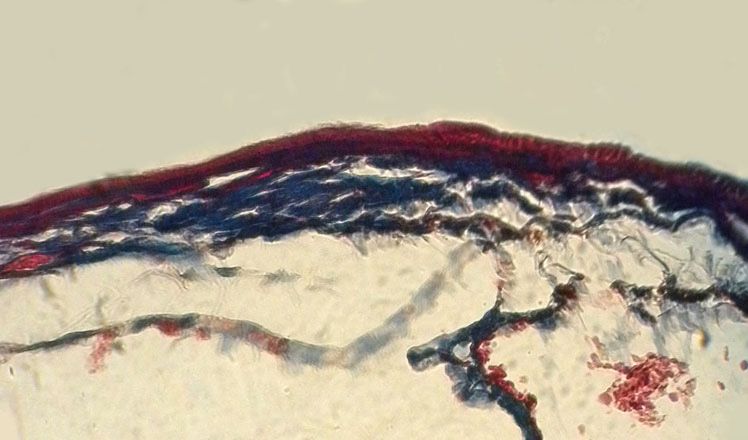
Microscopic study of the gall bladder of the chukar partridge (Alectoris chukar)

variation in these features compared to               Bancroft, 1984). Histological studies on
other bird species.                                   stained sections were carried out by light
                                                      microscopy.
MATERIALS AND METHODS
                                                      RESULTS
Sixteen clinically healthy Iranian chukar
partridges (Alectoris chukar) from both               The gall bladder in Alectoris chukar was
sexes were used to determine the histo-               composed of tunica mucosa, tunica mus-
logical structures of the gall bladder.               cularis and tunica serosa (for the free sur-
They were reared in a floor-pen house                 face) and tunica adventitia (for the at-
from hatch to 20 weeks of age. The                    tached surface) in both sexes. The tunica
chuckar chicks received feed and water                mucosa of the gall bladder was mainly
ad libitum. The animals were euthanized.              lined by non-ciliated simple columnar
The gall bladders were removed from the               epithelium. However, in some regions it
donors, placed in physiological saline and            varied from cuboidal to tall columnar
cut open therein to expel intravesical bile           (Fig. 1). The apical cytoplasm of these
which is injurious to the epithelial tissues.         cells was covered by a continuous striated
The gall bladders were fixed in 10% buff-             border of microvilli (Fig. 2). No goblet
ered formalin solution for 12 to 24 h, de-            cells were observed in epithelium. Tunica
hydrated and embedded in paraffin in                  mucosa forms some simple folds lined
routine manners. Tissue samples were                  with tall columnar epithelium, which ap-
stained by a variety of techniques for                peared to be regularly distributed over the
general observations and types of fibres in           whole luminal surface of the gall bladder.
the connective tissues: 1) haematoxylin               The mucosal folds were almost isometric.
eosin, 2) Masson’s trichrome, 3) Ver-                 Deep invaginations of the surface epithe-
hoeff’s, 4) Gomori’s method for reticu-               lium were observed to have grown down
lum (Luna, 1968), 5) alcian blue (pH 1.0),            into the underlying loose connective tis-
6) Periodic acid-Schiff (PAS) (Cook &                 sue, showing a tubular gland like appear-

                                                                                 M
                                                                             LP-S

                                                               TA

 Fig. l. The gall bladder of twenty-week-old Iranian chukar partridges: epithelium (E), simple fold
 (arrows), epithelial invaginations (I), lymphatic aggregations (L), lamina propria (LP-SM), tunica
                muscularis (TM), tunica adventitia (TA). Haematoxylin eosin, × 400.

74                                                                                   BJVM, 15, No 2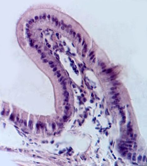
B. Mobini

                                                  E

                                             LP-SM

                                      TM

                                        TA

 Fig. 2. Reticular fibres (arrows) in lamina propria-submucosa (LP-SM), and tunica serosa (TA) of
chukar gall bladder, epithelium (E), tunica muscularis (TM). Gomori’s staining for reticulum, ×400.

ance (Fig. 1). The surface cells and those                All epithelial cells reacted positively
lining folds and the epithelial invagina-             to periodic acid Schiff (PAS) (Fig. 3), and
tions exhibited an oval nucleus located in            alcian blue stains (Fig. 4).
the basal cytoplasm. However, in some                     The lamina muscularis mucosa was
regions with cuboidal epithelium, the nu-             absent. The thin lamina propria-submu-
cleus was more spherical.                             cosa contained loose connective tissue
                                                      which consisted of reticular (Fig. 2), col-
                                                      lagenous (Fig. 5) and elastic fibres (Fig.
                                                      6), and numerous diffuse or nodular lym-
                                                      phatic tissues, but no glands were ob-
                                                      served (Fig. 1). The tunica muscularis
                                                      was composed of a layer of circularly
                                                      arranged muscle fibres (Fig. 1).
                                                          The outermost tunica of the free sur-
                                                      face of the gall bladder was the serosa,
                                  E                   which loose connective tissue invested by
                                                      mesothelium, whereas in attached surface
                                                      adventitia and mesothelium were absent.
                                                      The loose connective tissue was made up
                     LP-SM                            of adipose tissues, blood vessels (Fig. 6),
                                                      parasympathetic ganglia with nerve bun-
         TM                                           dles (Fig. 1), reticular, collagenous and,
                                                      elastic fibres, but glands were absent in
                                                      tunica serosa (Fig. 2, 5, 6).
Fig. 3. PAS-positive material is present in the
apical part of the cytoplasm of the epithelial        DISCUSSION
cells (arrows), epithelium (E), lamina propria-
submucosa (LP-SM), tunica muscularis (TM).            The wall of the gall bladder in Alectoris
PAS, ×1000.                                           chukar was composed of tunica mucosa,

BJVM, 15, No 2                                                                                   75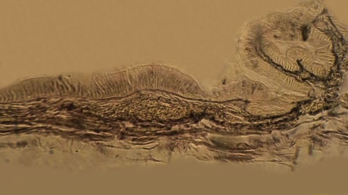
Microscopic study of the gall bladder of the chukar partridge (Alectoris chukar)
lamina propria-submucosa, tunica muscu- The apical cytoplasm of epithelial cells,
laris and tunica serosa or adventitia, which was covered by a continuous stri-
which was similar to those of guinea fowl ated border of microvilli agree with the
(Sivagnanam & Geetha, 2008). Although results obtained from Yamada (1974) and
some variations were observed in the epi- Dellmann (1993). The simple isometric
thelium of tunica mucosa, it was mainly folds were regularly distributed over the
lined by non-ciliated simple columnar whole gall bladder luminal surface, which
cells similarly to the findings of Yamada was similar to the previous findings (Ya-
& Hoshino (1972) in chickens and Sivag- mada & Hoshino, 1972; Gheri et al.
nanam & Geetha (2008) in guinea fowl. 1988).
E
LP-SM
TM
TA
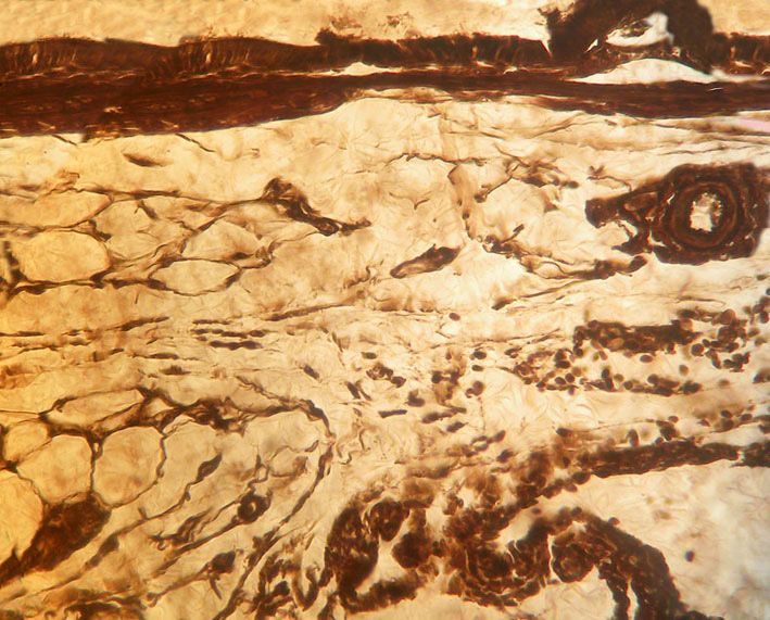
Fig. 4. Acidophilic mucosubstances are present in all surface epithelial cells (arrows),
epithelium (E), lamina propria-submucosa (LP-SM), tunica muscularis (TM),
tunica adventitia (TA). Alcian blue, ×400.
E
LP-SM
TM
TA
Fig. 5. Collagenous fibres (arrows) in lamina propria-submucosa (LP-SM) and tunica serosa (TA) of
chukar gall bladder, epithelium (E), tunica muscularis (TM). Masson’s trichrome, × 400.
76 BJVM, 15, No 2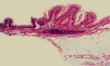
B. Mobini

                                   E
                                                                     LP-SM
                              TM

                                                                        BV

                                                      TA

                        A

    Fig. 6. Elastic fibres (arrows) in lamina propria-submucosa (LP-SM) and tunica serosa (TA)
         of chukar gall bladder. epithelium (E), tunica muscularis (TM), adipose tissue (A),
                                blood vessels (BV). Verhoeff’s, × 400.
    In tunica mucosa, downgrowths of the             the concentration of the bile (Hayward,
surface epithelium which exhibited a tu-             1968), others have noticed that they ap-
bular gland like appearance were in                  pear to play significant roles for maintain-
agreement with those reported previously             ing functionally important properties of
(Yamada & Hoshino, 1972; Gheri et al.,               membranes such as morphological con-
1988). All epithelial cells containing an            figurations, structural rigidity and perme-
oval nucleus were situated basally. Simi-            ability (Quinton & Philpott, 1973). This
lar results were also reported by Yamada             finding indicated that the epithelium of
& Hoshino (1972) in chick embryo and                 the gall bladder had a secretory function
Gheri et al. (1988) in adult fowl.                   (Dellmann, 1993) and suggested an asso-
    The mucosal histochemical reactions              ciation of neutral mucopolysaccharides
of the chukar gall bladder were similar to           with acid mucins (Gheri et al., 1988).
that of other poultry (Yamada & Hoshino,                 In our study, no lamina muscularis,
1972; Gheri et al., 1988; Madrid et al.,             goblet cells and gland were observed in
1989), which indicated that the muco-                gall bladder mucosa of the chukar part-
substances consist of acid and neutral gly-          ridges, which is in agreement with the
cosaminoglycan complexes (Yamada &                   results of a previous study (Sivagnanam
Hoshino, 1972). While some investigators             & Geetha, 2008).
have reported that the presence of mucous                Reported lymphatic aggregations of
secretion might be of importance in rela-            the lamina propria-submucosa in chicken
tion to the water absorbing function of the          gall bladder were in agreement with our
gall bladder epithelium (the mucus might             results (Ciobotaru & Militaru, 2002).
form a water-absorbing surface gel) for

BJVM, 15, No 2                                                                                   77
Microscopic study of the gall bladder of the chukar partridge (Alectoris chukar)
The tunica muscularis of gall bladder Staining Methods of the Armed Forces in
in guinea fowl was composed of an outer Statute of Pathology. 3rd edn, McGraw-
longitudinal layer and an inner layer that Hill, New York, pp. 87–88, 94–95, 158–
consisted of an outer circular and an inner 160, 163–164.
longitudinal muscle fibres (Sivagnanam Madrid, J. F., J. Ballesta, T. Galera, M.T. Cas-
& Geetha, 2008). In chukar partridges tells & R. Perez-Tomas, 1989. Histochem-
istry of glycoconjugates in the gallbladder
however, there was a layer of circularly
epithelium of ten animal species, Histo-
arranged muscle fibres. chemistry, 91, 437–443.
The tunica serosa and adventitia of
Quinton, P. M. & C. W. Philpott, 1973. A role
gall bladder in chukars are in agreement
for anionic sites in epithelial architecture.
with the results obtained from Sivagna- Effects of cationic polymers on cell mem-
nam & Geetha (2008). brane structure. The Journal of Cell Biol-
In conclusion, the microscopic anatomy ogy, 56, 787–796.
of the gall bladder of the Iranian chukar Sivgnanam, S. & R. Geetha, 2008. Histologi-
partridge (Alectoris chukar) was similar to cal studies on the gall bladder and biliary
that in chickens and guinea fowl. system in Guinea fowl. Indian Journal of
Veterinary Anatomy, 20, 60–61.
REFERENCES Stornelli, M. R., M. P. Ricciardi, E. Giannessi
& A. Coli, 2006. Morphological and histo-
logical study of the ostrich (Struthio came-
Abidu-Figueiredo, M., B. Xavier-Silva, F. V.
lus L.) liver and biliary system. Archivio
C. Bath, M. A. Babinski & M. A. Chagas,
Italiano Di Anatomia Ed Embriologia.
2006. Morphological and topographical
111, 1–7.
aspects of the ostrich (Struthio camelus)
liver. Revista Portuguesa de Ciências Vet- Yamada, K. 1974. Acid mucosaccharide-con-
erinárias, 101, 41–43. taining structures in the gall bladder epithe-
lium of the rabbit as seen with the electron
Ciobotaru, E. & M. Militaru, 2002. Researches
microscope. Histochemistry, 39, 351–360.
regarding the structure and reactivity of
lymphoid formation associated to gall Yamada, K. & M. Hoshino, 1972. Morpho-
bladder in chicken. Revista Romana de chemical analysis of the epithelial muco-
Medicina Veterinara, 12, 117–127. substances in the gall bladder of the fowl
(gallus domesticus). Histochemie, 29, 120–
Cook, H. & J. Bancroft, 1984. Manual of His-
128.
tological Techniques. Churchill Living-
stone Edinburgh, London, pp. 102–103,
111–112. Paper received 21.12.2011; accepted for
Dellmann, H. D., 1993. Textbook of Veteri- publication 23.04.2012
nary Histology. 4th edn, Lea and Febiger,
Philadelphia, pp. 189.
Correspondence:
Gheri, G., S. Gheri Bryk & G. E. Orlandini,
1988. Histochemistry of mucosubstances
Behzad Mobini DVM, PhD
in the gallbladder epithelium of the chick
Associate Professor of Anatomical Sciences,
embryo. Histochemistry, 88, 519–524.
Faculty of Veterinary Medicine,
Hayward, A. F., 1968. The structure of gall- Islamic Azad University, Shahrekord branch,
bladder epithelium. International Review P.O.Box 166 Shahrekord, Iran
of General and Experimental Zoology, 3, e-mail: dr.mobini@iaushk.ac.ir
205–239.
Luna, L. G., 1968. Manual of Histological
78 BJVM, 15, No 2You can also read