Mitocryptide-2: Identification of Its Minimum Structure for Specific Activation of FPR2-Possible Receptor Switching from FPR2 to FPR1 by Its ...
←
→
Page content transcription
If your browser does not render page correctly, please read the page content below
International Journal of
               Molecular Sciences

Article
Mitocryptide-2: Identification of Its Minimum Structure for
Specific Activation of FPR2–Possible Receptor Switching from
FPR2 to FPR1 by Its Physiological C-terminal Cleavages
Takayuki Marutani † , Kodai Nishino, Tomoyuki Miyaji, Keisuke Kamada, Koji Ohura, Yoshiaki Kiso
and Hidehito Mukai *

                                          Laboratory of Peptide Science, Graduate School of Bio-Science, Nagahama Institute of Bio-Science and
                                          Technology, Nagahama, Shiga 526-0829, Japan; b210052@nagahama-i-bio.ac.jp (T.M.);
                                          b112116@m.nagahama-i-bio.ac.jp (K.N.); b116169@m.nagahama-i-bio.ac.jp (T.M.);
                                          b116046@m.nagahama-i-bio.ac.jp (K.K.); b116028@m.nagahama-i-bio.ac.jp (K.O.);
                                          y_kiso@nagahama-i-bio.ac.jp (Y.K.)
                                          * Correspondence: hmukai-endo@umin.ac.jp; Tel.: +81-749-64-8113
                                          † Supported by JSPS Research Fellowship for Young Scientists.

                                          Abstract: Mitocryptides are a novel family of endogenous neutrophil-activating peptides originating
                                          from various mitochondrial proteins. Mitocryptide-2 (MCT-2) is one of such neutrophil-activating
                                peptides, and is produced as an N-formylated pentadecapeptide from mitochondrial cytochrome b.

                                          Although MCT-2 is a specific endogenous ligand for formyl peptide receptor 2 (FPR2), the chemical
Citation: Marutani, T.; Nishino, K.;      structure within MCT-2 that is responsible for FPR2 activation is still obscure. Here, we demonstrate
Miyaji, T.; Kamada, K.; Ohura, K.;        that the N-terminal heptapeptide structure of MCT-2 with an N-formyl group is the minimum
Kiso, Y.; Mukai, H. Mitocryptide-2:
                                          structure that specifically activates FPR2. Moreover, the receptor molecule for MCT-2 is suggested to
Identification of Its Minimum
                                          be shifted from FPR2 to its homolog formyl peptide receptor 1 (FPR1) by the physiological cleavages
Structure for Specific Activation of
                                          of its C-terminus. Indeed, N-terminal derivatives of MCT-2 with seven amino acid residues or longer
FPR2–Possible Receptor Switching
                                          caused an increase of intracellular free Ca2+ concentration in HEK-293 cells expressing FPR2, but
from FPR2 to FPR1 by Its
Physiological C-terminal Cleavages.
                                          not in those expressing FPR1. Those MCT-2 derivatives also induced β-hexosaminidase secretion in
Int. J. Mol. Sci. 2021, 22, 4084.         neutrophilic/granulocytic differentiated HL-60 cells via FPR2 activation. In contrast, MCT-2(1–4), an
https://doi.org/                          N-terminal tetrapeptide of MCT-2, specifically activated FPR1 to promote those functions. Moreover,
10.3390/ijms22084084                      MCT-2 was degraded in serum to produce MCT-2(1–4) over time. These findings suggest that MCT-2
                                          is a novel critical factor that not only initiates innate immunity via the specific activation of FPR2,
Academic Editor: Andrey                   but also promotes delayed responses by the activation of FPR1, which may include resolution and
V. Kuznetsov                              tissue regeneration. The present results also strongly support the necessity of considering the exact
                                          chemical structures of activating factors for the investigation of innate immune responses.
Received: 2 March 2021
Accepted: 12 April 2021
                                          Keywords: cryptide; mitocryptide; N-formylated peptide; neutrophil; formyl peptide receptor 1;
Published: 15 April 2021
                                          formyl peptide receptor 2; inflammation

Publisher’s Note: MDPI stays neutral
with regard to jurisdictional claims in
published maps and institutional affil-
                                          1. Introduction
iations.
                                               Neutrophils are a type of leukocyte that are involved in the innate defense system [1–3].
                                          Neutrophils comprise the majority of peripheral leukocytes and normally exist in the
                                          bloodstream to monitor for infection and tissue damage. When tissue injury occurs due
Copyright: © 2021 by the authors.
                                          to bacterial infections or internal tissue damage, neutrophils immediately migrate to
Licensee MDPI, Basel, Switzerland.
                                          and infiltrate the injury site. The infiltrated neutrophils are then activated and exert
This article is an open access article
                                          their functions, including superoxide production and phagocytosis of invading bacterial
distributed under the terms and           components and toxic substances.
conditions of the Creative Commons             Bacterial N-formylated proteins and peptides, including formyl-Met-Leu-Phe (fMLF) [4,5],
Attribution (CC BY) license (https://     complement related factors such as component 5a [6,7], and some chemokines such as
creativecommons.org/licenses/by/          IL-8 [8,9] chemoattract and activate neutrophils to promote inflammatory reactions. More-
4.0/).                                    over, various mitochondrial-derived peptides that also activate neutrophils were recently

Int. J. Mol. Sci. 2021, 22, 4084. https://doi.org/10.3390/ijms22084084                                          https://www.mdpi.com/journal/ijms
Int. J. Mol. Sci. 2021, 22, 4084 2 of 21
identified in mammalian tissues [10–14]. Namely, we isolated and identified the novel
neutrophil-activating peptides mitocryptide-1, mitocryptide-2 (MCT-2), and mitocryptide-
CYC, which are cleaved from mitochondrial cytochrome c oxidase subunit VIII, cytochrome b,
and cytochrome c, respectively, from porcine heart [10–13]. In addition, we found the possi-
ble presence of many mitocryptides that were derived from various mitochondrial proteins
to induce the migration and activation of neutrophils [10,14].
Recently, there has also been interest in the roles of mitochondria and their derived
substances in innate immunity. Indeed, various tissues damaged by trauma or burns
release mitochondria and their contents into the bloodstream [15–21]. Mitochondria
and their contents are also released from a variety of cells damaged by bacterial infec-
tion [21–25]. In addition, it is suggested that mitochondria and their derived substances
are leaked into the cerebrospinal fluid from damaged tissues in patients with Alzheimer’s
disease [26,27]. These released mitochondria and their derived factors, which are called
mitochondrial damage-associated molecular patterns (mtDAMPs), promote various innate
immune responses, including induction of neutrophil and macrophage migration and acti-
vation [15,17,21,28,29]. MtDAMPs also stimulate the secretion of inflammatory mediators
from mast cells [30].
N-formylated peptides may constitute one family of the activating factors in mt-
DAMPs because inhibitors against the functions of N-formylated peptides prevent the
activity of mtDAMPs [15,17,28]. Specifically, as 13 proteins are encoded in mitochondrial
DNA and are translated in mitochondria as N-formylated forms [31,32], these protein-
derived N-formylated peptides may exist in mtDAMPs, and are thought to cause proin-
flammatory responses including neutrophil infiltration and activation. As MCT-2 is the
only endogenous N-formylated peptide that has been isolated from mammalian tissues so
far and its complete chemical structure has been determined, it is a leading candidate for
such activating factors in mtDAMPs.
Formyl peptide receptor 1 (FPR1, formerly referred to as formyl peptide receptor)
and formyl peptide receptor 2 (FPR2, formerly referred to as formyl peptide receptor-
like 1) recognize not only N-formylated proteins and peptides derived from bacteria
but also endogenous N-formylated peptide MCT-2 and putative peptides derived from
mitochondrial N-formylated proteins [33–38]. These receptors are expressed mainly by
inflammatory immune cells such as neutrophils and macrophages, and their activation
induces not only the infiltration of neutrophils and macrophages into injury sites but also
the phagocytosis of toxic debris, superoxide generation, and the production of inflamma-
tory cytokines [35,37,38]. FPR2 is also expressed by endothelial cells, and its activation
causes an increase in vascular permeability to promote further neutrophil infiltration from
the bloodstream to injury sites during the initial stage of inflammation [38,39]. Moreover,
FPR1 is proposed to be involved in the processes of resolution and wound healing/tissue
regeneration. Indeed, FPR1 activation induces the proliferation of various cells, including
epithelial cells and hepatocytes, to promote tissue regeneration [40–44]. In this way, FPR1
and FPR2 are thought to participate throughout the initiation of the proinflammatory
response by tissue damage, healing, and regeneration.
However, it is unknown how FPR2 and FPR1 recognize various endogenous N-
formylated peptides, including MCT-2. FPR2 specifically recognizes MCT-2 and its N-
terminal derivatives longer than ten amino acid residues to activate neutrophils [36,45].
Putative endogenous N-formylated peptides derived from mitochondrial DNA-encoded
proteins activate FPR2 and/or FPR1, i.e., NADH dehydrogenase subunit 4(1–20) and
NADH dehydrogenase subunit 5(1–28) specifically activate FPR2, whereas NADH dehy-
drogenase subunit 6(1–6) specifically activates FPR1, and cytochrome c oxidase subunit
I(1–13) activates FPR1 and FPR2 [46]. However, the chemical structure within endogenous
N-formylated peptide MCT-2 that is specifically recognized by FPR2 as well as those within
endogenous N-formylated peptides recognized by FPR1 or FPR2 are unclear, although the
N-formyl group of those peptides is known to be essential for recognition by FPR2 and
FPR1 [45].Int. J. Mol. Sci. 2021, 22, 4084 3 of 21
In this study, we investigated the structure–activity relationships of MCT-2 and its
derivatives to elucidate how FPR2 recognizes MCT-2. We also explored the time-dependent
alterations of the molecular forms of MCT-2 in serum and attempted to elucidate the
meaning of these alterations in innate immune responses.
2. Results
2.1. Effects of MCT-2(1–15) and Its Derivatives on [Ca2+ ]i in HEK-293 Cells Stably Expressing
FPR1 or FPR2
An endogenous pentadecapeptide, MCT-2 [MCT-2(1–15)], specifically binds to and ac-
tivates FPR2, but it neither interacts with nor activates FPR1 [36]. In addition, MCT-2(1–10)
or N-terminal derivatives longer than it specifically activate FPR2 to cause β-arrestin recruit-
ment and superoxide production [45]. However, the precise structure within MCT-2(1–15)
that is responsible for the specific activation of FPR2 is not known. Here, increases in the
concentration of intracellular free Ca2+ ([Ca2+ ]i ) promoted by MCT-2(1–15) and its deriva-
tives were assessed in HEK-293 cells stably expressing FPR1 or FPR2 (Figure 1). The Gα16
type of G protein was also stably co-expressed in these cells because this protein interacts
with various G protein-coupled receptors and effectively induces an agonist-promoted
increase of [Ca2+ ]i [47]. As a result, 100 µM MCT-2(1–10), MCT-2(1–9), MCT-2(1–8), and
MCT-2(1–7) as well as 10 µM MCT-2(1–15) induced an increase of [Ca2+ ]i in HEK-293
cells expressing FPR2, but not in FPR1-expressing cells (Figure 1A–E). In contrast, 100 µM
MCT-2(1–5) induced an increase of [Ca2+ ]i in FPR1- and FPR2-expressing cells (Figure 1F).
Moreover, 100 µM MCT-2(1–4) promoted an increase of [Ca2+ ]i in FPR1-expressing cells,
but not in FPR2-expressing cells (Figure 1G). In addition, stimulation with MCT-2(1–6) did
not induce an increase of [Ca2+ ]i in FPR2- or FPR1-expressing cells, even at a concentration
of 100 µM (data not shown). These results demonstrate that the N-terminal derivative MCT-
2(1–7) and its longer derivatives specifically activate FPR2, in contrast with MCT-2(1–4),
which specifically activates FPR1.
2.2. Effects of C- or N-Terminal Truncations of MCT-2(1–15) on β-Hexosaminidase Release from
Neutrophilic/Granulocytic Differentiated HL-60 Cells
The effects of N- or C-terminal truncations of MCT-2 on β-hexosaminidase released
from neutrophilic/granulocytic differentiated HL-60 cells were investigated to elucidate the
minimum structure that was required for the activation of their receptor molecules. As we
reported previously [12,14], a pentadecapeptide MCT-2 [MCT-2(1–15)] dose-dependently
promoted β-hexosaminidase secretion from differentiated HL-60 cells (EC50 : 20 ± 7 nM,
Figure 2 and Table 1), and the maximum response was observed at concentrations greater
than 1 µM.
To elucidate the necessity of the N-formyl group for this stimulation, we examined
the effects of its removal from MCT-2(1–15). Des-N-formyl MCT-2(1–15) did not induce
β-hexosaminidase release, even at a concentration of 100 µM (Figure 2A), demonstrating
that the formyl group at the N-terminus of MCT-2(1–15) is crucial for this process.
Next, we examined the importance of the C-terminal sequence of MCT-2(1–15) for the
stimulation of β-hexosaminidase release from differentiated HL-60 cells by performing
C-terminal truncations. Truncations of one to eight amino acid residues from the C-
terminus of MCT-2(1–15) caused consecutive increases in the EC50 values without affecting
the maximum response. Specifically, the EC50 value of MCT-2(1–14) was increased by
approximately five-fold compared with that of MCT-2(1–15) (Figure 2B and Table 1).
MCT-2(1–13) and MCT-2(1–12) also exhibited decreased activity when compared with
MCT-2(1–15), but MCT-2(1–11) had almost the same potency as MCT-2(1–12) (Figure 2B
and Table 1). In addition, the activities of MCT-2(1–10), MCT-2(1–9), and MCT-2(1–8)
were sequentially attenuated compared with that of MCT-2(1–11), but with the same
maximum effect as MCT-2(1–15), and MCT-2(1–7) had almost the same potency as MCT-
2(1–8). However, MCT-2(1–6), a derivative that was truncated by nine C-terminal amino
acid residues, exhibited a significant decrease of the maximum response compared withInt. J. Mol. Sci. 2021, 22, 4084 4 of 21
MCT-2(1–15) (maximum response: 90 ± 3%) with a 5.7-fold increase in the EC50 value
compared with MCT-2(1–7). These results indicate that MCT-2(1–7) with an N-formyl
group is the minimum structure that is required for maximum stimulation via the activation
of FPR2 because the minimum sequence that gave the same level of maximum response as
MCT-2(1–15) was MCT-2(1–7), which was also the minimum structure for inducing specific
FPR2 activation to cause an increase of [Ca2+ ]i (Figure 1). In addition, the MCT-2(8–15)
structure within MCT-2(1–15) may contribute to the binding affinity between MCT-2(1–15)
and FPR2, since the removal of one to eight amino acid residues from the C-terminus
Int. J. Mol. Sci. 2021, 22, x FOR PEER REVIEW
did not affect the maximum response, but caused a consecutive increase in EC50 values4 of 22
(Figure 2B and Table 1).
1. Changes
FigureFigure in [Cain2+ ] in2+HEK-293 cells stably expressing FPR1 (formyl peptide receptor 1) or FPR2 (formyl peptide
1. Changes [Ca
i ]i in HEK-293 cells stably expressing FPR1 (formyl peptide receptor 1) or FPR2 (formyl peptide
receptor 2) induced
receptor by MCT-2
2) induced by MCT-2(Mitocryptide-2)(1–15)
(Mitocryptide-2)(1–15) and and
its derivatives. Fura-2-loaded
its derivatives. Fura-2-loadedcells were
cells stimulated
were stimulatedwith
with1010 μM
MCT-2(1–15)(A)
µM MCT-2(1–15) (A)oror100
100µMμMMCT-2(1–10)
MCT-2(1–10)(B), (B),MCT-2(1–9)
MCT-2(1–9)(C),(C), MCT-2(1–8)
MCT-2(1–8) (D), MCT-2(1–7) (E), MCT-2(1–5)
MCT-2(1–5) (F),
(F),and
andMCT-
2(1–4) (G).
MCT-2(1–4) (G). The
The changes
changes in in the
the fluorescence
fluorescence ratio
ratio of
of Fura-2
Fura-2 (excitation
(excitationwavelengths,
wavelengths,340 340nm
nmandand380
380nm;
nm;emission
emissionwave-
length, 500
wavelength, 500 nm)
nm) were
were recorded
recordedby byaafluorometer
fluorometerCAF-100.
CAF-100.Horizontal
Horizontalaxes
axesshow
showthethetimes
timesafter
afterstimulation.
stimulation.Vertical
Vertical axes
depictthe
axes depict thefluorescence
fluorescenceratiosratios at at excitation
excitation wavelengths
wavelengths of 340
of 340 nmnmandand
380 380
nm. nm. Arrows
Arrows indicate
indicate the timing
the timing of peptide
of peptide
administration.
administration.
2.2. Effects of C- or N-Terminal Truncations of MCT-2(1–15) on β-Hexosaminidase Release from
Neutrophilic/Granulocytic Differentiated HL-60 Cells
The effects of N- or C-terminal truncations of MCT-2 on β-hexosaminidase released
from neutrophilic/granulocytic differentiated HL-60 cells were investigated to elucidate
the minimum structure that was required for the activation of their receptor molecules.
As we reported previously [12,14], a pentadecapeptide MCT-2 [MCT-2(1–15)] dose-de-
pendently promoted β-hexosaminidase secretion from differentiated HL-60 cells (EC50: 20
± 7 nM, Figure 2 and Table 1), and the maximum response was observed at concentrations
greater than 1 μM.Int.
Int. J.J. Mol.
Mol. Sci.
Sci.2021,
2021, 22,
22, x4084
FOR PEER REVIEW 55 of
of 22
21
Figure
Figure 2.2.Effects of N-terminal
Effects (A) or(A)
of N-terminal C-terminal (B,C) truncations
or C-terminal of MCT-2(1–15)
(B,C) truncations on β-hex- on β-
of MCT-2(1–15)
osaminidase release by differentiated HL-60 cells. The differentiated HL-60 cells
hexosaminidase release by differentiated HL-60 cells. The differentiated HL-60 cells were stimulated
were stim-
by MCT-2(1–15) or its derivatives at 37 °C for 10 min,
◦ and the amount of the released
ulated by MCT-2(1–15) or its derivatives at 37 C for 10 min, and the amount of the released β-hex-
osaminidase was quantified
β-hexosaminidase as described
was quantified in “Materials
as described and Methods”.
in “Materials The ability
and Methods”. of each
The ability of peptide
each pep-
to induce β-hexosaminidase release is expressed as a percentage of enzyme secretion promoted by
tide to induce β-hexosaminidase release is expressed as a percentage of enzyme secretion promoted
10 μM MCT-2(1–15). Data are expressed as the mean ± SE of four to six independent experiments.
by 10 µM MCT-2(1–15). Data are expressed as the mean ± SE of four to six independent experiments.
Table 1. Amino acid sequences of MCT-2(1–15) and its C-terminal or N-terminal derivatives and their EC50 values and
The effect of C-terminal truncation was examined further because MCT-2(1–6) could
maximum effects on the stimulation of β-hexosaminidase release from differentiated HL-60 cells.
still induce β-hexosaminidase release. As a result, MCT-2(1–5), surprisingly, had 6.5-fold
Peptide higher activity than MCT-2(1–6) (Figure 2C and TableEC
Sequence 1)50with
(nM)the same level of
Maximum maximum
Effect (%) a
1 response as MCT-2(1–15). Moreover,
2 3 4 5 6 7 8 9 10 11 12 13 14 15 the activity of MCT-2(1–4) was further reinforced
MCT-2(1–15) formyl - M compared
T P M with R KMCT-2(1–5)
I N PasLwell M as
K MCT-2(1–6)
L I N (Table 20.0 ±1); nevertheless, the
3.39 100 maximum
MCT-2(1–14) formyl - M response of MCT-2(1–4) was
T P M R K I N P L M K L I significantly reduced by approximately
96.7 ± 17.6 10% compared
102 with
MCT-2(1–15). Taken together with the observations that MCT-2(1–5) induced the activation
MCT-2(1–13) formyl - M T P M R K I N P L M K L 186 ± 33.6 100 ± 3
of FPR2 and FPR1, and MCT-2(1–4) specifically activated FPR1 for the increases in [Ca2+ ]i
MCT-2(1–12) formyl - M T P M R K I N P L M K 633 ± 153 98 ± 3
described above (Figure 1), these findings suggest that MCT-2(1–5) effectively promotes
MCT-2(1–11) formyl - M T P M R K I N P L M 547 ± 93.9 103 ± 3
the stimulation of β-hexosaminidase release via FPR1 and FPR2 activation. Furthermore,
MCT-2(1–10) formyl - M T P M R K I N P L 1800 ±
MCT-2(1–4) specifically activates FPR1 to promote stimulation. 173 98 ± 3
MCT-2(1–9) formyl - M T P M R K I N P 10,760 ± 850 104 ± 3
MCT-2(1–8) formyl - M T P M R K I N 41,333 ± 2517 100 ± 2Int. J. Mol. Sci. 2021, 22, 4084 6 of 21
Table 1. Amino acid sequences of MCT-2(1–15) and its C-terminal or N-terminal derivatives and their EC50 values and
maximum effects on the stimulation of β-hexosaminidase release from differentiated HL-60 cells.
Maximum
Peptide Sequence EC50 (nM)
Effect (%) a
1 2 3 4 5 6 7 8 9 10 11 12 13 14 15
MCT-2(1–15) formyl - M T P M R K I N P L M K L I N 20.0 ± 3.39 100
MCT-2(1–14) formyl - M T P M R K I N P L M K L I 96.7 ± 17.6 102
MCT-2(1–13) formyl - M T P M R K I N P L M K L 186 ± 33.6 100 ± 3
MCT-2(1–12) formyl - M T P M R K I N P L M K 633 ± 153 98 ± 3
MCT-2(1–11) formyl - M T P M R K I N P L M 547 ± 93.9 103 ± 3
MCT-2(1–10) formyl - M T P M R K I N P L 1800 ± 173 98 ± 3
MCT-2(1–9) formyl - M T P M R K I N P 10,760 ± 850 104 ± 3
MCT-2(1–8) formyl - M T P M R K I N 41,333 ± 2517 100 ± 2
MCT-2(1–7) formyl - M T P M R K I 35,500 ± 500 101 ± 2
MCT-2(1–6) formyl - M T P M R K 202,667 ± 2309 90 ± 2 **
MCT-2(1–5) formyl - M T P M R 26,000 ± 1581 104 ± 2
MCT-2(1–4) formyl - M T P M 18,000 ± 1323 88 ± 2 ***
Des-N-formyl MCT-2 M T P M R K I N P L M K L I N >1,000,000 0
aThe ability of each peptide to cause β-hexosaminidase release is expressed as a percentage of enzyme secretion promoted by 10 µM
MCT-2(1–15). Data are expressed as the mean ± SE of four to six independent experiments. ** p < 0.01; *** p < 0.001, values significantly
different from MCT-2(1–15).
2.3. Involvement of FPR1 and FPR2 in β-Hexosaminidase Release Stimulated by MCT-2(1–15)
and Its Derivatives
To further elucidate the involvement of FPR1 and FPR2 in β-hexosaminidase release
from differentiated HL-60 cells stimulated with MCT-2(1–15) and its derivatives, we ex-
amined the inhibitory effects of inhibitors against FPR1 [cyclosporin H (CysH)] and FPR2
(PBP10) on this process. Stimulation with MCT-2(1–15) at 40 nM, which induced approxi-
mately 60% of the maximum response, was dose-dependently inhibited by PBP10, and 1
µM PBP10 completely inhibited this activity, but 1 µM CysH did not influence MCT-2(1–15)-
induced enzyme release (Figure 3A,B). Similarly, the responses induced by MCT-2(1–10),
MCT-2(1–9), MCT-2(1–8), and MCT-2(1–7) at a concentration that caused a 60% response
of the maximum effect were also abolished in a dose-dependent manner by PBP10 but
not CysH (Figure 3A,B). In contrast, stimulation with MCT-2(1–6) and MCT-2(1–5) was
partially inhibited by CysH or PBP10, and the combination of inhibitors (1 µM CysH
and 1 µM PBP10) almost completely prevented these responses (Figure 3C). Moreover, β-
hexosaminidase release induced by MCT-2(1–4) was completely abolished by 1 µM CysH,
but was unaffected by 1 µM PBP10 (Figure 3A,B). These results also support the notion
that MCT-2(1–7) and its longer N-terminal derivatives induce β-hexosaminidase release
from differentiated HL-60 cells via the specific activation of FPR2; in contrast, MCT-2(1–4)
specifically activates FPR1, and MCT-2(1–6) and MCT-2(1–5) activate both FPR1 and FPR2
(Figure 3D).and 1 μM PBP10) almost completely prevented these responses (Figure 3C). Moreover, β-
hexosaminidase release induced by MCT-2(1–4) was completely abolished by 1 μM CysH,
but was unaffected by 1 μM PBP10 (Figure 3A,B). These results also support the notion
that MCT-2(1–7) and its longer N-terminal derivatives induce β-hexosaminidase release
from
Int. J. Mol. Sci. 2021, 22, 4084differentiated HL-60 cells via the specific activation of FPR2; in contrast, MCT-2(1– 7 of 21
4) specifically activates FPR1, and MCT-2(1–6) and MCT-2(1–5) activate both FPR1 and
FPR2 (Figure 3D).
Figure 3. Involvement
Figure 3. Involvement of FPR1 andofFPR2 FPR1 in and FPR2 in β-hexosaminidase
β-hexosaminidase release byrelease by differentiated
differentiated HL-60 cellsHL-60 cells
stimulated with
stimulated with MCT-2(1–15) and its derivatives. (A,B) Concentration-dependent
MCT-2(1–15) and its derivatives. (A,B) Concentration-dependent inhibitory effects of inhibitors against FPR1 (CysH) (A) inhibitory ef-
or FPR2 (PBP10)fects(B)of on
inhibitors against FPR1release
β-hexosaminidase (CysH)induced
(A) or FPR2 (PBP10) (B) and
by MCT-2(1–15) on β-hexosaminidase
its derivatives. (C)release in- effects of
Inhibitory
duced by MCT-2(1–15) and its derivatives. (C) Inhibitory effects of CysH (1 μM; black), PBP10 (1
CysH (1 µM; black), PBP10 (1 µM; gray), or a combination of both inhibitors (1 µM PBP10 and 1 µM CysH; slash lines)
μM; gray), or a combination of both inhibitors (1 μM PBP10 and 1 μM CysH; slash lines) on the
on the stimulation of β-hexosaminidase release by MCT-2(1–15) and its derivatives. White bars indicate the stimulation
stimulation of β-hexosaminidase release by MCT-2(1–15) and its derivatives. White bars indicate
of MCT-2(1–15) theand its derivatives
stimulation in the absence
of MCT-2(1–15) and itsofderivatives
the inhibitor. The
in the stimulatory
absence of the concentrations of MCT-2(1–15) and
inhibitor. The stimulatory
its derivativesconcentrations
were the concentrations that caused a 60% response of the maximum effect.
of MCT-2(1–15) and its derivatives were the concentrations that caused a 60% The ability of each
re- peptide in
the absence or the presence
sponse of the inhibitors
of the maximum wasability
effect. The expressed as peptide
of each a percentage
in theof the total
absence or enzyme activity,
the presence which was the
of the
enzyme activityinhibitors was disruption
leaked after expressed as of athe
percentage
cells with of the total
0.05% Tritonenzyme activity, which
X-100 (%/Total). Data was the enzyme
are expressed mean ± SE
activ-
as the
ity leaked after disruption of the cells with 0.05% Triton X-100 (%/Total). Data are expressed
of four to six independent experiments. * p < 0.05, values significantly different from each peptide in the absence of the as the
inhibitors. (D)mean ± SE preference
Receptor of four to six independent and
of MCT-2(1–15) experiments. * p < 0.05;
its derivatives. ** p < 0.01;
N-terminal *** p < 0.001,
derivatives valueswith
of MCT-2 signifi-
seven amino
cantly different from each peptide in the absence of the inhibitors. (D) Receptor preference
acid residues [MCT-2(1–7)] or longer than it specifically activate FPR2, whereas MCT-2(1–4) induces the specific activation of
MCT-2(1–15) and its derivatives. N-terminal derivatives of MCT-2 with seven amino acid residues
of FPR1, as well as MCT-2(1–6) and MCT-2(1–5), which activate both FPR1 and FPR2.
[MCT-2(1–7)] or longer than it specifically activate FPR2, whereas MCT-2(1–4) induces the specific
activation of FPR1, as well as MCT-2(1–6) and MCT-2(1–5), which activate both FPR1 and FPR2.
2.4. Effects of Substituting Ala for Each Amino Acid Residue of MCT-2(1–15) on
β-Hexosaminidase Release by Differentiated HL-60 Cells
We examined the effect of substituting Ala for each amino acid residue in MCT-2(1–15)
on the stimulation of β-hexosaminidase release to elucidate the contribution of each side
chain structure to this process. As the MCT-2(1–7) structure with an N-formyl group was
indicated as crucial for the activation of FPR2 (Figures 1–3), we investigated the effect of
substituting Ala for each amino acid residue in positions one to seven within MCT-2(1–15).
The replacement of Met1 of MCT-2(1–15) with Ala caused a 30% reduction of the maximum
response with a dramatic (>500-fold) increase in the EC50 value compared with MCT-
2(1–15) (Figure 4A and Table 2), indicating that the Met1 side chain is important for not
only the affinity of MCT-2(1–15) to FPR2, but also for FPR2 activation. The substitution of
Met4 , Arg5 , Lys6 , or Ile7 with Ala also caused an increase in the EC50 value with the same
level of maximum response as MCT-2(1–15). In contrast, the replacement of Thr2 with Ala
promoted a remarkable decrease in the EC50 value without affecting the maximum response,
although the substitution of Pro3 with Ala had no effect on the EC50 value. These results
suggest that the side chains of Met4 , Arg5 , Lys6 , and Ile7 , and Thr2 within MCT-2(1–15)
contribute positively and negatively, respectively, to its affinity to bind to FPR2.not only the affinity of MCT-2(1–15) to FPR2, but also for FPR2 activation. The substitution
of Met4, Arg5, Lys6, or Ile7 with Ala also caused an increase in the EC50 value with the same
level of maximum response as MCT-2(1–15). In contrast, the replacement of Thr2 with Ala
promoted a remarkable decrease in the EC50 value without affecting the maximum re-
sponse, although the substitution of Pro3 with Ala had no effect on the EC50 value. These
Int. J. Mol. Sci. 2021, 22, 4084 8 of 21
results suggest that the side chains of Met4, Arg5, Lys6, and Ile7, and Thr2 within MCT-2(1–
15) contribute positively and negatively, respectively, to its affinity to bind to FPR2.
Figure
Figure4.4.Effects
Effectsofofsubstituting
substitutingAla
Alafor
foreach
eachamino
aminoacid
acidresidue
residue in
in the
the MCT-2(1–7)
MCT-2(1–7) (A) or the MCT-2
MCT-
2(8–15)
(8–15)(B)
(B)sequence
sequencewithin
within MCT-2(1–15) on β-hexosaminidase release by differentiated
MCT-2(1–15) on β-hexosaminidase release by differentiated HL-60 cells. HL-60
cells. The differentiated
The differentiated HL-60HL-60 cells were
cells were stimulated
stimulated by MCT-2(1–15)
by MCT-2(1–15) or its derivatives
or its derivatives atfor
at 37 ◦ C 37 10
°C min,
for
10 min, and the amount of the released β-hexosaminidase was quantified as described in “Materials
and the amount of the released β-hexosaminidase was quantified as described in “Materials and
and Methods”. The ability of each peptide to cause β-hexosaminidase release is expressed as a per-
Methods”. The ability of each peptide to cause β-hexosaminidase release is expressed as a percentage
centage of enzyme secretion promoted by 10 μM MCT-2(1-15). Data are expressed as the mean ± SE
ofoffour
enzyme
to sixsecretion promoted
independent by 10 µM MCT-2(1-15). Data are expressed as the mean ± SE of four
experiments.
to six independent experiments.
Table 2. Amino acid sequences of MCT-2(1–15) and its Ala-substituted derivatives and their EC50 values and maximum
effects on the induction of β-hexosaminidase release from differentiated HL-60 cells.
Peptide Sequence EC50 (nM) Maximum Effect (%) a
1 2 3 4 5 6 7 8 9 10 11 12 13 14 15
MCT-2(1–15) formyl - M T P M R K I N P L M K L I N 20.0 ± 3.39 100
[Ala1 ]MCT-2 formyl - A T P M R K I N P L M K L I N >10,000 70 ± 2 ***
[Ala2 ]MCT-2 formyl - M A P M R K I N P L M K L I N 0.61 ± 0.07 106 ± 2
[Ala3 ]MCT-2 formyl - M T A M R K I N P L M K L I N 16.3 ± 0.29 102 ± 1
[Ala4 ]MCT-2 formyl - M T P A R K I N P L M K L I N 68.0 ± 2.65 100 ± 2
[Ala5 ]MCT-2 formyl - M T P M A K I N P L M K L I N 61.3 ± 0.58 104 ± 1
[Ala6 ]MCT-2 formyl - M T P M R A I N P L M K L I N 64.0 ± 3.00 103 ± 2
[Ala7 ]MCT-2 formyl - M T P M R K A N P L M K L I N 94.7 ± 3.21 104 ± 2
[Ala8 ]MCT-2 formyl - M T P M R K I A P L M K L I N 31.0 ± 3.97 106 ± 1
[Ala9 ]MCT-2 formyl - M T P M R K I N A L M K L I N 3.02 ± 1.21 105 ± 1
[Ala10 ]MCT-2 formyl - M T P M R K I N P A M K L I N 73.0 ± 13.3 100 ± 2
[Ala11 ]MCT-2 formyl - M T P M R K I N P L A K L I N 36.7 ± 11.0 102 ± 1
[Ala12 ]MCT-2 formyl - M T P M R K I N P L M A L I N 12.3 ± 0.29 106 ± 3
[Ala13 ]MCT-2 formyl - M T P M R K I N P L M K A I N 83.7 ± 3.54 107 ± 3
[Ala14 ]MCT-2 formyl - M T P M R K I N P L M K L A N 76.7 ± 11.9 103 ± 3
[Ala15 ]MCT-2 formyl - M T P M R K I N P L M K L I A 29.3 ± 1.49 104 ± 3
aThe ability of each peptide to cause β-hexosaminidase release is expressed as a percentage of enzyme secretion promoted by 10 µM
MCT-2. Data are expressed as the mean ± SE of four to six independent experiments. *** p < 0.001, values significantly different from
MCT-2(1–15).Int. J. Mol. Sci. 2021, 22, 4084 9 of 21
We also examined the effect of substituting Ala for each amino acid residue in the
MCT-2(8–15) sequence within MCT-2(1–15) because this structure may be important for the
binding affinity between MCT-2(1–15) and FPR2, but not the receptor activation described
above. The substitution of Leu10 , Leu13 , or Ile14 with Ala caused an increase in the EC50
value with the same level of maximum response as MCT-2(1–15) (Figure 4B and Table 2).
In contrast, the replacement of Pro9 with Ala promoted a remarkable decrease in the EC50
value without affecting the maximum response [EC50 : 3 ± 2 nM, Figure 4B and Table 2],
although the substitution of Asn8 , Met11 , Lys12 , or Asn15 with Ala had no effect. These
results suggest that the Leu10 , Leu13 , or Ile14 , and Pro9 side chains within MCT-2(1–15)
contribute positively and negatively, respectively, to its affinity to bind to FPR2.
2.5. Circular Dichroic Spectra of MCT-2(1–15) and Its Derivatives
It was suggested that the Thr2 , Met4 , Arg5 , Lys6 , Ile7 , Pro9 , Leu10 , Leu13 , and Ile14
side chains within MCT-2(1–15) had an effect on its binding affinity to FPR2 and that the
MCT-2(8–15) structure was important for binding to FPR2. Thus, the secondary structures
of MCT-2(1–15) and its derivatives were analyzed using circular dichroic (CD) spectra,
which is an excellent tool for the rapid investigation of secondary structures. As we
reported previously [45], the CD spectrum of MCT-2(1–15) exhibited two minima at 225 nm
and 205 nm in TFE solution, suggesting that MCT-2(1–15) predominantly contained an
α-helical structure in hydrophilic circumstance (Figure 5A). Similarly, the spectra of the
N-terminal MCT-2 derivatives that were truncated by one to six amino acid residues
from the C-terminus of MCT-2(1–15) at 100 µM also showed two minima at 225 nm and
202–208 nm in TFE solution, although these minima were consecutively attenuated by the
C-terminal truncations (Figure 5B–G). Moreover, the spectra of MCT-2(1–8) and MCT-2(1–7)
displayed a minimum at approximately 200 nm in TFE solution (Figure 5H,I), proposing
that MCT-2(1–8) and MCT-2(1–7) do not contain defined secondary structures, even under
hydrophilic conditions. In contrast, the CD spectra of all of these derivatives including
MCT-2(1–15) at 100 µM exhibited a minimum at approximately 200 nm in a hydrophilic
phosphate buffer (Figure 5), proposing that MCT-2(1–15) and its derivatives did not form
defined secondary structures in hydrophilic conditions.
2.6. Time-Dependent Alterations of the Molecular Forms of MCT-2(1–15) in Serum
We examined the alterations of the molecular forms of MCT-2(1–15) in serum to
elucidate the time-dependent degradation of MCT-2(1–15). MCT-2(1–15) was incubated
with mouse serum, and the production of MCT-2-derived peptides was analyzed by reverse
phase (RP)-HPLC. The molecular masses of the newly produced peaks were measured by
MALDI-TOF-mass spectrometry (MS) to identify the molecular forms of the MCT-2-related
peptides. As a result, the amount of MCT-2(1–15) in serum was reduced by approximately
50% and 80% at 1 and 2 h after incubation, respectively, and MCT-2(1–15) completely
disappeared at 4 h (Figure 6), indicating that its half-life in serum was approximately 1 h.
In contrast, MCT-2(1–11), MCT-2(1–10), and MCT-2(1–4) were produced simultaneously
with the decrease in the amount of MCT-2(1–15), that is, the production of MCT-2(1–11)
and MCT-2(1–10) was detected at 1 h, and the maximum amounts of MCT-2(1–11) and
MCT-2(1–10) were observed at 1 and 2 h after incubation, respectively (Figure 6B,C). MCT-
2(1–11) and MCT-2(1–10) were not found at 4 h after incubation (Figure 6D). MCT-2(1–4)
was detected initially at 1 h after incubation but at a low level, and its maximum level was
observed at 4 h (Figure 6B–F). The amount of MCT-2(1–4) was then reduced gradually
over time, but it was still present at 48 h (Figure 6F). In addition, no fragmented peptides
derived from MCT-2(1–15) were observed at any time points. These results suggest that
MCT-2(1–15) is degraded by various proteases in serum, and MCT-2(1–11), MCT-2(1–10),
and MCT-2(1–4) are produced simultaneously. Moreover, it is considered that MCT-2(1–4)
may be produced by the degradation of MCT-2(1–15), MCT-2(1–11), or MCT-2(1–10), and
was present in serum for more than 48 h. These findings suggest that MCT-2(1–15) exerts its
functions not only as MCT-2(1–15) but also as MCT-2(1–11), MCT-2(1–10), and MCT-2(1–4).Int. J. Mol. Sci. 2021, 22, 4084 10 of 21
Int. J. Mol. Sci. 2021, 22, x FOR PEER REVIEW 11 of 22
mum level was observed at 4 h (Figure 6B–F). The amount of MCT-2(1–4) was then re-
duced gradually over time, but it was still present at 48 h (Figure 6F). In addition, no
fragmented peptides derived from MCT-2(1–15) were observed at any time points. These
results suggest that MCT-2(1–15) is degraded by various proteases in serum, and MCT-
Figure 5. CD spectra of MCT-2(1–15) and its derivatives. The CD spectra of MCT-2(1–15) (A), MCT-2(1–14) (B), MCT-2(1–13)
2(1–11), MCT-2(1–10), and MCT-2(1–4) are produced simultaneously. Moreover, it is con-
(C), MCT-2(1–12) (D), MCT-2(1–11)
sidered(E), MCT-2(1–10)
that MCT-2(1–4) (F),may
MCT-2(1–9) (G), MCT-2(1–8)
be produced (H), and MCT-2(1–7)
by the degradation (I) in MCT-2(1–
of MCT-2(1–15), 50% TFE
buffer (solid lines) or 10 mM11),
phosphate buffer (pH 7.4) (dashed lines) were measured with a CD spectrometer.
or MCT-2(1–10), and was present in serum for more than 48 h. These findings CDsuggest
data
with peptide solutions (final concentration: 100 µM) were the average of five scans collected at 1 nm intervals.
that MCT-2(1–15) exerts its functions not only as MCT-2(1–15) but also as MCT-2(1–11), The results
are expressed as optical rotation (mdeg).
MCT-2(1–10), and MCT-2(1–4).
Figure
Figure 6. Time-dependent
6. Time-dependent alteration
alteration of the
of the molecular
molecular forms
forms of MCT-2(1–15)
of MCT-2(1–15) in serum.
in serum. TheThe alteration
alteration of the
of the molecular
molecular forms
forms of MCT-2(1–15) in serum was examined as described in the Materials and Methods. Briefly, MCT-2(1–15) was incu-
of MCT-2(1–15) in serum was examined as described in the Materials and Methods. Briefly, MCT-2(1–15) was incubated in
bated in mouse serum at 37 °C for 0, 1, 2, 4, 24, and 48 h, and samples at those time points were analyzed by RP-HPLC.
mouse serum at 37 ◦ C for 0, 1, 2, 4, 24, and 48 h, and samples at those time points were analyzed by RP-HPLC. The panels
The panels show the analytical RP-HPLC profile of each sample at 0 (A), 1 (B), 2 (C), 4 (D), 24 (E), or 48 (F) h after incuba-
show the
tion. analytical
Black and redRP-HPLC profile
lines indicate of each that
the samples sample
wereatincubated
0 (A), 1 (B),
with2 (C), 4 (D), 24MCT-2(1–15),
and without (E), or 48 (F)respectively.
h after incubation. Black
Analytical
1
andconditions:
red lines indicate
column, the
5C18samples
column that
(4.6 ×were incubated
150 mm); with
elution and
with without
a linear MCT-2(1–15),
gradient from 10%respectively. Analytical
to 60% CH3CN/0.1% conditions:
trifluoroa-
cetic acid
column, 5C18for 50 min;(4.6
column flow
× rate, 1 mL/min;
150 mm); elutiondetection
with a wavelength,
linear gradient 214from
nm. The
10%arrows
to 60%indicate the peaks
CH3 CN/0.1% containing MCT-
trifluoroacetic acid for
2(1–15)
50 min; or its
flow related
rate, peptides
1 mL/min; that were
detection identified by214
wavelength, MALDI-TOF-MS.
nm. The arrows indicate the peaks containing MCT-2(1–15) or its
related peptides that were identified by MALDI-TOF-MS.
3. Discussion
3.1. Recognition Mechanisms of MCT-2(1–15) and Its Derivatives by Formyl-Peptide Receptors
FPR1, which recognizes N-formylated peptides, and its homolog FPR2 are expressed
in neutrophils and neutrophilic differentiated HL-60 cells [33–35,38,48–51], and the en-
dogenous pentadecapeptide MCT-2(1–15) specifically activates FPR2 but not FPR1 [36,45]
(see also Figures 1–3). At first, we examined the structure–activity relationships of MCT-Int. J. Mol. Sci. 2021, 22, 4084 11 of 21
3. Discussion
3.1. Recognition Mechanisms of MCT-2(1–15) and Its Derivatives by Formyl-Peptide Receptors
FPR1, which recognizes N-formylated peptides, and its homolog FPR2 are expressed
in neutrophils and neutrophilic differentiated HL-60 cells [33–35,38,48–51], and the endoge-
nous pentadecapeptide MCT-2(1–15) specifically activates FPR2 but not FPR1 [36,45] (see
also Figures 1–3). At first, we examined the structure–activity relationships of MCT-2(1–15)
to identify the minimum structure that is required for specific FPR2 activation. We found
that the N-formyl group of MCT-2(1–15) was crucial for the induction of β-hexosaminidase
release stimulated by the peptide, and suggested that the Met1 side chain contributed
not only to the affinity of MCT-2(1–15) to bind to FPR2 but also to receptor activation
(Figures 1–4).
We also showed that MCT-2(1–4), which is an N-terminal tetrapeptide of MCT-2(1–15),
induced β-hexosaminidase release, but surprisingly, MCT-2(1–4) specifically activated
FPR1 (Figures 1–3). Specifically, the evidence that MCT-2(1–4) promoted an increase of
[Ca2+ ]i in HEK-293 cells expressing FPR1 but not in those expressing FPR2, as well as the
evidence that its stimulation for β-hexosaminidase secretion was specifically inhibited by
the FPR1 inhibitor CysH but not FPR2 inhibitor PBP10 indicates that MCT-2(1–4) induces β-
hexosaminidase secretion in differentiated HL-60 cells via the specific activation of FPR1. In
addition, MCT-2(1–5), a derivative that was extended by C-terminal one amino acid residue
to MCT-2(1–4), also exhibited the induction of β-hexosaminidase release (Figure 2C), but
in contrast with MCT-2(1–4), MCT-2(1–5) caused an increase of [Ca2+ ]i in both FPR1- and
FPR2-expressing HEK-293 cells (Figure 1F,G). These findings suggest that MCT-2(1–5)
promotes β-hexosaminidase secretion via the activation of FPR1 and FPR2. The idea
that MCT-2(1–5) activates FPR1 and FPR2 to induce β-hexosaminidase secretion was also
supported by the use of selective inhibitors against FPR1 and FPR2; the stimulation of
MCT-2(1–5) was partially inhibited by CysH or PBP10 and was completely prevented by a
combination of both inhibitors (Figure 3C).
In contrast with MCT-2(1–4) and MCT-2(1–5), MCT-2(1–7) induced β-hexosaminidase
release via the specific activation of FPR2. Indeed, MCT-2(1–7) promoted β-hexosaminidase
secretion in differentiated HL-60 cells and caused an increase of [Ca2+ ]i in HEK-293 cells
expressing FPR2 but not in those expressing FPR1 (Figures 1E and 2B). MCT-2(1–7) was
also demonstrated to be a full agonist for FPR2 because the maximum response of MCT-
2(1–7) to potentiate β-hexosaminidase secretion was at the same level as that induced by
MCT-2(1–15), an endogenous specific agonist for FPR2. Moreover, N-terminal derivatives
longer than MCT-2(1–7) also exhibited the same maximum effect as MCT-2(1–15) on the
stimulation of β-hexosaminidase release, and those EC50 values were simultaneously
reduced by C-terminal extension, that is, the activity of the derivatives was consecutively
potentiated (Figure 2B). In addition, similar to MCT-2(1–7), the N-terminal derivatives
MCT-2(1–8), MCT-2(1–9), and MCT-2(1–10) as well as MCT-2(1–15) caused an increase of
[Ca2+ ]i only in HEK-293 cells expressing FPR2 (Figure 1A–E). These findings indicate that
the N-terminal derivatives longer than MCT-2(1–7) are also specific full agonists for FPR2
to promote β-hexosaminidase secretion. This notion concerning the receptor preference
of MCT-2(1–15) and its derivatives was also supported by experiments using selective
inhibitors against FPR1 and FPR2. Specifically, the stimulation of β-hexosaminidase release
by MCT-2(1–7) and its longer N-terminal derivatives was completely inhibited by the FPR2
inhibitor PBP10, but was unaffected by the FPR1 inhibitor CysH (Figure 3A,B).
MCT-2(1–6) did not induce an increase of [Ca2+ ]i in HEK-293 cells expressing ei-
ther FPR2 or FPR1, even at 100 µM; nevertheless, it caused β-hexosaminidase secretion
(Figure 2). The apparent discrepancy between the induction of β-hexosaminidase release
and increase in [Ca2+ ]i stimulated by MCT-2(1–6) may be a result of its weak stimulatory
activity for not only FPR1, but also FPR2; the weak stimulation of β-hexosaminidase release
by MCT-2(1–6) was presumably a consequence of the slight activation of both FPR1 and
FPR2, although this was not evident in HEK-293 cells expressing FPR1 or FPR2. The idea
that MCT-2(1–6) weakly activates FPR1 and FPR2 for the induction of β-hexosaminidaseInt. J. Mol. Sci. 2021, 22, 4084 12 of 21
secretion was also supported by the use of selective inhibitors against FPR1 or FPR2,
i.e., β-hexosaminidase release stimulated by MCT-2(1–6) was partially inhibited by ei-
ther CysH or PBP10, and was completely prevented by a combination of both inhibitors
(Figure 3A–C).
3.2. Secondary Structures of MCT-2(1–15) and Its Derivatives for the Interaction with FPR2
In the present study, we showed that the MCT-2(1–7) structure within MCT-2(1–15)
is required to induce the maximum response by FPR2 activation. What is the role of
the C-terminal MCT-2(8–15) sequence within MCT-2(1–15)? Truncation of one to eight
amino acid residues from the C-terminus of MCT-2(1–15) had no effect on the maximum
response for the stimulation of β-hexosaminidase release, but did cause a consecutive
increase of EC50 values (Figure 2B and Table 1), suggesting that the MCT-2(8–15) structure
contributes to the binding affinity of MCT-2(1–15) to FPR2, but is not essential for the
receptor activation itself. Especially, the removal of the Ile14 , Leu13 , Met11 , and Leu10 side
chains from MCT-2(1–15) significantly increased the EC50 values, suggesting that these
hydrophobic amino acid residues are important for the affinity of MCT-2(1–15) with FPR2.
Many bioactive peptides form amphipathic α-helical structures when interacting with the
cell membrane, and the hydrophobic side chains of those peptides influence their affinity
for the cell membrane and their receptors [52,53]. Indeed, MCT-2(1–15) exhibited α-helical
signals in CD spectra in hydrophilic conditions, and the truncation of one to eight amino
acid residues from its C-terminus caused a simultaneous decrease of the α-helical signals
(Figure 5). These findings propose that the α-helical structure of MCT-2(1–15) formed in
hydrophobic circumstance may also contribute to its interaction with FPR2. Here, the
substitution of Pro9 in MCT-2(1–15) with Ala remarkably decreased the EC50 value for
β-hexosaminidase release, i.e., its activity was potentiated by the exchange (Figure 4B).
Taken together with our previous findings that the replacement of Pro9 in MCT-2(1–15) with
Ala increased the α-helical content in hydrophilic conditions [45], the importance of the
amphiphilic α-helical structure of the C-terminal part of MCT-2(1–15) for the improvement
of its affinity for FPR2 was also supported by these results.
In this study, the Thr2 side chain was also suggested to contribute negatively to the
affinity of MCT-2(1–15) for FPR2, that is, the replacement of Thr2 in MCT-2(1–15) with Ala
promoted a remarkable decrease in the EC50 value (Table 2); nevertheless, this substitution
did not influence the α-helical content of MCT-2(1–15) [45]. Hence, the increase in activity
by its replacement may be a result of an increase in hydrophobicity at position two that
improves its binding affinity with FPR2.
3.3. Binding Characteristics of MCT-2(1–15) and Its Derivatives to FPR1 and FPR2
Recently, the tertiary structure of the FPR2-Gi complex analyzed by cryo-electron
microscopy (EM) has been reported, and that of the FPR1-Gi complex was also predicted
based on the structure of FPR2 by Zhuang et al. [54]. In addition, formylated peptides
containing fMLF were docked to those receptors. These findings suggested that the N-
formyl groups of those peptides interacted with the Asp106 , Arg201 , and Arg205 residues
distributed at the bottom of the ligand-binding cavities of FPR1 and FPR2 for the activation
of both receptors. Thus, we simulated the docking of MCT-2(1–15) and its derivatives to
FPR2 and FPR1 using the Glide program in Schrödinger, which was the program used by
Zhuang et al. [54] (Figure 7, Marutani et al., manuscript in preparation). In brief, the N-
formyl group of MCT-2(1–15) and its N-terminal derivatives longer than seven amino acid
residues were also shown to interact with Asp106 , Arg201 , and Arg205 of FPR2 but not those
of FPR1 because these peptides caused steric hindrance on binding to the cavity of FPR1
(Figure 7A and C vs. Figure 7E,G). Moreover, the Met1 side chains of those peptides filled
the space at the bottom of the ligand-binding cavity of FPR2 (Figure 7A,C), presumably
contributing to the stabilization of the interaction between the N-formyl groups of those
peptides and Asp106 , Arg201 , and Arg205 of FPR2 for receptor activation.Int. J. Mol. Sci. 2021, 22, 4084 13 of 21
Int. J. Mol. Sci. 2021, 22, x FOR PEER REVIEW 14 of 22
Figure 7. Molecular docking of MCT-2(1–7) and MCT-2(1–4) to FPR2 or FPR1. MCT-2(1–7) and MCT-2(1–4) are shown as
Figure 7. Molecular docking of MCT-2(1–7) and MCT-2(1–4) to FPR2 or FPR1. MCT-2(1–7) and
green sticks and orange sticks, respectively. (A,B) The docking model of MCT-2(1–7) (A) and MCT-2(1–4) (B) to FPR2.
MCT-2(1–4) are shown as green sticks and orange sticks, respectively. (A,B) The docking model
(C,D) Detail of the ligand-binding cavity of FPR2. The structure of the ligand-binding cavity of FPR2 is shown in blue.
(E,F) The docking modelof ofMCT-2(1–7)
MCT-2(1–7) (A) and MCT-2(1–4)
(E) and MCT-2(1–4) (F)
(B)to
toFPR1.
FPR2.(G,H)
(C,D)Detail
Detailofofthe
theligand-binding
ligand-binding cavity
cavity ofof FPR2.
FPR1.
The structurecavity
The structure of the ligand-binding of theof
ligand-binding
FPR1 is showncavity of FPR2
in yellow. is shownbonding
Hydrogen in blue. interactions
(E,F) The docking model
are shown asof
dashed lines. MCT-2(1–7) (E) and MCT-2(1–4) (F) to FPR1. (G,H) Detail of the ligand-binding cavity of FPR1. The
structure of the ligand-binding cavity of FPR1 is shown in yellow. Hydrogen bonding interactions
are shownInas
contrast, MCT-2(1–4) just fit into the binding cavity of FPR1 to promote the inter-
dashed lines.
action between its N-formyl group and Asp106, Arg201, and Arg205 (Figure 7F,H). However,
theIninteraction
contrast, MCT-2(1–4) just fit into and
between MCT-2(1–4) the binding cavity of FPR1
the ligand-binding to promote
cavity of FPR2 the
wasinterac-
not stabi-
tionlized
between its N-formyl group and Asp 106 , Arg201 , and Arg205 (Figure 7F,H). However,
due to the lack of hydrophobic and hydrogen bonding interactions as well as the
thelarge
interaction
binding between
cavity MCT-2(1–4)
(Figure 7B,D) and the ligand-binding
(docking cavity of FPR2model,
score: FPR1–MCT-2(1–4) was not sta-vs.
−4.84
bilized due to the lack of hydrophobic
FPR2–MCT-2(1–4) model, −3.84). and hydrogen bonding interactions as well as theInt. J. Mol. Sci. 2021, 22, 4084 14 of 21
large binding cavity (Figure 7B,D) (docking score: FPR1–MCT-2(1–4) model, −4.84 vs.
FPR2–MCT-2(1–4) model, −3.84).
In addition, the Arg5 or Lys6 residue of MCT-2(1–7) and the C-terminal carboxyl group
of MCT-2(1–4) were exhibited to form hydrogen bonds with the Asp281 residue of FPR2
and the Arg84 residue of FPR1 (Figure 7C,H), respectively, which are distributed at the
top of the binding cavities of those receptors, proposing that these hydrogen bonding
interactions are of importance for stabilizing further receptor–peptide binding. The present
simulation results can well explain those from structure–activity studies of the peptides in
the present study in which MCT-2(1–7) and its derivatives longer than seven amino acid
residues specifically activated FPR2, whereas MCT-2(1–4) specifically activated FPR1.
3.4. Alteration of the Molecular Forms of MCT-2(1–15) in the Bloodstream
Since the receptor preference of MCT-2(1–15) and its derivatives critically depends
on the length of its C-terminus, changes in the molecular forms of MCT-2(1–15) in serum
were investigated. MCT-2(1–15) in serum was detected within 4 h after incubation, and its
half-life was approximately 1 h (Figure 6). In addition, the production of MCT-2-related
FPR2 specific agonists MCT-2(1–11) and MCT-2(1–10) was observed at 1 h after incubation,
but they were no longer present at 4 h, similar to MCT-2(1–15) (Figure 6). Since MCT-2(1–15)
is proposed to be released into the bloodstream from injury tissues as discussed below,
these results suggest that MCT-2(1–15) released into the bloodstream initially activates
FPR2 for several hours. In contrast, the MCT-2-related FPR1 specific agonist MCT-2(1–4)
was found at low levels at 1 h after incubation; its levels then increased gradually over time,
and its maximum amount was observed at 4 h. Moreover, MCT-2(1–4) was still present at
48 h (Figure 6). These findings propose that MCT-2(1–15) released into the bloodstream is
degraded and the resulting product, MCT-2(1–4), induces the activation of FPR1 following
FPR2 activation and continues to activate FPR1 for over 48 h.
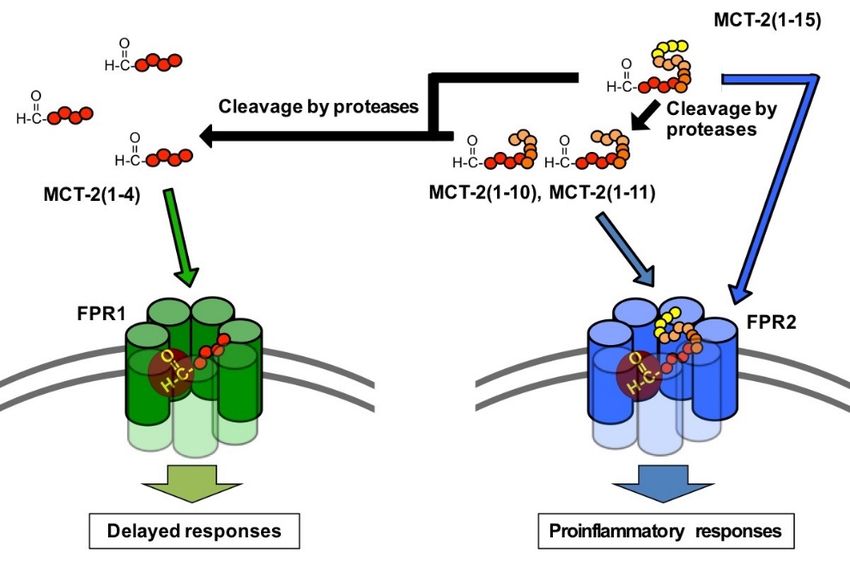
3.5. Possible Physiological Roles of the Receptor Preference Shift of MCT-2(1–15) from FPR2
to FPR1
The results of this study demonstrate that the receptor preference of MCT-2(1–15) is
shifted from FPR2 to FPR1 by the cleavage of its C-terminus. What is the physiological
significance of this shift in receptor preference?
FPR1 and FPR2 play critical roles in inflammation including proinflammatory re-
sponses, subsequent resolution, and wound healing/tissue regeneration. Specifically, FPR1
and FPR2 are expressed mainly by inflammatory immune cells including neutrophils,
monocytes, and monocyte-derived macrophage cells such as tissue-resident macrophages
and microglia [35,37,38], and FPR2 is also expressed by a variety of cells including microvas-
cular endothelial cells [38,39]. FPR1 and FPR2 have roles in the mechanisms concerning the
infiltration of neutrophils and macrophages into injury sites, and their activation causes
various inflammatory responses, including phagocytosis, superoxide generation, and in-
flammatory cytokine production [35,37,38]. In addition, there is evidence indicating that
the activation of FPR2 increases the vascular permeability of endothelial cells [38,39], sug-
gesting a further promotion of neutrophil infiltration from the bloodstream into injury
sites following receptor activation in the initial stage of inflammation. Thus, the activation
of FPR1 and FPR2 expressed by neutrophils, macrophages, and endothelial cells induces
various innate immune responses initiated by the infiltration and activation of neutrophils.
Oppositely, it is known that liganded FPR2 suppresses the production of inflammatory
cytokines following the acute proinflammatory responses [38,55–58]. Moreover, FPR1 acti-
vation has been demonstrated to promote wound healing/tissue regeneration, including
cell proliferation [40–44].
It is believed that mitochondrial-derived N-formylated peptides activate FPR1 and
FPR2 as endogenous activating factors. Indeed, mtDAMPs consisting of mitochondria and
their contents are released into the bloodstream as a result of sterile tissue damage, such as
trauma [15–21], and still unidentified endogenous N-formylated peptides in mtDAMPs
are thought to activate FPR1 and/or FPR2 to induce innate immune responses [15,17,28].You can also read


