Molecular Inverse Comorbidity between Alzheimer's disease and Lung Cancer: new insights from Matrix Factorization - bioRxiv
←
→
Page content transcription
If your browser does not render page correctly, please read the page content below
bioRxiv preprint first posted online May. 21, 2019; doi: http://dx.doi.org/10.1101/643890. The copyright holder for this preprint
 (which was not peer-reviewed) is the author/funder, who has granted bioRxiv a license to display the preprint in perpetuity.
                                  It is made available under a CC-BY 4.0 International license.

       Molecular Inverse Comorbidity between Alzheimer’s
       disease and Lung Cancer: new insights from Matrix
       Factorization
       Alessandro Greco1,2,3,4, Jon Sanchez Valle5, Vera Pancaldi5,6,7, Anaïs Baudot8,5, Emmanuel
       Barillot2,3,4, Michele Caselle1, Alfonso Valencia5,9, Andrei Zinovyev2,3,4, Laura Cantini10,2,3,4*

        1    Universitá degli Studi di Torino, Department of Physics and INFN, via P. Giuria 1, I-10125, Turin, Italy
        2    Institut Curie, PSL Research University, Paris, France
        3    INSERM U900, Paris, France
        4    CBIO-Centre for Computational Biology, Mines ParisTech, PSL Research University, Paris, France
        5    Barcelona Supercomputing Center (BSC), Barcelona, 08034 Spain
        6    Centre de Recherches en Cancérologie de Toulouse (CRCT), UMR1037 Inserm, ERL5294 CNRS, 2 Avenue
             Hubert Curien, 31037 Toulouse, France
        7    Université de Toulouse, Université Toulouse III Paul Sabatier, Toulouse, France
        8    Aix Marseille Univ, INSERM, MMG, CNRS, Marseille, France
        9    ICREA, Barcelona, 08010 Spain
        10   Computational Systems Biology Team, Institut de Biologie de l’Ecole Normale Supérieure, CNRS
             UMR8197, INSERM U1024, Ecole Normale Supérieure, Paris Sciences et Lettres Research University, Paris,
             75005, France

        * Correspondence: laura.cantini@ens.fr;

        Received: date; Accepted: date; Published: date

        Abstract: Matrix Factorization (MF) is an established paradigm for large-scale biological data
        analysis with tremendous potential in computational biology.

        We here challenge MF in depicting the molecular bases of epidemiologically described
        Disease-Disease (DD) relationships. As use case, we focus on the inverse comorbidity association
        between Alzheimer’s disease (AD) and lung cancer (LC), described as a lower than expected
        probability of developing LC in AD patients. To the day, the molecular mechanisms underlying
        DD relationships remain poorly explained and their better characterization might offer
        unprecedented clinical opportunities.

        To this goal, we extend our previously designed MF-based framework for the molecular
        characterization of DD relationships. Considering AD-LC inverse comorbidity as a case study, we
        highlight multiple molecular mechanisms, among which the previously identified immune system
        and mitochondrial metabolism. We then discriminate mechanisms specific to LC from those
        shared with other cancers through a pancancer analysis. Additionally, new candidate molecular
        players, such as Estrogen Receptor (ER), CDH1 and HDAC, are pinpointed as factors that might
        underlie the inverse relationship, opening the way to new investigations. Finally, some lung
        cancer subtype-specific factors are also detected, suggesting the existence of heterogeneity across
        patients also in the context of inverse comorbidity.

        Keywords: networks; Alzheimer’s disease; lung cancer; inverse comorbidity; transcriptome;
        matrix factorization

       1. Introduction
       Large-scale genomics projects, including for instance The Cancer Genome Atlas
       (TCGA, https://www.cancer.gov/tcga), are currently providing an overwhelming amount of omics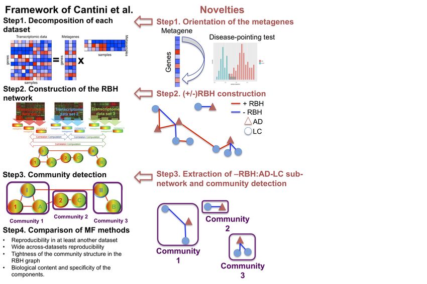
bioRxiv preprint first posted online May. 21, 2019; doi: http://dx.doi.org/10.1101/643890. The copyright holder for this preprint
 (which was not peer-reviewed) is the author/funder, who has granted bioRxiv a license to display the preprint in perpetuity.
                                  It is made available under a CC-BY 4.0 International license.

       Int. J. Mol. Sci. 2019, 20, x FOR PEER REVIEW                                                                                2 of 15

       data. The available data offer the opportunity to better understand biological systems and cancer in
       particular, but their high dimensionality poses considerable challenges typical of “Big Data” [1].

       A powerful approach to this problem is represented by Matrix Factorization (MF), a class of
       unsupervised methods that reduces high-dimensional data into low dimensional subspaces, while
       preserving as much information as possible [2–4]. Given a data matrix X, MF learns two sets of
       low-dimensional representations: “metagenes”, encoding molecular relationships, and
       “metasamples”, encoding sample-level relationships. Up to now, MF has been successfully used in
       a broad spectrum of applications: unsupervised clustering, especially in the context of cancer
       subtyping [5,6], molecular pattern discovery [7,8], mutational signatures definition [9,10] and
       tumour sample immune infiltration quantification [11]. Such results have been obtained by mining
       with MF single large-scale datasets, such as transcriptome or methylome. Recently, we designed a
       metric to infer univocal correspondences between the metagenes obtained by an MF algorithm on
       multiple independent datasets profiled from the same biological condition (e.g. same cancer tissue),
       and used this metric to design a methodological framework that revealed relevant pathways
       characteristic of colorectal cancer [12].

       We are here interested in investigating the molecular bases of previously documented
       Disease-Disease (DD) relationships. Indeed, several computational studies have inferred DD
       relationships, starting from the “Human Disease Network” where diseases were connected when
       sharing disease genes [13], to the “multiplex network of human diseases” composed by genotype-
       and phenotype-based layers that propose new disease-associations [14]. More importantly, DD
       relationships have also been systematically identified by epidemiological studies, working at the
       level of populations and looking for the co-occurrence of different diseases in the same patients by
       using medical claims [15], medical records [16] and insurance claims [17]. The higher than expected
       risk of developing pancreatic cancer in patients suffering for type II diabetes [18] and of developing
       lung cancer in asthma patients [19] are among the most renown examples of cancer-related
       comorbidities. Interestingly, it has also been described that patients suffering from certain diseases
       have a lower than expected risk of developing cancer, known as inverse comorbidity [20–22]. An
       example of these protective effects of one disease on the other is represented by the documented
       inverse comorbidity between Alzheimer's Disease (AD) and Lung Cancer (LC) [22–24]. Molecular
       and non-molecular factors (e.g. the environment, lifestyle or drug treatments) can be responsible for
       such DD relationships. The molecular mechanisms underlying these DD relationships are poorly
       understood and investigating them offers unprecedented opportunities to better understand the
       etiology and pathogenesis of diseases, with the hope of identifying opportunities for repositioning
       of pre-existing treatments.

       Recently, transcriptomic meta-analyses revealed sets of significantly up and down regulated genes
       that are shared across diseases displaying different patterns of direct and inverse comorbidities
       [25,26]. However, differential expression analysis only focuses on the predominant signals present
       in the data, failing to capture alternative signals and local behaviors [3]. These limitations are
       overcome by MF that learns metagenes, i.e. ranking of genes, without focusing on single sets of
       predominant genes. Moreover, contrarily to differential expression analysis, MF jointly provides
       metagenes and metasamples, i.e. also grouping samples together with their biological
       characterization. We hereby propose to use an MF approach to study the molecular bases of DD
       relationships. This, however, requires innovative adaptations. We thus propose to extend our
       previously defined MF framework for the particular study of DD relationships [12]. Moreover,
       given the existence of positive and negative DD connections, we also adapt the framework to
       distinguish molecular relationships concordantly and discordantly altered in datasets coming from
       different diseases.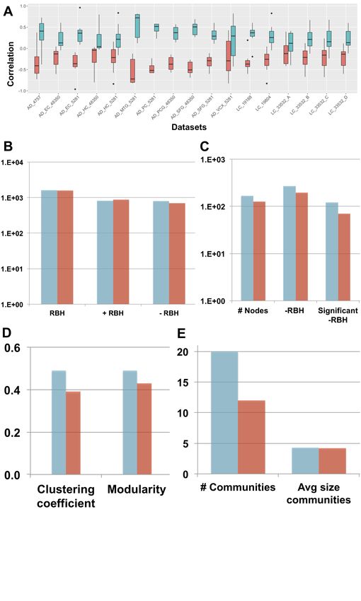
bioRxiv preprint first posted online May. 21, 2019; doi: http://dx.doi.org/10.1101/643890. The copyright holder for this preprint
 (which was not peer-reviewed) is the author/funder, who has granted bioRxiv a license to display the preprint in perpetuity.
                                  It is made available under a CC-BY 4.0 International license.

       Int. J. Mol. Sci. 2019, 20, x FOR PEER REVIEW                                                                                3 of 15

       Considering the inverse comorbidity between Alzheimer’s disease (AD) and lung cancer (LC) as a
       case study [22–24], we applied our MF framework to 17 transcriptomic datasets, including both LC
       and AD samples (total of 1367 samples), and we highlighted multiple molecular mechanisms
       possibly underlying the inverse comorbidity pattern. Through a pancancer analysis we categorized
       the processes here suggested to be involved in the AD-LC inverse comorbidity based on their
       presence in other cancers. The previously identified role of the immune system and mitochondrial
       metabolism in AD-LC inverse comorbidity is confirmed by our analysis. Additionally, new
       candidate molecular players, such as Estrogen Receptor (ER), CDH1 and Histone Deacetylase
       (HDAC), are identified as potentially involved in the inverse comorbidity considered. Finally, some
       lung cancer subtype-specific alterations are also detected suggesting the existence of heterogeneity
       across patients also in the context of inverse comorbidity.

       2. Results

       2.1. A new MF framework to study disease-disease relationships
             We previously defined the Reciprocal Best Hit (RBH) metric to infer univocal correspondences
       between the MF metagenes obtained on independent datasets measured from the same biological
       condition (e.g. same cancer tissue) [12]. Based on this metric we designed an RBH-based
       framework, structured in three sequential steps: (1) each transcriptomic dataset is independently
       decomposed in metagenes and metasamples with MF; (2) using the RBH metric, relationships
       between metagenes are inferred and a RBH network is constructed; (3) communities are detected in
       the RBH network. These communities of genes are then analyzed for functional relatedness and
       provide a biological interpretation of the principal factors that shape the transcriptomes. Here, we
       adapted the framework to the study of the molecular mechanisms underlying DD relationships, in
       order to infer univocal positive/negative correspondences between MF metagenes independently
       obtained on datasets measured from different diseases. Briefly, the main methodological novelties
       are: (i) the investigation of a methodology for the orientation of the metagenes (i.e. assign a sign to
       the metagenes, in order to express either direct or inverse similarity between them); (ii) a novel
       definition of Reciprocal Best Hit (RBH) network taking into account the orientation of the
       metagenes and (iii) the restriction of the community detection phase to the subnetwork of interest
       (e.g. subnetwork of negative links connecting metagenes of LC and AD in our case). The structure
       of the framework together with its novelties is summarized in Figure 1.

       2.1.1. Setp1: data decomposition and orientation of the components
       Each transcriptomic datasets is separately decomposed using MF. The framework here proposed
       can be combined with the MF algorithm of interest. In this work, we chose stabilized Independent
       Component Analysis (sICA) [12,27,28], a stabilized version of ICA [28–30]. SICA was indeed
       previously shown to outperform alternative MFs in the extraction of relevant biological knowledge
       from collections of transcriptomic datasets derived from the same biological condition (e.g. the
       same cancer type) [12]. Moreover, the ability of sICA to separate the various overlapping biological
       factors present in transcriptomic data, such as those linked to the tumor cells, the tumor
       microenvironment and non biological factors, linked to sample processing or data generation,
       makes this approach particularly promising for extracting relevant molecular factors from the
       numerous confounding factors involved in DD relationships.

       By applying sICA to a transcriptomic matrix X (n x m), with n genes in the rows and m samples in
       the columns, we reduce it to the product of an unknown mixing matrix A (n x k), whose columns
       are here denoted as “metagenes” and an unknown matrix of source signals S (k x m), whose rows
       are here denoted as “metasamples”. The metagene/metasample associated to the component i will
       thus provide the contribution of each gene/sample present in the matrix X to component i.
       Metasamples and metagenes are learned based upon the assumption that the number k of
bioRxiv preprint first posted online May. 21, 2019; doi: http://dx.doi.org/10.1101/643890. The copyright holder for this preprint
(which was not peer-reviewed) is the author/funder, who has granted bioRxiv a license to display the preprint in perpetuity.
It is made available under a CC-BY 4.0 International license.
Int. J. Mol. Sci. 2019, 20, x FOR PEER REVIEW 4 of 15
components occurring in the input matrix X is smaller than either its rows or columns. We here
selected the number k of components equal to 100 for those datasets having more than 100 samples
and equal to half of the samples for smaller datasets. These chosen values are higher than the
estimation of the optimal transcriptomic dimension, due to the fact that overdecomposition in sICA
was proven not detrimental for the interpretability of the resulting components [28].
To determine the orientation of the sICA metagenes two alternative approaches are considered:
“Long tail-pointing” and “disease-pointing”. The long tail-pointing approach, previously used for
other sICA applications [28], orients the metagenes such that the longest tail of their distribution
corresponds to their positive side. Indeed the sICA factors are identified by maximizing
non-gaussianity of the data point projection distributions. As a consequence, the longest-tails of
such distributions are those containing most of the biological information. We here introduce the
“disease-pointing” approach, which exploits the availability of cases and control samples to orient
the components. More specifically, the differential association of each metasample to case vs.
control is tested based on a Wilcoxon test and the couple metagene-metasample is oriented so that
the cases in the metasample are on the positive side.
2.1.2. Step 2: Construction of the signed Reciprocal Best Hits (sRBHs)
At Step 2, the Reciprocal Best Hit (RBH) network is constructed. A positive/negative RBH is defined
as follows: given two sets of metagenes !! . . . . !! and !! . . . . !! obtained from the
transcriptomic datasets ! !
and ! , respectively, we define !! and !! a positive Reciprocal Best
!
Hit (+RBH) iff
! !
!"# !"# !! , !! !!! = !"# !"# !! !!! , !! >0 (1)
and !! and !! a negative Reciprocal Best Hit (-RBH) iff
! !
!"# !"# !! , !! !!! = !"# !"# !! !!! , !!bioRxiv preprint first posted online May. 21, 2019; doi: http://dx.doi.org/10.1101/643890. The copyright holder for this preprint
(which was not peer-reviewed) is the author/funder, who has granted bioRxiv a license to display the preprint in perpetuity.
It is made available under a CC-BY 4.0 International license.
Int. J. Mol. Sci. 2019, 20, x FOR PEER REVIEW 5 of 15
alternative approaches were considered: “Long tail-pointing” and “disease-pointing”. We tested
how such choice impacts the following steps of the framework and, in particular, the structure of
the obtained RBH network. To do this, we selected a specific DD relationship, i.e., the inverse
comorbidity between Alzheimer’s disease (AD) and lung cancer (LC) as a case study [22–24].
17 transcriptomic datasets, spanning AD and LC patients and containing case and control
samples, were employed (see Methods for further details). Following our framework (Figure 1),
each dataset was decomposed separately through sICA (see Supp Table 1 for the number of
components) and the orientation of the components was established both with the
long-tail-pointing and the disease-pointing approaches. The resulting metagenes were then
compared according to multiple criteria (see Figure 2).
First, the correlation between the obtained metagenes and the case vs. control fold-change of
expression was considered. In fact, to associate a metagene to a specific biological function or
pathway, we need to perform enrichment tests using databases of functional annotations (e.g.
Reactome, GO). Generally this interpretation step is just aimed at associating a function to each
metagene, without taking into account the sign of activity of the identified pathways/processes.
However, when dealing with comorbidities it is important to not only associate a function to each
metagene, but also to infer the sign of activity of such pathways/functions. This task can be easily
achieved once the metagenes are positively correlated with the gene fold-change. As shown in
Figure 2A, the disease-pointing orientation produces metagenes that are significantly more
correlated with the genes fold-change than the long tail-pointing one (significance tested with
Wilcoxon test, resulting P-values available in Supp Table 1).
We have then applied the Step 2 of the framework and independently constructed an RBH
network for “long-tail-pointing” and “disease-pointing” oriented metagenes. In both cases, the
nodes of the network correspond to the metagenes independently identified in the 17 datasets (369
total nodes) and their links are +/-RBHs, defined as in equations (1,2). Changes in the orientation of
the metagenes alter the sign of the correlations giving rise to different RBH networks. We have thus
compared the “long-tail-pointing” vs. “disease-pointing” RBH networks based on their number of
links (Figure 2B). The “disease-pointing” method returns 1616 RBHs vs. the 1574 returned by the
“long-tail-pointing” method. Such result is due to the higher number of -RBHs identified with the
“disease-pointing” orientation (802 vs. 705).
In Step 3 we focused on the subnetwork composed of -RBHs and linking AD components with
LC ones and vice-versa, which in the following we call “-RBH AD/LC subnetwork”. These are in
fact the metagenes and RBHs of interest for the study of AD-LC inverse comorbidity. We studied
the topology of this subnetwork starting from its number of nodes and links (Figure 2C). The -RBH
AD/LC subnetwork based on the “disease-pointing” orientation includes a higher number of
metagenes (167 vs. 127 of “long-tail-pointing”) and a higher number of links (268 vs. 194 of
“long-tail-pointing”). Moreover, among the RBHs present in the subnetwork, those of the
“disease-pointing” tend to be more frequently connecting factors that are significantly differential
between case and control (112 vs. 70 of “long-tail-pointing”). Communities were then detected in
the “long-tail-pointing” and “disease-pointing” -RBH AD/LC subnetworks. As shown in Figure
2D, the “disease-pointing” -RBH AD/LC subnetwork has a higher modularity (0.49 vs. 0.43) and
higher clustering coefficient (0.49 vs. 0.39). Moreover 20 communities of size higher or equal to 4 are
detected in the “disease-pointing” -RBH AD/LC subnetwork vs. the 12 of the alternative approach
and the average size of the “disease-pointing” communities is 4.3 vs. the 4.2 of the alternative
approach (Figure 2E).
Overall our analysis indicates that the “disease-pointing” orientation tends to identify a higher
number of candidate molecular processes/pathways involved in AD-LC inverse comorbidity. ForbioRxiv preprint first posted online May. 21, 2019; doi: http://dx.doi.org/10.1101/643890. The copyright holder for this preprint
(which was not peer-reviewed) is the author/funder, who has granted bioRxiv a license to display the preprint in perpetuity.
It is made available under a CC-BY 4.0 International license.
Int. J. Mol. Sci. 2019, 20, x FOR PEER REVIEW 6 of 15
all these reasons, “disease-pointing” is the orientation approach that we selected for the following
analysis.
2.3. New biological insights on the inverse comorbidity between AD and LC
We hypothesize that the communities of the -RBH AD/LC subnetwork, obtained with the
“disease-pointing” orientation, could be related to the AD-LC inverse comorbidity. We thus
annotated the communities of the -RBH AD/LC subnetwork by using MsigDB signatures [33],
Microenvironment Cell Populations-counter (MCP-counter) signatures [34], predefined lung cancer
subtypes [35] and the metagenes computed in [27], here referred to as CIT, as described in
Methods. The obtained -RBH AD/LC subnetwork with the main biological information is illustrated
in Figure 3 and Supp Table 2.
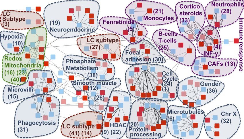
The majority of the communities present in the network are associated to the immune system and
mitochondrial functioning, confirming the results of previous transcriptomics meta-analyses on the
inverse comorbidity between AD and LC [25,26]. Interestingly, these processes are here deeply
partitioned into multiple communities, suggesting that we can detect more detailed aspects of their
involvement. Fibroblasts, Neutrophils, Monocytes, B and T cells are the immune cells showing an
inverse activity in LC and AD according to our analysis. Moreover, communities involved in the
regulation of two immune-system related drugs (fenretinide and corticosteroids) are identified.
Interestingly, corticosteroids are associated with less Alzheimer neuropathology [36], while their
use in LC patients is associated with lower overall survival [37]. At the same time, fenretinide has
been shown to inhibit growth in lung cancer cell lines [38] and it has been proposed as a potential
adjuvant for late onset Alzheimer’s disease [39]. Moreover, the fenretinide community is tightly
linked with the monocytes one, in agreement with its mechanisms of action involving the
regulation of the secretion of pro-inflammatory cytokines in human monocytes [40].
The communities associated to mitochondria span different processes related to their activity:
oxidation-reduction process (communities 16, 23, 40), hypoxia (community 10) and phosphate
metabolic process (community 38). Enrichment in hypoxia could correspond to a confounding
factor linked to the state of the profiled tissues (post-mortem for AD and fresh tissue biopsy for LC).
However, patients suffering from systemic or prenatal hypoxia have a higher risk of developing
Alzheimer’s disease [41,42] and targeting hypoxia seems to Improve lung cancer outcome [43],
indicating that such hypoxia-related community could also contain non-trivial information.
Additionally to mitochondria and immune system, community 36 has been associated to gender, in
line with the higher risk of females to develop Alzheimer’s disease, in opposition to lung cancer,
which is more frequent in men [44,45]. Histone Deacetylase (HDAC), associated to community 22,
confirms the known involvement of HDAC1 in both cancer and Alzheimer’s disease [46,47].
Community 30 is enriched in focal adhesion. The inhibition of focal adhesion kinase, which is
overexpressed in several cancers, decreases cell viability [48], while, in the case of Alzheimer’s
disease, amyloid-ß induces the inactivation of focal adhesion kinase [49]. Cell cycle and CDH1
targets are associated to community 24. Interestingly, growing evidence suggests that
dysregulation of APC/C-Cdh1 is involved in neurodegenerative diseases, potentially as a
consequence of amyloid-ß driven proteasome-dependent degradation of CDH1 [50]. On the other
hand, significantly higher methylation level of CDH1, inducing its inactivation, plays an important
role in lung cancer [51]. Community 20 is associated to protein processing and chaperone-mediated
protein folding. Protein misfolding is a known marker of AD [52,53]. At the same time, cell division,
migration, and invasion rely on microtubules and actin filament components and thus
chaperone-mediated protein folding activity is tightly linked to cancer [54]. Similar arguments
support the involvement of microtubules (community 6) to AD-LC inverse comorbidity. Moreover,
response to Estrogene Receptor (ER) (“ESR1 targets”) has been found enriched in 20 communities,
even if without clear association to a specific one. Interestingly, an inverse association has beenbioRxiv preprint first posted online May. 21, 2019; doi: http://dx.doi.org/10.1101/643890. The copyright holder for this preprint
(which was not peer-reviewed) is the author/funder, who has granted bioRxiv a license to display the preprint in perpetuity.
It is made available under a CC-BY 4.0 International license.
Int. J. Mol. Sci. 2019, 20, x FOR PEER REVIEW 7 of 15
shown between the use of estrogen and early onset of Alzheimer’s disease, suggesting that it might
be beneficial for the disease [55], potentially due to its inhibitory activity on neuroinflammation
[56]. On the other hand, the use of hormonal replacement therapy significantly increases LC
mortality, supporting a role of estrogen in lung cancer [57]. These inverse effects of regulation of
focal adhesion, CDH1 and estrogen receptor in cancer and AD are consistent with a possible
association of these pathways to the inverse comorbidity patterns observed.
We then explored if also lung cancer subtype-specific molecular mechanisms could be involved into
the AD-LC inverse comorbidity [35]. The main biologically-annotated network communities
resulted to be general of LC with no association to a specific subtype. At the same time, three
communities in our network (27, 14 and 3) mapped to the three predefined LC subtypes (proximal
proliferative, proximal inflammatory and terminal respiratory unit, respectively). Therefore, some
lung cancer subtype-specific regulatory programs seem to also be involved, suggesting the
existence of an across-patients heterogeneity, even if such phenomenon is not a predominant one.
Finally, AD has been shown to have comorbidity relationships at the epidemiological level not only
with LC, but also with other cancer types, with most of them being inverse comorbidities [13]. We
thus tested if some of the candidate biological processes that we here identified to be possibly
involved into AD-LC inverse comorbidity could be generalized to the comorbidity relationship
between AD and other cancers. With this aim, we considered metagenes previously computed on
TCGA transcriptomes for 32 different cancer types (pancancer metagenes) [28] and inferred their
RBHs with the metagenes of the -RBH AD/LC subnetwork. The presence of pancancer metagenes in
the communities of the -RBH AD/LC subnetwork has then been tested. If a community in the -RBH
AD/LC subnetwork is found to be correlated with some pancancer metagenes we can infer that
such process could also have a role into the relationship between AD and other cancers. We thus
quantified for each community of the -RBH AD/LC subnetwork the number of their connected
pancancer metagenes (see Supposed Table 2 for results). Of note, the orientation of the TCGA
pancancer metagenes has not been defined in [28], we thus cannot infer here if the activity of the
pancancer metagenes is concordant with that of the LC metagenes or of the AD ones.
As reported in Supp Table 2, the majority of the identified communities, corresponding to the
immune system-related signals, gender, chr X and mitochondrial activity, matches metagenes
obtained from other cancers, indicating a possible role of such processes in the co-morbidity of AD
with other cancers. On the opposite five communities (19%), corresponding to LC subtypes,
INF-Gamma and phosphate metabolism, are found to be specific to AD-LC inverse comorbidity.
3. Discussion
Matrix Factorization (MF) is a prominent solution for high-dimensional omics data analysis with a
vast range of applications in computational biology.
We were here interested in investigating disease-disease relationships, representing an
unprecedented opportunity to exploit mechanistic knowledge and repurpose treatments from one
disease to the other. We thus proposed a computational framework for the application of MF to the
study of disease-disease relationships. Considering the inverse comorbidity between Lung Cancer
(LC) and Alzheimer’s Disease (AD) as a case study, different methodologies for the orientation of
the metagenes were tested and the “Disease-pointing” one, orientating metagenes based on the case
vs. control behavior of metasamples, proved to give better performance.
The framework here proposed and applied to the study of the inverse comorbidity between LC and
AD, can be used to investigate direct/inverse comorbidity relations among other combinations of
diseases. More complex patterns of direct and/or inverse comorbidities, involving more than two
diseases, could also be studied. Moreover, we here chose sICA as MF algorithm and we employedbioRxiv preprint first posted online May. 21, 2019; doi: http://dx.doi.org/10.1101/643890. The copyright holder for this preprint
(which was not peer-reviewed) is the author/funder, who has granted bioRxiv a license to display the preprint in perpetuity.
It is made available under a CC-BY 4.0 International license.
Int. J. Mol. Sci. 2019, 20, x FOR PEER REVIEW 8 of 15
transcriptomic data. However, the framework here proposed can also be implemented with other
MF approaches (e.g. NMF, PCA) or different omics data types (e.g. methylome, proteome). More
generally, also multi-omics factors, obtained with approaches such as Multi-Omics Factor Analysis
(MOFA) or tensorial ICA (tICA), could also be considered as input of our analysis [58,59].
Finally, we performed a functional analysis of the genes involved in the subnetwork containing
negative links between AD and LC factors. Our results confirmed previously identified molecular
mechanisms underlying this inverse comorbidity, such as the involvement of the immune system
and mitochondrial processes, plus new candidate factors have been identified. Overall, our results
suggest that the MF RBH-based extended approach can be of biological and medical relevance once
investigating the molecular bases of DD relationships.
4. Materials and Methods
4.1 Data collection
3 microarray datasets from NCBI Gene Expression Omnibus (GEO)
(https://www.ncbi.nlm.nih.gov/geo/) were collected for Alzheimer’s disease: GSE4757; GSE48350
obtained from 4 brain regions: hippocampus, entorhinal cortex, superior frontal cortex, post-central
gyrus and GSE5281 obtained from six brain regions: entorhinal cortex, hippocampus, medial
temporal gyrus, posterior cingulate, superior frontal gyrus and primary visual cortex. The last two
datasets were split based on the region of the brain in which the samples were collected, obtaining a
total of 11 AD datasets composed of both case and control samples. Concerning lung cancer, 3
microarray datasets from NCBI GEO were collected: GSE19188, GSE19804 and GSE33532. The last
one, involving 4 biopsies from the same sample, was split in 4 datasets. We thus obtained a total of
6 LC datasets composed of case and control samples. Additionally, the RNA-seq Lung dataset
downloaded from The Cancer Genome Atlas (TCGA; https://tcga-data.nci.nih.gov/tcga/) was added
to the analysis.
4.2 Biological characterization of the communities
We characterized the communities obtained in the -RBH AD/LC subnetwork using the following
annotations: MSigDB signatures [33], Microenvironment Cell Populations-counter (MCP-counter)
signatures [34], predefined TCGA lung cancer subtypes [35] and the metagenes computed in [27],
here referred to as CIT. Concerning subtypes association we employed the metasamples obtained
from the TCGA lung cancer data.
We tested the significance of the association with the predefined LC subtypes by performing a
two-sided Wilcoxon test (cancer subtype vs. all other samples) and corrected for multiple testing
using Bonferroni. For all the other biological annotations involving genes we employed the
metagenes contained in each community. We associated to each community of the -RBH AD/LC
subnetwork a “consensus metagene” corresponding to the average of all the metagenes contained
in the community, paying attention to first concordantly orientate all the metagenes of the
community based on the signs of their correlations (all the metagenes in the community were
oriented based on the direction of LC). We then defined as top-contributing genes of a community
those genes having a weight in the consensus metagene higher than 3 standard deviations in
absolute value. The top-contributing genes were then divided into up and down based on their sign
in the consensus metagene and tested for their intersection with the various collections of
signatures. For cell types specific signatures we used a Fisher’s exact test with Bonferroni
correction, for MsigDB we employed its default enrichment test [33].
After testing the association of each community with all the considered annotations (MSigDB
signatures, MCP counter cell types signatures, the lung cancer subtypes available for TCGA data
and the CIT metagenes), we associate to each community the annotation that is more consistentlybioRxiv preprint first posted online May. 21, 2019; doi: http://dx.doi.org/10.1101/643890. The copyright holder for this preprint
(which was not peer-reviewed) is the author/funder, who has granted bioRxiv a license to display the preprint in perpetuity.
It is made available under a CC-BY 4.0 International license.
Int. J. Mol. Sci. 2019, 20, x FOR PEER REVIEW 9 of 15
found across the different tests. In Supp Table 2 the annotations associated to each community
together with their associated P-values are more extensively described.
Finally, to test the reproducibility of the identified consensus metagenes in other cancers, we used
the metagenes computed with sICA on TCGA transcriptomics data from 32 different cancer types
[28]. Then, for each community in the -RBH AD/LC subnetwork, we computed the number of
cancers having at least one correlated metagene. The resulting values are reported in Supp Table 2.
Supplementary Materials: Supplementary materials can be found at www.mdpi.com/xxx/s1.
Author Contributions: conceptualization, L.C. and A.G.; methodology, L.C. and A.G.; formal analysis, L.C.
and A.G.; biological curation, J.S, V.P, A.B. and A.V.; writing—original draft preparation, L.C. and A.B.;
writing—review and editing, L.C., V.P., A.B.; supervision, L.C.; project administration, L.C.; funding
acquisition, E.B. and A.Z.
Funding: This work is supported by the “Departments of Excellence 2018–2022” Grant awarded by the Italian
Ministry of Education, University and Research (MIUR) (L.232/2016). This work is also supported by a PhD
Fellowship (BES-2016-077403) and funded by the Spanish Ministry of Economics and Competitiveness
(BFU2015-71241-R).
Acknowledgments: We thank the Bioinfo4Women program for partially funding V.P. and A.B. The results
shown here are in part based upon data generated by the TCGA Research Network:
https://www.cancer.gov/tcga.
Conflicts of Interest: The authors declare no conflict of interest.
Abbreviations
AD Alzheimer’s disease
LC Lung Cancer
RBH Reciprocal Best Hit
DD Disease-Disease
References
1. Bell, G.; Hey, T.; Szalay, A. COMPUTER SCIENCE: Beyond the Data Deluge. Science 2009, 323,
1297–1298.
2. Stein-O’Brien, G.L.; Arora, R.; Culhane, A.C.; Favorov, A.V.; Garmire, L.X.; Greene, C.S.; Goff,
L.A.; Li, Y.; Ngom, A.; Ochs, M.F.; et al. Enter the Matrix: Factorization Uncovers Knowledge from
Omics. Trends Genet. 2018, 34, 790–805.
3. Devarajan, K. Nonnegative Matrix Factorization: An Analytical and Interpretive Tool in
Computational Biology. PLoS Computational Biology 2008, 4, e1000029.
4. Meng, C.; Zeleznik, O.A.; Thallinger, G.G.; Kuster, B.; Gholami, A.M.; Culhane, A.C.
Dimension reduction techniques for the integrative analysis of multi-omics data. Briefings in
Bioinformatics 2016, 17, 628–641.
5. De Sousa E Melo, F.; Wang, X.; Jansen, M.; Fessler, E.; Trinh, A.; de Rooij, L.P.M.H.; de Jong,
J.H.; de Boer, O.J.; van Leersum, R.; Bijlsma, M.F.; et al. Poor-prognosis colon cancer is defined by a
molecularly distinct subtype and develops from serrated precursor lesions. Nature Medicine 2013,
19, 614–618.
6. Sadanandam, A.; Lyssiotis, C.A.; Homicsko, K.; Collisson, E.A.; Gibb, W.J.; Wullschleger, S.;
Ostos, L.C.G.; Lannon, W.A.; Grotzinger, C.; Del Rio, M.; et al. A colorectal cancer classification
system that associates cellular phenotype and responses to therapy. Nat. Med. 2013, 19, 619–625.bioRxiv preprint first posted online May. 21, 2019; doi: http://dx.doi.org/10.1101/643890. The copyright holder for this preprint
(which was not peer-reviewed) is the author/funder, who has granted bioRxiv a license to display the preprint in perpetuity.
It is made available under a CC-BY 4.0 International license.
Int. J. Mol. Sci. 2019, 20, x FOR PEER REVIEW 10 of 15
7. Kong, W.; Mou, X.; Hu, X. Exploring matrix factorization techniques for significant genes
identification of Alzheimer’s disease microarray gene expression data. BMC Bioinformatics 2011, 12
Suppl 5, S7.
8. Brunet, J.-P.; Tamayo, P.; Golub, T.R.; Mesirov, J.P. Metagenes and molecular pattern discovery
using matrix factorization. Proceedings of the National Academy of Sciences 2004, 101, 4164–4169.
9. Alexandrov, L.B.; Ju, Y.S.; Haase, K.; Van Loo, P.; Martincorena, I.; Nik-Zainal, S.; Totoki, Y.;
Fujimoto, A.; Nakagawa, H.; Shibata, T.; et al. Mutational signatures associated with tobacco
smoking in human cancer. Science 2016, 354, 618–622.
10. Australian Pancreatic Cancer Genome Initiative; ICGC Breast Cancer Consortium; ICGC
MMML-Seq Consortium; ICGC PedBrain; Alexandrov, L.B.; Nik-Zainal, S.; Wedge, D.C.; Aparicio,
S.A.J.R.; Behjati, S.; Biankin, A.V.; et al. Signatures of mutational processes in human cancer. Nature
2013, 500, 415–421.
11. Hackl, H.; Charoentong, P.; Finotello, F.; Trajanoski, Z. Computational genomics tools for
dissecting tumour-immune cell interactions. Nat. Rev. Genet. 2016, 17, 441–458.
12. Cantini, L.; Kairov, U.; de Reyniès, A.; Barillot, E.; Radvanyi, F.; Zinovyev, A. Assessing
reproducibility of matrix factorization methods in independent transcriptomes. Bioinformatics 2019.
13. Goh, K.-I.; Cusick, M.E.; Valle, D.; Childs, B.; Vidal, M.; Barabási, A.-L. The human disease
network. Proc. Natl. Acad. Sci. U.S.A. 2007, 104, 8685–8690.
14. Halu, A.; De Domenico, M.; Arenas, A.; Sharma, A. The multiplex network of human diseases.
NPJ Syst Biol Appl 2019, 5, 15.
15. Hidalgo, C.A.; Blumm, N.; Barabási, A.-L.; Christakis, N.A. A dynamic network approach for
the study of human phenotypes. PLoS Comput. Biol. 2009, 5, e1000353.
16. Beck, M.K.; Jensen, A.B.; Nielsen, A.B.; Perner, A.; Moseley, P.L.; Brunak, S. Diagnosis
trajectories of prior multi-morbidity predict sepsis mortality. Sci Rep 2016, 6, 36624.
17. Wang, K.; Gaitsch, H.; Poon, H.; Cox, N.J.; Rzhetsky, A. Classification of common human
diseases derived from shared genetic and environmental determinants. Nat. Genet. 2017, 49, 1319–
1325.
18. Eibl, G.; Cruz-Monserrate, Z.; Korc, M.; Petrov, M.S.; Goodarzi, M.O.; Fisher, W.E.; Habtezion,
A.; Lugea, A.; Pandol, S.J.; Hart, P.A.; et al. Diabetes Mellitus and Obesity as Risk Factors for
Pancreatic Cancer. J Acad Nutr Diet 2018, 118, 555–567.
19. Qu, Y.-L.; Liu, J.; Zhang, L.-X.; Wu, C.-M.; Chu, A.-J.; Wen, B.-L.; Ma, C.; Yan, X.-Y.; Zhang, X.;
Wang, D.-M.; et al. Asthma and the risk of lung cancer: a meta-analysis. Oncotarget 2017, 8, 11614–
11620.
20. Musicco, M.; Adorni, F.; Di Santo, S.; Prinelli, F.; Pettenati, C.; Caltagirone, C.; Palmer, K.;
Russo, A. Inverse occurrence of cancer and Alzheimer disease: a population-based incidence study.
Neurology 2013, 81, 322–328.
21. Freedman, D.M.; Wu, J.; Chen, H.; Kuncl, R.W.; Enewold, L.R.; Engels, E.A.; Freedman, N.D.;
Pfeiffer, R.M. Associations between cancer and Alzheimer’s disease in a U.S. Medicare population.
Cancer Med 2016, 5, 2965–2976.
22. Driver, J.A.; Beiser, A.; Au, R.; Kreger, B.E.; Splansky, G.L.; Kurth, T.; Kiel, D.P.; Lu, K.P.;
Seshadri, S.; Wolf, P.A. Inverse association between cancer and Alzheimer’s disease: results from
the Framingham Heart Study. BMJ 2012, 344, e1442.
23. Tavares, A.R.; de Melo, A.C.; Sternberg, C. Cancer linked to Alzheimer disease but not vascularbioRxiv preprint first posted online May. 21, 2019; doi: http://dx.doi.org/10.1101/643890. The copyright holder for this preprint
(which was not peer-reviewed) is the author/funder, who has granted bioRxiv a license to display the preprint in perpetuity.
It is made available under a CC-BY 4.0 International license.
Int. J. Mol. Sci. 2019, 20, x FOR PEER REVIEW 11 of 15
dementia. Neurology 2010, 75, 1215–1216; author reply 1216.
24. Ganguli, M. A reduced risk of Alzheimer’s disease in those who survive cancer. BMJ 2012, 344,
e1662.
25. Ibáñez, K.; Boullosa, C.; Tabarés-Seisdedos, R.; Baudot, A.; Valencia, A. Molecular evidence for
the inverse comorbidity between central nervous system disorders and cancers detected by
transcriptomic meta-analyses. PLoS Genet. 2014, 10, e1004173.
26. Sánchez-Valle, J.; Tejero, H.; Ibáñez, K.; Portero, J.L.; Krallinger, M.; Al-Shahrour, F.;
Tabarés-Seisdedos, R.; Baudot, A.; Valencia, A. A molecular hypothesis to explain direct and
inverse co-morbidities between Alzheimer’s Disease, Glioblastoma and Lung cancer. Sci Rep 2017, 7,
4474.
27. Biton, A.; Bernard-Pierrot, I.; Lou, Y.; Krucker, C.; Chapeaublanc, E.; Rubio-Pérez, C.;
López-Bigas, N.; Kamoun, A.; Neuzillet, Y.; Gestraud, P.; et al. Independent component analysis
uncovers the landscape of the bladder tumor transcriptome and reveals insights into luminal and
basal subtypes. Cell Rep 2014, 9, 1235–1245.
28. Kairov, U.; Cantini, L.; Greco, A.; Molkenov, A.; Czerwinska, U.; Barillot, E.; Zinovyev, A.
Determining the optimal number of independent components for reproducible transcriptomic data
analysis. BMC Genomics 2017, 18, 712.
29. Engreitz, J.M.; Daigle, B.J.; Marshall, J.J.; Altman, R.B. Independent component analysis:
Mining microarray data for fundamental human gene expression modules. Journal of Biomedical
Informatics 2010, 43, 932–944.
30. Hyvärinen, A.; Oja, E. Independent component analysis: algorithms and applications. Neural
Networks 2000, 13, 411–430.
31. van Dongen, S.; Abreu-Goodger, C. Using MCL to extract clusters from networks. Methods Mol.
Biol. 2012, 804, 281–295.
32. Enright, A.J.; Van Dongen, S.; Ouzounis, C.A. An efficient algorithm for large-scale detection of
protein families. Nucleic Acids Res. 2002, 30, 1575–1584.
33. Liberzon, A.; Subramanian, A.; Pinchback, R.; Thorvaldsdottir, H.; Tamayo, P.; Mesirov, J.P.
Molecular signatures database (MSigDB) 3.0. Bioinformatics 2011, 27, 1739–1740.
34. Becht, E.; Giraldo, N.A.; Lacroix, L.; Buttard, B.; Elarouci, N.; Petitprez, F.; Selves, J.;
Laurent-Puig, P.; Sautès-Fridman, C.; Fridman, W.H.; et al. Estimating the population abundance of
tissue-infiltrating immune and stromal cell populations using gene expression. Genome Biology 2016,
17.
35. Cancer Genome Atlas Research Network Comprehensive molecular profiling of lung
adenocarcinoma. Nature 2014, 511, 543–550.
36. Beeri, M.S.; Schmeidler, J.; Lesser, G.T.; Maroukian, M.; West, R.; Leung, S.; Wysocki, M.; Perl,
D.P.; Purohit, D.P.; Haroutunian, V. Corticosteroids, but not NSAIDs, are associated with less
Alzheimer neuropathology. Neurobiol. Aging 2012, 33, 1258–1264.
37. Scott, S.C.; Pennell, N.A. Early Use of Systemic Corticosteroids in Patients with Advanced
NSCLC Treated with Nivolumab. J Thorac Oncol 2018, 13, 1771–1775.
38. Ohlmann, C.-H.; Jung, C.; Jaques, G. Is growth inhibition and induction of apoptosis in lung
cancer cell lines by fenretinide [N-(4-hydroxyphenyl)retinamide] sufficient for cancer therapy? Int.
J. Cancer 2002, 100, 520–526.
39. Goodman, A.B. Retinoid receptors, transporters, and metabolizers as therapeutic targets in latebioRxiv preprint first posted online May. 21, 2019; doi: http://dx.doi.org/10.1101/643890. The copyright holder for this preprint
(which was not peer-reviewed) is the author/funder, who has granted bioRxiv a license to display the preprint in perpetuity.
It is made available under a CC-BY 4.0 International license.
Int. J. Mol. Sci. 2019, 20, x FOR PEER REVIEW 12 of 15
onset Alzheimer disease. J. Cell. Physiol. 2006, 209, 598–603.
40. Lin, C.-H.; Lee, S.-Y.; Zhang, C.-C.; Du, Y.-F.; Hung, H.-C.; Wu, H.-T.; Ou, H.-Y. Fenretinide
inhibits macrophage inflammatory mediators and controls hypertension in spontaneously
hypertensive rats via the peroxisome proliferator-activated receptor gamma pathway. Drug Des
Devel Ther 2016, 10, 3591–3597.
41. Peers, C.; Pearson, H.A.; Boyle, J.P. Hypoxia and Alzheimer’s disease. Essays Biochem. 2007, 43,
153–164.
42. Nalivaeva, N.N.; Turner, A.J.; Zhuravin, I.A. Role of Prenatal Hypoxia in Brain Development,
Cognitive Functions, and Neurodegeneration. Frontiers in Neuroscience 2018, 12.
43. Salem, A.; Asselin, M.-C.; Reymen, B.; Jackson, A.; Lambin, P.; West, C.M.L.; O’Connor, J.P.B.;
Faivre-Finn, C. Targeting Hypoxia to Improve Non-Small Cell Lung Cancer Outcome. J. Natl.
Cancer Inst. 2018, 110.
44. Mazure, C.M.; Swendsen, J. Sex differences in Alzheimer’s disease and other dementias. Lancet
Neurol 2016, 15, 451–452.
45. Zang, E.A.; Wynder, E.L. Differences in Lung Cancer Risk Between Men and Women:
Examination of the Evidence. JNCI Journal of the National Cancer Institute 1996, 88, 183–192.
46. Patra, S.; Panigrahi, D.P.; Praharaj, P.P.; Bhol, C.S.; Mahapatra, K.K.; Mishra, S.R.; Behera, B.P.;
Jena, M.; Bhutia, S.K. Dysregulation of histone deacetylases in carcinogenesis and tumor
progression: a possible link to apoptosis and autophagy. Cell. Mol. Life Sci. 2019.
47. Janczura, K.J.; Volmar, C.-H.; Sartor, G.C.; Rao, S.J.; Ricciardi, N.R.; Lambert, G.; Brothers, S.P.;
Wahlestedt, C. Inhibition of HDAC3 reverses Alzheimer’s disease-related pathologies in vitro and
in the 3xTg-AD mouse model. Proceedings of the National Academy of Sciences 2018, 115, E11148–
E11157.
48. Zhang, H.; Shao, H.; Golubovskaya, V.M.; Chen, H.; Cance, W.; Adjei, A.A.; Dy, G.K. Efficacy
of focal adhesion kinase inhibition in non-small cell lung cancer with oncogenically activated
MAPK pathways. Br. J. Cancer 2016, 115, 203–211.
49. Lachén-Montes, M.; González-Morales, A.; de Morentin, X.M.; Pérez-Valderrama, E.; Ausín, K.;
Zelaya, M.V.; Serna, A.; Aso, E.; Ferrer, I.; Fernández-Irigoyen, J.; et al. An early dysregulation of
FAK and MEK/ERK signaling pathways precedes the β-amyloid deposition in the olfactory bulb of
APP/PS1 mouse model of Alzheimer’s disease. J Proteomics 2016, 148, 149–158.
50. Fuchsberger, T.; Martínez-Bellver, S.; Giraldo, E.; Teruel-Martí, V.; Lloret, A.; Viña, J. Aβ
Induces Excitotoxicity Mediated by APC/C-Cdh1 Depletion That Can Be Prevented by Glutaminase
Inhibition Promoting Neuronal Survival. Sci Rep 2016, 6, 31158.
51. Yu, Q.; Guo, Q.; Chen, L.; Liu, S. Clinicopathological significance and potential drug targeting
of CDH1 in lung cancer: a meta-analysis and literature review. Drug Des Devel Ther 2015, 9, 2171–
2178.
52. Ashraf, G.M.; Greig, N.H.; Khan, T.A.; Hassan, I.; Tabrez, S.; Shakil, S.; Sheikh, I.A.; Zaidi, S.K.;
Akram, M.; Jabir, N.R.; et al. Protein misfolding and aggregation in Alzheimer’s disease and type 2
diabetes mellitus. CNS Neurol Disord Drug Targets 2014, 13, 1280–1293.
53. Selkoe, D.J. Cell biology of protein misfolding: the examples of Alzheimer’s and Parkinson’s
diseases. Nat. Cell Biol. 2004, 6, 1054–1061.
54. Vallin, J.; Grantham, J. The role of the molecular chaperone CCT in protein folding and
mediation of cytoskeleton-associated processes: implications for cancer cell biology. Cell StressbioRxiv preprint first posted online May. 21, 2019; doi: http://dx.doi.org/10.1101/643890. The copyright holder for this preprint
(which was not peer-reviewed) is the author/funder, who has granted bioRxiv a license to display the preprint in perpetuity.
It is made available under a CC-BY 4.0 International license.
Int. J. Mol. Sci. 2019, 20, x FOR PEER REVIEW 13 of 15
Chaperones 2019, 24, 17–27.
55. Slooter, A.J.; Bronzova, J.; Witteman, J.C.; Van Broeckhoven, C.; Hofman, A.; van Duijn, C.M.
Estrogen use and early onset Alzheimer’s disease: a population-based study. J. Neurol. Neurosurg.
Psychiatry 1999, 67, 779–781.
56. Vegeto, E.; Benedusi, V.; Maggi, A. Estrogen anti-inflammatory activity in brain: a therapeutic
opportunity for menopause and neurodegenerative diseases. Front Neuroendocrinol 2008, 29, 507–
519.
57. Rodriguez-Lara, V.; Hernandez-Martinez, J.-M.; Arrieta, O. Influence of estrogen in non-small
cell lung cancer and its clinical implications. J Thorac Dis 2018, 10, 482–497.
58. Argelaguet, R.; Velten, B.; Arnol, D.; Dietrich, S.; Zenz, T.; Marioni, J.C.; Buettner, F.; Huber,
W.; Stegle, O. Multi-Omics Factor Analysis—a framework for unsupervised integration of multi-
omics data sets. Molecular Systems Biology 2018, 14, e8124.
59. Teschendorff, A.E.; Jing, H.; Paul, D.S.; Virta, J.; Nordhausen, K. Tensorial blind source
separation for improved analysis of multi-omic data. Genome Biology 2018, 19.
Figures
Figure 1. Schematic view of the framework and the novelties introduced with respect to [12].bioRxiv preprint first posted online May. 21, 2019; doi: http://dx.doi.org/10.1101/643890. The copyright holder for this preprint
(which was not peer-reviewed) is the author/funder, who has granted bioRxiv a license to display the preprint in perpetuity.
It is made available under a CC-BY 4.0 International license.
Int. J. Mol. Sci. 2019, 20, x FOR PEER REVIEW 14 of 15
Figure 2. “Long-tail-pointing” (red) vs. “disease-pointing” (blue) orientation of the sICA
factors. (A) The two methods of factors orientation are compared based on the correlation of the
obtained metagenes with the case vs. control genes’ fold change. (B) The two methods are
compared based on the number of links present in their RBH network. Total RBHs (RBH), positive
RBHs (+RBH), negative RBHs (-RBH). (C,D,E) The two methods are compared based on thebioRxiv preprint first posted online May. 21, 2019; doi: http://dx.doi.org/10.1101/643890. The copyright holder for this preprint
(which was not peer-reviewed) is the author/funder, who has granted bioRxiv a license to display the preprint in perpetuity.
It is made available under a CC-BY 4.0 International license.
Int. J. Mol. Sci. 2019, 20, x FOR PEER REVIEW 15 of 15
structure of their -RBH AD/LC subnetwork, relevant for the study of inverse comorbidity. In (C) the
number of nodes and links in the subnetwork are compared. Number of nodes (# Nodes ), negative
RBHs connecting an AD component with a LC component (-RBH) and negative RBHs connecting
an AD component with a LC component that are associated to nodes with significant differential
behaviour (Wilcoxon p-value < 0.05) between case and control (significant -RBH). In (D) The
clustering coefficient and modularity of the subnetwork are considered. In (E) The number of
communities and their average size in the subnetwork is taken into account.
Figure 3. -RBH AD/LC subnetwork with biological annotations. Each node in the network
corresponds to a metagene, the list of metagenes associated to each community ID is reported in
Supp Table 3. Colours are linked to the diseases: red for AD and blue for LC. In AD datasets
obtained from the same region of the brain are denoted with different shades of red (normal and
light red). The nodes are organized into communities. Each community is denoted with a number
corresponding to its ID and the main biological annotation associated to them (see Supp Table 2 for
an extensive report).bioRxiv preprint first posted online May. 21, 2019; doi: http://dx.doi.org/10.1101/643890. The copyright holder for this preprint
(which was not peer-reviewed) is the author/funder, who has granted bioRxiv a license to display the preprint in perpetuity.
It is made available under a CC-BY 4.0 International license.
Supp. Table 1. Comparison of
correlation values between fold
change of expression and metagenes
oriented with long-tail vs disease-
pointing approach.
dataset Wilcoxon P-value
GSE4757 0.003
EC_GSE48350 0.001
EC_GSE5281 4.11E-05
HC_GSE48350 0.007
HC_GSE5281 5.05E-04
MTG_GSE5281 0.007
PC_GSE5281 0.029
PCG_GSE48350 5.83E-04
SFG_GSE48350 0.007
SFG_GSE5281 1.55E-04
VCX_GSE5281 0.024
GSE19188 3.11E-04
GSE19804 1.48E-06
GSE33532_A 0.161
GSE33532_B 0.003
GSE33532_C 0.0207
GSE33532_D 0.002bioRxiv preprint first posted online May. 21, 2019; doi: http://dx.doi.org/10.1101/643890. The copyright holder for this preprint
(which was not peer-reviewed) is the author/funder, who has granted bioRxiv a license to display the preprint in perpetuity.
It is made available under a CC-BY 4.0 International license.
Supp. Table 2. Annotations of the communities in the -RBH AD-LC subnetwork. For each community the table reports the ID, the associated annotation reported in the Figure, the
correlated CIT metagene, the MsigDB enriched signatures with the associated FDR q-values, the number of TCGA cancer having a metagene correlated with them, the lung cancer
subtype/clinical annotation with the Bonferroni corrected Wilcoxon P-value and the MCP counter based cell type with the Bonferroni corrected Fisher's exact test P-value.
LC subtype/clinical
Community Associated Number of MCP counter cell type with
CIT MsigDB signatures (FDR q-value) annotation (Wilcoxon P-
ID annotation other cancers P-value
value)
Proximal
3 "GO_INNATE_IMMUNE_RESPONSE" (3.18E-35) 0 Proximal Inflammatory 0.034
Inflammatory
"GO_INFLAMMATORY_RESPONSE" (6.32E-49),
4 INF-Gamma CIT-8 "HALLMARK_INTERFERON_GAMMA_RESPONSE" (1.05E-45) 0
Fenretinide
5 "FERRARI_RESPONSE_TO_FENRETINIDE_DN" (2.05E-03), "" 3
response
"GO_CILIUM" (4.75E-38),
6 Microtubules "GO_MICROTUBULE_BUNDLE_FORMATION" (6.48E-23), 3
"GO_MICROTUBULE_BASED_PROCESS" (3.55E-17)
"GO_IMMUNE_RESPONSE" (2.68E-23),
7 Monocytes CIT-8 "GSE29618_BCELL_VS_MONOCYTE_DAY7_FLU_VACCINE_UP 3 Monocytes 0.002
" (6.37E-08)
"ELVIDGE_HYPOXIA_UP" (3.17E-05), "HALLMARK_HYPOXIA"
10 Hypoxia (4.10E-04) 2
"GO_EXTRACELLULAR_STRUCTURE_ORGANIZATION" (5.38E-
99),
13 Fibroblasts CIT-8 "GO_POSITIVE_REGULATION_OF_IMMUNE_SYSTEM_PROCE 6 Fibroblasts 2.71e-08
SS" (1.36E-03)
"GO_MICROVILLUS" (3.79E-09),
15 Microvilli "GO_REGULATION_OF_MICROVILLUS_ORGANIZATION" 2
(5.63E-08)
Neuro endocrine
19 CIT-18 1
tumors
"GSE34156_TLR1_TLR2_LIGAND_VS_NOD2_AND_TLR1_TLR2
21 Monocytes CIT-8 _LIGAND_24H_TREATED_MONOCYTE_UP" (7.43E-58), 10 Monocytes 2.04 e-6
"GO_IMMUNE_SYSTEM_PROCESS" (6.86E-132)
"GSE22886_NAIVE_CD8_TCELL_VS_MONOCYTE_DN" (7.96E-
30), "GSE22886_NAIVE_CD4_TCELL_VS_MONOCYTE_DN"
25 B and T cells CIT-8 (2.96E-28), "GSE22886_NAIVE_BCELL_VS_NEUTROPHIL_DN" 6 B cells 0.03
(1.72E-23), "GSE22886_NAIVE_BCELL_VS_MONOCYTE_DN"
(2.15E-20)
"GSE22886_NAIVE_BCELL_VS_NEUTROPHIL_DN" (6.29E-52),
28 Neutrophils CIT-8 "HALLMARK_INFLAMMATORY_RESPONSE" (3.25E-67) 7 Neutrophilis 0.006
"KEGG_FOCAL_ADHESION" (1.64E-03),
30 Focal adhesion "GO_CELL_JUNCTION_ORGANIZATION" (1.34E-03), 2
"GO_ANCHORING_JUNCTION" (8.14E-04)
"GO_PHAGOCYTOSIS_RECOGNITION" (3.80E-09),
31 Phagocytosis "GO_PHAGOCYTOSIS" (3.80E-09) 5
"DISTECHE_ESCAPED_FROM_X_INACTIVATION" (1.56E-06),
32 chr X "RUNNE_GENDER_EFFECT_UP" (2.50E-20) 8
Corticosteroids "GO_RESPONSE_TO_CORTICOSTEROID" (1.69E-04),
33 "GO_IMMUNE_SYSTEM_PROCESS" (6.22E-44) 7
response
36 Gender 6 Gender 10^-37
Phosphate "GO_REGULATION_OF_PHOSPHORUS_METABOLIC_PROCES
38 S" (7.81E-03), 0
metabolism
Terminal Respiratory Terminal Respiratory unit
14 and 41 0
unit 2.92e-6; 0.021
“GO_CHAPERONE_MEDIATED_PROTEIN_FOLDIN” (8.225E-5),
20 protein processing “GO_PROTEIN_TRANSPORT” (1.625E-3) 1
22 and 9 HDAC "HELLER_HDAC_TARGETS_UP" (4.21E-12), "" 6
"GO_OXIDATION_REDUCTION_PROCESS" (2.00E-06),
"GO_OXIDOREDUCTASE_ACTIVITY_ACTING_ON_NAD_P_H_Q
23, 16 and 40 Redox/Mithocondria CIT-4 UINONE_OR_SIMILAR_COMPOUND_AS_ACCEPTOR" (2.65E- 2
06), "GO_SMALL_MOLECULE_METABOLIC_PROCESS" (2.77E-
14)
"FISCHER_G2_M_CELL_CYCLE" (5.04E-08),
24 and 1 Cell Cycle CIT-7 "GO_MITOTIC_CELL_CYCLE" (9.59E-08), 10
"ONDER_CDH1_TARGETS_2_UP" (1.34E-07)
"GO_EXTRACELLULAR_MATRIX" (1.37E-85),
26 and 12 Smooth muscle CIT-3 "GO_EXTRACELLULAR_STRUCTURE_ORGANIZATION" (5.48E- 5
70)
Proximal Proximal Proliferative 0.016;
27 and 17 0
Proliferative 0.035You can also read