Molecular Pathology of Breast Cancer - Elena Provenzano Lead Breast Pathologist Addenbrookes Hospital
←
→
Page content transcription
If your browser does not render page correctly, please read the page content below
Molecular Pathology of Breast
Cancer

             Elena Provenzano
             Lead Breast Pathologist
             Addenbrookes Hospital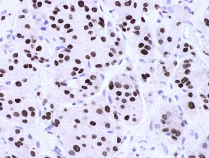
Biology of breast cancer
 • Breast cancer is not a single disease but a collection of
   diseases with different molecular characteristics and clinical
   outcomes
Why do we need molecular
characterisation of breast
cancer
• To identify patients whose prognosis is so good that
  adjuvant therapy after local surgery would not be beneficial
• To identify patients whose prognosis is so poor that a more
  aggressive adjuvant approach would be warranted
• To identify patients likely to be responsive or resistant to
  particular forms of therapy ( = predictive factors)
• => Individualised patient management
Traditional classification of
breast cancer
 • Assessment of the extent to which the appearance of a
   carcinoma resembles normal breast glandular tissue

 • Tumour type

 • Histological Grade

 • ER and HER2 status
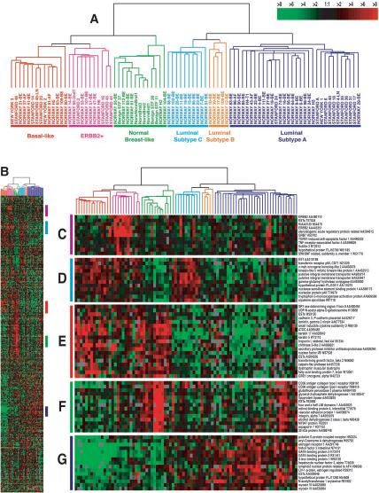
Newer classification systems for invasive
breast cancer – Intrinsic Subtypes
                      • Gene expression patterns of
                        breast carcinomas distinguish
                        tumour subclasses with clinical
                        implications
                      • ‘Intrinsic subtypes’
                      • Sørlie, Perou et al.
                      • PNAS, 2001; 98(19 ):10869–74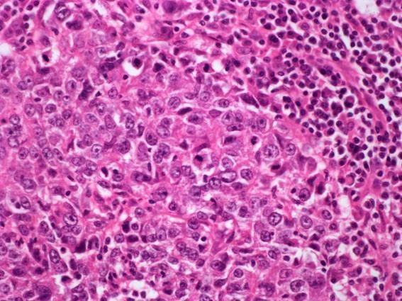
Newer classification systems for invasive
breast cancer – Intrinsic Subtypes
  •   Immunohistochemical correlates
  •   ER positive - Luminal breast cancers
  •   type A – PgR +, HER2 -, low proliferation
  •   type B – PgR +/-, HER2 +/-, high proliferation

  • ER negative
  • HER2 positive
  • Basal breast cancers – ER/PgR/HER2 -, CK5/ CK14 / EGFR +
Newer classification systems for invasive
 breast cancer – Intrinsic Subtypes
• Intrinsic subtypes are reproducible across platforms,
  however assignment of individual cancers to a molecular
  subtype shows only moderate reproducibility
• Dependent upon platform used, expression thresholds, and
  composition of the population
• Basal-like group most reproducible, luminal B and HER2
  enriched least reproducible
Newer classification systems for invasive
  breast cancer – Intrinsic Subtypes
• Concordance between gene expression and IHC defined
  subtypes modest at best
• Luminal A versus Luminal B – Ki67 cut point of 14% [Cheang
  et al., JNCI 2009]. Sensitivity 72%, specificity 77%
• Follow up study looking at 2 large clinical series [Prat et al.,
  JCO 2013]:
• 81-85% of luminal A correctly identified
• 35-52% of luminal B misclassified as Luminal A
• Improvement if include PR with cut off of 20%
Newer classification systems for invasive
breast cancer – Intrinsic Subtypes

                      Prat A and Perou C, Mol Oncol 2011
Newer classification systems for invasive
breast cancer – Intrinsic Subtypes
• Heterogeneity within HER2 positive disease, largely driven by ER
  status
• Clinically HER2 + and – tumours within each intrinsic subtype differ
  only in expression of genes in or near the HER2 amplicon on 17q
• Highest levels of HER2 pathway activation in cHER2+ HER2 enriched
  tumours

                                               Prat et al., JNCI 2014;106(8).
Newer classification systems for invasive
breast cancer – Intrinsic Subtypes
• Retrospective analysis of NOAH study looking at PAM50 subtypes
• Only 55% of HER2+ tumours HER2-E subtype; 21% luminal, 7% basal-
like, 18% normal-like
• Better pCR rates in HER2-E vs luminal HER2+ tumours (53% v 29%)
with larger improvement in EFS with addition of Trastuzumab
Prat et al., Clin Cancer Res 2014;20(2):511-21.Pereira et al., Nature Comms 2016.
A new molecular taxonomy of breast cancer

                           1.0                                    PAM50 1.0                                                   IntClust

                           0.8                                              0.8
                Survival

                           0.5                                              0.5

                                 Subtype (subjects/events)                                     Subtype (subjects/events)
                           0.3       Luminal A (2202/430)                   0.3       IntClust 1 (400/134)        IntClust 6 (189/69)
                                     Luminal B (1253/450)                             IntClust 2 (167/69)         IntClust 7 (497/103)
                                     HER2 (538/215)                                   IntClust 3 (959/198)        IntClust 8 (709/168)
                                     Basal (825/273)                                  IntClust 4 (938/229)        IntClust 9 (376/139)
                           0.0       Normal-like (404/92)                   0.0       IntClust 5 (455/189)        IntClust 10 (548/168)

                                 0                 5         10         15       0                  5                10                   15
                                                                  Follow-up (years)
Integrative clusters
IntClust Molecular Features Prognosis
1 Amp 17q23, mut GATA3, High instability Intermed
2 Amp cyclin D1. Poor
High instability
3 Mut PIK3CA and CDH1, low instability Good
4 Low instability, immune upregulation Good
5 HER2 Amp Poor
6 Amp ZNF703, high instability Intermed
7 16p gain, 16q loss, amp 8q Good
8 1q gain, 16q loss Good
9 8q gain, 20q amp, high instability Intermed
10 TNBC; high instability, DNA damage repair Poor
Mukherjee et al., NPJ Breast Ca 2018Integrative clusters
Mukherjee et al., NPJ Breast Ca 2018Integrative clusters
Mukherjee et al., NPJ Breast Ca 2018Percent
100
20 40 60 80
0
Ba
sa
l
H
ER
2
N
or
m
al
Lu
m
in
al
B
Lu
m
in
al
A
100
No pCR
In 20 40 60 80
tC 0
lu
s
In t 10
tC
lu
In s t 5
tC
pCR
lu
In s t 4
tC
lu
In s t 1
tC
lu
In s t 9
tC
lu
In s t 6
tC
lu
In s t 3
tC
lu
In s t 7
tC
lu
In s t 8
Different clinical behaviour
tC
lu
st
2ILC - Molecular
• Distinct morphology but genetic heterogeneity
• Gene expression profiling and copy number analysis reveals two
subgroups
• Immune related –immune signalling and cytokine pathways, with
upregulation of immune response inhibitors
• 78% have moderate to severe lym infiltrate
• Hormone related –ESR1 and PGR, cell cycle and ER target genes
Michaut et al., Nature Scientific Reports
Jan 2016.ILC - Molecular • Mutations: • CDH1 – 43-63% • PIK3CA – 35-48% • TP53 – 5-27% • PTEN – 14% (more common than NST – 3%) • GATA 3 – 3-5% (less common than NST – 20%) • ERBB2 – 4-18% • Remainder low Ciriello et al., Cell 2015;163:506-19. frequency
Case 1
• 63 y.o. female
• Presented clinically with
  a lump in the left UOQ
• Core biopsy – grade 3
  invasive cancer, mixed
  NST and micropapillary;
  ER/ PR 8, HER2
  borderline/ FISH non-
  amp
Case 1
•   Proceeded to WLE and SLNB
•   Final histology:
•   19 mm grade 3 invasive ca, mixed NST and micropapillary
•   Clear of margins
•   SLN – 0/2
Case 1

         10 year chemotherapy benefit
         = 4.5% -> discuss
Gene based prognostic tests
• Gene expression profiling
• cDNA arrray or RT-PCR based
• Several gene signatures have been proposed
• 21 gene – Oncotype Dx® - TAILORx
• 70 gene – Mammaprint® - MINDACT
• PAM50 – uses intrinsic subtypes
• 12 gene - EndoPredict
• Little overlap in specific genes that make up the signatures,
but all include genes involved in proliferation and ER
signallingOncotype DX
INVASION GROUP
PROLIFERATION
Cathepsin L2
GROUP
HER2 GROUP Stromelysin 3
KI67
HER2 GRB7
REF GROUP STK-15
ER GROUP Beta-actin GAPDH SURVIVIN
ER PgR RPLPO GUS CYCLIN B1
Bcl2 SCUBE2 TFRC MYBL2
Combine results in an algorithm to get the recurrence score:
30 30.5%Case 1 Recurrence score = 12 – low risk Endocrine therapy and RT only WGS as part of PBCP – validated PIK3CA mutation
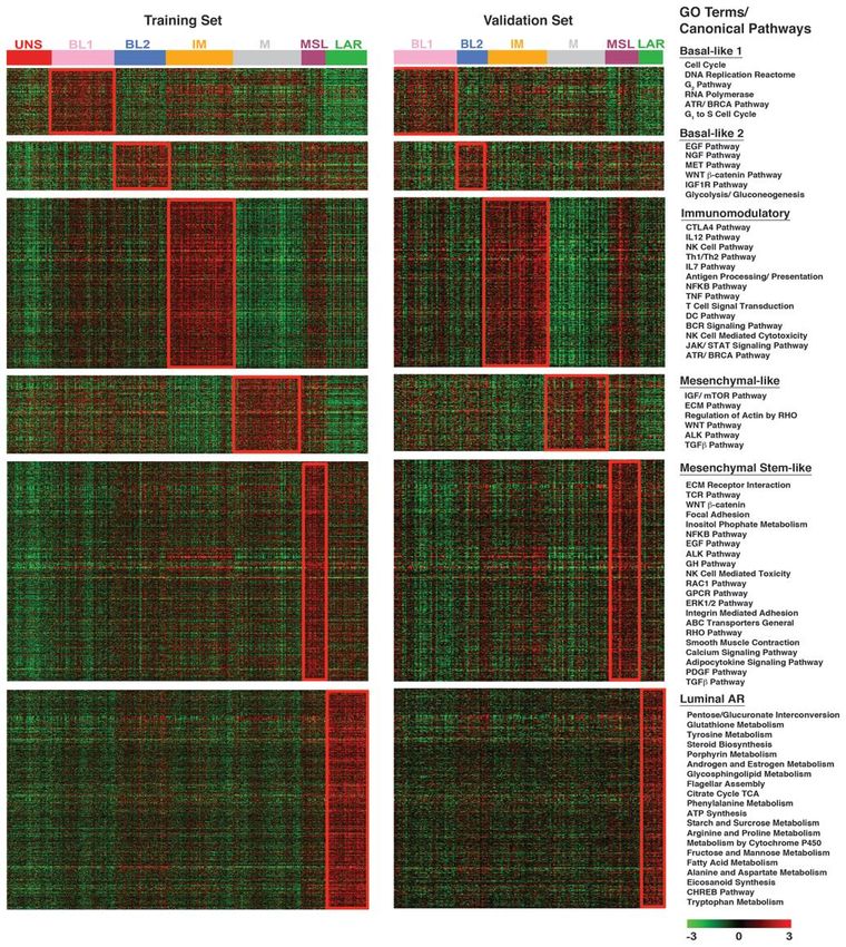
TNBC – Genomic Subtypes
• Gene expression array analysis of TNBC
  identified 6 subtypes, now refined to 4
  (immunomodulatory group reflects TILs
  and MSL reflects stromal contamination)
• Basal like 1 – elevated cell cycle and
  damage response genes
• Basal like 2 – growth factor signalling and
  myoepithelial genes
• Mesenchymal – epithelial-mesenchymal
  transition and growth factor pathways
• Luminal Androgen Receptor
   • luminal gene expression driven by AR
   • lower grade
   • higher incidence of lymph node and bone
     metastasis
   • high incidence PIK3CA mutations (40%)
                                               Lehmann et al. J Clin Inv 2011;121(7):2750-67.
TNBC – response to NACT • Masuda et al., Clin Ca Res 2013;19(19):5533-40 • Different rates of pCR between subtypes • No difference in OS – LAR group had low pCR rate but best survival at 3 years
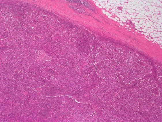
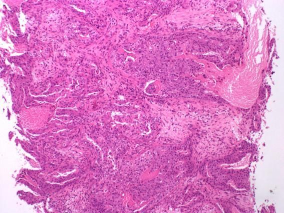
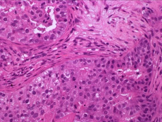
Basal Breast Cancers
• ‘Triple negative’ – ER, PgR and HER2 negative
• Express basal cytokeratins – CK 5/6, CK14
• Express EGFR
• Distinct morphology – high grade, central acellular
  zones, necrosis, high mitotic count
• Heterogeneous group – medullary-like, metaplastic,
  adenoid cystic carcinoma
• Associated with BRCA1 mutations in young women
• Associated with worse prognosis and distant metastasis,
  particularly visceral metastases
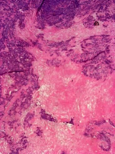
Basal Breast Cancers
Basal Breast Cancers
Carcinoma with medullary
features
• Circumscribed tumour with
  pushing rather than infiltrating
  margin
• Interconnecting sheets of large,
  bizarre and pleomorphic
  carcinoma cells forming a
  syncytial network
• Prominent lymphocytic
  inflammatory cell infiltrate
• Usually ER/ HER2 negative
• Association with BRCA1
  mutations
TNBC – LAR Subtype
 • Luminal Androgen Receptor
    • luminal gene expression driven by AR
    • lower grade, higher incidence of lymph node and bone metastasis
    • high incidence PIK3CA mutations (40%)
• Molecular Apocrine Subtype
• Farmer et al., Oncogene2005
• 3 groups; luminal (ER+), basal and an ‘intermediate’ group -> ER- but
  with a luminal keratin expression pattern
• 50% HER2 positive
• Androgen receptor positive with expression of metabolism related
  signatures and increased androgen signalling
• Review of histology – apocrine features but not classical apocrine
  carcinomas
Carcinoma with Apocrine Features • Large cells with abundant granular eosinophilic cytoplasm • Round nuclei with prominent nucleoli • Pure apocrine carcinoma incidence 1-5% • Older women • AR and GCDFP15 positive • ER/ PR negative • 10-60% HER2 positive
Androgen Receptor
• AR is the most commonly expressed hormone receptor in
  breast cancer
• Up to 90% of breast cancers are positive depending upon
  methods and cut offs used (literature 60-90%)
• ER + - 85-95% AR+ (LumA – 91%, LumB – 68%)
• ER - - 15-70% AR+ depending on series
• ER-/ HER2+ - 50-66% AR+
• TNBC – 32% AR+ (20-55%)
Apocrine Carcinoma and AR
Apocrine
Ca
AR+ TNBC
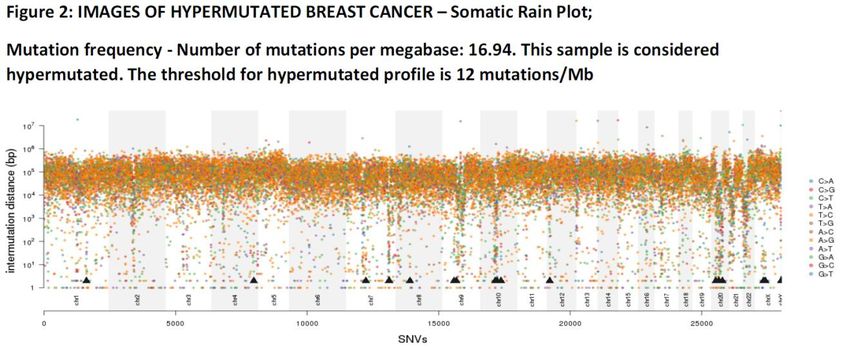
BCFuture Classification • Hypermutated tumour • Associated with increased expression of neoantigens -> host immune response with TILS • BASKET OF BASKETS trial – eligible for immune therapy
Case
• 49 year old female presented with a painful right breast lump
• Core biopsy – grade 3 invasive carcinoma NST, ER/ PR/ HER2
  negative
Case
• Enrolled into PARTNER trial using platinum based chemotherapy
  with a novel targeted agent, olaparib.
• The patient also consented to WGS in the Personalised Breast
  Cancer Program
• No germline mutations including BRCA1/2
• Cosmic signature 3 – defective double stranded DNA break repair
  by homologous recombination
• Likely to respond well to trial therapy with platinum and targeted
  DNA damage repair agent
Case

• Complete radiological
  response on
  midtreatment MRI
• Final histology: pCR
  following neoadjuvant
  chemotherapy
Personalised Breast Cancer Programme – whole genome sequencing looking
  for mutations and copy number alterations

Actionable mutations:
Highly Actionable (Tier 1) - Robust evidence
- Genomic alteration validated in clinical trials
– Clinical evidence of association with response to therapy

Potentially Actionable (Tier 2)
- Evidence mutation is activating (oncogene) or non-activating (tumour
   suppressor gene) in models
- Pre clinical evidence of association with response to treatment but clinical
   evidence lacking/ insufficient

                                 Dr Jean Abraham and Prof Carlos Caldas
Future Classification • Whole genome sequencing -> mutational signatures • Cosmic signatures – distinct patterns of mutations are associated with different mechanisms of DNA damage • Signature 1 = age related • Signature 2 = APOBEC family of cytosine deaminases; C>T • Signature 3 = double stranded DNA damage repair; BRCA1 and 2 mutation carriers • Concept of Basket studies – eligibility for trial dependent upon mutational profile or presence of specific mutations rather than tissue type
You can also read