MOTOR NEURON DISEASE DUE TO NEUROPATHY TARGET ESTERASE GENE MUTATION: CLINICAL FEATURES OF THE INDEX FAMILIES
←
→
Page content transcription
If your browser does not render page correctly, please read the page content below
MOTOR NEURON DISEASE DUE TO NEUROPATHY TARGET ESTERASE
GENE MUTATION: CLINICAL FEATURES OF THE INDEX FAMILIES
SHIRLEY RAINIER, PhD,1 JAMES W. ALBERS, MD, PhD,1 PETER J. DYCK, MD,2 O. PETTER ELDEVIK, MD, PhD,3
SANDRA WILCOCK, RN,1 RUDY J. RICHARDSON, ScD,1,4 and JOHN K. FINK, MD1,5
1
Department of Neurology, University of Michigan, 5013 BSRB, 109 Zina Pitcher Place, Ann Arbor, Michigan 48109, USA
2
Department of Neurology, Mayo Clinic, Rochester, Minnesota, USA
3
Department of Radiology, University of Michigan, Ann Arbor, Michigan, USA
4
Toxicology Research Training Program, Department of Environmental Health Sciences, University of Michigan,
Ann Arbor, Michigan, USA
5
Geriatric Research, Education, and Clinical Center, Veterans Affairs Medical Center, Ann Arbor, Michigan, USA
Accepted 27 April 2010
ABSTRACT: Recently, we reported that mutations in the neu- institutional review board at the University of
ropathy target esterase (NTE) gene cause autosomal recessive Michigan.
motor neuron disease (NTE-MND). We describe clinical, neuro-
physiologic, and neuroimaging features of affected subjects in Sensory and motor nerve conduction studies
the index families. NTE-MND subjects exhibited progressive were performed in the dominant upper and lower
lower extremity spastic weakness that began in childhood and extremities of subject 2 using standard percutane-
was later associated with atrophy of distal leg and intrinsic
hand muscles. NTE-MND resembles Troyer syndrome, except ous nerve stimulation and surface recordings. Sen-
that short stature, cognitive impairment, and dysmorphic fea- sory and motor response amplitudes and latencies
tures, which often accompany Troyer syndrome, are not fea- were measured and conduction velocities were cal-
tures of NTE-MND. Early onset, symmetry, and slow
progression distinguish NTE-MND from typical amyotrophic lat- culated. F-response latencies were obtained for
eral sclerosis. NTE is implicated in organophosphorus com- motor nerves. Skin temperatures were maintained
pound–induced delayed neurotoxicity (OPIDN). NTE-MND between 32.0 and 34.0 C. Neuromuscular trans-
patients have upper and lower motor neuron deficits that are
similar to OPIDN. Motor neuron degeneration in subjects with mission was evaluated by repetitive stimulation (2
NTE mutations supports the role of NTE and its biochemical HZ) of the median and ulnar motor nerves before
cascade in the molecular pathogenesis of OPIDN and possibly and after brief volitional exercise. Concentric nee-
other degenerative neurologic disorders.
Muscle Nerve 43: 19–25, 2011 dle electromyography (EMG) was performed on
selected proximal and distal muscles of the upper
We recently reported the discovery of neuropathy and lower extremities, and insertional activity and
target esterase (NTE) gene mutations in a consan- motor unit potential recruitment and configura-
guineous family (family 1, Fig. 1A) and a non-con- tion were evaluated.
sanguineous family (family 2, Fig. 1B) in which Brain magnetic resonance imaging (MRI) and
affected subjects exhibited slowly progressive spas- cervical and thoracic spine MRI were performed in
tic paraplegia and distal muscle atrophy.1 Herein subject 2 without intravenous gadolinium using a
we provide a detailed description of the clinical 1.5-Tesla scanner. Axial T1, axial T2, and sagittal
features1 and results of electrodiagnostic and neu- T2 sequences obtained of the brain, and sagittal
roimaging studies of affected subjects in these T1, sagittal T2, and axial T2 sequences were
families. obtained of the cervical and thoracic spine.
Quantitative sensation testing (QST) of the
METHODS
hand and foot was performed in subject 2 using
We examined 7 members of a consanguineous CASE III or IV (WR Medical Electronics, Stillwater,
South American kindred of Ashkenazi Jewish Minnesota).
descent (Fig. 1) and 5 members of a non-consan-
guineous Caucasian family of German ancestry. RESULTS
Participation in this study was approved by the Clinical Descriptions. Clinical findings and results
of diagnostic studies are summarized in Table 1.
Abbreviations: CMAP, compound muscle action potential; DTR, deep Antenatal and perinatal histories (available from
tendon reflex; EMG, electromyography; HSP, hereditary spastic paraple- the parents of subjects 2, 3, and 4) were unremark-
gia; MND, motor neuron disease; MRI, magnetic resonance imaging;
NCS, nerve conduction study; NTE, neuropathy target esterase; OP, orga- able, and developmental milestones were attained
nophosphorus; OPIDN, organophosphorus compound–induced delayed normally. Each subject had a slowly progressive
neuropathy; PKA, cyclic AMP-dependent protein kinase; SPG, spastic
paraplegia genetic locus; SWS, Swiss Cheese gene; TOCP, tri-ortho-cre- gait disturbance that began in early childhood
syl phosphate; UE, upper extremities (subject 1) around age 4 years (subjects 2 and 4)
Key words: motor neuron disease, neuropathy target esterase,
neurotoxicity, organophosphorus compound, paraplegia, spasticity, spinal or around age 7 years (subject 3). Progressive
cord disorder, Troyer syndrome weakness and atrophy in the hands became evident
Correspondence to: J.K. Fink; e-mail: jkfink@umich.edu
in the late teenage years (subjects 1 and 2),
V
C 2010 Wiley Periodicals, Inc.
Published online 15 December 2010 in Wiley Online Library
between age 12 and 21 years (subject 3), or
(wileyonlinelibrary.com). DOI 10.1002/mus.21777 around age 20 years (subject 4). Each subject
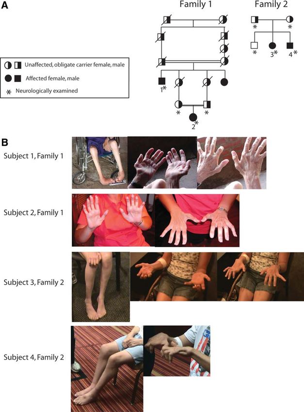
Clinical Features in NTE-MND MUSCLE & NERVE January 2011 19FIGURE 1. (A) NTE-MND syndrome families (modified from Rainier et al.1). (B) Distal atrophy in NTE-MND syndrome.
reported urinary urgency beginning in adulthood. notably in the thenar group and dorsal interossei)
Subjects did not report cognitive impairment, vis- and distal lower extremities. Although intrinsic
ual disturbance, swallowing difficulties, sensory hand muscles were markedly weak, strength was
loss, or symptoms of autonomic disturbance such preserved in wrist extensors, wrist flexors, and prox-
as postural light-headedness or impaired sweating. imal upper extremity muscles. Subject 4 had ham-
Subject 1 had mild dysarthria dating to early adult- string muscle contractures. Other subjects had mod-
hood that was slowly progressive. erate to marked spasticity in hamstring muscles,
Neurologic examinations were performed at 82 moderate to marked weakness of iliopsoas
(subject 1), 48 (subject 3), and 34 (subject 4) years muscles, and marked weakness of tibialis anterior
of age. Subject 2 was examined six times between muscles. Upper extremity deep tendon reflexes were
ages, 8 and 35 years. brisk (3þ) in subjects 1, 2, and 3, but they were
Each subject exhibited marked atrophy and diminished in subject 4. Deep tendon reflexes at the
weakness of intrinsic muscles of the hands (most knees were hyperactive in subjects 1, 2, and 4, but
20 Clinical Features in NTE-MND MUSCLE & NERVE January 2011Table 1. Clinical features and diagnostic studies of NTE-MND subjects.
Subject 1 Subject 2 Subject 3 Subject 4
Antenatal, perinatal, Unavailable Unremarkable Unremarkable Unremarkable
and early
childhood history
Age of onset of Early childhood 4 years 7 years 4 years
slowly progressive
gait disturbance
Age of onset of Teenage years Teenage years Teenage years 20 years
weakness and atrophy
in the hands
Age of onset of Adulthood Adulthood Adulthood Adulthood
urinary urgency
Age at neurologic 82 years Serial exams 48 years 38 years
examination ages 8–35 years
Gait impairment Severe: Marked but Moderate; uses Marked; barely
non-ambulatory ambulates four-pronged ambulatory with
without aid cane a walker
Weakness and atrophy Marked Marked Marked Marked
of intrinsic
hand muscles
Lower extremity Marked Moderate Moderate Marked
weakness spasticity
and distal atrophy
Deep tendon reflexes 3þ UE and 3þ UE and knees; 2þ 3þ UE and knees; 3þ UE and knees;
knees; absent ankle DTRs markedly diminished markedly diminished
ankle DTRs ankle DTRs ankle DTRs
Plantar response Extensor Extensor Extensor Extensor
Light touch and Diminished Normal Normal Normal
pin-prick sensation
Distal vibration Reduced Reduced Reduced Reduced
sensation
NCS Not available Serial NCS (ages Motor NCS (age 12.5 Normal at
8–30 years) showed years): decreased age 4 years
progressive motor amplitude upper
neuropathy and normal and lower motor
sensory NCS action potentials;
increased distal
latency of median
response; normal
sensory NCS
Electromyography Not available Serial EMG (ages EMG (age 12.5 years): Not available
8–35 years): progressive, chronic denervation
chronic denervation, in first dorsal
initially affecting interosseous and
distal lower extremity tibialis anterior
muscles and later distal
upper extremity and proximal
lower extremity muscles
MRI brain and Not available MRI (age 30 years): normal Not available Not available
total spine brain; significant thoracic
spinal cord atrophy
DTR, deep tendon reflex; EMG, electromyography; MRI, magnetic resonance imaging; NCS, nerve conduction studies; UE, upper extremities.
they were absent in subject 4. Ankle jerks were dimin- two families. Subject 1 was unable to walk (when
ished (subjects 2 and 3) or absent (subjects 1 and 4). examined at age 82) because of severe lower ex-
All subjects exhibited extensor plantar responses. Vi- tremity spasticity, weakness, and hamstring tendon
bratory sensation was diminished in the toes. Light contractures. His niece (subject 2, age 38 years)
touch and pinprick sensations were normal in sub- had prominent spastic gait and scissoring but was
jects 3 and 4, but were diminished distally in subject ambulatory without assistance. There was some evi-
1 (who was examined at 82 years of age). dence of intrafamily variation. Whereas subject 4
There was moderate to severe functional had marked spastic paraparesis and was marginally
impairment that was generally similar between the ambulatory even with a walker at 38 years of age,
Clinical Features in NTE-MND MUSCLE & NERVE January 2011 21his older sister (age 48) had moderate spastic para- sural sensory nerve action potential. Median and
paresis, but was fully ambulatory with a four- ulnar CMAP amplitudes were markedly reduced
pronged cane. (0.3 and 2.5 mV, respectively), and peroneal and
Serial evaluations of subject 2 at age 8, 11, 12, tibial CMAPs were unobtainable. The median
17, 20, and 35 years disclosed an evolving pheno- motor distal latency was slightly prolonged, and
type. Initial examination (age 8 years) and subse- the conduction velocity was reduced to a degree
quent examinations showed a spastic gait that consistent with loss of large motor axons. There
worsened progressively. Lower extremity hyperre- was no evidence of motor conduction block or
flexia and extensor plantar responses were present abnormal temporal dispersion. The ulnar motor
from the initial evaluation (age 8). Tendon distal latency and conduction velocity were normal,
reflexes in the upper extremities were normal, and as was the F-wave latency. There was no evidence
corticobulbar signs were absent at each evaluation. of impaired neuromuscular transmission. Needle
Although examinations through 17 years of age EMG at 30 years of age showed chronic distal par-
showed normal upper extremity strength, examina- tial denervation and reinnervation characterized by
tions at ages 20 and 35 years showed definite weak- small-amplitude fibrillation potentials (some mi-
ness and atrophy of thenar, hypothenar, and inter- nute) and decreased recruitment of large-ampli-
osseous muscles. These muscles were markedly tude motor unit potentials. True myotonic dis-
weak at the last examination (age 35 years). Quan- charges were recorded from a single fibrotic
titative sensation testing (performed in the hand muscle (anterior tibialis), which also showed severe
and foot using CASE III or IV (WR Medical Elec- neurogenic changes. The study was interpreted as
tronics, Stillwater, Minnesota) was performed on showing evidence of a moderately severe motor
each occasion and was normal until the last exami- neuronopathy characterized by clinically symmetric
nation (age 35 years), when decreased vibration distal involvement. The myotonic discharges
was detected at the great toe and index finger. recorded from a single fibrotic muscle were noted
to be a non-specific finding associated on occasion
Neurophysiologic and Neuroimaging Studies and with chronic denervation.
Muscle Biopsy. Motor nerve conduction studies Serial needle EMG examinations showed evolu-
(NCSs) and needle EMG were normal in subject 4 tion of this chronic dennervation. Whereas exami-
when they were performed at age 4 years. Motor nation at age 8 years showed slightly increased
NCSs performed in subject 3 at age 12.5 years insertional activity, fibrillation potentials, and
showed decreased amplitude ulnar, median, and decreased recruitment characterized by large poly-
peroneal motor responses and a slightly increased phasic motor unit potentials only in the intrinsic
median motor distal latency. Sensory NCSs were foot muscles, examination at age 11 showed
normal. EMG findings (subject 3, age 12.5 years) increased insertional activity, fasciculation poten-
were interpreted as showing neurogenic atrophy in tials, and alteration of motor unit potentials
distal muscles. For example, there was markedly (increased size and duration) in distal lower ex-
decreased motor unit potential recruitment, large- tremity muscles. At 20 years of age, these abnor-
amplitude and long-duration motor unit poten- malities included the anterior tibial muscle. At
tials, increased insertional activity, and 2þ fibrilla- 35 years of age, intrinsic hand muscles were simi-
tion potentials in the first dorsal interosseous mus- larly affected, and very mild but similar changes
cle. EMG of the tibialis anterior demonstrated a were found in anterior thigh muscles.
‘‘mixed population’’ of large and small motor unit Muscle biopsy obtained from the extensor digi-
potentials. torum communis muscle of subject 3 at age
Serial nerve conduction studies in subject 2 at 12 years of age was judged to be ‘‘non-diagnostic.’’
age 8, 11, 12, 17, and 20 years documented pro- There was no significant histologic evidence of
gressive motor neuropathy or neuronopathy. Pero- chronic denervation despite EMG findings from
neal and tibial compound muscle action potential the first dorsal interosseous, tibialis anterior, and
(CMAP) amplitudes were already reduced at 8 adductor digiti quinti muscles that were consistent
years of age (0.8 and 1.6 mV, respectively), further with chronic denervation at this age.
reduced by age 12 (0.5 and 0.3 mV, respectively), Magnetic resonance imaging of the brain and
and absent at age 20. The ulnar CMAP amplitude spinal cord were performed in subject 2 (Fig. 2).
was 4.2 mV at 8 years of age (normal value >6.0 No significant intracranial abnormality was identi-
mV). This borderline-low amplitude decreased pro- fied with appropriate cerebellar and midbrain vol-
gressively (3.6, 2.2, and 0.7 mV at age 11, 12, and umes, and there was no evidence of corpus cal-
20 years, respectively). losum atrophy or agenesis. MRI of the spinal cord
NCSs performed in subject 2 at age 30 years showed significant cord atrophy beginning at the
showed normal sensory studies, including a normal T3–4 level and extending down to T11, where the
22 Clinical Features in NTE-MND MUSCLE & NERVE January 2011FIGURE 2. MRI of brain and spinal cord in NTE-NMD syndrome. (A) No significant intracranial abnormality is identified. The corpus
callosum (not shown here) was present without evidence of atrophy. Significant spinal cord atrophy is present beginning at the T3–4
level where the cord again expands at the conus medullaris. Although atrophic, the spinal cord was not compressed, and there were
no spinal cord aberrations. (B) Cord transverse area (circle) at T9 measured 25 mm2 at the T9 level (compared with control subjects,
not shown) that measure 39.3 6 8.1 mm2.
cord expanded at the conus medullaris (Fig. 3). motor neurons. Such lower motor neuron signs
Thoracic spinal cord cross-sectional area2 at T9 was occur as a constant or variable feature in many
reduced (25 mm2) compared with control subjects forms of complicated HSP (SPG7, SPG10, SPG14,
(39.3 6 8.1 mm2). There was no evidence of SPG15, SPG17, SPG20, and SPG26 HSP; see Fink3
intrinsic spinal cord signal aberration, compres- for review), and in SPG3A,4,5 a common childhood-
sion, or significant narrowing of the central canal. onset form of HSP that is usually uncomplicated.
Among the various forms of HSP, NTE-MND
DISCUSSION most closely resembles SPG20 HSP (Troyer syn-
The salient features of subjects with NTE-MND drome), a complicated form of autosomal recessive
are: (1) childhood onset of slowly progressive spas- HSP that was initially described in Old Order
tic paraplegia; (2) progressive atrophy of intrinsic Amish due to SPG20/spartin gene mutation.6,7
muscles of the hands and distal lower extremities Although both Troyer syndrome and NTE-MND
beginning in early through late adolescence; (3) have progressive spastic paraplegia and distal mus-
mild diminution of distal vibration sensation; and cle atrophy, it is notable that many subjects with
(4) family history consistent with autosomal reces- the SPG20/spartin mutation have neurologic signs
sive inheritance. EMG and nerve conduction stud- that are not present in the SPG39/NTE-mutation
ies were consistent with progressive motor nerve subjects identified to date. In addition to progres-
impairment. Peripheral sensory nerve studies were sive spastic paraplegia, subjects with SPG20/spartin
normal. Neuroimaging identified prominent tho- mutation have delayed milestones (15 of 21 exam-
racic spinal cord atrophy in the one patient ined subjects), cognitive impairment (in 17 of
studied. 21 examined subjects), emotional lability (in 17 of
NTE-MND may be classified as a form of hered- 21 subjects), cerebellar signs (16 of 21 examined
itary spastic paraplegia (designated SPG39).1 Dur- subjects), spastic dysarthria (19 of 21 examined sub-
ing childhood and early adolescence, NTE-MND jects), skeletal abnormalities (all 21 examined sub-
presents as an ‘‘uncomplicated’’ hereditary spastic jects), ataxia (3 of 17 examined subjects), choreoa-
paraplegia (HSP) syndrome. The subsequent thetoid movements (3 of 21 examined subjects),
appearance (in early through late adolescence) of and dysphagia (2 of 21 examined subjects).8 More-
progressive atrophy of distal upper and lower ex- over, 3 of 21 subjects with SPG20/spartin mutation
tremity muscles signals transition to a complicated did not have distal muscle atrophy, indicating that
HSP syndrome involving both upper and lower clinical diagnosis cannot rest on this major feature
Clinical Features in NTE-MND MUSCLE & NERVE January 2011 23alone. Our observations suggest that the presence jects reduce NTE activity and alter its enzyme
or absence of these additional signs (cognitive, bul- kinetics in vitro.22 It is unknown, however, if reduc-
bar, cerebellar, emotional, and extrapyramidal def- tion in phospholipase activity of NTE is sufficient
icits and skeletal abnormalities) help distinguish to cause neurodegeneration in NTE-MND. The
SPG20/spartin-mutation/Troyer syndrome subjects impact of NTE-MND mutations (and irreversible
from those with NTE mutations. organophosphorylation) on other emerging NTE
The NTE-MND subjects just described were functions has not been assessed. Specifically, there
ascertained as familial clusters suggesting autoso- is recent evidence that the NTE Drosophila homo-
mal recessive inheritance. Because subjects with log Swiss Cheese (SWS) binds to and regulates
autosomal recessive disorders often have no previ- cyclic AMP-dependent protein kinase (PKA).23 Dis-
ous family history of the disorder, it is possible covering that human NTE regulates PKA in a simi-
that NTE mutations contribute to motor neuron lar manner would suggest additional mechanisms
disease among subjects without a family history of (including altered phosphorylation of target pro-
the disease. teins and/or cAMP-regulated gene expression) by
NTE is an important factor in OPIDN,9–14 a dis- which NTE disturbance leads to neurodegenera-
tal axonopathy that, like NTE-MND, is character- tion. It is also possible that NTE-mediated motor
ized in late-stage disease by spastic paraparesis with neuron degeneration is due to NTE protein mis-
distal weakness and atrophy. Causes of OPIDN folding. Abnormal protein folding is a mechanism
include occupational and accidental exposures to common to many neurodegenerative disorders,24
neuropathic organophosphorus compounds.15–20 including Parkinson disease,25 polyglutamine-expan-
One example of OPIDN is a disorder sometimes sion disorders,26 and amyotrophic lateral sclero-
referred to as ‘‘jake leg palsy,’’ in reference to the sis.27,28 Increased recognition of NTE-MND and elu-
accompanying ingestion of Jamaican ginger cidating its molecular pathogenesis will provide
(‘‘jake’’) and to the characteristic gait displayed by insight into possible gene–environment interactions
afflicted individuals reflecting a combination of that contribute to motor neuron disease and
upper and lower motor neuron involvement. The advance our ability to treat these disorders.
disorder may have affected as many as 50,000 indi-
This research was supported by grants from the National Institutes
viduals in the Prohibition-era United States. Jake of Health (NINDS R01NS053917), the Department of Veterans
leg palsy was attributed to recreational con- Affairs (Merit Review Awards), the Spastic Paraplegia Foundation,
sumption of Jamaican ginger extract, a patent and the generous support of the Paul and Lois Katzman Family
medicine adulterated with tri-ortho-cresyl phosphate Fund (to J.K.F.). We gratefully acknowledge the expert secretarial
assistance of Lynette Girbach, and the participation of patients
(TOCP).21 In addition to the clinical similarity
and their relatives without whom this research would not have
between OPIDN and NTE-MND, neuropathologic been possible.
findings in the chicken model of OPIDN (includ-
ing axon degeneration of corticospinal tracts and REFERENCES
dorsal column fibers)13 overlap the predominant 1. Rainier S, Bui M, Mark E, Thomas E, Tokarz D, Ming L, et al. Neu-
ropathy target esterase gene mutations cause motor neuron disease.
localization of neurologic signs in subjects with Am J Hum Genet 2008;82:780–785.
2. Hedera P, Eldevik OP, Maly P, Rainier S, Fink JK. Spinal cord mag-
NTE-MND mutation. These observations suggest netic resonance imaging in autosomal dominant hereditary spastic
that motor neuron syndrome associated with the paraplegia. Neuroradiology 2005;47:730–734
3. Fink JK. Hereditary spastic paraplegia. Curr Neurol Neurosci Rep
organophosphorus compound–NTE interaction 2006;6:65–76.
shares a common molecular pathogenesis with 4. Dalpozzo F, Rossetto MG, Boaretto MS, et al. Infancy onset heredi-
tary spastic paraplegia associated with a novel atlastin mutation. Neu-
motor neuron disease attributed to homozygous rology 2003;61:580–581.
(or compound heterozygous)1 NTE mutations. 5. Ivanova N, Claeys KG, Deconinck T, et al. Hereditary spastic paraple-
gia 3A associated with axonal neuropathy. Arch Neurol 2007;64:
This observation supports the hypothesis that some 706–713.
apparently sporadic (or genetically occult) MNDs 6. Cross HE, McKusick VA. The Troyer syndrome. A recessive form of
spastic paraplegia with distal muscle wasting. Arch Neurol 1967;16:
could be due to exposure to some neuropathic 473–485.
organophosphorus compounds, either alone or to- 7. Patel H, Cross H, Proukakis C, Hershberger R, Bork P, Cicarelli FD,
et al. SPG20 is mutated in Troyer syndrome, an hereditary spastic
gether with predisposing NTE gene mutations or paraplegia. Nat Genet 2002;31:347–348.
genetic variation in factors that regulate or interact 8. Proukakis C, Cross H, Patel H, Patton MA, Valentine A, Crosby AH.
Troyer syndrome revisited. A clinical and radiological study of a
with NTE. complicated hereditary spastic paraplegia. J Neurol 2004;251:
The molecular mechanisms by which NTE dis- 1105–1110.
9. Winrow CJ, Hemming ML, Allen DM, Quistad GB, Casida JE, Barlow
turbance (both due to mutations in the NTE ester- C. Loss of neuropathy target esterase in mice links organophosphate
ase domain as well as irreversible organophosphor- exposure to hyperactivity. Nature Genet 2003;33:477–485.
10. Glynn P. Neuropathy target esterase. Biochem J 1999;344:625–631.
ylation of the NTE active site serine) lead to axon 11. Johnson MK. The primary biochemical lesion leading to the delayed
degeneration that particularly involves central and neurotoxic effects of some organophosphorus esters. J Neurochem
1974;23:785–789.
peripheral motor neurons are not clear. Recently, 12. Glynn P. Neural development and neurodegeneration: two faces of
we showed that the mutations in NTE-MND sub- neuropathy target esterase. Prog Neurobiol 2000;61:61–74.
24 Clinical Features in NTE-MND MUSCLE & NERVE January 201113. Abou-Donia MB. Organophosphorus ester-induced delayed neuro- mutations related to motor neuron disease have altered enzymatic
toxicity. Ann Rev Pharmacol Toxicol 1981;21:511–548. properties. Toxicol Lett 2010;196:67–73.
14. Wijeyesakere SJ, Richardson RJ. Neuropathy target esterase. In: 23. Bettencourt da Cruz A, Wentzell J, Kretzschmar D. Swiss cheese, a
Krieger R, editor. Hayes’ handbook of pesticide toxicology, 3rd ed. protein involved in progressive neurodegeneration, acts as a nonca-
San Diego, CA: Elsevier/Academic Press; 2010. p 1435–1478. nonical regulatory subunit for PKA-C3. J Neurosci 2008;28:
15. Smith HV, Spalding JM. Outbreak of paralysis in Morocco due to 10885–10892.
ortho-cresyl phosphate poisoning. Lancet 1959;5:1019–1021. 24. Luo GR, Le WD. Collective roles of molecular chaperones in protein
16. Taylor P. Anticholinesterase agents. Goodman and Gilman’s pharma- degradation pathways associated with neurodegenerative diseases.
cologic basis of therapeutics, 9th ed. New York: McGraw Hill; 1996. Curr Pharm Biotechnol 2010;11:180–187.
17. Sorokin M. Orthocresyl phosphate neuropathy: report of an out- 25. Bandopadhyay R, de Belleroche J. Pathogenesis of Parkinson’s dis-
break in Fiji. Med J Austral 1969;10:506–508. ease: emerging role of molecular chaperones. Trends Mol Med
18. Srivastava AK, Das M, Khanna SK. An outbreak of tricresyl phosphate 2010;16:27–36.
poisoning in Calcutta, India. Food Chem Toxicol 1990;28:303–304. 26. Williams AJ, Paulson HL. Polyglutamine neurodegeneration: protein
19. Sarkar JK. Outbreaks of paralytic disease in West Bengal due to tri- misfolding revisited. Trends Neurosci 2008;31:521–528.
cresyl phosphate poisoning. J Indian Med Assoc 1974;63:359–361. 27. Johnson BS, Snead D, Lee JJ, McCaffery JM, Shorter J, Gitler AD.
20. Mehta RS, Dixit IP, Khakharia SJ. Toxic neuropathy in Raipur due to tri- TDP-43 is intrinsically aggregation-prone, and amyotrophic lateral
orthocresylphosphate (TOCP). J Assoc Physicians India 1975;23:133–138. sclerosis-linked mutations accelerate aggregation and increase toxic-
21. Morgan JP, Penovich P. Jamaica ginger paralysis. Forty-seven-year fol- ity. J Biol Chem 2009;284:20329–20339.
low-up. Arch Neurol 1978;35:530–532. 28. Kerman A, Liu HN, Croul S, et al. Amyotrophic lateral sclerosis is a
22. Hein ND, Stuckey JA, Rainier SR, Fink JK, Richardson RJ. Constructs non-amyloid disease in which extensive misfolding of SOD1 is
of human neuropathy target esterase catalytic domain containing unique to the familial form. Acta Neuropathol 2010;119:335–344.
Clinical Features in NTE-MND MUSCLE & NERVE January 2011 25You can also read