Mutant p53 regulates Survivin to foster lung metastasis
←
→
Page content transcription
If your browser does not render page correctly, please read the page content below
Downloaded from genesdev.cshlp.org on September 11, 2021 - Published by Cold Spring Harbor Laboratory Press

Mutant p53 regulates Survivin to foster
lung metastasis
Qiaosi Tang,1,2 Gizem Efe,2 Anna M. Chiarella,2 Jessica Leung,3 Maoting Chen,2 Taiji Yamazoe,1
Zhenyi Su,2 Jason R. Pitarresi,1 Jinyang Li,1 Mirazul Islam,4 Tatiana Karakasheva,1
Andres J. Klein-Szanto,5 Samuel Pan,2 Jianhua Hu,2 Shoji Natsugoe,6 Wei Gu,2 Ben Z. Stanger,1
Kwok-K Wong,7 J. Alan Diehl,8 Adam J. Bass,9 Hiroshi Nakagawa,2 Maureen E. Murphy,3
and Anil K. Rustgi2
1
 Abramson Cancer Center, University of Pennsylvania, Philadelphia, Pennsylvania 19104, USA; 2Herbert Irving Comprehensive
Cancer Center, Columbia University, New York, New York 10032, USA; 3Program in Molecular and Cellular Oncogenesis, The
Wistar Institute, Philadelphia, Pennsylvania 19104, USA; 4Department of Cell and Developmental Biology, Vanderbilt University,
Nashville, Tennessee 37235, USA; 5Department of Pathology, Cancer Biology Program, Fox Chase Cancer Center, Philadelphia,
Pennsylvania 19104, USA; 6Department of Digestive Surgery, Kagoshima University, Sakuragaoka, Kagoshima 890-0065, Japan;
7
 New York University Langone Center, New York, New York 10016, USA; 8Case Western University, Cleveland, Ohio 44106,
USA; 9Department of Medical Oncology, Dana-Farber Cancer Institute, Boston, Massachusetts 02215, USA

Esophageal squamous cell carcinoma (ESCC) is one of the most lethal cancers worldwide and evolves often to lung
metastasis. P53R175H (homologous to Trp53 R172H in mice) is a common hot spot mutation. How metastasis is reg-
ulated by p53R175H in ESCC remains to be investigated. To investigate p53R175H-mediated molecular mechanisms,
we used a carcinogen-induced approach in Trp53R172H/− mice to model ESCC. In the primary Trp53R172H/− tumor
cell lines, we depleted Trp53R172H (shTrp53) and observed a marked reduction in cell invasion in vitro and lung
metastasis burden in a tail-vein injection model in comparing isogenic cells (shCtrl). Furthermore, we performed
bulk RNA-seq to compare gene expression profiles of metastatic and primary shCtrl and shTrp53 cells. We identified
the YAP-BIRC5 axis as a potential mediator of Trp53 R172H-mediated metastasis. We demonstrate that expression of
Survivin, an antiapoptotic protein encoded by BIRC5, increases in the presence of Trp53R172H. Furthermore,
depletion of Survivin specifically decreases Trp53R172H-driven lung metastasis. Mechanistically, Trp53R172H but not
wild-type Trp53, binds with YAP in ESCC cells, suggesting their cooperation to induce Survivin expression. Fur-
thermore, Survivin high expression level is associated with increased metastasis in several GI cancers. Taken to-
gether, this study unravels new insights into how mutant p53 mediates metastasis.
[Keywords: mutant p53; metastasis; Survivin; YAP]
Supplemental material is available for this article.
Received May 15, 2020; revised version accepted February 15, 2021.

Metastases contribute to the majority of cancer-associated                  instability, facilitate invasion to the stroma, and are cru-
deaths (Steeg 2016). The complex invasion-metastasis cas-                   cial driving forces of metastases (Steeg 2016).
cade involves multiple steps, including cancer cell inva-                      Across all human cancers, TP53 is the most frequently
sion, intravasation into circulation, extravasation from                    mutated gene, and alterations in p53 functions and its
circulation, and metastatic colonization into a distant or-                 downstream signaling pathways have been validated in
gan (e.g., liver and lung). However, despite a large number                 diverse cancers (Muller and Vousden 2014). Unlike other
of studies demonstrating key mechanisms and treatment                       tumor suppressor proteins that harbor frameshift or non-
strategies in primary tumors, the mechanisms underlying                     sense mutations that lead to loss of expression, the major-
tumor metastasis are still in evolution (Lambert et al.                     ity of cancer-associated TP53 alterations encompass
2017). Increasing evidence has shown that tumor intrinsic                   missense mutations (Olivier et al. 2010; Freed-Pastor
factors such as genomic, genetic, and epigenetic aberra-                    and Prives 2012). Missense TP53 mutations occur fre-
tions are important contributors to metastasis. Specifi-                    quently on six residues, namely R175, G245, R248,
cally, tumor intrinsic oncogenic events induce genomic                      R249, R273, and R282, and are designated as “hot spot”
                                                                            mutations (Rivlin et al. 2011). Functionally, hot spot
Corresponding author: akr2164@cumc.columbia.edu
Article published online ahead of print. Article and publication date are   © 2021 Tang et al. This article, published in Genes & Development, is
online at http://www.genesdev.org/cgi/doi/10.1101/gad.340505.120. Free-     available under a Creative Commons License (Attribution-NonCommer-
ly available online through the Genes & Development Open Access             cial 4.0 International), as described at http://creativecommons.org/licens-
option.                                                                     es/by-nc/4.0/.

                  GENES & DEVELOPMENT 35:1–14 Published by Cold Spring Harbor Laboratory Press; ISSN 0890-9369/21; www.genesdev.org                  1
Downloaded from genesdev.cshlp.org on September 11, 2021 - Published by Cold Spring Harbor Laboratory Press

Tang et al.

p53 mutations have been demonstrated to promote tu-                 mutant p53 ESCC cells. Furthermore, we determined
morigenesis (Freed-Pastor and Prives 2012). For example,            that Survivin expression is dependent upon p53R175H
mice with germline Trp53 (equivalent to TP53 in human)              and is necessary and specific to mediate p53R175H-driven
mutations have a different tumor spectrum compared                  lung metastasis. Mechanistically, we revealed that
with Trp53-null mice. Trp53R172H/− (equivalent to human             Trp53R172H, but not wild-type Trp53, interacts physically
p53R175H/−) mice have increased incidence of carcinomas,            with YAP (Yes-associated protein) in ESCC cells. We also
whereas Trp53−/− mice frequently develop lymphomas                  discovered that Trp53R172H enhances the binding of YAP
and sarcomas (Olive et al. 2004). Compared with                     on the BIRC5 promoter, suggesting their cooperation to
Trp53+/− mice, Trp53R172H/+ mice harbor metastatic oste-            induce Survivin expression. Additionally, we found that
osarcoma and epithelial carcinomas, whereas tumors in               there is up-regulation of Survivin expression in ESCC
Trp53+/− mice typically do not metastasize (Lang et al.             based upon a human ESCC tissue microarray and TCGA
2004). Furthermore, in several types of cancers, mutant             analysis. There is correlation of high Survivin expression
p53, especially p53R175H, promotes cell invasion, motility,         with p53 nuclear accumulation in ESCC. Furthermore,
and metastasis through the regulation of different path-            there is Survivin up-regulation with increased metastasis
ways (Bossi et al. 2006; Dong et al. 2009; Muller et al.            in other GI cancers (pancreatic and colon). Taken togeth-
2009, 2013; Tabach et al. 2010; Weissmueller et al.                 er, this study underscores the novel finding that BIRC5 is
2014). Furthermore, analysis of clinical data has validated         a novel mediator of p53R175H in promoting lung metasta-
these findings. For example, in head and neck squamous              sis, thereby offering new insights into how mutant p53
cell carcinoma (HNSCC), patients harboring p53R175H                 promotes metastasis and opening up new avenues for
have poor clinical outcomes, including increased metasta-           translational therapeutics.
sis and decreased overall survival (Neskey et al. 2015).
Taken together, mutant p53 contributes to an aggressive
tumor phenotype characterized by enhanced cell motility,            Results
invasion, and metastatic capacity in several cancer types.
                                                                    Mutant p53 is required for invasion and metastasis
   Esophageal cancer is a highly aggressive cancer. Global-
                                                                    in ESCC
ly, it was estimated that approximately 570,000 people
were diagnosed with esophageal cancer in 2018 (Uhlen-               To understand the function of mutant p53 in ESCC,
hopp et al. 2020). The major subtype of esophageal cancer           a mutant p53-driven ESCC mouse model was generated
worldwide is esophageal squamous cell carcinoma                     combining genetic and carcinogenic approaches. This
(ESCC), which typically has a poor prognosis, since                 model recapitulates human ESCC grossly and histologi-
many cases are not detected until late stages (Pennathur            cally (Tang et al. 2021). Additionally, we generated
et al. 2013; Rustgi and El-Serag 2014). At the time of diag-        Trp53R172H/− and Trp53−/− cell lines from these primary
nosis, local invasion and distant metastasis, especially            tumors and only used early passage cells (Tang et al.
lung metastasis, are frequently detected (Rustgi and El-            2021). To elucidate the role of mutant p53 in tumor cell
Serag 2014). Thus, understanding the mechanisms under-              invasion and metastasis, we generated an isogenic control
lying metastasis in ESCC is crucial to improvements in              system through stably expressing shRNA targeting
treatment strategies and patient outcomes, with potential           Trp53 in Trp53R172H/− cells (shTrp53) and compared their
therapeutic applications to other genomically related squ-          biological behavior with control shRNA-expressing
amous cell cancers (SCC) arising from the head and neck,            Trp53R172H/− cells (shCtrl) (Fig. 1A; Supplemental Fig.
lung, and anogenital tract (Dotto and Rustgi 2016; Camp-            S1A). We also generated a “rescue” cell line by introduc-
bell et al. 2018). The most frequent genetic alteration in          ing shTrp53-resistant Trp53R172H (Fig. 1A). The CRISPR/
ESCC is TP53 mutation, occurring in 83% of ESCC pa-                 Cas9 system was also used to deplete Trp53 in
tients (Song et al. 2014), and as such, ESCC represents             Trp53R172H/− cells (sgTrp53), comparing with an empty
one of the top cancers that harbor TP53 mutations (Olivier          vector-expressing Trp53R172H/− cells (Vector) (Supplemen-
et al. 2010). In ESCC, R175H mutation is one of the top             tal Fig. S1B). In addition, we ectopically expressed
three hot spot TP53 mutations, which leads to distortion            Trp53R172H in Trp53−/− cells (R172H) to compare with
of the global protein structure and is referred to as confir-       Trp53−/− cells expressing an empty vector (Ctrl) (Fig. 1A).
mation mutation (IARC database) (Olivier et al. 2010).                We showed that tumor cell invasion is dependent on
How mutant p53, especially the R175H mutation, con-                 Trp53R172H using a transwell invasion assay. Depletion
tributes to ESCC invasion and metastasis remains to be              of Trp53R172H abrogated cell invasion, whereas reintro-
understood, and discoveries therein can be applied to oth-          duction of shTrp53-resistant Trp53R172H or ectopic ex-
er cancers, especially SCC.                                         pression of Trp53R172H enhanced invasion capacity (Fig.
   In this study, we focused on the oncogenic role of mu-           1B; Supplemental Fig. S1C). To evaluate whether alter-
tant p53 in driving lung metastasis. Through in vitro               ations in proliferation or apoptosis might influence inva-
and in vivo studies, we demonstrated that p53R175H pro-             sion, we performed the carboxyfluorescein succinimidyl
motes ESCC cell motility, invasion, and lung metastasis.            ester (CFSE) proliferation assay and observed no signifi-
To investigate the underlying mechanisms, we profiled               cant difference in proliferation upon Trp53 R172H depletion
gene expression signatures in metastatic and primary                (Supplemental Fig. S1D). Additionally, no significant
ESCC cells. We identified BIRC5 (encoding an antiapopto-            change was observed in apoptosis (Supplemental Fig.
sis protein Survivin) to be highly enriched in metastatic           S1E). The impact of Trp53R172H in cell invasion was

2       GENES & DEVELOPMENT
Downloaded from genesdev.cshlp.org on September 11, 2021 - Published by Cold Spring Harbor Laboratory Press

                                                                                                          Mutant p53 and Survivin in metastasis

                         A

                         B

                         C                                                         D

                         E                                                         F

Figure 1. Mutant p53 promotes ESCC invasion and lung metastasis. (A, left) Trp53R172H is depleted by shRNA in Trp53R172H/− cells.
shRNA-resistant Trp53R172H is reintroduced to Trp53R172H/− shTrp53 (middle), and Trp53R172H is ectopically expressed in Trp53−/− cells
(right). Relative intensity densitometry of TRP53 results are shown at the bottom. (B) Depletion of Trp53R172H suppresses cell invasion,
whereas reintroduction of shRNA-resistant Trp53R172H or ectopic expression of Trp53R172H promotes cell invasion in a Boyden chamber
assay. Trp53R172H/− shCtrl versus shTrp53, n = 4 per group, P = 0.0035, unpaired t-test; Trp53R172H/− shRNA + Ctl versus + Trp53R172H, n = 3
per group, P = 0.0398, unpaired t-test; Trp53−/− Ctrl versus R172H, n = 3 per group, P = 0.0094, unpaired t-test. Error bars represent SEM. (C,
D) YFP+ ESCC cells were injected into the tail vein to induce lung metastatic nodules. Mice were sacrificed 8 wk after injection. Lung
metastasis colony number in Trp53R172H/− shCtrl injected mice was increased greatly compared with shTrp53 injected mice.
Trp53R172H/− shCtrl versus shTrp53, n = 3 per group, P < 0.0001, unpaired t-test. Scale bar, 1 mm. (E,F ) YFP+ Trp53R172H/− shTrp53 cells
reintroduced with shRNA-resistant Trp53R172H were used to repeat the tail-vein injection assay. Mice were sacrificed 8 wk after injection.
Trp53R172H/− shTrp53-rescued cell line-injected mice had a trend of increased lung metastatic colony number. Trp53R172H/− shTrp53+Ctrl
(n = 4 per group) versus shTrp53+ Trp53 R172H (n = 3 per group), P = 0.0709, unpaired t-test. Scale bar, 1 mm.

determined further using 3D organotypic culture (OTC),                      Next, we examined whether Trp53R172H is required for
an air–liquid interface model system (Kalabis et al.                      ESCC metastatic potential through the injection of shCtrl
2012). In OTC with epithelial Trp53R172H depletion, there                 and shTrp53 cells into the lateral tail veins of athymic
is reduced cell invasion in the stroma (Supplemental Fig.                 nude mice (NCr nude, Taconic). Lung metastases were de-
S1F). Taken together, Trp53R172H promotes tumor cell                      tected at 2 wk, 5 wk, and 8 wk postinjection, and the num-
invasion.                                                                 ber and size of metastases were scored. At each time point,

                                                                                                             GENES & DEVELOPMENT             3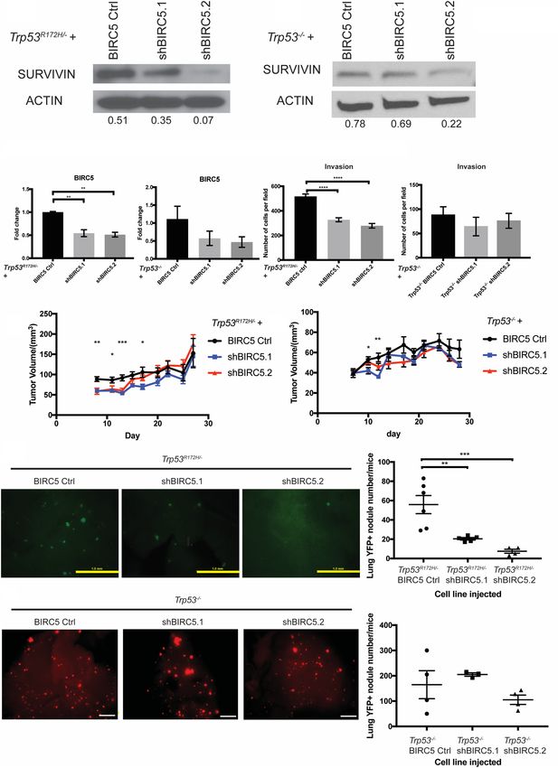
Downloaded from genesdev.cshlp.org on September 11, 2021 - Published by Cold Spring Harbor Laboratory Press

Tang et al.

the metastatic burden was decreased significantly upon              with a pool of shRNAs in Trp53R172H/− ESCC cells
Trp53R172H depletion (Fig. 1C,D; Supplemental Fig. S2A,             (shYAP) and compared its oncogenic behaviors with non-
B). In addition, we performed a “rescue” experiment and             target control (shNT) (Supplemental Fig. S3A). Depletion
observed a partial rescue of the metastatic phenotype fol-          of YAP in Trp53R172H/− ESCC cells significantly reduced
lowing introduction of shTrp53-resistant Trp53R172H (Fig.           invasion capacity (Supplemental Fig. S3B). In addition,
1E,F; Supplemental Fig. S2C,D). As independent corrobo-             YAP depletion also significantly decreased subcutaneous
ration, we repeated the tail-vein injection assay with              tumor growth (Supplemental Fig. S3C,D). Taken together,
R172H and Ctrl cell lines and observed a trend of in-               these data underscore YAP as an important oncogenic fac-
creased metastatic nodule number in the presence of ec-             tor in ESCC. Oncogenic functions of YAP have also been
topic Trp53R172H expression (Supplemental Fig. S2E,F).              investigated in several other cancers, including its effects
Taken together, these results underscore that Trp53R172H            in fostering metastasis (Wang et al. 2018; Lee et al. 2019).
enhances ESCC metastatic capability to the lungs.                   Furthermore, YAP has been suggested to interact with
                                                                    mutant p53 and other p53 family members in cell lines
                                                                    (Di Agostino et al. 2016; Mello et al. 2017; Furth et al.
Mutant p53-mediated reprogramming of pathways in
                                                                    2018). However, whether the interaction of mutant p53
lung metastasis
                                                                    and YAP promotes metastasis remains to be elucidated.
Following the generation and characterization of our lung           Taken together, we hypothesize that YAP signaling may
metastatic model, we generated further a panel of meta-             function as a mediator of mutant p53-driven metastasis.
static cell lines (shTrp53-M and shCtrl-M; M indicates                To investigate further the activation of YAP signaling in
metastatic) from lung metastases at week 8 through YFP              shCtrl-M cells, we found that a YAP downstream target,
+ FACS sorting and confirmed that Trp53R172H depletion              BIRC5, was enriched significantly in shCtrl-M cells (Fig.
was maintained in shTrp53-M cells (Fig. 2A). To under-              3B; Muramatsu et al. 2011). To validate the RNA-seq re-
stand the molecular basis of Trp53R172H-driven ESCC                 sults, we confirmed by qRT-PCR that BIRC5 mRNA
lung metastasis, we performed RNA-seq analysis on                   was up-regulated significantly in shCtrl-M cells compared
shCtrl-M (n = 5), shTrp53-M (n = 3), shCtrl (n = 1), and            with shTrp53-M cells (Fig. 3C). Next, we examined the ex-
shTrp53 (n = 1) cell lines. Principal component analysis            pression of Survivin, the protein encoded by BIRC5, in 3D
(PCA) revealed that primary tumor cells (shCtrl and                 organoids generated from shCtrl and shTrp53 cells. In-
shTrp53) clustered away from metastatic cells (shCtrl-M             deed, Survivin expression is up-regulated in shCtrl com-
and shTrp53-M) (Fig. 2B). Compared with two primary tu-             pared with shTrp53 3D organoids (Fig. 3D). In addition,
mor cell lines shCtrl and shTrp53, metastatic cell lines            we examined Survivin expression in the esophagi of
shCtrl-M and shTrp53-M exhibited a distinct gene expres-            Trp53R172H/− and Trp53−/− 4NQO mouse models. Survi-
sion pattern, suggesting that gene expression signatures            vin expression trended to be higher in the Trp53R172H/−
are changed during metastasis (Fig. 2C). In metastatic              esophagi (Fig. 3E). Finally, we assessed Survivin expres-
cells, we identified 112 genes significantly up-regulated           sion in metastatic lung lesions formed by shCtrl and
and 170 genes down-regulated in shCtrl-M cells compared             shTrp53 cells. In metastatic lung lesions formed by shCtrl
with shTrp53-M cells (Padj < 0.01 and |log2 fold change| >          cells, Survivin expression is increased compared with
2). Among these, Trp53 is the most significantly up-regu-           metastatic lesions formed by shTrp53 cells (Fig. 3F). In
lated gene, which served to validate our system (Fig. 2D,           summary, our complementary in vitro and in vivo results
E). Gene set enrichment analysis (GSEA) Hallmark re-                reveal for the first time that Trp53R172H enhances BIRC5
vealed 13 down-regulated pathways and 20 up-regulated               gene expression in lung metastasis.
pathways in shCtrl-M cells. Epithelial–mesenchymal
transition (EMT), an up-regulated pathway, has been
                                                                    Depletion of BIRC5 expression reduces Trp53 R172H- but
shown previously to play an important role in metastasis
                                                                    not Trp53−/−-driven lung metastases
(Fig. 2F; Tsai and Yang 2013; Dongre and Weinberg 2019).
In addition, other signaling pathways such as MYC,                  Given our finding that Trp53R172H induces BIRC5 gene
mTORC1, and glycolysis are also known to be involved                expression, we next investigated the latter’s direct role
in regulating metastasis (Fig. 2F; Wolfer and Ramaswamy             in mediating the phenotypic effects of Trp53R172H. As a re-
2011; Huang and Zhou 2012; Payen et al. 2016).                      sult, we depleted BIRC5 with two different shRNAs in
                                                                    mouse primary Trp53R172H/− or Trp53−/− ESCC cells
                                                                    (Fig. 4A–D). Depletion of BIRC5 significantly reduced
Trp53R172H promotes BIRC5 function
                                                                    cell invasion in Trp53R172H/− but not Trp53−/− ESCC cells
To identify and characterize candidate genes that mediate           (Fig. 4E,F). Furthermore, depletion of BIRC5 significantly
Trp53R172H/− functional effects in the promotion of lung            decreased early subcutaneous tumor growth (Fig. 4G,H).
metastasis, we focused on pathways and genes that are en-           To assess the effect of BIRC5 in lung metastasis, we
riched in shCtrl-M cells. We found that YAP (Yes-associ-            performed tail-vein injection assays with Trp53R172H/−
ated protein) signaling was up-regulated in shCtrl-M                BIRC5 Ctrl, Trp53R172H/− shBIRC5.1, or Trp53R172H/−
cells compared with shTrp53-M cells (Fig. 3A). YAP sig-             shBIRC5.2 cell lines. At both 5 wk and 8 wk postinjection,
naling has been demonstrated as a crucial oncogenic path-           the number and size of lung metastases were reduced
way (Muramatsu et al. 2011; Cui and Li 2018). To                    drastically in Trp53R172H/− shBIRC5.1 or Trp53R172H/−
investigate the role of YAP in ESCC, we depleted YAP                shBIRC5.2 cell lines injected mice (Fig. 4I; Supplemental

4       GENES & DEVELOPMENT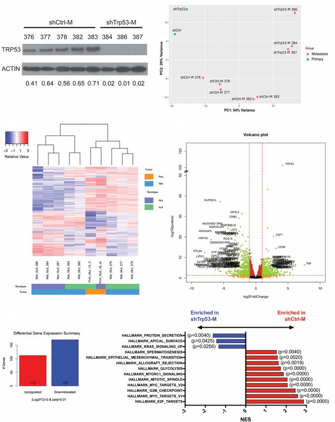
Downloaded from genesdev.cshlp.org on September 11, 2021 - Published by Cold Spring Harbor Laboratory Press

                                                                                                      Mutant p53 and Survivin in metastasis

            A                                                   B

            C
                                                                    D

            E                               F

Figure 2. Depletion of Trp53R172H in ESCC cells alters the transcriptome during lung metastasis. (A) Depletion of Trp53R172H is main-
tained in isolated metastatic cell lines. Relative intensity densitometry of TRP53 results are shown at the bottom. (B) Principle compo-
nent analysis (PCA) demonstrated distinct gene expression profiles depending upon p53 status and primary tumor versus metastatic
tumor status. (C) Heat map of differentially regulated genes upon Trp53R172H depletion in primary and metastatic cells, profiled by
RNA-seq. (D) Volcano plot of differentially regulated genes in metastatic cells. (E) Numbers of significantly (Padj < 0.01, |log2FC| > 2)
changed genes in metastatic Trp53R172H/− cells (n = 5) compared with Trp53−/− cells (n = 3). (F ) GSEA reveals hallmark pathways signifi-
cantly enriched in shCtrl-M and shTrp53-M cells. (M) Metastatic.

Fig. S4A). However, this effect is not observed in week 5               (Fig. 4J; Supplemental Fig. S4B). Taken together, these re-
and week 8 lung metastases formed by Trp53−/− BIRC5                     sults suggest that BIRC5 is a critical direct mediator of
Ctrl, Trp53−/− shBIRC5.1, or Trp53−/− shBIRC5.2 cell lines              Trp53R172H-driven ESCC invasion and lung metastasis.

                                                                                                         GENES & DEVELOPMENT             5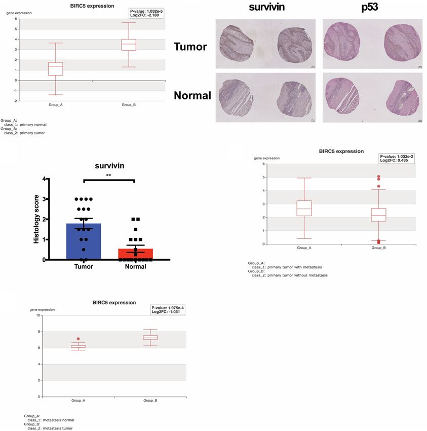
Downloaded from genesdev.cshlp.org on September 11, 2021 - Published by Cold Spring Harbor Laboratory Press
Tang et al.
A B Figure 3. The expression of SURVIVIN de-
pends upon Trp53R172H status. (A) GSEA re-
veals enrichment of the YAP signature in
Trp53R172H/− shCtrl-M cells. (B) RNA-seq
C shows that BIRC5, a YAP target gene, is in-
creased significantly in Trp53R172H/− shCtrl-
M cells. (C ) QRT-PCR of BIRC5 confirms
its up-regulation in Trp53R172H/− shCtrl-M
compared with shTrp53-M cells.
Trp53R172H/− shCtrl-M (n = 5) versus
shTrp53-M (n = 3), P = 0.0287, unpaired t-
test. Error bars represent SEM. (D) Immuno-
histochemical staining of SURVIVIN in 3D
D organoids form by Trp53R172H/− shCtrl and
shTrp53 cells. Trp53R172H/− shCtrl (n = 10
organoids) versus shTrp53 (n = 14 organoids),
P = 0.016, unpaired t-test. Error bars represent
SEM. Scale bar, 100 µm. (E) Immunohisto-
chemical staining of p53 (top) and SURVIVIN
(bottom) in the esophagus epithelium of
Trp53R172H/− andTrp53−/− mice treated with
4NQO. P53 staining: Trp53R172H/− versus
Trp53−/−, n = 8 each group. (∗ ) P = 0.0168, un-
E paired t-test. SURVIVIN staining:
Trp53R172H/− versus Trp53−/−, n = 8 each
group. Error bars represent SEM. Scale bar,
100 µm. (F) Immunohistochemical staining
of SURVIVIN in Trp53R172H/− shCtrl and
shTrp53 in metastatic lung lesions.
Trp53R172H/− shCtrl (n = 15 tumors) versus
shTrp53 (n = 10 tumors), P = 0.0178, unpaired
t-test. Error bars represent SEM. Scale bar,
100 µm.
F
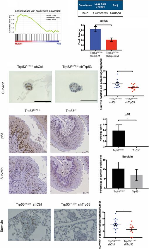
Trp53R172H, but not wild-type Trp53, binds with YAP and that is used to demonstrate direct protein–protein interac-
fosters Survivin expression in ESCC cells tions in a quantitative fashion, representing a substantial
enhancement for what has been reported (Di Agostino
We next tested whether there might be direct interaction et al. 2016). This is based on the premise that YAP has
between Trp53R172H and YAP proteins in murine ESCC been demonstrated to be an inducer of Survivin (Mura-
cells. First, we evaluated the endogenous expression of matsu et al. 2011). Both endogenously and ectopically ex-
YAP and p53 in wild-type p53, Trp53R172H/− shCtrl pressed Trp53R172H associate with YAP in murine ESCC
/shTrp53, and Trp53−/− Ctrl/R172H cell lines (Fig. 5A). cells (Fig. 5B,C). However, in two murine ESCC cell lines
Then, we performed the proximity ligation assay (PLA) expressing wild-type Trp53, binding of endogenous wild-
6 GENES & DEVELOPMENT
Downloaded from genesdev.cshlp.org on September 11, 2021 - Published by Cold Spring Harbor Laboratory Press

                   A                                                       B

                   C                         D                         E                       F

                   G                                                    H

                   I

                   J

Figure 4. Depletion of BIRC5 attenuates Trp53R172H-driven lung metastasis. (A,B) SURVIVIN expression is depleted or decreased signif-
icantly in Trp53R172H/− or Trp53−/− cells with two independent shRNAs (shBIRC5.1 and shBIRC5.2). Relative intensity densitometry of
SURVIVIN results are shown at bottom. (C,D) BIRC5 depletion in Trp53R172H/− or Trp53−/− cells is confirmed by qPCR. Trp53R172H/−
BIRC5 Ctrl versus shBIRC5.1 versus shBIRC5.2, n = 3 per group, (∗∗ ) P = 0.0021 Trp53R172H/− BIRC5 Ctrl versus shBIRC5.1, (∗∗ ) P =
0.0015 Trp53R172H/− BIRC5 Ctrl versus shBIRC5.2, one-way ANOVA. Trp53−/− BIRC5 Ctrl versus shBIRC5.1 versus shBIRC5.2, n = 3
per group. Error bars represent SEM. (E,F) Depletion of BIRC5 significantly decreased cell invasion in Trp53R172H/− but not Trp53−/− cells.
Trp53R172H/− BIRC5 Ctrl versus shBIRC5.1 versus shBIRC5.2, n = 9 per group, (∗∗∗∗ ) P < 0.0001 Trp53R172H/− BIRC5 Ctrl versus shBIRC5.1,
(∗∗∗∗ ) P < 0.0001 Trp53R172H/− BIRC5 Ctrl versus shBIRC5.2, one-way ANOVA. Error bars represent SEM. (G,H) BIRC5 depletion signifi-
cantly reduces subcutaneous tumor growth at early stage. Trp53R172H/− BIRC5 Ctrl (n = 6 flanks) versus shBIRC5.1 (n = 8 flanks) versus
shBIRC5.2 (n = 6 flanks). (∗∗ ) P = 0.0042 day 8; (∗ ) P = 0.00214 day 11; (∗∗∗ ) P = 0.0004 day 13; (∗ ) P = 0.0261 day 17, one-way ANOVA. Error
bars represent SEM. Trp53−/− BIRC5 Ctrl (n = 6 flanks) versus shBIRC5.1 (n = 6 flanks) versus shBIRC5.2 (n = 6 flanks). (∗ ) P = 0.0322 day 10;
(∗∗ ) P = 0.0016 day 12, one-way ANOVA. Error bars represent SEM. (I) Depletion of BIRC5 in Trp53R172H/− ESCC cells significantly reduces
lung metastasis burden number detected at week 8 after tail-vein injection. BIRC5 Ctrl (n = 6) versus shBIRC5.1 (n = 5), P = 0.0057; BIRC5
Ctrl (n = 6) versus shBIRC5.2 (n = 4), P = 0.0009, one-way ANOVA. Error bars represent SEM. Scale bar, 1 mm. (J) Depletion of BIRC5 in
Trp53−/− ESCC cells does not significantly reduce lung metastasis burden number detected at week 8 after tail-vein injection. BIRC5
Ctrl (n = 4) versus shBIRC5.1 (n = 3) versus shBIRC5.2 (n = 4). Error bars represent SEM. Scale bar, 1mm.

                                                                                                               GENES & DEVELOPMENT             7
Downloaded from genesdev.cshlp.org on September 11, 2021 - Published by Cold Spring Harbor Laboratory Press
A
B C
D E
G
F
Figure 5. Trp53R172H, not wild-type p53, binds to YAP and fosters Survivin expression in murine ESCC cells. (A, left) Endogenous expres-
sion level of YAP and p53 in wild type, Trp53R172H/− shCtrl, shTrp53, Trp53−/− Ctrl, and Trp53−/− R172H cells. (Right) Expression levels of
YAP and p53 in LS123 nontarget control (NT), Crispr knockout (Crispr) cell lines, and H1299 empty vector control (control) and p53R175H
(R175H) ectopic expressed cell lines. (B) Proximity ligation assay (PLA) for YAP and Trp53R172H. Scale bar, 50 µm. (C) Quantification of the
average number of PLA signals shows significantly high levels of signals in Trp53R172H/− shCtrl cells (vs. shTrp53 cells, n > 100 cells per
group, P < 0.0001, unpaired t-test, error bars represent SEM) and Trp53−/− R172H cells (vs. Ctrl cells, n > 100 cells per group, P < 0.0001,
unpaired t-test, error bars represent SEM). (D) Immunoprecipitation-Western blot analysis of Trp53R172H-HA with YAP-DDK in 293 FT
cells. (E) Chromatin immunoprecipitation (ChIP) with YAP antibody in WT1, Trp53R172H/− shCtrl, and shTrp53 cells demonstrates in-
creased level of YAP on the BIRC5 promoter in the presence of Trp53 R172H. shCtrl versus WT1 cells, n = 3 replicates per group, P =
0.0052, unpaired t-test; shCtrl versus shTrp53 cells, n = 3 replicates per group, P = 0.0107, unpaired t-test. Error bars represent SEM. (F)
Proximity ligation assay for YAP and p53R175H. Scale bar, 20 µm. (G) Quantification of the average number of PLA signals shows signifi-
cantly high levels of signals in LS123 NT cells (vs. Crispr cells, n > 100 cells per group, P < 0.0001, unpaired t-test, error bars represent SEM)
and H1299 R175H cells (vs. control cells, n > 100 cells per group, P < 0.0001, unpaired t-test, error bars represent SEM).
8 GENES & DEVELOPMENTDownloaded from genesdev.cshlp.org on September 11, 2021 - Published by Cold Spring Harbor Laboratory Press
Mutant p53 and Survivin in metastasis
type Trp53 with YAP is not detected (Fig. 5B). The binding the oncogenic role of a hot spot TP53 mutation,
of YAP and Trp53R172H is confirmed further by a coimmu- p53R175H, in invasion and lung metastasis. Through gene
noprecipitation (co-IP) assay (Fig. 5D). These results indi- expression profiling of ESCC lung metastatic cells and pri-
cate the interaction with YAP is specific to Trp53R172H mary tumor cells, we nominate BIRC5, an antiapoptotic
and suggests this specific YAP-Trp53R172H protein–pro- gene, as a novel effector of p53R175H-driven lung metasta-
tein interaction fosters Survivin expression. To test this sis. We demonstrated for the first time in both human and
hypothesis, we performed chromatin immunoprecipita- mouse cell lines that BIRC5 expression is dependent
tion (ChIP) analysis in wild-type p53, Trp53R172H/− shCtrl, directly upon p53R175H, indicating that BIRC5 may be a
and shTrp53 cell lines. We verified binding of YAP to the novel mutant p53 target. We demonstrated that
BIRC5 promoter in Trp53R172H/− shCtrl, but not in wild- p53R175H, not wild-type TRP53, interacts directly with
type p53 and shTrp53 cell lines (Fig. 5E). In addition, we YAP, and this interaction induces BIRC5 gene expression.
used the human colorectal adenocarcinoma cell line Survivin is also up-regulated in human ESCC, as well as
LS123, which expresses p53R175H, and used the CRISPR/ metastatic PDAC and metastatic CRC when compared
Cas9 system to deplete endogenous p53R175H. As an addi- with their matched primary tumors, suggesting a possible
tional approach, we used a p53−/− non-small cell lung car- basis for metastasis across common GI cancers.
cinoma cell line, H1299, with ectopic expression of Mutant p53 has been demonstrated to promote tumor
p53R175H (Fig. 5A; Basu et al. 2018). We confirmed by cell motility, invasion, and metastasis through gain-of-
PLA that YAP and p53R175H binding is also detected in function mechanisms (Tang et al. 2020). For example, in
these two human cancer cell lines (Fig. 5F,G). Taken to- vivo evidence has revealed that Trp53R172H/+ mice
gether, these results suggest that mutant p53 might en- develop metastatic osteosarcomas and carcinomas. How-
able YAP to bind the BIRC5 promoter, thereby inducing ever, Trp53+/− mice do not harbor metastatic tumors
Survivin expression. (Lang et al. 2004). In different types of cancers, mutant
p53 modulates certain pathways; for example, up-regula-
tion of PDGFRβ and enhancement of mitochondrial func-
High Survivin expression is detected in human ESCC and tion (Weissmueller et al. 2014; Basu et al. 2018). In
metastases in other GI cancers addition, mutant p53 has been reported to promote lung
Analysis of TCGA revealed that Survivin expression is metastasis through regulating glycoprotein folding
significantly elevated in human ESCC compared with ad- (Vogiatzi et al. 2016). Furthermore, one recent study dem-
jacent normal tissues (P < 0.0001) (Fig. 6A). In addition, we onstrated that tumor cells harboring mutant p53 can es-
examined Survivin expression in human ESCC tissue mi- tablish proinvasive niches by secreting exosomes,
croarrays (TMAs). We observed that Survivin expression is indicating the oncogenic role of mutant p53 in altering
elevated significantly in ESCC compared with adjacent the tumor microenvironment (Cooks et al. 2018). Here,
normal tissues (P = 0.0014) (Fig. 6B,C). Next, we examined we provided the first evidence that p53R175H regulates
the correlation of Survivin expression level with p53 nu- the YAP-BIRC5 axis to affect lung metastasis. Further in-
clear accumulation in ESCC. We found that high Survivin vestigation is required to determine whether the regula-
expression is associated significantly with p53 nuclear ac- tion of the YAP-BIRC5 axis by mutant p53 affects lung
cumulation in ESCC (P = 0.027). Because TP53 mutations metastasis in other GI cancers.
have been shown to cause aggregation and accumulation In this study, we identified Survivin, encoded by BIRC5,
of mutant p53 protein in cells (Ano Bom et al. 2012; Silva as a novel downstream effector of mutant p53 to promote
et al. 2018), our observation indicates that up-regulation of lung metastasis. We propose a model whereby p53R175H
Survivin may be correlated with TP53 mutations in ESCC interacts with YAP and regulates survivin expression
patient tumor tissues. Finally, we investigated the as- through binding of YAP to the BIRC5 promoter, which ul-
sociation of Survivin with metastases in other gastrointes- timately promotes lung metastasis. Survivin, encoded by
tinal (GI) cancers. In pancreatic ductal adenocarcinoma BIRC5, is a key regulator of mitosis and programmed cell
(PDAC), Survivin expression is up-regulated significantly death. Survivin is expressed highly in transformed cells
in primary PDAC tumors with metastases compared with and cancers, such as ESCC, lung, breast, and pancreatic
primary PDAC tumors without metastases (P = 0.01) (Fig. cancers (Mita et al. 2008). Several studies have revealed
6D). Furthermore, in colorectal cancer (CRC) lung metas- mechanisms underlying Survivin up-regulation, includ-
tases, Survivin RNA expression is increased significantly ing gene amplification, exon demethylation, promoter ac-
compared with adjacent normal tissue (P = 0.0002) (Fig. tivation, and phosphatidylinositol-3-kinase (PI3K) and
6E). Taken together, these results underscore the role of MAPK signaling activation (Mita et al. 2008). Survivin
Survivin in ESCC tumorigenesis and indicate its potential has been suggested as a transcriptional target of YAP
contribution to metastasis of other GI cancers, such as (Muramatsu et al. 2011). YAP overexpression is detected
PDAC and colorectal cancer. frequently in cells and primary tumors (Muramatsu
et al. 2011). In breast cancer cell lines, YAP and mutant
p53 cooperate to up-regulate proproliferative genes such
Discussion as cyclin A, cyclin B, and CDK1 (Di Agostino et al.
2016). Survivin enhances tumor angiogenesis and chemo-
TP53 mutation is the predominant genetic alteration in resistance (Mita et al. 2008). Previous studies demonstrat-
ESCC (Song et al. 2014). In this study, we demonstrated ed the role of Survivin in tumor cell invasion (Mehrotra
GENES & DEVELOPMENT 9Downloaded from genesdev.cshlp.org on September 11, 2021 - Published by Cold Spring Harbor Laboratory Press
Tang et al.
A B
C D
E
Figure 6. High survivin expression is detected in human ESCC and metastases and in other GI cancers. (A) Analysis of TCGA-ESCA da-
tabase (n = 143) revealed significant increase of survivin expression in primary esophageal carcinoma tissue compared with normal tissue.
P < 0.0001. (B) IHC staining of survivin and p53 in tissue microarrays (TMAs) of paired ESCC primary tumor and normal tissue. Scale bar,
100 µm. (C ) Increased level of survivin is detected in ESCC primary tumor compared with paired normal tissue. n = 17 pairs, P = 0.0014,
Wilcoxon matched-pairs signed rank test. Scoring standards: (0.5) 50% positive cells positive nuclei. Error bars represent SEM. Increased expression
of survivin is correlated with p53 nuclear accumulation in ESCC. n = 59, P = 0.027, Spearman’s rank correlation test. (D) Analysis of TCGA
pancreatic ductal adenocarcinoma (PDAC) data set (TCGA-PAAD, n = 185) showed that survivin expression is significantly elevated in
PDAC tumors with metastasis. P = 0.01. (E) Analysis of RNA-seq (GSE68468, n = 27) in colorectal cancers reveals increased survivin levels
in lung metastases compared with adjacent normal tissues. P = 0.0002.
et al. 2010; Rivadeneira et al. 2015). Given the important geting Survivin in advanced ESCC may be a novel thera-
roles of Survivin in cancers, therapeutic strategies have peutic approach.
been designed to target Survivin. These strategies include In summary, we have demonstrated an oncogenic role
RNAi, antisense oligonucleotides, small molecule inhibi- of p53R175H in promoting invasion and lung metastasis
tors, and developing a cancer vaccine (Mita et al. 2008; through up-regulation of Survivin in ESCC. This study
Santarelli et al. 2018). Notably, several Survivin peptide also provides a prism through which to view the molecu-
vaccine therapies are currently in clinical trials lar basis of p53R175H-driven lung metastasis and offers a
(NCT03349450, NCT03762291, and NCT01416038). platform to identify novel therapeutic targets for metasta-
Given the role of Survivin in ESCC lung metastasis, tar- ses harboring mutant p53.
10 GENES & DEVELOPMENTDownloaded from genesdev.cshlp.org on September 11, 2021 - Published by Cold Spring Harbor Laboratory Press
Mutant p53 and Survivin in metastasis
Materials and methods upper surface with a cotton swab, and the remaining cells were
fixed in 70% ethanol for 10 min at room temperature. Next, the
Cell culture invading cells were stained with 2% Crystal-Violet for 10 min
at room temperature. Finally, the invading cell number was
Murine ESCC cells were cultured in keratinocyte serum-free me-
counted using a Keyence bright-field microscope at 10× with
dium (KSFM; Gibco) without CaCl2, supplemented with 0.018
four fields per chamber. Invasion was calculated by the number
mM CaCl2, 50 µg/mL bovine pituitary extract (Gibco), 5 ng/mL
of invaded cells per field. For 3D organotypic culture assays, inva-
human recombinant EGF (Gibco), and 1% penicillin/streptomy-
sion was determined by quantitating the area of invasive regions
cin (Gibco). Cells were cultured at 37°C in a 5% CO2 humidified
divided by the total epithelial length.
incubator. To achieve Trp53 knockdown, cells were infected with
mouse lentiviral shRNA against Trp53 (pMSCV puro, Clontech)
or nontargeting control shRNA. Cells were selected in 2 µg/mL
puromycin 48 h postinfection. Quantitative PCR, immunofluo- 3D organotypic culture and 3D organoid culture assays
rescence staining, and Western blotting were performed to con-
firm knockdown efficiency. Alternatively, CRISPR/Cas9 The protocol to culture esophageal cells in 3D organotypic cul-
against Trp53 (pLentiCRISPR v2, GenScript) or an empty vector ture was described previously (Kalabis et al. 2012). 3T3 cells
control were used for infection, and puromycin selection and were used to establish the fibroblast layer for mouse organotypic
Trp53 expression validation were performed as described. For cultures. For 3D organoid culture, 3000 cells were resuspended
YAP knockdown, a pool of three to five YAP-specific shRNAs in 50 µL of Matrigel in a 24-well dish followed by 1-h solidifica-
(Santa Cruz Biotechnology sc-38638-SH) or nontargeting control tion to generate the organoids. After organoids solidified, the
shRNA (Santa Cruz Biotechnology sc-108060) were used. For media (DMEM/F12, 1× Glutamax, 1× HEPES, 1× N2 supplement,
BIRC5 knockdown, mouse lentiviral shRNA against BIRC5 1× B27 supplement, 0.1 mM N-acetylcysteine, 50 ng/mL re-
(pLKO.1-CMV-neo, Sigma) or nontargeting control shRNA was combinant EGF, Noggin/R-Spondin conditioned media, 10 μM
used to infect cells. Neomycin selection was performed at a Y27632) was suppled at 500 µL per well and replaced every 2
concentration of 250 µg/mL 48 h postinfection. Knockdown d. Organoids were allowed to grow for 10–14 d. To harvest organo-
validation was performed as described. To ectopically express ids, Matrigel was dissolved by pipetting several times. Recovered
Trp53 R175H in Trp53−/− murine ESCC cells, we cloned organoids were fixed in 4% PFA (Sigma 158127) overnight at 4°C
Trp53 R175H cDNA into the pUltra-Chili lentiviral construct and were embedded in 2% Bacto-Agar:2.5% gelatin.
(Addgene, plasmid 48687). pUltra-chili-Trp53 R175H was then
used to infect cells, followed by RFP flow cytometry sorting for
cell selection and Western blotting to confirm expression level.
Proliferation assay
The human cell line LS123 was purchased from ATCC (ATCC
CCL-255) and was cultured in Eagle’s minimum essential medi- Carboxyfluorescein diacetate succinimidyl ester (CFSE) is a fluo-
um (EMEM; ATCC 30-2003). LS123 cells were infected with rescent dye to measure cell division. Cells were labeled with 5 µM
the human TP53 CRISPR/Cas9 construct (pLentiCRISPR v2) or CFSE (Life Technologies C34554) for 30 min at 37°C. Then, cells
nontargeting control. Puromycin selection and p53 expression were centrifuged at 1000 rpm for 5 min, the supernatant was
validation were performed as described. Human cell lines removed, and cells were seeded in six-well plates in KSFM over-
H1299 control and R175H overexpression were a gift from Dr. night at 37°C or 4°C. On the following day, cells were trypsinized
Maureen Murphy (Basu et al. 2018) and were cultured in RPMI- and fluorescence was measured with BD Accuri C6. FlowJo was
1640 medium (Thermo). The shRNA sequences used were mouse used to analyze results.
Trp53 (CCACTACAAGTACATGTGTAATAG), mouse Birc5
(TRCN0000054613; GAAGAACTAACCGTCAGTGAA), and
mouse Birc5 (TRCN0000054616; CAAAGACTACCCGTCAGT
CAA). The gRNA sequences used were mouse Trp53 (AGTGAA Apoptosis assay
GCCCTCCGAGTGTC), mouse Trp53 (AACAGATCGTCCAT To examine apoptosis, cells were plated in 12-well plates. Apo-
GCAGTG), and human TP53 (CCATTGTTCAATATCGTCCG). ptosis was detected with the APC-AnnexinV and 7-AAD kit
(BioLegend 640930), following the manufacturer’s instructions.
BD Accuri C6 was used to acquire data and results were analyzed
Western blot
with FlowJo.
Primary antibodies p53 (1:2000; Leica NCL-L-p53-CM5p), YAP
(1:1000; Cell Signaling Technology 14074S), and β-actin
(1:10,000; Sigma A5316), were incubated overnight at 4°C, and
horseradish peroxidase (HRP)-conjugated secondary antibodies Animal models
(anti-mouse: 1:10,000 [GE Healthcare LNA931V/AH]; and anti- All animal studies were approved by our Institutional Animal
rabbit: 1:10,000 [GE Healthcare LNA934V/AH]) were incubated Care and Use Committee. To establish the lung metastasis mod-
for 1 h at room temperature. Western blots were imaged using el, 5-wk-old female athymic nude mice were purchased (Taconic).
ECL prime (Sigma GERPN 2232). At the age of 7 wk, 106 cells were resuspended in 100 µL of cold
Dulbecco’s phosphate-buffered saline (DPBS) and injected into
the distal end of the lateral tail vein with a 30-gauge 0.5-in needle.
Invasion assay
After injection, mice were monitored daily for signs of pain or dis-
Corning BioCoat Matrigel invasion chambers were used for inva- comfort and weight loss. To detect the formation of lung meta-
sion assays. Chambers were rehydrated in serum-free KSFM for 2 static lesions, mice were euthanized at 2, 5, and 8 wk by CO2
h at 37°C. Cells (105) resuspended in 500 µL of serum-free KSFM and cervical dislocation. To identify lung metastases, the mouse
were seeded in the upper chamber, and 500 µL regular KSFM was lungs were flushed with 20 mL of Heparin-PBS to deplete red
added to the bottom of the well. Cells were incubated at 37°C for blood cells and were imaged with an Olympus IX71 fluorescent
24 h. After incubation, noninvading cells were removed from the microscope at 4×.
GENES & DEVELOPMENT 11Downloaded from genesdev.cshlp.org on September 11, 2021 - Published by Cold Spring Harbor Laboratory Press
Tang et al.
Lung metastatic cell isolation (1:400; Cell Signaling Technology 2808S), p53 (1:200; Abcam
ab1101).
Prior to cell isolation, all tools and solutions were autoclaved or
For immunofluorescence, cells were cultured on cover slips,
filter-sterilized. Mouse lungs were harvested and placed in cold
rinsed with PBS, and fixed with 4% PFA for 15 min at room
DPBS and were physically dissociated with scissors. To digest,
temperature. Permeabilization was performed with 100% meth-
dissociated tissues were mixed with 50 mL of 1 mg/mL solution
anol for 10 min at −20°C. Cells were blocked with blocking solu-
of collagenase type V (Sigma Aldrich 9263), dissolved in pre-
tion (PBS, 1% BSA, 0.3% Triton X, 5% donkey serum) for 1 h at
warmed KSFM, and the mixture was stirred with a magnet for
room temperature. Cells were then incubated with primary
20 min at 37°C. After digestion, the mixture was centrifuged
antibodies overnight at 4°C, washed, incubated with secondary
twice at 1000 rpm for 5 min., resuspended in KSFM, and plated
antibody (1:600; JacksonImmunoresearch) for 1 h at room temper-
on a 10-cm cell culture dish. Cultured cells were washed with
ature in the dark, stained with DAPI (Invitrogen D1306) for 3 min,
DPBS and replaced with new media every other day. After expan-
and mounted (KPL 71-00-16). Fluorescent imaging was performed
sion, cells were trypsinized and flow cytometry-sorted for YFP,
with a Nikon E600 fluorescent microscope. The primary anti-
followed by Western blotting to confirm p53 expression.
body was against p53 (1:200; Leica NCL-L-p53-CM5p).
RNA isolation and qRT-PCR
Proximity ligation assay (PLA)
Total RNA was extracted with the GeneJET RNA purification kit
(Thermo Fisher K0731) or RNAqueous kit (Invitrogen AM1912). A Duolink In Situ Red Starter kit (Sigma DUO92101) was used to
cDNA was synthesized with a High-Capacity cDNA reverse perform PLA assays, according to the manufacturer’s instruc-
Transcription kit (Thermo Fisher 4374966). qRT-PCR was per- tions. ImageJ was used to quantify PLA signals. Primary antibod-
formed with the StepOnePlus real-time PCR system (Applied Bio- ies included the following: p53(1C12) (1:2000; Cell Signaling
systems) using Power SYBR Green PCR Master Mix (Applied Technology 2524), YAP (1:100; Cell Signaling Technology
Biosystems 4367659). Primer sequences used were as follows: 14074).
Trp53 (forward: CCCCGCAAAAGAAAAAACCAC; reverse:
AGCTGGAGTGAGCCCTGC), Birc5 (forward: GAACCCGAT
GACAACCCGAT; reverse: TGGTCTCCTTTGCAATTTTGTT
Chromatin immunoprecipitation (ChIP)
CT), and Actin (forward: ACCCAGATCATGTTTGAGACC; re-
verse: AGAGCATAGCCCTCGTAGAT). ChIP was performed as previously described (Chiarella et al. 2018)
with ESCC cells using 10 million cells per replicate and experi-
mental condition. YAP (Novus Biologicals NB110-58358) and
RNA-seq IgG (Cell Signaling 5415S) antibodies were used. Following
ChIP, qPCR was performed with SYBR Green (Thermo Scientific
RNA was extracted from early passage cells as described above.
4367659). Enrichment levels were normalized to the background
Libraries were prepared with Illumina TruSeq stranded mRNA
levels detected with IgG. For each experimental type, three bio-
sample preparation kits from 500 ng of purified total RNA accord-
logical replicates were analyzed. The following Survivin primers
ing to the manufacturer’s protocol. The finished dsDNA libraries
(5′ –3′ ) were used: (+)TGGACTGGTGAGGTTTAGGA and (−)
were quantified by Qubit fluorometer, Agilent TapeStation 2200,
ACATAGGCAGCTGGAACAAG.
and qRT-PCR using the Kapa Biosystems library quantification
kit, according to the manufacturer’s instructions. Uniquely in-
dexed libraries were pooled in equimolar ratios and sequenced
on an Illumina NextSeq500 with single-end 75-bp reads at the Human tissues and databases
Dana-Farber Cancer Institute Molecular Biology Core Facility.
Well-annotated tissue microarrays (TMAs) representing paired
Sequenced reads were aligned to the UCSC mm10 reference ge-
primary ESCC tumors and adjacent normal mucosa from IRB-ap-
nome assembly, and gene counts were quantified using STAR
proved and de-identified therapy-naïve patients (n = 39) have been
(v2.5.1b). Differential gene expression testing was performed by
previously described (Liu et al. 2013; Natsuizaka et al. 2017).
DESeq2 (v1.10.1) and normalized read counts (FPKM) were calcu-
TCGA-ESCA, TCGA-PAAD, and GEO68468 were accessed and
lated using cufflinks (v2.2.1).
analyzed through the Human Cancer Metastasis Database
RNA-seq data were deposited at the NCBI Gene Expression
(https://hcmdb.i-sanger.com).
Omnibus (https://www.ncbi.nlm.nih.gov/geo). All data sets are
available on request.
Statistical analysis
Immunohistochemistry and immunofluorescence
Sample size and replicate number have been described in the text
Formalin-fixed, paraffin-embedded tissue was sectioned, and an- and figure legends. All data were presented as mean ± standard er-
tigen retrieval was performed through pressure cooking in citric ror of the mean (SEM). Statistical analysis including paired Stu-
acid buffer (pH 6). Endogenous peroxidases were quenched by dent’s t-test, unpaired Student’s t-test, and ANOVA was
3% peroxide, followed by avidin (Sigma Aldrich A9275), biotin performed by GraphPad Prism 7 (GraphPad Software). P < 0.05
(Sigma Aldrich B4501) blocking and protein blocking buffer was reported as statistically significant.
(Thermo Fisher) at room temperature. Primary antibodies were
incubated overnight at 4°C, and biotinylated secondary antibod-
ies (1:600; Vector Laboratories) for 30 min at 37°C. Sections
Data availability
were then incubated with the ABC reagent (Vector Laboratories
PK-6100) for 30 min at 37°C, and slides were treated with DAB RNA-seq data from this article have been deposited at the NCBI
substrate (Vector Laboratories SK4100) and counterstained with Gene Expression Omnibus (https://www.ncbi.nlm.nih.gov/geo/
hematoxylin. The primary antibody was against Survivin query/acc.cgi?acc=GSE143696).
12 GENES & DEVELOPMENTDownloaded from genesdev.cshlp.org on September 11, 2021 - Published by Cold Spring Harbor Laboratory Press
Mutant p53 and Survivin in metastasis
Competing interest statement YAP enhances the pro-proliferative transcriptional activity
of mutant p53 proteins. EMBO Rep 17: 188–201. doi:10
The authors declare no competing interests. .15252/embr.201540488
Dong P, Xu Z, Jia N, Li D, Feng Y. 2009. Elevated expression of
p53 gain-of-function mutation R175H in endometrial cancer
Acknowledgments cells can increase the invasive phenotypes by activation of
the EGFR/PI3K/AKT pathway. Mol Cancer 8: 103. doi:10
We thank the Flow Cytometry and Cell Sorting Core at the Uni-
.1186/1476-4598-8-103
versity of Pennsylvania Perelman School of Medicine; the Imag-
Dongre A, Weinberg RA. 2019. New insights into the mecha-
ing Facility at the Wistar Institute; the Molecular Pathology,
nisms of epithelial–mesenchymal transition and implications
Genetically Engineered Mouse Model, Flow Cytometry, and Bio-
for cancer. Nat Rev Mol Cell Biol 20: 69–84. doi:10.1038/
statistics Shared Resources at the Herbert Irving Comprehensive
s41580-018-0080-4
Cancer Center (HICCC) of Columbia University Irving Medical
Dotto GP, Rustgi AK. 2016. Squamous cell cancers: a unified per-
Center; and the Molecular Biology Core Facility (MBCF) at the
Dana-Farber Cancer institute (DFCI). This work was funded by spective on biology and genetics. Cancer Cell 29: 622–637.
National Institutes of Health grant P01-CA098101 (A.K.R.) and doi:10.1016/j.ccell.2016.04.004
the American Cancer Society Research Professorship (A.K.R.). Freed-Pastor WA, Prives C. 2012. Mutant p53: one name, many
Author contributions: Q.T., G.E., A.M.C., M.C., and J.L. per- proteins. Genes Dev 26: 1268–1286. doi:10.1101/gad.190678
formed experiments and data analysis. T.Y., Z.S., T.K., and J.L. .112
assisted with experiments. A.J.K.-S. performed pathology scoring. Furth N, Aylon Y, Oren M. 2018. P53 shades of Hippo. Cell Death
J.R.P., M.I., S.P., and J.H. performed RNA-seq analysis. S.N. col- Differ 25: 81–92. doi:10.1038/cdd.2017.163
lected TMA tissues. W.G., B.Z.S., K.-K.W., J.A.D., A.J.B., Huang S, Zhou H. 2012. Role of mTOR signaling in tumor cell
M.E.M., and H.N. designed the experiments and reviewed the motility, invasion and metastasis. Curr Protein Pept Sci 12:
manuscript. A.K.R. designed the experiments, performed data 30–42. doi:10.2174/138920311795659407
analysis, and wrote the manuscript. Q.T. also wrote the Kalabis J, Wong GS, Vega ME, Natsuizaka M, Robertson ES, Her-
manuscript. lyn M, Nakagawa H, Rustgi AK. 2012. Isolation and character-
ization of mouse and human esophageal epithelial cells in 3D
organotypic culture. Nat Protoc 7: 235–246. doi:10.1038/nprot
References .2011.437
Lambert AW, Pattabiraman DR, Weinberg RA. 2017. Emerging
Ano Bom APD, Rangel LP, Costa DCF, De Oliveira GAP, Sanches
biological principles of metastasis. Cell 168: 670–691. doi:10
D, Braga CA, Gava LM, Ramos CHI, Cepeda AOT, Stumbo
.1016/j.cell.2016.11.037
AC, et al. 2012. Mutant p53 aggregates into prion-like amyloid
Lang GA, Iwakuma T, Suh YA, Liu G, Rao VA, Parant JM, Valen-
oligomers and fibrils: implications for cancer. J Biol Chem
tin-Vega YA, Terzian T, Caldwell LC, Strong LC, et al. 2004.
287: 28152–28162. doi:10.1074/jbc.M112.340638
Gain of function of a p53 hot spot mutation in a mouse model
Basu S, Gnanapradeepan K, Barnoud T, Kung C, Tavecchio M,
of Li-Fraumeni syndrome. Cell 119: 861–872. doi:10.1016/j
Scott J, Watters A, Chen Q, Kossenkov AV, Murphy ME.
.cell.2004.11.006
2018. Mutant p53 controls tumor metabolism and metastasis
Lee Ck, Jeong Sh, Jang C, Bae H, Kim YH, Park I, Kim SK, Koh GY.
by regulating PGC-1α. Genes Dev 32: 230–243. doi:10.1101/
2019. Tumor metastasis to lymph nodes requires YAP-depen-
gad.309062.117
dent metabolic adaptation. Science 363: 644–649. doi:10
Bossi G, Lapi E, Strano S, Rinaldo C, Blandino G, Sacchi A. 2006.
Mutant p53 gain of function: reduction of tumor malignancy .1126/science.aav0173
of human cancer cell lines through abrogation of mutant Liu K, Jiang M, Lu Y, Chen H, Sun J, Wu S, Ku WY, Nakagawa H,
p53 expression. Oncogene 25: 304–309. doi:10.1038/sj.onc Kita Y, Natsugoe S, et al. 2013. Sox2 cooperates with inflam-
.1209026 mation-mediated stat3 activation in the malignant transfor-
Campbell JD, Yau C, Bowlby R, Liu Y, Brennan K, Fan H, Taylor mation of foregut basal progenitor cells. Cell Stem Cell 12:
AM, Wang C, Walter V, Akbani R, et al. 2018. Genomic, path- 304–315. doi:10.1016/j.stem.2013.01.007
way network, and immunologic features distinguishing squa- Mehrotra S, Languino LR, Raskett CM, Mercurio AM, Dohi T,
mous carcinomas. Cell Rep 23: 194–212.e6. doi:10.1016/j Altieri DC. 2010. IAP regulation of metastasis. Cancer Cell
.celrep.2018.03.063 17: 53–64. doi:10.1016/j.ccr.2009.11.021
Chiarella AM, Quimby AL, Mehrab-Mohseni M, Velasco B, Mello SS, Valente LJ, Raj N, Seoane JA, Flowers BM, McClendon
Kasoji SK, Davis IJ, Dayton PA, Hathaway NA, Pattenden J, Bieging-Rolett KT, Lee J, Ivanochko D, Kozak MM, et al.
SG. 2018. Cavitation enhancement increases the efficiency 2017. A p53 super-tumor suppressor reveals a tumor suppres-
and consistency of chromatin fragmentation from fixed cells sive p53-Ptpn14-Yap axis in pancreatic cancer. Cancer Cell
for downstream quantitative applications. Biochemistry 57: 32: 460–473.e6. doi:10.1016/j.ccell.2017.09.007
2756–2761. doi:10.1021/acs.biochem.8b00075 Mita AC, Mita MM, Nawrocki ST, Giles FJ. 2008. Survivin: key
Cooks T, Pateras IS, Jenkins LM, Patel KM, Robles AI, Forshew T, regulator of mitosis and apoptosis and novel target for cancer
Appella E, Gorgoulis VG, Harris CC, Morris J. 2018. Mutant therapeutics. Clin Cancer Res 14: 5000–5005. doi:10.1158/
p53 cancers reprogram macrophages to tumor supporting 1078-0432.CCR-08-0746
macrophages via exosomal miR-1246. Nat Commun 9: 771. Muller PAJ, Vousden KH. 2014. Perspective mutant p53 in can-
doi:10.1038/s41467-018-03224-w cer: new functions and therapeutic opportunities. Cancer
Cui M, Li Z. 2018. Downregulation of YAP inhibits proliferation Cell 25: 304–317. doi:10.1016/j.ccr.2014.01.021
and induces apoptosis in Eca‐109 cells. Exp Ther Med 15: Muller PAJ, Caswell PT, Doyle B, Iwanicki MP, Tan EH, Karim S,
1048–1052. Lukashchuk N, Gillespie DA, Ludwig RL, Gosselin P, et al.
Di Agostino S, Sorrentino G, Ingallina E, Valenti F, Ferraiuolo M, 2009. Mutant p53 drives invasion by promoting integrin recy-
Bicciato S, Piazza S, Strano S, Del Sal G, Blandino G. 2016. cling. Cell 139: 1327–1341. doi:10.1016/j.cell.2009.11.026
GENES & DEVELOPMENT 13You can also read