Nanomedicines to Deliver mRNA: State of the Art and Future Perspectives - MDPI
←
→
Page content transcription
If your browser does not render page correctly, please read the page content below
nanomaterials

Review
Nanomedicines to Deliver mRNA: State of the Art
and Future Perspectives
Itziar Gómez-Aguado , Julen Rodríguez-Castejón , Mónica Vicente-Pascual ,
Alicia Rodríguez-Gascón , María Ángeles Solinís * and Ana del Pozo-Rodríguez *
 Pharmacokinetics, Nanotechnology & Gene Therapy Group (PharmaNanoGene), Faculty of Pharmacy,
 Centro de investigación Lascaray ikergunea, University of the Basque Country UPV/EHU,
 Paseo de la Universidad 7, 01006 Vitoria-Gasteiz, Spain; itziar.gomez@ehu.eus (I.G.-A.);
 julen.rodriguez@ehu.eus (J.R.-C.); monica.vicente@ehu.eus (M.V.-P.); alicia.rodriguez@ehu.eus (A.R.-G.)
 * Correspondence: marian.solinis@ehu.eus (M.Á.S.); ana.delpozo@ehu.eus (A.d.P.-R.);
   Tel.: +34-945013469 (M.Á.S.); +34-945014498 (A.d.P.-R.)

 Received: 31 January 2020; Accepted: 16 February 2020; Published: 20 February 2020

 Abstract: The use of messenger RNA (mRNA) in gene therapy is increasing in recent years, due to its
 unique features compared to plasmid DNA: Transient expression, no need to enter into the nucleus
 and no risk of insertional mutagenesis. Nevertheless, the clinical application of mRNA as a therapeutic
 tool is limited by its instability and ability to activate immune responses; hence, mRNA chemical
 modifications together with the design of suitable vehicles result essential. This manuscript includes
 a revision of the strategies employed to enhance in vitro transcribed (IVT) mRNA functionality and
 efficacy, including the optimization of its stability and translational efficiency, as well as the regulation
 of its immunostimulatory properties. An overview of the nanosystems designed to protect the mRNA
 and to overcome the intra and extracellular barriers for successful delivery is also included. Finally,
 the present and future applications of mRNA nanomedicines for immunization against infectious
 diseases and cancer, protein replacement, gene editing, and regenerative medicine are highlighted.

 Keywords: in vitro transcribed messenger RNA (IVT mRNA); gene therapy; nanomedicine;
 immunotherapy; infectious disease vaccines; cancer immunotherapy; Chimeric Antigen Receptor
 (CAR) T cells; dendritic cells; protein replacement; gene editing

1. Introduction
      According to the European Medicines Agency (EMA), a gene therapy medicinal product generally
consists of a vector or delivery formulation/system containing a genetic construct engineered to express
a specific transgene (‘therapeutic sequence’) for the regulation, repair, replacement, addition or deletion
of a genetic sequence [1]. Nevertheless, a broader perspective is usually accepted from a scientific point
of view, and the concept of gene therapy includes the therapeutic application of products containing
any nucleic acid.
      Gene therapy entered clinical trials in the early 1990s. Up to date around 17 nucleic acid-based
products have been approved worldwide, and almost 2700 gene therapy-based clinical trials have
been completed, are ongoing or have been approved for a broad range of applications. It is expected
that nucleic acid-based products will have a substantial impact on the biopharmaceutical market in
the near future [2]. Gene therapy has been clinically implemented primarily through two different
approaches: Ex vivo or in vivo. In ex vivo therapy, cells are harvested from the patient or a donor,
in vitro modified with the therapeutic nucleic acid and finally, re-infused into the patient. In vivo gene
therapy is applied by direct administration of the vector containing the nucleic acid into the patient [3].

Nanomaterials 2020, 10, 364; doi:10.3390/nano10020364                       www.mdpi.com/journal/nanomaterials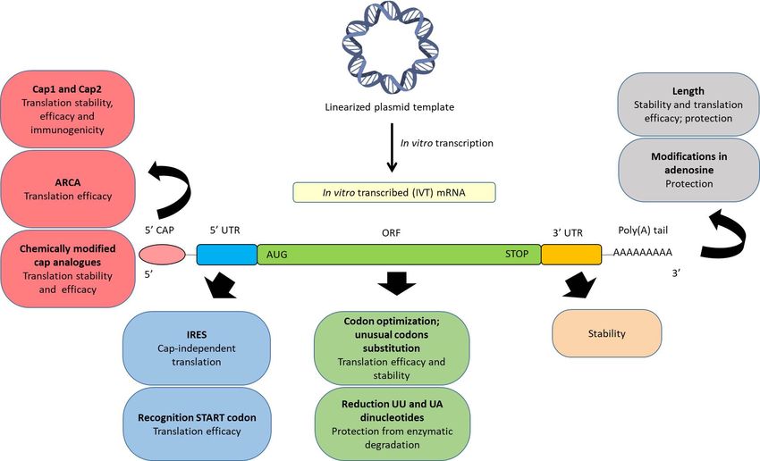
Nanomaterials 2020, 10, 364                                                                                               2 of 43

therapy    is applied
 Nanomaterials            by direct administration of the vector containing the nucleic acid into the patient
                2020, 10, 364                                                                                               2 of 42
[3].
      Depending on the final objective, gene therapy can be applied for gene augmentation, gene
       Depending on the final objective, gene therapy can be applied for gene augmentation, gene
silencing, or gene editing [4]. Diverse nucleic acids are being used to address the development of
 silencing, or gene editing [4]. Diverse nucleic acids are being used to address the development of these
these new medicinal products. DNA and messenger RNA (mRNA) induce protein expression,
 new medicinal products. DNA and messenger RNA (mRNA) induce protein expression, whereas,
whereas, small interfering RNA (siRNA), microRNA, oligonucleotides or aptamers provide gene
 small interfering RNA (siRNA), microRNA, oligonucleotides or aptamers provide gene silencing [2].
silencing [2]. Molecular scissor and gene editing approaches, such as zinc finger nucleases (ZFNs),
 Molecular scissor and gene editing approaches, such as zinc finger nucleases (ZFNs), transcription
transcription activator-like effector nucleases (TALENs), and clustered regularly interspaced short
 activator-like effector nucleases (TALENs), and clustered regularly interspaced short palindromic
palindromic repeats (CRISPR)-associated nuclease Cas9 (CRISPR/Cas9) are also being developed.
 repeats (CRISPR)-associated nuclease Cas9 (CRISPR/Cas9) are also being developed.
      The specific features of synthetic mRNA make it a promising alternative to DNA based products.
       The specific features of synthetic mRNA make it a promising alternative to DNA based products.
Firstly, mRNA does not need the machinery of the nucleus to be functional, as DNA therapies do [5–
 Firstly, mRNA does not need the machinery of the nucleus to be functional, as DNA therapies do [5–8].
8]. Therefore, once mRNA reaches the cytoplasm, translation of the encoding protein begins, being
 Therefore, once mRNA reaches the cytoplasm, translation of the encoding protein begins, being
effective in both, mitotic and non-mitotic cells [9–11]. Secondly, mRNA has a better safety profile,
 effective in both, mitotic and non-mitotic cells [9–11]. Secondly, mRNA has a better safety profile,
because it does not integrate into the host genome; hence, the risk of carcinogenesis and mutagenesis
 because it does not integrate into the host genome; hence, the risk of carcinogenesis and mutagenesis
usually associated with DNA is notably reduced [5–8,11]. In addition, it works without encoding
 usually associated with DNA is notably reduced [5–8,11]. In addition, it works without encoding
additional genes, (i.e., those related to antibiotic resistance) [9]. Thirdly, the synthesis of the encoded
 additional genes, (i.e., those related to antibiotic resistance) [9]. Thirdly, the synthesis of the encoded
protein   is fast, and its expression is temporary [6,9]. The onset of expression is usually detected within
 protein is fast, and its expression is temporary [6,9]. The onset of expression is usually detected within
1h after transfection with a peak in expression 5–7 h later [12]. Finally, the production of in vitro
 1h after transfection
transcribed     mRNA (IVT     with  a peak
                                 mRNA)         in expression
                                           is easier  than the 5–7    h later [12].ofFinally,
                                                                 manufacturing          DNA, and  the itproduction     of in vitro
                                                                                                          can be standardized,
 transcribed     mRNA       (IVT  mRNA)
maintaining its reproducibility [7].        is  easier than  the  manufacturing       of DNA,    and    it can be standardized,
 maintaining
      Nevertheless,its reproducibility
                          the use of IVT[7].mRNA for clinical purposes has been mostly limited by its physical
       Nevertheless,
instability,               the use ofcapacity,
               its immunogenic         IVT mRNA     andfor clinical
                                                         the        purposes
                                                              difficulty         has been
                                                                           in passing        mostly
                                                                                         through     thelimited  bymembrane,
                                                                                                           cellular  its physical
 instability,  its  immunogenic       capacity,   and  the  difficulty  in passing    through
due to the anionic nature of mRNA molecules [8,13,14]. The knowledge of mRNA structure has      the   cellular  membrane,     due
 to the anionic
boosted              nature of mRNA
            modifications        designed  molecules
                                               to improve[8,13,14]. The knowledge
                                                                stability                 of mRNA
                                                                            and translation              structure
                                                                                                   efficacy,    and has
                                                                                                                      to boosted
                                                                                                                          reduce
 modifications       designed    to improve      stability and   translation    efficacy,
immunogenicity; however, optimized IVT mRNA still has to overcome several extracellular andand  to  reduce    immunogenicity;
 however, optimized
intracellular       barriers.IVTAsmRNA      still has
                                     observed          to overcome
                                                   in Figure     1, IVT several
                                                                            mRNA  extracellular
                                                                                       has to avoidand intracellular
                                                                                                            degradation  barriers.
                                                                                                                            from
 As  observed      in  Figure  1, IVT   mRNA      has  to avoid   degradation      from   extracellular
extracellular degradative agents, such as ribonucleases, and interact with the target cell, cross the       degradative    agents,
 such as ribonucleases,
cytoplasmic       membraneand    andinteract
                                      diffusewith    thecytoplasm
                                                 in the   target cell,tocross thethe
                                                                          reach     cytoplasmic
                                                                                       ribosomesmembrane
                                                                                                      [7,15]. Theand    diffuse in
                                                                                                                    formulation
 the cytoplasm        to reach  the  ribosomes     [7,15].  The  formulation     of  IVT   mRNA
of IVT mRNA in suitable nanosystems or vectors is frequently a requirement for surpassing all these  in  suitable  nanosystems
 or vectorsNon-viral
barriers.     is frequently      a requirement
                             vectors,  and morefor      surpassinglipidic
                                                      specifically     all these   barriers.
                                                                               systems,     areNon-viral
                                                                                                 the mostvectors,
                                                                                                               studiedand   more
                                                                                                                          among
 specifically    lipidic   systems,
delivery systems for IVT mRNA.        are the   most  studied    among    delivery    systems   for   IVT   mRNA.

      Figure
       Figure1.1. Intracellular
                   Intracellularbarriers
                                 barriersfor
                                          forin
                                              invitro
                                                vitro transcribed
                                                       transcribed (IVT)
                                                                    (IVT) mRNA
                                                                          mRNA delivery:
                                                                                 delivery: (1)
                                                                                            (1)Interaction
                                                                                                Interactionbetween
                                                                                                            between
      the
       the delivery system and cell membrane, (2) endocytosis, and (3) endosomal escapeand
          delivery    system  and  cell membrane,   (2) endocytosis,  and (3) endosomal escape    andrelease
                                                                                                       releaseof
                                                                                                               ofthe
                                                                                                                  the
      mRNA    to   start the translation process.
       mRNA to start the translation process.
Nanomaterials 2020, 10, 364 3 of 42
The first step for efficient internalization of IVT mRNA is the interaction between the delivery
system and the cell membrane. The attachment to the cell surface may occur through electrostatic
interactions between the system and the membrane surface, which is favoured for those systems
presenting a cationic nature [16]. Cell binding can also be improved by incorporating ligands able to
interact with specific cell surface receptors [17,18] into the vectors.
The main mechanism of cell entry is endocytosis. It comprises a variety of complex processes
that determine the intracellular disposition of the mRNA. The vectors are included in endosomes
by the invagination of the cell membrane. Endosomes mature and fuse with lysosomes, where the
acidic environment and the presence of hydrolytic enzymes can degrade the vector and the nucleic
acid. Therefore, endosomal escape before degradation is considered a bottleneck for successful
mRNA therapy, and, as in the case of cellular internalization, the delivery system plays a crucial
role. The foremost proposed mechanisms of endosomal escape include endosome disruption, active
transport, or fusion of the delivery system with the endosomal membrane [19]. However, Patel et al.
have recently identified that late endosome/lysosome formation is essential for the functional delivery
of exogenously presented mRNA [20]. Therefore, for functional mRNA delivery, an appropriate
biodistribution is necessary, but not enough; understanding cell-type specific endosomal escape
mechanisms should help to design more appropriate strategies to optimize in vivo mRNA efficiency.
The present work reviews the strategies accomplished to optimize the functionality and efficacy
of IVT mRNA. Besides the chemical modifications in its structure, the nanosystems and technological
approaches developed for a successful IVT mRNA delivery will be described. Finally, the potential
applications of mRNA nanomedicines will be discussed: Vaccination against infectious diseases, cancer
immunotherapy, protein replacement, gene editing and regenerative medicine.
2. Structure of Synthetic IVT mRNA and Chemical Modifications
The production of IVT mRNA is usually carried out in cell-free systems, leading to easy
standardization of clinical-grade manufacturing, which can be performed under Good Manufacturing
Practices (GMPs). Fabrication costs of IVT mRNA under GMPs are substantially low as compared to
recombinant proteins produced in eukaryotic cells [21]. It is important to select an efficient purification
method of the IVT mRNA in order to eliminate aberrant (e.g., truncated) mRNA molecules, which are
highly immunogenic contaminants and may lower translation efficiency [22].
Manufacturing of IVT mRNA by a cell-free in vitro transcription system requires a linearized
DNA template which must contain a prokaryotic phage promoter sequence for the T3, T7, or SP6
RNA polymerases, the open reading frame (ORF) encoding the desired protein, the sequences
corresponding to the regulatory untranslated regions (UTRs), and optionally, to a polyadenylated tail
(poly(A) tail). [9,14,23]. When the poly(A) tail is not encoded directly in the DNA template, it can be
added post-transcriptionally by enzymatic reactions with recombinant poly(A)polymerase of E. coli
(E-PAP) [11,24]. Since the final IVT mRNA must be structurally similar to natural mRNA processed in
the cytoplasm of eukaryotic cells, it also needs to be capped in 5’.
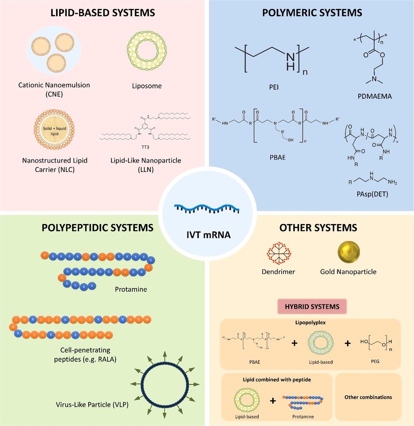
A synthetic IVT mRNA consists of the following five fundamental structures, which can be
chemically modified in order to optimize the translation process and the stability, and to regulate
the immunogenicity: (a) Cap in 5’; (b) 5’ UTR; (c) an ORF, which has the starting codon AUG and
the stop codon (UAA, UAG, UGA); (d) 3’ UTR; and (e) poly(A) tail. Figure 2 schematizes the main
chemical modifications of these structural elements described up to date. It is known that chemical
modifications influence in a specific manner the mRNA translation in different cell types. Therefore,
addressing the precise intracellular behaviour of mRNA in the cell of interest will lead to further
chemical modifications and extend the usefulness of mRNA as a biomedical product.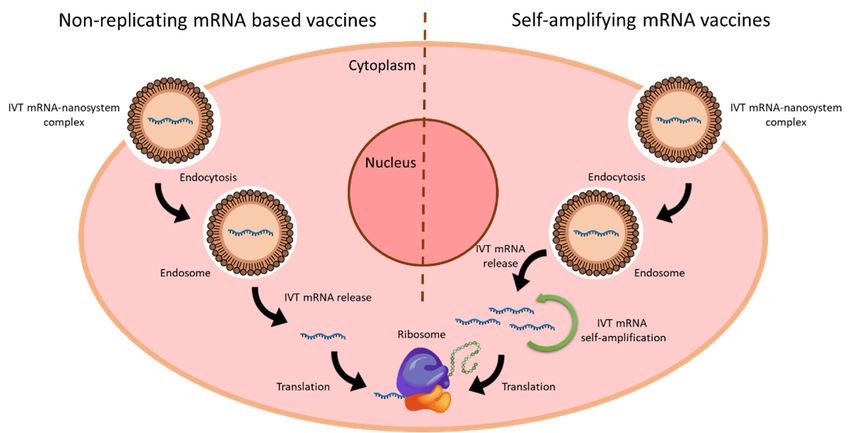
Nanomaterials 2020, 10, 364 4 of 42
Nanomaterials 2020, 10, 364 4 of 43
Figure2.2.Representative
Figure Representative scheme
scheme of IVT
of the the mRNA
IVT mRNA structure
structure and its and its principal
principal modifications
modifications to improveto
improve the efficacy and the stability, and to reduce the immunogenicity.
the efficacy and the stability, and to reduce the immunogenicity.
2.1.
2.1.5’5’Cap
Cap
Eukaryotic
Eukaryoticnative nativemRNAmRNApossesses
possessesaa5’5’cap capstructure,
structure,knownknownas ascap0,
cap0,formed
formedby bythe theunion
unionofof
inverted
inverted7-methyl
7-methylguanosine
guanosine(m7G) (m7G)to tothe
thefirst
firstnucleotide
nucleotideofofthe themRNAmRNAby byaa5’5’toto5’5’triphosphate
triphosphate
bridge during the transcription process. This capping occurs
bridge during the transcription process. This capping occurs by three consecutive by three consecutive enzymatic enzymatic
reactions,
when the first
reactions, when20–30
thenucleotides of mRNA have
first 20–30 nucleotides been transcribed
of mRNA in the nucleus.
have been transcribed in the nucleus.
Besides stabilizing the mRNA in the translation, splicing,
Besides stabilizing the mRNA in the translation, splicing, polyadenylation polyadenylation and nuclearand exportation
nuclear
processes, cap0 protects the mRNA from exonucleases. Additionally,
exportation processes, cap0 protects the mRNA from exonucleases. Additionally, cap0 interacts cap0 interacts with cap binding
with
proteins
cap binding(CBPs), essentials
proteins (CBPs),foressentials
the nuclear forexport of mRNA,
the nuclear exportand also with
of mRNA, andthealsotranslation initiation
with the translation
factor 4E (eIF4E)
initiation factorin4E the(eIF4E)
cytoplasm,in thecrucial for the initiation
cytoplasm, crucial for of translation
the initiation [11,25,26]. Moreover,[11,25,26].
of translation it could
be used as an innate immune marker, which differentiates the viral
Moreover, it could be used as an innate immune marker, which differentiates the viral RNA from RNA from cellular RNA [26].
In recent 0
cellular RNAyears,[26]. human enzymes that form two other types of 5 cap, cap1 and cap2, have been
identified [26,27]. In these cases, the m7G-specific 0 (20 O and
In recent years, human enzymes that form two2 other O methyltransferase
types of 5′ cap, cap1 MTase)cap2, methylates
have been
the second [26,27].
or thirdInribonucleotide 0 0
identified these cases, theatm7G-specific
the 2 O or 32′O O position of the riboses
methyltransferase generating
(2′O MTase) cap1 and
methylates the
cap2 structures, respectively. Cap1 and cap2 are less immunogenic
second or third ribonucleotide at the 2′O or 3′O position of the riboses generating cap1 and cap2 than cap0, and they enhance
translation
structures,efficiency [10].Cap1
respectively. Therefore,
and cap2the are
introduction of cap1 orthan
less immunogenic cap2cap0,
in the
and synthesis of IVTtranslation
they enhance mRNA is
aefficiency
crucial factor[10].inTherefore,
reducing the the introduction
immunogenicity for applications
of cap1 or cap2 in the in synthesis
which immune of IVTresponse
mRNA ishas to be
a crucial
minimized [24], but the incorporation of cap0 could be more useful
factor in reducing the immunogenicity for applications in which immune response has to be in therapies in which the immune
response
minimized is beneficial,
[24], but the such as vaccination.
incorporation of cap0 could be more useful in therapies in which the immune
In order
response to resemble
is beneficial, suchtheaschemical structure of eukaryotic mRNA, synthetic mRNA transcripts
vaccination.
can beIncapped after finishing the transcription
order to resemble the chemical structure (post-transcriptionally)
of eukaryotic mRNA, or during mRNA
synthetic the transcription
transcripts
(co-transcriptionally).
can be capped after finishing the transcription (post-transcriptionally) or during the transcription (co-
Post-transcriptional capping is usually carried out by using the enzyme machinery of recombinant
transcriptionally).
Vaccinia Virus to perform the
Post-transcriptional consecutive
capping enzymecarried
is usually reactions to add
out the cap0,
by using thecap1 or cap2.
enzyme machinery of
In co-transcriptional
recombinant Vaccinia Virus capping synthetic
to perform thecap analogues enzyme
consecutive are directly added to
reactions during
add thethe transcription.
cap0, cap1 or
This
cap2.process is simpler than the enzymatic capping, but it also presents some limitations. On the one
hand, Inall mRNA molecules obtained are not capped, due to the
co-transcriptional capping synthetic cap analogues are directly added duringcompetition between the cap analogue the
and the guanosine
transcription. Thistriphosphate
process is simpler(GTP), whichthan is theenzymatic
the initiator nucleotide
capping, [14,26].
but it also As apresents
consequence, some
limitations. On the one hand, all mRNA molecules obtained are not capped, due to the competition
between the cap analogue and the guanosine triphosphate (GTP), which is the initiator nucleotide
Nanomaterials 2020, 10, 364                                                                          5 of 42

the uncapped mRNAs are immediately digested by nucleases and can induce an immune response.
A strategy to reduce the stimulation of the immune system is the removal of the triphosphates of
the 50 end of uncapped IVT mRNA by using a phosphatase [9,28]. On the other hand, there is
the risk of reverse orientation of the cap analogues, which impede the binding to the CBP and the
posterior translation [11,24,29]. As an alternative, an anti-reverse cap analogue (ARCA) has been
developed [11,24]. ARCA consists of a methyl group in the 30 -OH of the m7G nucleotide that enables
the appropriate orientation of the cap and prevents the elongation of the mRNA along the wrong
site [24,26]. Another traditional limitation of co-transcriptional capping is the inability to incorporate
cap1 and cap2. However, recently, TriLink BioTechnologies has designed a new co-transcriptional
capping technology, CleanCap™ , able to incorporate cap1 or cap2 in the 94% of the mRNA molecules
during the transcription process [30].
      Finally, it also has to be taken into account that cytosolic decapping enzymes can remove mRNA
cap. In order to provide resistance to the IVT mRNA against these enzymes, and therefore, extend its
half-life, chemically modified cap analogues can be used. The resulting modified mRNAs can contain
a phosphorothioate, phosphorothiolate, imidiphosphate and boranophosphate, among others [31–36].

2.2. ORF
     The codon composition of the region that encodes the protein sequence, known as ORF, may
also influence the translation efficiency and stability of the mRNA. The reduction of the quantity of
UU and UA dinucleotides in the ORF has demonstrated to protect the IVT mRNA from decapping
enzymes [37,38]. Additionally, since most amino acids are encoded by different synonymous codons,
codon optimization strategies have been developed to improve the cost efficiency of recombinant
protein production [39]. Codon optimization is mainly based on the substitution of multiple rare codons
by other more frequent ones that encode the same amino acid. As a result, the rate and efficiency of the
protein translation are increased [14,23,40]. However, the clinical application in humans is controversial,
because in the last years it has been documented that synonymous mutations affect protein functions,
and alterations in translation kinetics can lead to alterations in protein conformation [40–43].

2.3. Poly(A) Tail
     The poly(A) tail in native eukaryotic mRNA is formed by 100–250 residues of adenosine [9,24,44].
It participates in the exportation process of mRNA molecules from the nucleus, interacts with initiation
factors of the translation and prevents the degradation by nucleases through the interaction with
poly(A)-binding protein, providing stability [45].
     The poly(A) tail can be added to IVT mRNA directly during the transcription, if the DNA template
encodes the poly(T) sequence, or post-transcriptionally by enzymatic reactions with recombinant
E-PAP [11,24]. The addition during transcription is preferable, because the length of the poly(A) tail is
more reproducible [14,23,24].
     The length of the poly(A) tail influences the stability and translation efficiency of the IVT
mRNA [46,47]; although a relatively long poly(A) tail seems to be appropriate, the optimal length may
vary depending on the target cell. In HeLa (epithelial human) cells the increase of adenosine residues
in the poly(A) tail from 14 to 98 improved the protein expression [48], whereas, in dendritic cells (DCs)
a number of 120 adenosine residues enhances the translation efficiency, the protection and the stability
of the IVT mRNA [49,50].

2.4. UTRs
     The ORF is limited by the UTRs in both 50 and 30 sides. These non-coding regions do not participate
directly in the codification of proteins, but their sequences, length and secondary structures are crucial
for the regulation of the translation of the mRNA and the protein expression [51]. 50 UTR is involved
in the initiation of translation, which is considered the most intricate step among the whole process,
whereas, 30 UTR influences the mRNA stability and the extent of protein expression [52].
Nanomaterials 2020, 10, 364 6 of 42
The presence of the internal ribosomal entry sites (IRES) in the 5’ UTR recruits the ribosome and
induce a cap-independent translation initiation [53–55]. In order to improve translation efficiency,
50 UTRs containing IRES from viral origin may be incorporated into IVT mRNA [37]. In these cases,
translation is not dependent on eIF4E, as it is with cap0, and IVT mRNA expression is extended to cells
where eIF4E levels are low [14]. However, the 50 cap is still necessary for the protection of the mRNA
against nucleases, and therefore, most IVT mRNAs contain both 50 cap and IRES in their structure.
The Kozak consensus sequence, located in the 50 UTR, also plays a major role in the initiation
of the translation process. The Kozak sequence, defined as RCCAUGG, where R is a purine (A or G)
was considered the preferred sequence for translation initiation in eukaryotes [56]. In this sequence,
some nucleotides are more important than others; particularly, the −3 and the +4 positions, relative
to the adenine of the starting codon AUG. To facilitate the recognition of the starting codon AUG, G
nucleotide must be in the position +4, and A/G nucleotides must be in the position −3 [57].
Regarding the 30 UTR, the presence of specific sequences of α-globin and β-globin mRNAs in this
region improves the stability of IVT mRNA and the duration of protein expression, respectively [58,59].
In addition, the length of the 30 UTR sequence may be modified to regulate the protein localization.
For instance, in the case of CD47 membrane protein, long 30 UTR induces the protein expression on
the cell surface, whereas, short 30 UTR results in the localization of the protein in the endoplasmic
reticulum [60].
2.5. Modified Nucleosides
The incorporation of modified nucleosides into mRNA is a common strategy to reduce its
immunostimulatory activity. Exogenous IVT mRNA induces innate immune responses by interacting
with pattern recognition receptors (PRRs), including Toll-like receptors (TLRs) and cytoplasmic RNA
sensors, such as retinoic acid-inducible protein I (RIG-I) [61,62]. It is known that uridine residues activate
TLR7, and GU- and AU-rich RNA strands activate TLR7 and TLR8 [63,64]. However, the incorporation
of modified nucleosides into the transcript, (i.e., pseudouridine (Ψ), N1-methylpseudouridine (N1mΨ),
5-methylcytidine (m5C), N6-methyladenosine (m6A), 5-methyluridine (m5U), or 2-thiouridine(s2U)),
avoids the activation of TLRs [65–69]. Some of them, such as Ψ and s2U, also abolish RIG-I activation [63].
In addition, the presence of m6A in the 5’ UTR have been proposed as an alternative to IRES, in order
to favour cap-independent translation [70].
3. mRNA Nanomedicines
A key challenge for the clinical application of nucleic acid medicinal products entails the availability
of delivery systems specifically adapted to their features and purpose. A vehicle for mRNA therapy, in
addition to protecting it and providing specificity to reach the target cell, must afford an adequate
intracellular disposition of the nucleic acid that enables the translation process, and all of this, preventing
the activation of the immune response [71,72].
Currently, about 70% of the clinical trials with nucleic acids use recombinant viruses as delivery
systems, such as retroviruses, lentiviruses, adenoviruses and adeno-associated viruses, among
others [73]. Viral systems are viruses genetically modified to prevent their replication in the host cell;
they present high transfection capacity, but also oncogenic and immunogenic potential. Moreover,
viral vectors present a limitation regarding the size of the nucleic acid they can transport [74]. Despite
the major advances achieved in DNA delivery with viral vectors, they do not play an important role in
the case of mRNA-based products [7,75–78]. Non-viral systems are safe, their production is simple,
economical and reproducible compared to viral vectors, and the size of nucleic acid to be packed is
not usually an obstacle. However, their transfection efficacy is still a limitation, although it has been
improved by different strategies and the efforts are still ongoing [75,77,79].
Therapeutic application of mRNA without the help of a delivery system presents important
drawbacks [66]. Naked mRNA has been mainly applied ex vivo by using physical methods, including
electroporation, microinjection and gene gun; these methods are able to disrupt the cell membraneNanomaterials 2020, 10, 364 7 of 42 and facilitate the entry of mRNA into the cell [80]. Electroporation consists in generating pores in a cell membrane through electric pulses. This technique has shown efficient mRNA delivery for tumor antigen loading of DCs [81]; even in some studies, mRNA electroporation has been more efficient than DNA electroporation, with faster and more homogeneous protein expression in vivo [82]. The direct injection of mRNA into the target cell by the use of microneedles (namely, microinjection) [83,84], and gene gun, in which naked mRNA is pneumatically shot into the target cell, have also been used for mRNA transfection [85–88]. In in vivo applications, intravenously administered naked mRNA is rapidly degraded by ribonucleases and the innate immune system can be activated. In fact, the half-life of naked mRNA has been estimated
Nanomaterials 2020, 10, 364                                                                                         8 of 42
  Nanomaterials 2020, 10, 364                                                                                   8 of 43

conventional
   aminopropane cationic   lipids [5].
                     (DODMA),      haveWhile
                                          beencationic
                                                developed lipids  present
                                                              as an         alkylated
                                                                     alternative        quaternary cationic
                                                                                  to conventional    ammonium lipidsgroups
                                                                                                                      [5].
andWhile  cationic
     retain         lipids present
             their cationic    naturealkylated   quaternary
                                        regardless             ammonium
                                                      of the pH,    ionizable groups
                                                                                lipidsand  retain positive
                                                                                         acquire  their cationic  nature
                                                                                                             charges,   due
   regardless  of the   pH,  ionizable  lipids  acquire   positive  charges,   due to  the protonation
to the protonation of free amines as pH decreases [13]. These new lipids are neutral at physiological    of free amines
pHasbut
      pHpositively
          decreases charged
                       [13]. These  newthe
                                 inside   lipids  are neutral
                                              endosome,         at physiological
                                                             when    pH values are pHbelow
                                                                                        but positively
                                                                                              its pKa. charged    inside
                                                                                                         The electrostatic
   the endosome,
interactions         whennaturally
               between       pH values    are below
                                       occurring       its pKa.
                                                    anionic      The in
                                                              lipids   electrostatic
                                                                          endosomal   interactions
                                                                                         membranes  between
                                                                                                       and the naturally
                                                                                                                 ionizable
   occurring
cationic lipidsanionic
                 have beenlipidsproposed
                                  in endosomal
                                             as the membranes
                                                    underlying and        the ionizable
                                                                    mechanism              cationic
                                                                                   of nucleic   acid lipids
                                                                                                      releasehave
                                                                                                               [88].been
                                                                                                                      These
   proposed   as  the   underlying   mechanism      of  nucleic   acid  release [88].  These
interactions are able to promote membrane lytic nonbilayer structures, such as the hexagonal Hinteractions   are ablephase,
                                                                                                                       to
                                                                                                                   II
   promote membrane lytic nonbilayer structures, such as the hexagonal HII phase, culminating in the
culminating in the intracellular mRNA delivery [98]. Nowadays, nanocarriers containing ionizable
   intracellular mRNA delivery [98]. Nowadays, nanocarriers containing ionizable cationic lipids are
cationic lipids are among the leading delivery system candidates with promising applications for
   among the leading delivery system candidates with promising applications for siRNA and mRNA
siRNA and mRNA delivery [78].
   delivery [78].

                     Figure 3. Representative scheme of chemical nanocarriers for mRNA delivery.
                   Figure 3. Representative scheme of chemical nanocarriers for mRNA delivery.

        Cationic
     Cationic      lipids
               lipids      canformulated
                      can be    be formulated      to prepare
                                          to prepare            cationic nanoemulsions
                                                       cationic nanoemulsions    (CNEs) and (CNEs)   and lipid
                                                                                              lipid nanoparticles
  nanoparticles   (LNPs)  [75]. CNEs   consist of a dispersion of an oil phase stabilized by an aqueous
(LNPs) [75]. CNEs consist of a dispersion of an oil phase stabilized by an aqueous phase containing      phase
  containing   the  cationic lipid. These nanoemulsions     present  a  droplet size distribution
the cationic lipid. These nanoemulsions present a droplet size distribution of about 200 nm, and  of about 200are
  nm,   and are  mainly   used   to formulate  mRNA     vaccines  [2,9]. For instance, a self-amplifying
mainly used to formulate mRNA vaccines [2,9]. For instance, a self-amplifying RNA (SAM) vaccine,          RNA
expressing Human Immunodeficiency Virus (HIV) type 1 envelope, formulated in a CNE induced
Nanomaterials 2020, 10, 364 9 of 42
potent immune responses in rhesus macaques [99]. Brito et al. [100] developed a well-tolerated and
immunogenic SAM vaccine based on CNEs. This SAM vaccine-elicited immune responses in a variety
of animal models (including mice, rats, rabbits, and nonhuman primates) at much lower doses than
those required for pDNA vaccines.
LNPs include liposomes and other lipid-based nanoparticles, and they are regarded as one of the
most developed systems for mRNA delivery. Currently, several LNP platforms are at the forefront
of clinical trials. Indeed, they are clinically validated delivery systems for RNA therapy. In the
beginning, LNPs were considerably promising for the delivery of siRNA, being their utility as mRNA
delivery agents more recent [101]. In this sense, Patisiran (ONPATTRO™), a siRNA formulated in
LNPs targeted to inhibit hepatic transthyretin protein, received a recent FDA approval, meaning
significant progress in the field [102]. This product contains a novel ionizable lipid, Dlin-MC3-DMA
(MC3) [103], and after its success, several groups have managed to use MC3 as a vehicle for transferring
mRNA [104]. The previous experience in siRNA formulation is benefiting the development of mRNA
nanosystems, although mRNA has a different structure which may interfere with the packing capacity
of nanoparticles [101]. For an optimal mRNA release, the delivery systems should contain less
ionizable lipid and cholesterol and more phospholipid and polyethylene glycol (PEG) [5] than in the
case of siRNA.
Liposome based formulations are amphiphilic spherical vesicles formed by one or more lipid
bilayers enveloping an aqueous core with size ranging from 20 nm to a few microns. They generally
contain a cationic lipid combined with: (a) A helper lipid that supports the bilayer structure and
facilitates the endocytosis; (b) cholesterol to stabilize the lipid bilayer of the LNP; and (c) a PEG-lipid.
PEG lends the nanoparticle a hydrating layer to improve colloidal stability, reduces protein adsorption
and non-specific uptake, and prevents reticuloendothelial clearance [13,90]. Additionally, helper lipids,
such as DSPC (1,2-distearoyl-sn-glycero-3-phosphocholine), DOPE and POPE (1-palmitoyl-2-oleoyl-
sn-glycero-3-phosphoethanolamine) are frequently used in these systems [44]. The in vivo delivery of
mRNA by using this kind of lipid-based system was already evaluated in 1994 [105], demonstrating
a comparable efficacy to liposome-DNA complexes in accomplishing in situ tumor transfection.
That study also showed that mRNA might be considered as an alternative to pDNA for genetic
immunopotentiation applications. More recently, LNPs containing an ionizable cationic lipid,
phosphatidylcholine, cholesterol and PEG-lipid were used to encapsulate nucleoside-modified
mRNA encoding the pre-membrane and envelope glycoproteins of a strain from Zika virus [106].
A single intradermal low-dose immunization elicited potent and durable neutralizing antibody
responses and protective immunity in mice and non-human primates. SAM vaccine platform is
another example of a synthetic mRNA delivered by LNPs. SAM vaccine encoding an influenza
H1HA antigen from N1H1 virus, and formulated with 1,2-dilinoleyloxy-3-dimethylaminopropane,
1,2-diastearoyl-sn-glycero-3-phosphocholine, cholesterol and PEG-DMG 2000, has demonstrated to
be immunogenic in mice at low doses, and to elicit antibody responses that were comparable to the
licensed vaccine [107]. The use of mRNA-liposomes in cancer therapy has increased dramatically, since
the first study in 1999. Zhou et al. [108] used hemagglutinating virus of Japan (HVJ)-based liposomes
for the synthesized mRNA encoding gp-100. Direct injection into the mouse spleen induced gp100
antibody expression and responses against B16 cells. Recently, the first-in-human, open label phase I
study in ovarian cancer patients has been approved. Patients will be vaccinated intravenously prior,
and during (neo)-adjuvant chemotherapy with a liposome formulated mRNA vaccine (W_ova1 vaccine)
which includes three ovarian cancer- tumor-associated antigens (TAAs) RNAs (ClinicalTrials.gov
Identifier: NCT04163094). CRISPR/Cas9 genome editing mediated by LNPs has recently provided
in vivo evidence in animal models. Finn et al. [109] developed an LNP-mediated delivery method
by using a biodegradable and ionizable lipid termed LP01 co-formulated with both Spy Cas9 mRNA
and single guide RNA (sgRNA). A single administration enabled significant editing of the mouse
transthyretin gene in the liver and resulted in >97% reduction in serum protein levels that persisted for
at least 12 months.Nanomaterials 2020, 10, 364 10 of 42
A few marketed transfection reagents useful for mRNA transfection (for instance, MegaFectinTM ,
TransITTM ) are cationic liposome formulations based on DOTAP, DOPE and cholesterol [90]. As an
example, expression was obtained after the intravenous injection to mice of MegaFectinTM containing
mRNA encoding red fluorescent protein [110]. Other cationic lipid-based transfection reagents
commercialized have been widely used for mRNA delivery, such as LipofectinTM , which is a mixture of
DOTMA and DOPE, and LipofectamineTM , which is a combination of DOSMA (the polycationic lipid
2,3-dioleyloxy-N-[2(sperminecarboxamido)ethyl]-N,N-dimethyl-1-propanaminium trifluoroacetate)
and DOPE [90]. However, their use is restricted to in vitro studies, due to the toxicity associated
with cationic charges, fast clearance, activation of immune response and low transfection efficacy
in vivo [90,98].
Lipidoids, a new class of lipid-like delivery molecules comprising multiple hydrophilic groups
and several lipid tails, were developed in 2008 as novel siRNA delivery agents [111]. Based on those
results, more recently, a new class of lipid-like materials termed zwitterionic amino lipids (ZALs) have
been applied for mRNA gene editing [112]. Intravenous co-delivery of Cas9 mRNA and targeted
sgRNA from a single ZAL nanoparticle enabled CRISPR/Cas9 gene editing in mice.
In recent years, new lipid derivate systems, known as lipid-like nanoparticles (LLNs), have been
developed for mRNA delivery. N1,N3,N5-tris(2-aminoethyl)benzene-1,3,5-tricarboxamide (TT) is
formed by a phenyl ring, three amide linkers and three aminolipids chains [44]. Among them, TT3
was recognized as the principal compound in the optimized formulation, which increased in 350-fold
the efficacy of luciferase-mRNA transfection [113]. More recently, TT3 LLNs delivered human factor IX
(hFIX) mRNA and recovered the normal levels of the protein in IX-deficient mice [113]. TT3 LLNs
were also optimized to deliver both Cas9 mRNA and sgRNA, demonstrating effective gene editing of
hepatitis B virus DNA and the proprotein convertase subtilisin/kexin type 9 (pcsk9) gene [114].
Nanostructured lipid carriers (NLCs) are another type of LNPs used to deliver mRNA for
vaccination. NLCs are colloidal structures composed by a core containing a mixture of solid and
liquid lipids, resulting in an unstructured lipid matrix [115]. An important advantage of NLCs is their
low toxicity respect to other lipid systems, as emulsions, which require high quantities of surfactants
and cosurfactants. Additionally, production and sterilization of NLCs are easy and cheap compared
to other systems. Specifically, sterilization of liposomal formulations is rather difficult, due to the
sensitivity of phospholipids to heat and radiation [23], their production is costly, and batch-to batch
reproducibility and large-scale manufacturing are difficult and expensive to achieve [116]. Better than
for nucleic acid delivery, NLCs have been studied mainly for increasing the oral bioavailability of
poorly soluble drugs [117]. Nevertheless, in a recent study [118], administration of replicating viral
mRNA encoding Zika virus antigens formulated in NLCs, completely protected mice against a lethal
Zika challenge; this achievement represents what might be the most potent approach to date of any
Zika vaccine.
3.2. Polymeric Systems
The use of polymer nanoparticles has been intensively investigated for pDNA delivery, although
few studies have addressed their use for mRNA. One key advantage of polymeric systems is the
possibility of modifying their chemical properties to adapt them to the active substance. The binding
of cationic polymers and nucleic acids leads to the formation of polyplexes [90]. Different cationic
polymers have been studied for mRNA complexation, including polyethyleneimine (PEI), polyacrylates,
poly(β-amino esters) (PBAEs) and poly(aspartamides) (PAsp), among others. However, despite the
significant number of polymeric materials available, polymeric systems are not as clinically advanced
as lipidic systems for mRNA-based therapies.
PEI was one of the first polymers used for nucleic acid delivery; it contains a large number
of amine groups in its structure conferring it a positive charge. PEI may present both a linear and
a branched conformation. Linear PEI contains secondary amino groups partially protonated at
physiological pH, whereas, branched PEI contains primary and secondary groups, and a small numberNanomaterials 2020, 10, 364 11 of 42
of tertiary amines. The presence of amino groups is responsible for the strong affinity to nucleic acids,
including mRNA, and the cationic charges facilitate the interaction of the polyplexes with the cell
membrane and the entry into the target cell. Moreover, amino groups allow ionization and confer high
buffering ability. This buffering capacity, although under discussion, is responsibility of the “proton
sponge effect” and enables the endosomal swelling and rupture by changing the osmolarity of acidic
vesicles [71]. Nevertheless, the high density of positive charges is also related to in vivo toxicity by
owing interactions with extra and intracellular proteins, destabilization of lipid cellular membranes
and activation of the immune system [119,120]. New PEI derivatives have been designed to improve
its biocompatibility and transfection efficiency. For example, jetPEI® , a linear PEI commercialized as
an in vivo transfection reagent in mice, was firstly evaluated for pDNA and siRNA transfection, and
later for mRNA delivery [121]. More recently, the administration of IVT mRNA with jetPEI® by direct
myocardial injection in the mouse demonstrated the expression of the protein in the lungs [122].
Polyacrylates have also been used for mRNA delivery, although with modifications in the side
chain, needed to interact electrostatically with nucleic acids. One of the most studied polyacrylates
is poly(2-dimethylaminoethyl methacrylate) (PDMAEMA), which presents lower affinity for mRNA
than for pDNA; however, its PEGylation improves mRNA binding and transfection efficiency [123].
The development of triblock copolymers, formed by modifications in PDMAEMA structure, has
demonstrated an improvement in transfection efficiency in both siRNA and mRNA systems.
The modifications include: (1) The addition of amphiphilic materials, such as PEG methacrylate
(PEGMA), to improve the stability and biocompatibility; (2) the incorporation of a hydrophobic butyl
methacrylate segment (BMA) for fusogenicity; and (3) a pH-responsive diethylaminoethyl methacrylate
(DEAEMA), to break the membrane of endosomes [71]. Triblock copolymer with PEGMA placed in the
center of the copolymer chain showed high transfection efficiency and stability in macrophages and DCs,
showing the potential of this system for mRNA-based vaccination approaches [124]. Oligoalkylamines
grafted to an 8000 Da poly(acrylic acid) (PAA8k) scaffold complexed with chemically modified mRNA
resulted in another kind of polyplexes with transfection capacity. Intravenous administration of
PAA8k-luciferase mRNA systems in mice showed different luminescence signal in liver depending on
their structure [125].
PBAEs are biodegradable and pH responsiveness copolymers synthesized by the addition of
amines and acrylates via Michael-type reaction. The tertiary amine of its structure can electrostatically
interact with the negative charge of nucleic acids. There is a wide variety of PBAEs delivery
systems, due to their compatibility with other polymers, such as PEG, poly(lactic acid) (PLA), and
poly(ε-caprolactone) (PCL) [126]. The use of PBAEs formulated with PEG-lipid improves serum
stability of mRNA after intravenous administration [127]. Recently, inhaled delivery of IVT mRNA by
hyperbranched PBAEs provided uniform distribution of luciferase mRNA in the lung, and protein
expression lasted 24 h [128].
PAsp are synthesized by polymerization of DL-aspartic acid in orthophosporic acid medium and
later addition of a nucleophilic amine [93]. The length of the amynoethylene side chain influences both
the cationic charge and buffering capacity of each PAsp construct. For example, PAsp(DET), which is
formed by the addition of 1,2-diaminoethane in the side chain to the PAsp, shows endosomal scape
capabilities and biodegradability [90,128]. An odd-even effect of the amynoethylene repeated units has
been described for different nucleic acid payload. PAsp containing even-numbered of amynoethylene
units showed higher transfection with pDNA, whereas, an odd-numbered unit sustainably increased
mRNA expression and enhanced resistance against [129,130]. PEG-PAsp polyplexes have displayed
enhanced stability and lower cytotoxicity; for instance, IVT mRNA complexed with PEG-PAsp(DET)
was intranasally administrated to mice for delivering brain-derived neurotrophic factor (BDNF),
showing protein expression in nasal tissues for nearly two days [131]. PEG-PAsp(DET) also has been
used to complex Bcl-2 IVT mRNA, being more effective than pDNA on reducing apoptosis in the liver
of mice with fulminant hepatitis [132].Nanomaterials 2020, 10, 364 12 of 42
In addition to the most used polymers discussed above, it has been reported transfection of IVT
mRNA with another kind of polyplexes. Nanoparticles formed by a core-shell structure of IVT mRNA
complexed with the peptide protamine surrounded by PCL layers improved the stability of mRNA [133].
Chitosan, a biocompatible cationic glycopolymer containing amines, formulated as chitosan/hyaluronic
acid nanoparticles, provided successful delivery of luciferase-encoding mRNA [134]. Self-immolative
polycarbonate-block-poly(α-amino)esters, also known as charge-altering releasable transporters
(CARTs), have demonstrated capacity to deliver mRNA thanks to their capacity to reduce chelative
electrostatic anion-binding ability with the cationic polymer and facilitate endosomal escape [135,136].
CARTs are effective for in vivo delivery of mRNA with minimal toxicity in different cell lines and
animal models via intramuscular, intratumoral, and intravenous administration. These CARTs have
shown strong antigen-specific immune response against mRNA-encoded viral epitopes in a mouse
model [137]. Another biodegradable ionizable amino polyesters (APEs) synthesized via ring opening
polymerization of lactones with tertiary amino-alcohols are tissue-selective for mRNA delivery [138].
3.3. Polypeptidic Systems
Polypeptides consist of one or various amino acids disposed of in block or random sequences.
They can provide biocompatibility and physicochemical properties to the delivery systems, thanks to
the biodegradable naturally occurring monomeric units. Another advantage of nucleic acid delivery is
the ability to adapt their cationic and endosomolytic properties, due to their structural flexibility [71].
Protamines are a family of small peptides with arginine-rich sequences obtained from fish sperm.
Arginines confer positive charge, facilitating electrostatic interactions with the negative charge of
the nucleic acid; in fact, protamine was described more than 50 years ago as an enhancer of RNA
uptake [139]. The condensation of mRNA with protamine protects it against ribonuclease degradation,
and the complex formed can activate TLRs acting as danger signals useful for vaccination [140].
Protamine-based formulations for IVT mRNA delivery are the second most used chemical systems in
clinical trials, although far from lipidic systems. RNActive® technology, developed by CureVac, is an
mRNA vaccine platform based on protamine/mRNA complexes currently under clinical evaluation
against rabies [138], and different cancers [141,142]. RNActive® platform also has been tested
preclinically against the influenza virus infection [143].
Cell-Penetrating Peptides (CPPs) have been used for nucleic acid delivery, due to their capacity to
overcome cell membranes. Although the mechanisms of cellular internalization are not fully known, it
is thought that CPPs may promote the grouping of negatively charged glycosaminoglycans of the cell
surface, triggering macropinocytosis and lateral diffusion or directly disrupting the lipid bilayer [13].
A cationic CPP containing the arginine-rich amphipathic RALA motif was used as an mRNA vector
for DCs. Nanocomplexes of RALA with Ψ and m5C modified IVT mRNA elicit potent cytolytic T cell
responses against the antigenic mRNA payload [144].
Artificial viral coat proteins formulated as virus-like particles (VLPs) have been used as vehicles
for transfection, due to their ability to assemble and protect mRNA. Li et al. [145] developed an
mRNA vaccine as therapy for prostate cancer based on recombinant bacteriophage MS2 VLPs.
This vaccine-induced strong humoral and cellular immune responses and protected mice against
prostate cancer. In another work, artificial viral coat protein consisting of an oligolysine (K12), a silk
protein-like midblock S10, and a long hydrophilic random coil block C was generated and complexed
with mRNA to form rod-shaped VLPs. This system transfected cells with both EGFP and luciferase,
but the efficacy was low compared to that obtained with a lipoplex transfection reagent [146]. More
recently, VLPs prepared by fusing protein G of Vesicular stomatitis virus (VSV-G) with a ribosomal
protein L7Ae of Archeoglobus fulgidus, resulted in the efficient delivery of EGFP in human induced
pluripotent stem cells (iPSCs) and monocytes [147].Nanomaterials 2020, 10, 364 13 of 42
3.4. Dendrimers
Dendrimers are highly branched polymeric macromolecules with well-defined uniform sizes
and shapes, and adaptable surface functionalities. Their basic structure encompasses a central
core, repetitive branching units, and terminal groups [148]. Modified dendrimers, derived from
polyamidoamine (PAMAM) have been extensively studied for nucleic acid delivery, due to their
hydrophilic, biocompatible and non-immunogenic properties. Chahal et al. [149] developed a
rapid-response and adjuvant-free vaccine based on a PAMAM dendrimer formulated in nanoparticles,
wherein the antigens were encoded by encapsulated mRNA replicons. Intramuscular injection to mice
of a single dose generated protective immunity against lethal Ebola, H1N1 influenza, and Toxoplasma
gondii challenges. In a later study, this dendrimer-based nanoparticle was used to create a vaccine
candidate that elicited Zika virus E protein-specific IgG responses. After immunization to mice, the
authors detected a CD8+ T cell response against a unique H-2Db-restricted epitope [150].
3.5. Gold Nanoparticles
Gold nanoparticles (AuNPs) present features that make them an appropriate platform for nucleic
acid delivery. AuNPs can be fabricated in a scalable fashion with low size dispersity, and they are
easily functionalized by the formation of multifunctional monolayers and the inclusion of different
moieties and targeting agents. Moreover, the in vivo toxicity and biodistribution, can be regulated
by optimizing the particle size and surface functionality [151]. Yeom et al. [152] injected into mice
xenograft tumors an IVT mRNA encoding Bcl-2-associated X (BAX) protein, a pro-apoptotic factor,
loaded on AuNP-DNA conjugates. The mRNA released produced BAX protein, and tumor growth
was inhibited.
3.6. Hybrid Systems
Hybrid systems are made up of the combination of various types of materials, including lipids,
polymers and peptides, among others. Thereby, the hybrid system takes advantage of all the benefits
of its individual components, offering greater functionality and flexibility [14,78], which may facilitate
their clinical translation in the near future. However, the careful optimization required by a combination
of such different components is an important becoming challenge in terms of scaling-up and clinical
utility [71].
The combination of cationic lipids and peptides has been commonly studied for mRNA delivery.
As an example, complexes formed by IVT mRNA, coding for the model antigen beta-galactosidase,
condensed with protamine and encapsulated in liposomes provided in vivo protein expression,
activation of cytotoxic T lymphocytes and production of IgG antibodies against the antigen [153].
In another study, the administration of lipid/protamine/IVT mRNA to mice bearing human lung
NCI-H460 carcinoma, demonstrated better results in both efficacy and toxicity than the equivalent
formulation with pDNA [154]. Lipofectamine® with CRPPR-R9, a peptide containing nine arginine
residues, efficiently transfected cultured mouse cardiac fibroblasts [155]. Cationic lipids have also been
combined with inorganic nanoparticles. The transfection efficacy of mRNA-DOTAP, mRNA-apatite
and mRNA-DOTAP-apatite was evaluated using an mRNA encoding the luciferase enzyme, being the
mRNA-DOTAP-apatite complex the most effective [156].
Lipopolyplexes, the complexation of nucleic acids with cationic polymers and lipids, were among
the first hybrids used for DNA and siRNA delivery, and later on for mRNA. Histidylated lipopolyplexes,
synthesized by the combination of PEGylated derivative of histidylated polylysine and L-histidine-
(N,N-di-n-hexadecylamine)ethylamide liposomes, incorporating a synthetic melanoma-associated antigen
MART1 mRNA have been administered to mice as an mRNA cancer vaccine. The histidylated
lipopolyplexes protected significantly injected mice against B16F10 melanoma tumor progression [157].
The subsequent mannosylation of the system targeted the mRNA into the DCs by the mannose
receptor [158]. mRNA nanocomplexes formed with the polymer PLGA (poly(lactic-co-glycolic acid)),Nanomaterials 2020, 10, 364 14 of 42
the cationic lipid-like compound (G0-C14) and a lipid-PEG were used to enhance the protein expression
of the tumor-suppressor gene PTEN (phosphatase and tensin homolog deleted on chromosome ten).
This hybrid system provided a high IVT mRNA PTEN transfection in prostate cancer cells, and led
to significant inhibition of tumor growth when delivered systemically in multiple mouse models of
prostate cancer [89]. A hybrid polymer-lipid nanoformulation for systemic delivery to the lung was
prepared by co-formulation of PBAEs with lipid-PEG. This degradable polymer–lipid nanoparticle showed
both enhanced serum stability and increased in vitro potency, delivering IVT mRNA in the lungs after
intravenous administration in mice [127].
A nanomicelle-based platform was prepared by mixing IVT mRNA encoding an anti-angiogenic
protein (sFlt-1), with PEG-polycation block copolymers. PAsp(TEP) was selected as the cationic
segment of the block copolymer, and a cholesterol moiety was attached by hydrophobic interaction.
PEG-PAsp(TEP)-cholesterol nanosystems produced efficient protein expression in tumor tissue, and
remarkable inhibition of the tumor growth [159].
Another example of a multi-component delivery system is that formed with poly(glycoamidoamine)
(PGAAs) brush nanoparticles. It has been used for intravenous administration of mRNA encoding
erythropoietin (EPO) in mice [160]. First, three different PGAA polymers based on tartarate, galactarate, or
glucarate sugars combined with three different amine-containing monomers were prepared. Polymer-brush
materials were synthesized through ring opening reactions between PGAAs and epoxides, and incorporated
into LNPs. Cholesterol, DSPC and mPEG2000-DMG (1,2-dimyristoyl-sn-glycero-3-phosphoethanolamine-
N-[methoxy-(polyethylene glycol)-2000]) were added via a microfluidic-based mixing device to form the
LNP polymer-brush nanoparticles.
Recently, DCs have been transfected with an mRNA delivery system combining both PLA-based
micelles and the cationic CPP RALA. This hybrid nanoplatform offers the possibility of further
multifunctionality by PLA core encapsulation [161].
4. Therapeutic Applications of mRNA
The growing knowledge of IVT mRNA design and manufacture, along with the advances in
nanotechnology have conducted to broaden the potential therapeutic applications of mRNA-based
medicines. According to preclinical and clinical trials, four major IVT mRNA applications can be
considered: Immunotherapy (against infectious diseases and cancer), protein replacement, gene
editing and regenerative medicine. Currently, all the clinical trials ongoing, both applying in vitro
and in vivo strategies, are still in Phase I or II, with most of them focusing on immunotherapy, and
especially on cancer therapy. Nevertheless, the successful transition of mRNAs from clinical studies to
commercialization in the form of medicinal products requires still an important consideration related
to the fabrication of large industrial batches: Optimization of IVT mRNA production and purification
processes with further cost reduction [71].
4.1. Immunotherapy
The induction of an immune response by using mRNA has been the main application among
mRNA-based therapies, with a number of mRNA vaccines being evaluated in clinical trials against
infectious diseases and multiple types of cancer. Apart from these applications, a proof of concept for
prevention of type 1 diabetes in mice, by administering modified T cells redirected against diabetogenic
CD8+ T cells, has emerged as a new mRNA-immunotherapy application [162].
Additionally, passive immunization by mRNA encoding monoclonal antibodies is showing
great biomedical interest. Given the rapidly growing market of therapeutic monoclonal antibodies,
and the high cost of this type of medicines, the pharmaceutical industry is looking for alternatives
approaches. mRNA is considered a good option, due to its simpler, faster and more cost-effective
synthesis. Until now, pre-clinical studies in small rodents have demonstrated antibody titters from the
first day after mRNA intravenous administration [163]. However, before moving on to the clinic, it isYou can also read