Natural Killer Cells and Current Applications of Chimeric Antigen Receptor-Modified NK-92 Cells in Tumor Immunotherapy - MDPI
←
→
Page content transcription
If your browser does not render page correctly, please read the page content below
International Journal of
           Molecular Sciences

Review
Natural Killer Cells and Current Applications of
Chimeric Antigen Receptor-Modified NK-92 Cells in
Tumor Immunotherapy
Jianguang Zhang, Huifang Zheng and Yong Diao *
 School of Medicine, Huaqiao University, Quanzhou 362021, Fujian, China; 1601116007@hqu.edu.cn (J.Z.);
 15136733530@163.com (H.Z.)
 * Correspondence: diaoyong@hqu.edu.cn; Tel.: +86-595-2269-2516

 Received: 15 November 2018; Accepted: 11 January 2019; Published: 14 January 2019

 Abstract: Natural killer (NK) cells are innate immune cells that can be activated rapidly to target
 abnormal and virus-infected cells without prior sensitization. With significant advancements in
 cell biology technologies, many NK cell lines have been established. Among these cell lines, NK-92
 cells are not only the most widely used but have also been approved for clinical applications.
 Additionally, chimeric antigen receptor-modified NK-92 cells (CAR-NK-92 cells) have shown strong
 antitumor effects. In this review, we summarize established human NK cell lines and their
 biological characteristics, and highlight the applications of NK-92 cells and CAR-NK-92 cells in
 tumor immunotherapy.

 Keywords: natural killer cell line; NK-92; chimeric antigen receptor; immunotherapy; tumor

1. Introduction
      Natural killer (NK) cells are innate immune cells that were first discovered in mice in 1975 [1].
NK cells account for approximately 10% of lymphocytes in human peripheral blood (PB). Owing to
the distinct chemokine receptors expressed in NK cells, the distributions of NK cells differ among
healthy tissues. Most NK cells are found in the PB, liver, spleen, and bone marrow, and a small portion
are also present in the lymph nodes [2–5]. NK cells are part of the first line of defense that protects
the body from pathogen invasion and malignant transformation. When normal cells are infected by
viruses, NK cells are rapidly activated to protect against abnormal and virus-infected cells, without
prior sensitization [5–7].
      In recent studies, as our understanding of tumor immunology has deepened, the basic research
and clinical applications of NK cells has become an interesting topic. Some studies have shown
that allogeneic NK cells have stronger tumor killing ability than autologous NK cells [8]. Moreover,
allogeneic NK cells can be obtained from many sources, such as bone marrow, human embryonic
stem cells, induced pluripotent stem cells, PB, and umbilical cord blood. However, these NK cells
are difficult to purify and expand in vitro. With the gradual advancement of cell cloning technology,
many NK cell lines have been established, including KHYG-1, NK-92, NKL, NKG, and YT cells.
All of these cell lines are characterized by a uniform phenotype, high purity, and function, consistent
with the general characteristics of NK cells. These cells can also be cultured at a large scale in vitro,
thus providing sufficient cells for research and clinical applications. Among these cell lines, NK-92
cells are the most widely used line that have been approved for clinical use. Additionally, chimeric
antigen receptor (CAR)-modified NK-92 (CAR-NK-92) cells have shown strong antitumor effects.
      In this review, we summarize the established human NK cell lines and their biological
characteristics and highlight the applications of NK-92 cells and CAR-NK-92 cells in
tumor immunotherapy.

Int. J. Mol. Sci. 2019, 20, 317; doi:10.3390/ijms20020317                        www.mdpi.com/journal/ijms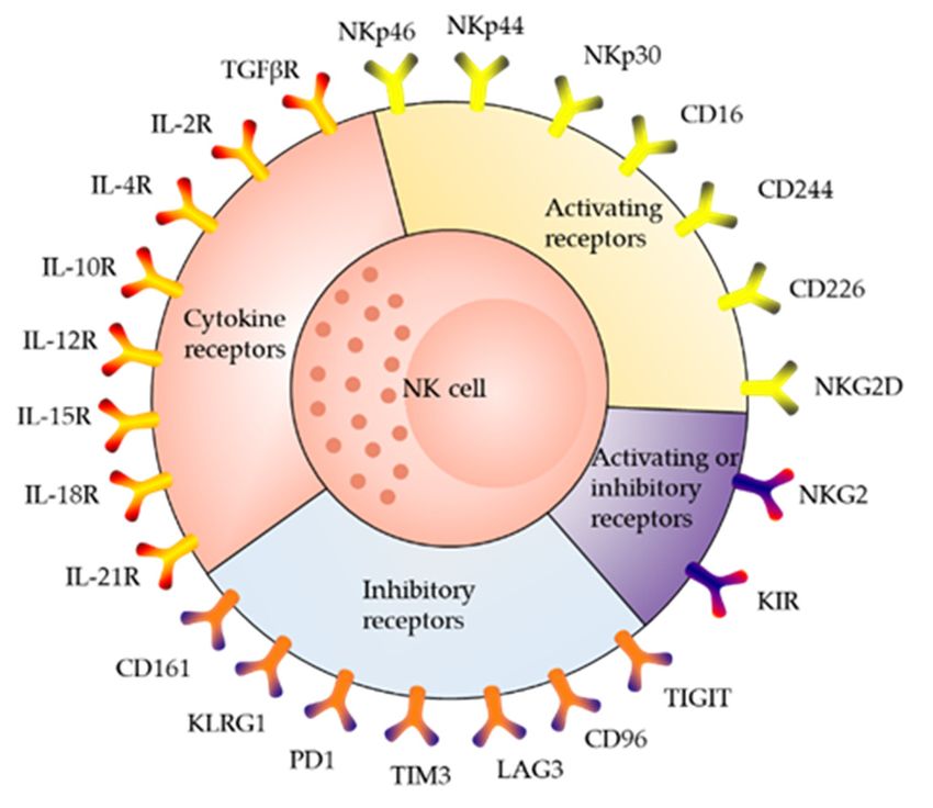
Int. J. Mol. Sci. 2019, 20, 317 2 of 20
Int. J. Mol. Sci. 2019, 20, x 2 of 20
2. Receptor
2. Receptor Distribution
Distribution and
and Killing
Killing Mechanism
Mechanism of
of NK
NK Cells
Cells
As the body's
As body’s first
firstline
lineofofdefense,
defense,many
many surface
surfacemolecules
molecules (Figure 1) are
(Figure expressed
1) are expressed on NK on cells
NK
[9,10],[9,10],
cells whichwhich
are characterized by the expression
are characterized of CD56 and
by the expression CD16 and
of CD56 cell surface markers,
CD16 cell surface andmarkers,
absence
of T-cell
and absencereceptor (TCR)
of T-cell and B-cell
receptor receptor.
(TCR) According
and B-cell to differences
receptor. Accordinginto CD56 and CD16
differences expression
in CD56 and
density,
CD16 two majordensity,
expression subsetstwoof NK cellssubsets
major can be distinguished,
of NK cells can i.e.,
beCD56bright
distinguished,and i.e.,
CD56dim
CD56bright [3,5,11,12].
and
CD56dim [3,5,11,12].
CD56dim NK cells are fully mature,
CD56dim account
NK cells for approximately
are fully mature, account90% for of the NK cells 90%
approximately in the of PB, and
the NK
predominantly mediate cytotoxicity responses [5]. CD56dim cells can play
cells in the PB, and predominantly mediate cytotoxicity responses [5]. CD56dim cells can play roles roles in antibody-
dependent
in cell-mediated
antibody-dependent cytotoxicity (ADCC)
cell-mediated through
cytotoxicity (ADCC)the surface
through expression
the surface of expression
CD16 (FcγRIII) [13–
of CD16
15]. CD56bright
(FcγRIII) [13–15].cells are immature,
CD56bright account
cells are for approximately
immature, 5–15% of the NK
account for approximately 5–15%cellsofinthe
theNK PB,cells
and
are predominant in tissues and secondary lymphoid organs [16]. CD56bright
in the PB, and are predominant in tissues and secondary lymphoid organs [16]. CD56bright cells cells express high levels
of CD56,high
express CD94/NKG2A, L-selectin,
levels of CD56, and a high-affinity
CD94/NKG2A, L-selectin,interleukin (IL)-2 receptor,
and a high-affinity but little
interleukin (IL)-2 to receptor,
no CD16
and killer-cell immunoglobulin-like receptor. Therefore, CD56bright cells function
but little to no CD16 and killer-cell immunoglobulin-like receptor. Therefore, CD56bright cells function primarily to
secrete cytokines,
primarily to secretesuch as interferon-γ,
cytokines, tumor necrosis
such as interferon-γ, factor
tumor (TNF)-α,
necrosis and(TNF)-α,
factor granulocyteandmacrophage-
granulocyte
colony-stimulating factor [11,17].factor [11,17].
macrophage-colony-stimulating
Figure
Figure 1. Majorreceptors
1. Major receptorsexpressed
expressedonon thethe surface
surface of natural
of natural killer
killer (NK)(NK)
cells.cells. NKp46,
NKp46, natural
natural killer
killer cell p46-related protein; NKp44, natural killer cell p44-related protein; NKp30,
cell p46-related protein; NKp44, natural killer cell p44-related protein; NKp30, natural killer cell p30- natural
killer cellprotein;
related p30-related
CD, protein;
Cluster CD, Cluster of differentiation;
of differentiation; NKG2, also NKG2, knownalsoas known
CD159; as CD159;
KIR, KIR,
killer-cell
killer-cell immunoglobulin-like receptor; TIGIT, T cell immunoreceptor with
immunoglobulin-like receptor; TIGIT, T cell immunoreceptor with Ig and ITIM domains; LAG3, Ig and ITIM domains;
LAG3, lymphocyte
lymphocyte activation
activation gene gene 3 protein;
3 protein; TIM3,TIM3,
T cellT cell immunoglobulinmucin
immunoglobulin mucinreceptor
receptor 3;
3; PD1,
PD1,
programmed cell death protein 1; KLRG1, killer cell lectin-like receptor subfamily G
programmed cell death protein 1; KLRG1, killer cell lectin-like receptor subfamily G member 1; IL-member 1; IL-2R,
interleukin-2 receptor;
2R, interleukin-2 TGFβR,
receptor; transforming
TGFβR, growth
transforming growthfactor beta beta
factor receptors.
receptors.
NK cells do not express antigen-specific recognition receptors. There are two receptors with
NK cells do not express antigen-specific recognition receptors. There are two receptors with
opposite functions on their surface. The first type of receptor can bind to its corresponding ligand
opposite functions on their surface. The first type of receptor can bind to its corresponding ligand on
on the surface of the target cell, activating the killing effects of NK cells. This receptor is called the
the surface of the target cell, activating the killing effects of NK cells. This receptor is called the
activating NK cell receptor [18]. The other type of receptor, called the inhibitory NK cell receptor,
activating NK cell receptor [18]. The other type of receptor, called the inhibitory NK cell receptor,
inhibits the killing effect of NK cells [18].
inhibits the killing effect of NK cells [18].
Both the activating receptor and inhibitory receptor can recognize classical or nonclassical major
Both the activating receptor and inhibitory receptor can recognize classical or nonclassical major
histocompatibility complex (MHC) class I molecules expressed on the surface of normal cells. Inhibitory
histocompatibility complex (MHC) class I molecules expressed on the surface of normal cells.
receptors play a primary role in preventing NK cells from killing normal cells [19,20]. In virus-infected
Inhibitory receptors play a primary role in preventing NK cells from killing normal cells [19,20]. In
cells and tumor cells, MHC class I molecules on the cell surface are lost or downregulated; activating
virus-infected cells and tumor cells, MHC class I molecules on the cell surface are lost or
NK cell receptors play a leading role in this process.
downregulated; activating NK cell receptors play a leading role in this process.
Activated NK cells exert cytotoxic effects mainly through three pathways (Figure 2). The first
Activated NK cells exert cytotoxic effects mainly through three pathways (Figure 2). The first
pathway is the perforin/granzyme pathway [9]. In this pathway, granzymes and perforin stored in
pathway is the perforin/granzyme pathway [9]. In this pathway, granzymes and perforin stored in
the cytoplasmic granules of NK cells are released by activated NK cells together into the intercellular
the cytoplasmic granules of NK cells are released by activated NK cells together into the intercellular
space. The structure of perforin is similar to that of complement, which can form a transmembrane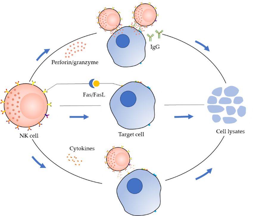
Int. J. Mol. Sci. 2019, 20, 317 3 of 20
Int. J. Mol. Sci. 2019, 20, x 3 of 20
space. The structure of perforin is similar to that of complement, which can form a transmembrane
channel
channeldirectly
directly on on
the the
target cell membrane,
target resulting
cell membrane, in increased
resulting permeability
in increased of the cell membrane
permeability of the cell
and ultimately
membrane andcausing osmotic
ultimately lysis
causing of the lysis
osmotic targetofcell. The perforation
the target channel formed
cell. The perforation channel byformed
perforin by
facilitates
perforin the entry ofthe
facilitates granzyme
entry ofinto the targetinto
granzyme cell. the
Additionally,
target cell. perforin causes redistribution
Additionally, perforin causes of
granzymes
redistributionin the
of cytoplasm
granzymes and nucleus
in the of target
cytoplasm andcells, allowing
nucleus granzymes
of target to accumulate
cells, allowing granzymesat theto
cleavage site; at
accumulate this promotes
the cleavagethe lysis
site; of promotes
this target cellsthe
andlysis
ultimately apoptosis.
of target cells andThe second pathway
ultimately apoptosis. is the
The
Fas/FasL pathwayis[21,22].
second pathway The Fas
the Fas/FasL molecule,
pathway also known
[21,22]. The Fas asmolecule,
Apo-1 or CD95, is a type-I
also known transmembrane
as Apo-1 or CD95, is
protein,
a type-Ⅰ andtransmembrane
Fas ligand (FasLprotein,
or CD95L)andisFas
a type-II
ligandtransmembrane
(FasL or CD95L) protein that belongs
is a type-Ⅱ to the TNF
transmembrane
family.
proteinWhen FasL binds
that belongs to theto TNF
Fas, Fas can When
family. deliverFasL
a “death
bindssignal”
to Fas, to
Fasthe
cancell, and the
deliver target signal”
a “death cell thento
undergoes
the cell, andapoptosis within
the target cell athen
fewundergoes
hours. In the third pathway,
apoptosis within the
a fewcytokine
hours. pathway,
In the thirdNKpathway,
cells secretethe
cytokine e.g.,
cytokines, pathway,
TNF-αNK [9], cells
whichsecrete cytokines,
can alter e.g.,ofTNF-α
the stability [9], in
lysosomes which
targetcan alter
cells, the stability
leading to leakageof
lysosomes
of in target affecting
various hydrolases, cells, leading to leakage
cell membrane of various metabolism,
phospholipid hydrolases, and affecting cell target
activating membranecell
phospholipid metabolism, and activating target cell endonuclease to degrade genomic DNA.
endonuclease to degrade genomic DNA.
Figure2.2.Mechanisms
Figure Mechanismsofofcytotoxicity
cytotoxicityby
bynatural
naturalkiller
killer(NK)
(NK)cells.
cells
3.3.Currently
CurrentlyKnown
KnownNK NKCell
CellLines
Lines
NK
NKcell
celllines
linesare
aremonoclonal
monoclonalcells
cellsthat
thatcan
cansurvive
survivepermanently
permanentlyininvitro.
vitro.Here,
Here,we
wesummarize
summarize
currently established primary NK cell lines (Table 1).
currently established primary NK cell lines (Table 1).
3.1. NK3.3 Cells
3.1. NK3.3 Cells
NK3.3 cells are a normal NK-derived cell line obtained by Kornbluth and co-workers in 1982 [23].
NK3.3 cells are a normal NK-derived cell line obtained by Kornbluth and co-workers in 1982
These cells are dependent on Interleukin-2 (IL-2) and are positive for CD2, CD11a, CD38, CD45, CD16,
[23]. These cells are dependent on Interleukin-2 (IL-2) and are positive for CD2, CD11a, CD38, CD45,
and CD56 [24]. The morphology, immunohistochemistry, and phenotype of NK3.3 cells are similar to
CD16, and CD56[24]. The morphology, immunohistochemistry, and phenotype of NK3.3 cells are
those of large granular lymphocytes (LGLs). NK3.3 cells have very strong cytotoxicity, which can kill
similar to those of large granular lymphocytes (LGLs). NK3.3 cells have very strong cytotoxicity,
NK-sensitive target cells, such as K562 and MOLT-4 cells [23,25,26].
which can kill NK-sensitive target cells, such as K562 and MOLT-4 cells [23,25,26].
3.2. YT Cells
3.2. YT Cells
The YT cell line was established in 1983 by Yodoi and co-workers, derived from a 15-year-old
The YT
pericardial cell line
effusion ofwas established
a Japanese man in 1983 byfrom
suffering Yodoi andlymphoma
acute co-workers,and
derived from[27].
thymoma a 15-year-old
YT cells
pericardial effusion of a Japanese man suffering from acute lymphoma and thymoma [27]. YT cells
show varying sizes, irregular nuclei, and abundant vacuoles, and azurophilic particles in the cytoplasm.
show varying sizes, irregular nuclei, and abundant vacuoles, and azurophilic particles
Additionally, YT cells are positive for CD56 and negative for CD2, CD3, and CD16. Importantly, in YT
the
cytoplasm. Additionally, YT cells are positive for CD56 and negative for CD2, CD3,
cells can undergo long-term in vitro expansion without the need for conditioned medium or IL-2. and CD16.
Importantly,
Moreover, YT cells
YT cells havecan undergo
toxic long-term
effects on in vitro
cancer cells, such expansion withoutHPB-ALL,
as K562, MOLT-4, the need and
for HSB-2
conditioned
cells.
medium or IL-2. Moreover, YT cells have toxic effects on cancer cells, such as K562, MOLT-4, HPB-
ALL, and HSB-2 cells.Int. J. Mol. Sci. 2019, 20, 317 4 of 20
YT2C2 and YTC3 cells are two subclones of YT cells [28]. These two cell subclones do
not mediate ADCC. However, these cell lines can kill 721.221 cells with high expression of
B7.1 [29]. Cytotoxicity analysis showed that YT2C2 and YTC3 cells primarily rely on cell surface
receptor-mediated cytotoxicity.
3.3. NKL Cells
The NKL cell line was established in 1996 by Robertson and co-workers and was derived from the
PB of a patient with LGL leukemia [30]. NKL cells require IL-2 for sustained growth and die if deprived
of IL-2 for more than 7 days. The morphology of NKL cells is similar to that of activated NK cells, which
have natural killing activity and can mediate ADCC. According to phenotypic analysis, NKL cells
overexpress CD2, CD6, CD11a, CD27, CD29, CD38, CD43, CD58, CD94, and CD95, but are negative
for CD3, CD4, CD5, CD8, CD14, CD19, CD20, and CD28. As the culture time in vitro increased, the
cell surface expression of CD16, CD56, and CD57 and the antigen density were significantly reduced.
3.4. HANK1 Cells
The HANK1 cell line was established in 1998 by Kagami and co-workers [31]. This cell line
was derived from a 46-year-old woman with CD56+ NK/T-cell lymphoma. HANK1 cells are large
polymorphic cells with irregular nuclei and azurophilic granules in the cytoplasm. Immunophenotypic
analysis showed that HANK1 cells were positive for CD2, CD3ε, CD56, and CD25. Moreover,
HANK1 cells exhibit IL-2 dependence during in vitro culture and retain the biological characteristics
of the original tumor cells, allowing them to be used as a model of abnormal nasal-type NK/T-cell
lymphoma [31].
3.5. NK-YS Cells
The NK-YS cell line was established by Tsuchiyama and co-workers in 1996 [32]. This cell line
was derived from a 19-year-old woman with leukemic-state nasal angiocentric NK cell lymphoma
with systemic skin infiltration. Immunophenotypic analysis showed that NK-YS cells retain the
characteristics of prototypic NK lymphoma cells and positively express CD2, CD5, CD7, and CD56,
but are negative for CD3, CD16, and CD57.
3.6. KHYG-1 Cells
The KHYG-1 cell line was established by Yagita and co-workers in 1997 and was derived from
a 45-year-old woman with aggressive NK cell leukemia [33]. KHYG-1 cells have the morphological
characteristics of LGLs, i.e., large nuclei, rough chromosomes, obvious nucleoli, abundant cytoplasm,
basophilic features, and azurophilic granules. Additionally, these cells rely on IL-2 during in vitro
culture. The immunophenotype of KHYG-1 cells is as follows: positive for CD2, CD3ε, CD7, CD8αα,
CD33, CD56, CD122, and CD132, and negative for CD1, CD3, CD16, CD25, CD34, and CD57. Suck and
co-workers [34] found that the KHYG-1 cell line is more cytotoxic to K562 cells and leukemia cells lines,
such as EM2, EM3, and HL60. Additionally, KHYG-1 cells are most likely to regulate cell lysis with
perforin by granzyme M (instead of granzymes A and B). Researchers have predicted that KHYG-1
may induce apoptosis in tumor cells through a novel granzyme/perforin pathway.
3.7. SNK-6 and SNT-8 Cells
SNK-6 and SNT-8 cells are nasal NK/T-cell lymphoma cells established in 2001 by Nagata and
co-workers in Japan [35]. SNK-6 cells were derived from a 62-year-old Japanese man, and SNT-8
cells were derived from a 48-year-old Japanese woman. These two cell lines were established using a
single-cell suspension culture method. Tumor cells from male patients were cultured for 20 months
and named SNK-6, and tumor cells from female patients were cultured for 17 months and named
SNT-8. Both cell lines rely on IL-2 for growth. Immunophenotypic analysis showed that SNK-6 cellsInt. J. Mol. Sci. 2019, 20, 317 5 of 20
are positive for CD56 and negative for CD3, CD4, CD8, CD19, and TCR (TCRα/β and TCR γ/δ),
whereas SNT-8 cells are positive for CD3, CD56, and TCRγ/δ, and negative for CD4, CD8, CD19,
and TCRα/β. These two cell lines have been shown to have important applications in studies of cell
necrosis caused by nasal T/NK cell lymphoma.
3.8. IMC-1 Cells
The IMC-1 cell line was established in 2004 by Chen and colleagues [36]. These cells are an
IL-2-dependent leukemia cell line obtained from an adult patient with invasive NK cell leukemia.
IMC-1 cells express CD56, CD2, CD11a, CD38, and HLA-DR cell surface antigens and lyse target cells
in a non-MHC-restricted and antibody-independent manner.
3.9. NK-92 Cells
The NK-92 cell line was isolated and successfully established by Klingemann’s group in 1992
from a 50-year-old man with malignant non-Hodgkin’s lymphoma [37]. NK-92 cells express CD2 and
CD56 and are negative for CD3, CD4, CD8, and CD16. The growth of NK-92 cells is dependent on the
presence of recombinant IL-2; once IL-2 is withdrawn, the cells will die within 72 h. NK-92 cells tend to
grow in an aggregated pattern and easily die when dispersed [24,37]. Similar to NK cells, NK-92 cells
can kill tumor cells without prior sensitization. Additionally, NK-92 cells lack the CD16 receptor and
therefore cannot mediate ADCC. However, these cells still exhibit a high degree of cytotoxic activity
toward a variety of cancers cells [38–40]. NK-92MI and NK-92CI cells are derived from NK-92 cells
and have biological characteristics similar to those of NK-92 cells, but do not rely on IL-2 [38].
Table 1. Currently known NK cell lines.
Doubling Viral Primary
Cell Line Year Disease Diagnosis Patient Cytokine
Time Status Reference
NK3.3 1982 NR NR NR EBV− IL-2-dependent [23]
Acute lymphoblastic
15-year-old Independent
YT 1983 lymphoma (with 40–50 h EBV+ [27]
male of IL-2
thymoma)
63-year-old
NKL 1996 NK-LGLL 24–48 h NR IL-2-dependent [30,41]
male
Nasal-like NK/T-cell 46-year-old
HANK1 1998 3 day EBV+ IL-2-dependent [31]
lymphoma female
NK cell lymphoma,
Nasal angiocentric,
19-year-old
NK-YS 1996 Leukemic state with 48 h EBV+ IL-2-dependent [32]
female
systemic skin
infiltration
Aggressive NK 45-year-old
KHYG-1 1997 24–48 h EBV− IL-2-dependent [33]
leukemia female
Nasal NK/T-cell 62-year-old
SNK-6 1998 NR EBV+ IL-2-dependent [35]
lymphoma male
Nasal NK/T-cell 48-year-old
SNT-8 1998 NR EBV+ IL-2-dependent [35]
lymphoma female
Aggressive NK cell 42-year-old
IMC-1 2004 24–36 h EBV− IL-2-dependent [36]
leukemia male
IL-2-dependent;
50-year-old
NK-92 1992 LGL-NHL 24 h EBV− Growth [37]
male
stimulation:IL-7
NR, not reported; EBV, Epstein-Barr virus; NK, natural killer cell; LGL: large granular lymphocyte; LGLL: large
granular lymphocyte leukemia; NHL: non-Hodgkin’s lymphoma.
4. Progress in the Application of NK-92 Cells
Among the several NK cell lines currently established, NK-92 cells lack almost all inhibitory killing
receptors and express a series of activating receptors [42]. Additionally, NK-92 cells are abundant in
perforin and granzyme, suggesting a broad spectrum of cytotoxic effects. Accordingly, these cells haveInt. J. Mol. Sci. 2019, 20, 317 6 of 20
become a critical NK cell line for preclinical research and are the only NK cell line approved by the US
Food and Drug Administration (FDA) for phase I and II clinical studies [43–47].
After the establishment of NK-92 cells, many studies were carried out to demonstrate the potential
applications of NK-92 cells in the treatment of tumors. Yan and co-workers [43–48] studied the
cell-killing effects of NK-92 cells on primary tumor cells derived from various leukemia patients
using chromium 51 release tests. The results showed that NK-92 cells were cytotoxic to leukemia cells
both in vitro and in vivo. Swift and co-workers studied the cytotoxicity of NK-92 cells to bulk and
clonogenic multiple myeloma and evaluated the effects of NK-92 cell treatment in a xenograft mouse
model. The results demonstrated that NK-92 cells could effectively kill clonogenic and bulk multiple
myeloma cells and could significantly reduce tumor burden in vivo [49].
The growth of NK-92 cells is dependent on the presence of IL-2; however, systemic administration
of IL-2 can result in unexpected side effects, such as high toxicity and nonlocalized administration [50].
To make NK-92 cells more suitable for clinical applications, Konstantinidis and co-workers used
gene editing to express IL-2 in the endoplasmic reticulum of NK-92 cells; the engineered NK-92 cells
could grow normally independent of IL-2 and exhibited the same function and cytotoxicity as NK-92
cells [50].
In combination with current good manufacturing practice (cGMP)-compliant expansion
methodologies [51], NK-92 cells are approved for analysis in clinical trials to determine their utility
in the treatment of some types of malignant tumors [47,52]. For example, Tam and co-workers [51]
demonstrated that when NK-92 cells were irradiated with 500 cGy gamma rays, the proliferation of
NK-92 cells was prevented, and their high killing activity was maintained. Moreover, even when
the irradiation dose was increased to 1000 cGy, the cytotoxic effects of NK-92 cells were maintained
within 48 h. Additionally, Tonn and co-workers [47] conducted a comprehensive study of the possible
limitations of NK-92 cells in clinical applications and determined the optimal culture conditions
and operating procedures for large-scale expansion under GMP conditions. Subsequently, a phase I
trial was performed in seven patients with advanced cancer; the NK-92 cells were infused twice per
patient with an interval of 48 h, and individual patients developed low-heat, back pain, and other
uncomfortable reactions after treatment. Generally, it was found to be safe to infuse 5 × 10⁹ /m2 of
NK-92 cells per patient. Further experiments are needed to determine the efficacy of NK-92 cells in
advanced cancer [47].
5. Structure of CARs and Their Applications in NK-92 Cells
In recent studies, major breakthroughs in the use of CAR-T cells for relapsed and refractory B cell
malignancies targeting CD19 have been made [53–62], and two CD19 CAR-T cell products have been
approved by the US FDA [63,64].
However, there are many limitations to the use of CAR-T cells, including off-target effects
and cytokine storms. Therapy with CAR-T cells has not yet been successful in patients with solid
tumors, and the production of autologous cells also has many limitations in the clinical setting [65].
The production of CAR-T cells requires a certain time period, which makes it challenging to prepare a
sufficient number of CAR-T cells within a short time for patients whose disease progresses faster. It is
also difficult to collect a sufficient number of healthy T cells from the patient. “Off-the-shelf” allogeneic
T cells can overcome these difficulties, but may cause severe graft-versus-host disease (GVHD) [66].
CAR-NK-92 cells are highly cytotoxic and can be harvested as phenotypically homogeneous
cells, with production of large numbers of cells within a short period. Additionally, compared with
CAR-T cells, CAR-NK-92 cells cannot proliferate after irradiation; thus, the survival time in vivo is
shorter, avoiding some off-target effects [67]. Even if the targeted antigen on the tumor is rapidly lost,
the CAR-NK-92 cells can still be activated by their activating receptors, conferring CAR-NK-92 cells
with significant advantages.Int. J. Mol. Sci. 2019, 20, x 7 of 20
the CAR-NK-92 cells can still be activated by their activating receptors, conferring CAR-NK-92 cells
Int. J. Mol. Sci. 2019, 20, 317 7 of 20
with significant advantages.
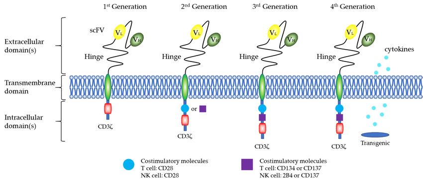
5.1.
5.1.Structure
StructureofofCARs
CARs
The
Thetraditional
traditionalCARCARvector
vectorstructure
structureconsists
consistsof ofthree
threeparts
parts(Figure
(Figure3), 3),including
includingan anextracellular
extracellular
antigen recognitionregion,
antigen recognition region, a transmembrane
a transmembrane region,
region, and intracellular
and intracellular signal which
signal domain, domain, which
determine
determine the specificity and functionality of the CAR modification. Current
the specificity and functionality of the CAR modification. Current CAR technology is in the fourth CAR technology is in
the fourth generation of development [68]. The first generation of CARs consisted
generation of development [68]. The first generation of CARs consisted of a single-chain variable of a single-chain
variable
fragmentfragment (scFv)
(scFv) that that recognizes
recognizes tumor tumor
surfacesurface
antigens,antigens,
and anand an immunoreceptor
immunoreceptor tyrosine-
tyrosine-based
based
activation motif (ITAM, usually CD3ζ) [69]. However, the first generation of CARs only causedcaused
activation motif (ITAM, usually CD3ζ) [69]. However, the first generation of CARs only T cells
Ttocells to proliferate
proliferate for aperiod
for a short short and
period
didand did not provide
not provide long-term long-term T-cell expansion
T-cell expansion signals tosignals
maintainto
maintain
antitumorantitumor
effects. effects.
According
According to to the
the dual-signal
dual-signalmodel
modelofof T-cell
T-cell activation,
activation, the the second
second and and
thirdthird generations
generations of CARsof
CARs introduced CD28, CD134 (OX40), CD137 (4-1BB) [70], and other costimulatory
introduced CD28, CD134 (OX40), CD137 (4-1BB) [70], and other costimulatory molecules. The aim molecules. The
aim
waswas to enhance
to enhance the antitumor
the antitumor cytotoxicity
cytotoxicity and and proliferative
proliferative capacity
capacity of T of T cells
cells in vivo.
in vivo.
To
To more completely remove tumor cells, Chmielewski and co-workers [71,72] designed
more completely remove tumor cells, Chmielewski and co-workers [71,72] designed thethe
“fourth
“fourth generation”
generation” CAR CAR structure.
structure. The
The fourth
fourth generation
generation of of CARs
CARs contains
contains twotwo costimulatory
costimulatory
molecules
molecules(CD28,
(CD28, CD134,
CD134, or orCD137)
CD137) and
and induces
induces thethesecretion
secretion ofof IL-12
IL-12 from
from the
the cell.
cell. In
Inthe
thetreatment
treatment
of
of solid cancer, fourth-generation CAR-T cells release IL-2 and activate innate immune cells
solid cancer, fourth-generation CAR-T cells release IL-2 and activate innate immune cells to
to
eliminate
eliminate antigen-negative cancer cells,
antigen-negative cancer cells,thereby
therebyincreasing
increasingthe theantitumor
antitumoreffecteffectofof these
these cells
cells in in vivo
vivo by
by several
several fold.
fold.
Generationsof
Figure3.3.Generations
Figure ofCAR
CARdesign.
design.The
Thetraditional
traditionalCAR
CARvector
vectorstructure
structureconsists
consistsof
ofthree
three parts:
parts: an
an
extracellularantigen
extracellular antigenrecognition
recognitionregion,
region,aatransmembrane
transmembraneregion,
region,and
andananintracellular
intracellularsignal
signaldomain.
domain.
The extracellular domain of CAR includes an scFv region (H [heavy] and L [light] chain) that is spliced
The extracellular domain of CAR includes an scFv region (H [heavy] and L [light] chain) that is spliced
by a linker. A hinge ensures flexibility and connects to the transmembrane domain. The intracellular
by a linker. A hinge ensures flexibility and connects to the transmembrane domain. The intracellular
domain includes a CD3ζ signaling domain and costimulatory domains, such as CD28, CD134, CD137,
domain includes a CD3ζ signaling domain and costimulatory domains, such as CD28, CD134, CD137,
and 2B4.
and 2B4.
CARs of NK Cells
5.1.1. CARs of NK Cells
Similar to CAR-T cells, the goal of CAR-NK cells is to establish a new activation pathway to
Similar
enhance the to CAR-T effects
antitumor cells, the goal
of the of CAR-NK
cells cells istumor
and to improve to establish a new CAR-NK
cell targeting. activationhave
pathway to
the basic
enhance the antitumor effects of the cells and to improve tumor cell targeting. CAR-NK
framework of CAR-T (Figure 3), including an extracellular antigen recognition region, a transmembrane have the
basic
region,framework of CAR-Tsignal
and an intracellular (Figure 3), including an extracellular antigen recognition region, a
domain.
transmembrane region, and an intracellular
CD3ζ is a classical intracellular signal segment signal domain.
of the CAR structure [73] and plays an important
CD3ζ is a classical intracellular signal
role in NK cells [74]. CAR-NK generally uses CD3ζ as segment of the
the CAR structure
first signal [73]
motif and plays an important
(first-generation CAR) and
role in NK cells [74]. CAR-NK generally uses CD3ζ as the first signal motif
then a costimulatory molecular motif (second-generation CAR), such as CD28 or CD137 (4-1BB) (first-generation CAR)
[75],
and then a costimulatory molecular
to form an intracellular signal region. motif (second-generation CAR), such as CD28 or CD137 (4-1BB)
[75], to form anisintracellular
NKG2D signal region.
a crucial activating receptor expressed on most CD8+ T cells and NK cells and is a
relatively unique activating receptor in NK cells. The NKG2D receptor binds to DAP10 or DAP12Int. J. Mol. Sci. 2019, 20, 317 8 of 20
transfer proteins to provide different activation signals [76]. Both signals can activate the cytotoxicity
of NK cells, but only the activation signal transmitted by DAP12 can promote the production of
cytokines by NK cells [77]. In one study [78], researchers linked DAP10 and CD3ζ to the NK
cell activation receptor NKG2D. In an osteosarcoma mouse model, the cytotoxic potential of NK
cells against a wide spectrum of tumor subtypes could be markedly enhanced by expression of
CAR-NKG2D-DAP10-CD3ζ receptor.
CD244, also known as the NK cell receptor 2B4, is a signal transduction lymphocyte-activating
molecule-related receptor expressed in all NK cells [79]. This protein is an important regulator of NK
cell activation and was shown to have robust costimulatory roles in a study in which NK cells were
used as effector cells to target CD19 or GD2 [80].
5.2. Preclinical Studies of CAR-NK-92 Cells
NK-92 cells are an ideal CAR carrier with natural antitumor properties and are easy to cultivate
and modify in vitro. The first generation of CAR has been widely applied in CAR-NK-92 cells
(Table 2) [16].
Uherek and co-workers [81] reported the expression of an antigen recognition receptor on the
surface of NK-92 cells by gene editing. This receptor recognizes the tumor-associated antigen ErbB2,
which is overexpressed in various epithelial tumors. Moreover, they used a first-generation technique
similar to CAR-T cells. The entire CAR structure included the extracellular region ErbB2-specific scFv
(FRP5) antibody fragment, a CD8 hinge region, a transmembrane region, and an intracellular CD3ζ
chain. In vitro experiments indicated that compared with the parental cells, the genetically modified
NK-92-scFv (FRP5)-CD3ζ cells were able to specifically recognize and effectively kill ErbB2-expressing
tumor cells from different sources. Subsequently, CAR-NK-92, the first-generation CAR structure
of different targets, showed good effects against hematomas and solid tumors. This structure also
included CD19 and CD20 for B cell leukemia and lymphoma [82–85], CD138 for myeloma [86],
human epidermal growth factor receptor 2 (HER2) and epidermal growth factor receptor for brain
metastasis [87–89], HER2 and EGFR for breast cancer [81,90–94], and GD2 for neuroblastoma [95,96].
Additionally, NK-92 cells do not express CD16 and therefore do not mediate the effects of ADCC.
In recent years, methods for expressing CD16 have been established, and expression of CD16 in
NK-92 cells can effectively enhance the antitumor effects of the cells [97]. Boissel and co-workers
demonstrated that CAR-NK-92 cells have the ability to clear chronic lymphocytic leukemia cells to a
greater extent than NK-92 cells expressing CD16 [85].
The efficacy and safety of CAR-NK-92 cells based on second- and third-generation CARs (Table 3)
were also confirmed in preclinical trials.
In one study, Oelsner and co-workers [83] compared the cytotoxic effects of
CAR-CD19-CD3ζ-NK-92, CAR-CD19-CD28-CD3ζ-NK-92, and CAR-CD19-CD137-CD3ζ-NK-92
on established B-cell leukemia and lymphoma cells. The results showed that all three
CD19-specific CAR-NK-92 cell lines were effective at killing B cell malignancies. However,
CAR-CD19-CD137-CD3ζ-NK-92 cells were less effective than CAR-CD19-CD3ζ-NK-92 and
CAR-CD19-CD28-CD3ζ-NK-92 cells at cell killing and cytokine production, indicating the differential
effects of the costimulatory CD28 and CD137 domains. In a recent study [98], researchers at
the University of California, San Diego evaluated the effects of different CAR constructs on NK
cell-mediated killing. They designed nine CAR constructs that target mesothelin. The killing
experiments in vitro revealed that a CAR containing the transmembrane domain of NKG2D, the 2B4
costimulatory domain, and the CD3ζ signaling domain of CAR could mediate strong antigen-specific
NK cell signaling. Subsequently, the effects of CAR-NK and CAR-T cells were compared in a mouse
model. The results showed that CAR-NK cells had in vivo activities similar to those of as CAR-T cells,
but with less toxicity. This research indicated that CAR-NK cell therapy may be safer than CAR-T
cell therapy.Int. J. Mol. Sci. 2019, 20, 317 9 of 20
5.3. Ongoing Clinical Trials
Clinical trials examining CAR-NK-92 cells for the treatment of tumors are being carried out
(Table 4). However, compared with CAR-T cells that have been applied in clinical studies, fewer clinical
studies have been performed for CAR-NK-92 cells [8,99], and little clinical data have been published.
In a recent clinical trial (NCT02944162), Tang and co-workers [100] revealed the efficacy and
safety of CAR-NK-92 cells in the treatment of relapsed and refractory acute myeloid leukemia. In this
study, a total of three patients received therapy with CD33-CAR NK-92 cells. The first patient was a
14-year-old girl and the second patient was a 24-year-old male, who both received doses of 3 × 10⁸ , 6
× 108 , and 1 × 10⁹ cells on days 1, 3, and 5, respectively. Another patient was a 49-year-old woman
who received doses of 1 × 10⁹ , 3 × 10⁹ , and 5 × 10⁹ cells on days 1, 4 and 7, respectively. All patients
had mild symptoms after treatment, including fever and cytokine release syndrome, but returned to
normal the next day. This is the first phase I clinical trial of CD33-CAR NK-92 cells in patients with
relapsed and refractory acute myeloid leukemia. Although the treatment effect was not significant, the
findings of this clinical study showed that CAR-NK-92 cells can be safely used.Int. J. Mol. Sci. 2019, 20, 317 10 of 20
Table 2. Preclinical studies with the first generation of CAR-NK-92 cells.
Antigen Intracellular Genetic Modification
Cancer Type Hinge TM Effector Cell Year References
Targeted Signal Domain Method
Multiple myeloma CD138 CD8 CD3ζ CD3ζ lentiviral vector NK-92MI 2014 [86]
B-cell malignancies CD19 CD8 NR CD3ζ Retrovirus NK-92 2016 [82]
B-cell malignancies CD19 CD8 CD28 CD3ζ Lentiviral NK-92 2017 [83]
CLL CD19 CD8 CD3ζ CD3ζ Electroporation NK-92 2009 [84]
ALL CD19
NR NR CD3ζ Lentivirus NK-92 2014 [85]
CLL CD20
B-cell malignancies CD20 CD8 CD3ζ CD3ζ Retroviral NK-92 2008 [101]
Prostate cancer EpCAM CD8 CD3ζ CD3ζ Retrovirus NK-92 2009 [102]
Prostate cancer EpCAM CD8 CD3ζ CD3ζ Retrovirus NK-92 2011 [103]
Neuroblastoma GD2 CD8 CD3ζ CD3ζ Retrovirus NK-92 2012 [96]
Neuroblastoma GD2 CD8 CD3ζ CD3ζ Retrovirus NK-92 2015 [95]
Melanoma GPA7 NR HLA-A2 CD3ζ Electroporation NK-92MI 2013 [104]
Brain metastasis HER2 CD8α CD3ζ CD3ζ Retrovirus NK-92 2016 [88]
Brain metastasis HER2 CD8 CD3ζ CD3ζ Retrovirus NK-92 2013 [89]
Breast cancer HER2 CD8 CD3ζ CD3ζ Retrovirus NK-92 2005 [91]
Breast cancer HER2 CD8 CD3ζ CD3ζ Retrovirus NK-92 2008 [92]
Breast/ovarian cancer HER2 CD8 CD3ζ CD3ζ Retrovirus NK-92 2002 [81]
Breast cancer, Ovarian cancer,
Melanoma HER2 CD8 CD3ζ CD3ζ Lentiviral NK-92 2015 [93]
Renal cell carcinoma
Ovarian cancer
Mesothelin CD8 NKG2D CD3ζ Transposon plasmids NK-92 2018 [98]
Mesothelin-expressing tumorsInt. J. Mol. Sci. 2019, 20, 317 11 of 20
Table 3. Preclinical studies with the second and third generations of CAR-NK-92 cells.
Intracellular
Cancer Type Antigen Targeted Hinge TM Genetic Modification Method Effector Cell Year References
Signal Domain
CD28-CD3ζ
B-cell malignancies CD19 CD8 CD28 Lentiviral NK-92 2017 [83]
CD137-CD3ζ
Multiple myeloma CS1 NR NR CD28-CD3ζ Lentivirus NK-92 2014 [105]
EBV+ cells EBNA3C NR NR CD137-CD3ζ Retrovirus NK-92MI 2012 [75]
EGFR
Glioblastoma NR CD28 CD28-CD3ζ Lentivirus NK-92 and NKL 2015 [106]
EGFRvIII
Brain metastasis EGFR NR NR CD28-CD3ζ Lentivirus NK-92 2016 [87]
EGFR
Glioblastoma CD8 CD28 CD28-CD3ζ Lentivirus NK-92 2015 [107]
EGFRvIII
Breast cancer EpCAM CD8 CD28 CD28-CD3ζ Lentivirus NK-92 2012 [90]
Breast cancer
CD28 CD28-CD3ζ
Renal cell carcinoma HER2 CD8 Lentiviral NK-92 2015 [93]
CD137 CD137-CD3ζ
Ovarian carcinoma Melanoma
Glioblastoma HER2 CD8 CD28 CD28-CD3ζ Lentiviral NK-92 2016 [108]
Breast cancer HER2 CD8 CD28 CD28-CD3ζ Electroporation NK-92 2015 [94]
Ovarian cancer
Mesothelin CD8 CD16 2B4-CD3ζ Transposon plasmids NK-92 2018 [98]
Mesothelin-expressing tumors
Ovarian cancer DAP10-CD3ζ
Mesothelin CD8 NKp44 Transposon plasmids NK-92 2018 [98]
mesothelin-expressing tumors 2B4-CD3ζ
Ovarian cancer 2B4-CD3ζ
Mesothelin CD8 NKG2D Transposon plasmids NK-92 2018 [98]
Mesothelin-expressing tumors CD137-CD3ζ
Ovarian cancer
Mesothelin CD8 CD28 CD28-CD137-CD3ζ Transposon plasmids NK-92 2018 [98]
Mesothelin-expressing tumors
2B4-DAP12-CD3ζ
Ovarian cancer
Mesothelin CD8 NKG2D 2B4-DAP10-CD3ζ Transposon plasmids NK-92 2018 [98]
Mesothelin-expressing tumors
CD137-2B4-CD3ζ
Aggressive T cell malignancies CD3 CD8 CD8 CD28-CD137-CD3ζ Lentivirus NK-92 2016 [109]
Aggressive T-cell malignancies CD5 CD8 CD8 CD28-CD137-CD3ζ Lentivirus NK-92 2017 [110]
NR, not reported; ALL, acute lymphoblastic leukemia; B-ALL, B-cell acute lymphoblastic leukemia; CLL, chronic lymphocytic leukemia; EGFR, epidermal growth factor receptor; HER2,
human epidermal growth factor receptor 2; TM, transmembrane domain; TAMs, tumor-associated macrophages.Int. J. Mol. Sci. 2019, 20, 317 12 of 20
Table 4. Clinical trials with CAR-NK-92 cells.
Estimated
NCT Number NK Cell Source Target Antigen Disease Phase Age Location References
Enrollment
Acute Myeloid
Leukemia;Precursor T-Cell
Lymphoblastic
Leukemia-Lymphoma; T-cell
Prolymphocytic Leukemia; T-cell
Large Granular Lymphocytic
18 Years and
Leukemia; Peripheral T-cell Phase 1
NCT02742727 NK-92 CD7 10 participants older (Adult, China NR
Lymphoma, NOS; Phase 2
Older Adult)
Angioimmunoblastic T-cell
Lymphoma Extranodal NK/T-cell
Lymphoma, Nasal Type;
Enteropathy-type Intestinal T-cell
Lymphoma; Hepatosplenic T-cell
Lymphoma
Acute Lymphocytic Leukemia;
Chronic Lymphocytic Leukemia; 3 Years to 80
Follicular Lymphoma; Mantle Cell Phase 1 Years (Child,
NCT02892695 NK-92 CD19 10 participants China NR
Lymphoma; B-cell Prolymphocytic Phase 2 Adult, Older
Leukemia; Diffuse Large Cell Adult)
Lymphoma;
Acute Myelogenous Leukemia;
Acute Myeloid Leukemia; Acute 3 Years to 80
Myeloid Leukemia with Phase 1 Years (Child,
NCT02944162 NK-92 CD33 10 participants China [100]
Maturation; Acute Myeloid Phase 2 Adult, Older
Leukemia Without Maturation; Adult)
ANLL
18 Years and
NCT03383978 NK-92 HER2 Glioblastoma Phase 1 30 participants older (Adult, Germany NR
Older Adult)
18 Years to 75
NCT03656705 NK-92 NR Non-small Cell Lung Cancer Phase 1 5 participants Years (Adult, China NR
Older Adult)
NR, not reported; Data are from http://www.clinicaltrials.gov.Int. J. Mol. Sci. 2019, 20, 317 13 of 20
5.4. Advantages of CAR-NK-92 Cells
CAR-NK cells are potential competitors for CAR-T cells. CAR-NK-92 cells do not cause
GVHD [111] and have greater cytotoxicity than ADCC [85]. Indeed, CAR-NK-92 cells have many
advantages, as follows: (1) CAR-NK-92 cells can target tumor cells and directly activate NK-92 cells to
kill target cells; (2) even if the targeted antigen on the tumor is rapidly lost, the CAR-NK-92 cells can
still be activated by their activating receptors [8]; (3) the inhibitory receptors are expressed at low levels
on the surface [37] and deletion of inhibitory receptors makes NK-92 cells more resistant to solid tumors
than other immune cells; and (4) NK-92 cells are immortalized cell lines with a uniform phenotype,
allowing them to be cultured in vitro for a long time to proliferate. CAR-NK-92 cells also have this
advantage, which can compensate for the decline in immune cell viability in patients with advanced
cancer. Depending on the type of tumor, corresponding CAR-NK-92 cells can be directly expanded for
treatment, thereby shortening the treatment cycle and reducing the cost of treatment [112].
5.5. Challenges and Coping Strategies
Although CAR-NK-92 cells have good antitumor effects, there are still some problems that need
to be solved to make them more suitable for clinical treatment.
5.5.1. Tumor-Producing and Potential Epstein-Barr (EB) Virus Susceptibility
NK-92 cells are derived from patients with malignant non-Hodgkin’s lymphoma; they may cause
secondary tumorigenesis and potential EB virus susceptibility after injection. Therefore, for safety
reasons, NK-92 cells need to be lethally irradiated before clinical application [16].
5.5.2. NK-92 Cells Have A Short Life Cycle after Irradiation
After irradiation, CAR-NK-92 cells survive in vivo for a short period. This method reduces the
side effects of CAR-NK-92 cells on the body, while also reducing the antitumor effects; thus, therapy
may require multiple injections.
5.5.3. Defects in the Transfected Vector
Genetic modification of cells is a critical step to realizing CAR expression. Virus transduction
is the most common method used for genetic modification. These viral vectors include retroviral
vectors, lentiviral vectors, and adenoviral vectors, among which retroviral vectors and lentiviral
vectors are most widely used. Viral vectors are capable of ensuring stable expression of the transgene.
However, the viral vector itself may be related to high production costs, a long reproduction cycle,
and complexity. Additionally, viral vectors alone cannot accurately insert genes into the desired
location, posing potential risks; for example, they may affect the expression of normal genes or cause
the inserted genes to be abnormally regulated. Accordingly, it is essential to develop non-viral vectors.
The CRISPR-Cas9 system can be combined with electroporation techniques to precisely insert genes
into the genome and achieve stable expression. Roth and co-workers [113] successfully demonstrated
that the system can successfully edit the genome at low cost and complete T-cell modification in just a
few weeks.
5.5.4. Off-Target Effects
CAR-NK-92 cells are target dependent and mainly kill cells with high expression of specific
antigens. However, once the antigens are also expressed in normal tissues and cells, off-target toxicity
can occur. Currently, there are no specific clinical data to assess the severity of the off-target toxicity
of CAR-NK-92 cells. Accordingly, it is necessary to consider dose control and other factors (e.g.,
introducing suicide genes) in order to reduce the risk of toxicity [114].Int. J. Mol. Sci. 2019, 20, 317 14 of 20
6. Conclusions and Perspectives
The antitumor ability of NK cells confers them with broad potential applications in cell therapy.
NK cells modified by CAR have also been shown to be promising as effector cells. However, the
exploration of CAR-NK cells is still at a preliminary stage. It is necessary to examine which types
of CAR structures are most effective for activating NK cells and which intracellular signal domains
can maintain the killing ability of NK cells for a long time. Although currently used viral vectors can
be stably integrated into the genome, there is a risk of random insertion. Thus, development of new
non-viral transfection methods is necessary. NK-92 cells, an immortalized cell line, can be quickly
and easily obtained in the clinical setting. Additionally, the cells must be subjected to lethal radiation
before use in order to avoid the risk of secondary tumorigenicity. Controllable NK-92 cells, such as
those expressing a combination of suicide genes, should be further explored. With advancements in
CAR-NK technology and the accumulation of clinical experience, combination of CAR-NK cells with
other anticancer therapies may also show efficacy. Although CAR-NK cells induce a low incidence of
cytokine storms and induce few side effects compared with CAR-T cells, further studies are needed
to fully assess the safety of these cells. As preclinical trials and clinical studies proceed, NK cells are
expected to play important roles in the treatment of cancer.
Author Contributions: Y.D. and J.Z. conceived and wrote this manuscript, H.Z. contributed additional material
and editing help. All the authors have approved the manuscript.
Funding: This work was supported financially by National Natural Science Foundation of China (81371669),
Project of Science and Technology of Quanzhou of China (Grant No. 2016Z006), Major project of University
and Industry Cooperation in Fujian Province of China (Grant No. 2018Y4009), and the Subsidized Project for
Cultivating Postgraduates’ Innovative Ability in Scientific Research of Huaqiao University (Grant No. 1601116007).
Acknowledgments: We thank Qingwang Han for his valuable discussion and advice.
Conflicts of Interest: The authors declare no conflict of interest.
Abbreviations
ADCC Antibody dependent cell mediated cytotoxicity
CAR Chimeric antigen receptor
cGMP Current good manufacturing practice
EB Epstein-Barr
FDA Food and Drug Administration
GVHD Graft-versus-host disease
HER2 Human epidermal growth factor receptor 2
IL-2 Interleukin-2
ITAMs Immunoreceptor tyrosine-based activation motif
KIRs Killer immunoglobulin-like receptor
LGLs Large granular lymphocytes
MHC Major histocompatibility complex
NK Natural killer
PB Peripheral blood
scFv Single-chain variable fragment
TCR T-cell receptor
TNF Tumor necrosis factor
References
1. Fang, F.; Xiao, W.; Tian, Z. NK cell-based immunotherapy for cancer. Semin. Immunol. 2017, 31, 37–54.
[CrossRef] [PubMed]
2. Hazenberg, M.D.; Spits, H. Human innate lymphoid cells. Blood 2014, 124, 700–709. [CrossRef] [PubMed]
3. Caligiuri, M.A. Human natural killer cells. Blood 2008, 112, 461–469. [CrossRef] [PubMed]Int. J. Mol. Sci. 2019, 20, 317 15 of 20
4. Rezvani, K.; Rouce, R.; Liu, E.; Shpall, E. Engineering Natural Killer Cells for Cancer Immunotherapy.
Mol. Ther. 2017, 25, 1769–1781. [CrossRef] [PubMed]
5. Campbell, K.S.; Hasegawa, J. Natural killer cell biology: An update and future directions. J. Allergy Clin.
Immun. 2013, 132, 536–544. [CrossRef] [PubMed]
6. Cerwenka, A.; Lanier, L.L. Natural killer cell memory in infection, inflammation and cancer. Nat. Rev.
Immunol. 2016, 16, 112–123. [CrossRef]
7. Hammer, Q.; Ruckert, T.; Romagnani, C. Natural killer cell specificity for viral infections. Nat. Immunol. 2018,
19, 800–808. [CrossRef]
8. Mehta, R.S.; Rezvani, K. Chimeric Antigen Receptor Expressing Natural Killer Cells for the Immunotherapy
of Cancer. Front. Immunol. 2018, 9, 283. [CrossRef]
9. Chiossone, L.; Dumas, P.Y.; Vienne, M.; Vivier, E. Natural killer cells and other innate lymphoid cells in
cancer. Nat. Rev. Immunol. 2018, 18, 671–688. [CrossRef]
10. Handgretinger, R.; Lang, P.; André, M.C. Exploitation of natural killer cells for the treatment of acute
leukemia. Blood 2016, 127, 3341–3349. [CrossRef]
11. Romagnani, C.; Juelke, K.; Falco, M.; Morandi, B.; D’Agostino, A.; Costa, R.; Ratto, G.; Forte, G.; Carrega, P.;
Lui, G.; et al. CD56brightCD16- Killer Ig-Like Receptor- NK Cells Display Longer Telomeres and Acquire
Features of CD56dim NK Cells upon Activation. J. Immunol. 2007, 178, 4947–4955. [CrossRef] [PubMed]
12. Fehniger, T.A.; Cooper, M.A.; Nuovo, G.J.; Cella, M.; Facchetti, F.; Colonna, M.; Caligiuri, M.A. CD56bright
natural killer cells are present in human lymph nodes and are activated by T cell-derived IL-2: A potential
new link between adaptive and innate immunity. Blood 2003, 101, 3052–3057. [CrossRef] [PubMed]
13. Clynes, R.A.; Towers, T.L.; Presta, L.G.; Ravetch, J.V. Inhibitory Fc receptors modulate in vivo cytotoxicity
against tumor targets. Nat. Med. 2000, 6, 443–446. [CrossRef] [PubMed]
14. Cooper, M.A.; Fehniger, T.A.; Caligiuri, M.A. The biology of human natural killer-cell subsets. Trends
Immunol. 2001, 22, 633–640. [CrossRef]
15. Romain, G.; Senyukov, V.; Rey-Villamizar, N.; Merouane, A.; Kelton, W.; Liadi, I.; Mahendra, A.; Charab, W.;
Georgiou, G.; Roysam, B.; et al. Antibody Fc engineering improves frequency and promotes kinetic boosting
of serial killing mediated by NK cells. Blood 2014, 124, 3241–3249. [CrossRef] [PubMed]
16. Lin, C.; Zhang, J. Reformation in chimeric antigen receptor based cancer immunotherapy: Redirecting
natural killer cell. Biochim. Biophys. Acta Rev. Cancer 2018, 1869, 200–215. [CrossRef] [PubMed]
17. De Maria, A.; Bozzano, F.; Cantoni, C.; Moretta, L. Revisiting human natural killer cell subset function
revealed cytolytic CD56(dim)CD16+ NK cells as rapid producers of abundant IFN-gamma on activation.
Proc. Natl. Acad. Sci. USA 2011, 108, 728–732. [CrossRef] [PubMed]
18. Lanier, L.L. Up on the tightrope: Natural killer cell activation and inhibition. Nat. Immunol. 2008, 9, 495–502.
[CrossRef] [PubMed]
19. Sivakumar, P.V.; Gunturi, A.; Salcedo, M.; Schatzle, J.D.; Lai, W.C.; Kurepa, Z.; Pitcher, L.; Seaman, M.S.;
Lemonnier, F.A.; Bennett, M.; et al. Cutting edge: Expression of functional CD94/NKG2A inhibitory
receptors on fetal NK1.1+Ly-49- cells: A possible mechanism of tolerance during NK cell development.
J. Immunol. 1999, 162, 6976–6980.
20. Kumar, S. Natural killer cell cytotoxicity and its regulation by inhibitory receptors. Immunology 2018, 154,
383–393. [CrossRef]
21. Wajant, H. The Fas Signaling Pathway: More Than a Paradigm. Science 2002, 296, 1635–1636. [CrossRef]
[PubMed]
22. Waring, P.; Mullbacher, A. Cell death induced by the Fas/Fas ligand pathway and its role in pathology.
Immunol. Cell Biol. 1999, 77, 312–317. [CrossRef] [PubMed]
23. Kornbluth, J.; Flomenberg, N.; Dupont, B. Cell surface phenotype of a cloned line of human natural killer
cells. J. Immunol. 1982, 129, 2831–2837. [PubMed]
24. Le Bouteiller, P.; Barakonyi, A.; Giustiniani, J.; Lenfant, F.; Marie-Cardine, A.; Aguerre-Girr, M.; Rabot, M.;
Hilgert, I.; Mami-Chouaib, F.; Tabiasco, J.; et al. Engagement of CD160 receptor by HLA-C is a triggering
mechanism used by circulating natural killer (NK) cells to mediate cytotoxicity. Proc. Natl. Acad. Sci. USA
2002, 99, 16963–16968. [CrossRef] [PubMed]
25. Umehara, H.; Huang, J.Y.; Kono, T.; Tabassam, F.H.; Okazaki, T.; Bloom, E.T.; Domae, N. Involvement of
protein tyrosine kinase p72syk and phosphatidylinositol 3-kinase in CD2-mediated granular exocytosis in
the natural killer cell line, NK3.3. J. Immunol. 1997, 159, 1200–1207. [PubMed]Int. J. Mol. Sci. 2019, 20, 317 16 of 20
26. Mahle, N.H.; Radcliff, G.; Sevilla, C.L.; Kornbluth, J.; Callewaert, D.M. Kinetics of cellular cytotoxicity
mediated by a cloned human natural killer cell line. Immunobiology 1989, 179, 230–243. [CrossRef]
27. Yodoi, J.; Teshigawara, K.; Nikaido, T.; Fukui, K.; Noma, T.; Honjo, T.; Takigawa, M.; Sasaki, M.; Minato, N.;
Tsudo, M.; et al. TCGF (IL 2)-receptor inducing factor(s). I. Regulation of IL 2 receptor on a natural killer-like
cell line (YT cells). J. Immunol. 1985, 134, 1623–1630.
28. Yoneda, N.; Tatsumi, E.; Kawano, S.; Teshigawara, K.; Oka, T.; Fukuda, M.; Yamaguchi, N. Detection of
Epstein-Barr virus genome in natural-killer-like cell line, YT. Leukemia 1992, 6, 136–141.
29. Chen, X.; Allan, D.; Krzewski, K.; Ge, B.; Kopcow, H.; Strominger, J.L. CD28-stimulated ERK2
phosphorylation is required for polarization of the microtubule organizing center and granules in YTS
NK cells. Proc. Natl. Acad. Sci. USA 2006, 103, 10346–10351. [CrossRef]
30. Robertson, M.J.; Cochran, K.J.; Cameron, C.; Le, J.M.; Tantravahi, R.; Ritz, J. Characterization of a cell line,
NKL, derived from an aggressive human natural killer cell leukemia. Exp. Hematol. 1996, 24, 406–415.
31. Kagami, Y.; Nakamura, S.; Suzuki, R.; Iida, S.; Yatabe, Y.; Okada, Y.; Kobayashi, T.; Tsurumi, T.; Seto, M.;
Ogura, M.; et al. Establishment of an IL-2-dependent cell line derived from ‘nasal-type’ NK/T-cell lymphoma
of CD2+, sCD3-, CD3epsilon+, CD56+ phenotype and associated with the Epstein-Barr virus. Br. J. Haematol.
1998, 103, 669–677. [CrossRef] [PubMed]
32. Tsuchiyama, J.; Yoshino, T.; Mori, M.; Kondoh, E.; Oka, T.; Akagi, T.; Hiraki, A.; Nakayama, H.; Shibuya, A.;
Ma, Y.; et al. Characterization of a novel human natural killer-cell line (NK-YS) established from natural
killer cell lymphoma/leukemia associated with Epstein-Barr virus infection. Blood 1998, 92, 1374–1383.
33. Yagita, M.; Huang, C.L.; Umehara, H.; Matsuo, Y.; Tabata, R.; Miyake, M.; Konaka, Y.; Takatsuki, K. A novel
natural killer cell line (KHYG-1) from a patient with aggressive natural killer cell leukemia carrying a p53
point mutation. Leukemia 2000, 14, 922–930. [CrossRef] [PubMed]
34. Suck, G.; Branch, D.R.; Smyth, M.J.; Miller, R.G.; Vergidis, J.; Fahim, S.; Keating, A. KHYG-1, a model for the
study of enhanced natural killer cell cytotoxicity. Exp. Hematol. 2005, 33, 1160–1171. [CrossRef] [PubMed]
35. Nagata, H.; Konno, A.; Kimura, N.; Zhang, Y.; Kimura, M.; Demachi, A.; Sekine, T.; Yamamoto, K.; Shimizu, N.
Characterization of novel natural killer (NK)-cell and gammadelta T-cell lines established from primary
lesions of nasal T/NK-cell lymphomas associated with the Epstein-Barr virus. Blood 2001, 97, 708–713.
[CrossRef] [PubMed]
36. Chen, I.; Whalen, M.; Bankhurst, A.; Sever, C.E.; Doshi, R.; Hardekopf, D.; Montgomery, K.; Willman, C.L.
A new human natural killer leukemia cell line, IMC-1. A complex chromosomal rearrangement defined
by spectral karyotyping: Functional and cytogenetic characterization. Leukemia Res. 2004, 28, 275–284.
[CrossRef]
37. Gong, J.H.; Maki, G.; Klingemann, H.G. Characterization of a human cell line (NK-92) with phenotypical
and functional characteristics of activated natural killer cells. Leukemia 1994, 8, 652–658. [PubMed]
38. Tam, Y.K.; Maki, G.; Miyagawa, B.; Hennemann, B.; Tonn, T.; Klingemann, H.G. Characterization
of genetically altered, interleukin 2-independent natural killer cell lines suitable for adoptive cellular
immunotherapy. Hum. Gene Ther. 1999, 10, 1359–1373. [CrossRef] [PubMed]
39. Tam, Y.K.; Miyagawa, B.; Ho, V.C.; Klingemann, H.G. Immunotherapy of malignant melanoma in a SCID
mouse model using the highly cytotoxic natural killer cell line NK-92. J. Hematother. 1999, 8, 281. [CrossRef]
[PubMed]
40. Klingemann, H.G.; Miyagawa, B. Purging of malignant cells from blood after short ex vivo incubation with
NK-92 cells. Blood 1996, 87, 4913–4914.
41. Isobe, Y.; Sugimoto, K.; Yang, L.; Tamayose, K.; Egashira, M.; Kaneko, T.; Takada, K.; Oshimi, K. Epstein-Barr
virus infection of human natural killer cell lines and peripheral blood natural killer cells. Cancer Res. 2004,
64, 2167–2174. [CrossRef] [PubMed]
42. Maki, G.; Klingemann, H.G.; Martinson, J.A.; Tam, Y.K. Factors regulating the cytotoxic activity of the human
natural killer cell line, NK-92. J. Hematother. Stem Cell. Res. 2001, 10, 369–383. [CrossRef] [PubMed]
43. Boyiadzis, M.; Agha, M.; Redner, R.L.; Sehgal, A.; Im, A.; Hou, J.; Farah, R.; Dorritie, K.A.; Raptis, A.;
Lim, S.H.; et al. Phase 1 clinical trial of adoptive immunotherapy using “off-the-shelf” activated natural
killer cells in patients with refractory and relapsed acute myeloid leukemia. Cytotherapy 2017, 19, 1225–1232.
[CrossRef] [PubMed]You can also read