Neuropeptide Y: An Update on the Mechanism Underlying Chronic Intermittent Hypoxia-Induced Endothelial Dysfunction
←
→
Page content transcription
If your browser does not render page correctly, please read the page content below
REVIEW
                                                                                                                                                   published: 27 August 2021
                                                                                                                                            doi: 10.3389/fphys.2021.712281

                                                Neuropeptide Y: An Update on the
                                                Mechanism Underlying Chronic
                                                Intermittent Hypoxia-Induced
                                                Endothelial Dysfunction
                                                Mei-mei Li 1, Yan-li Zheng 1, Wan-da Wang 1, Shu Lin 1,2,3* and Hui-li Lin 1*
                                                1
                                                  Department of Cardiology, The Second Affiliated Hospital of Fujian Medical University, Quanzhou, China, 2 Centre of
                                                Neurological and Metabolic Research, The Second Affiliated Hospital of Fujian Medical University, Quanzhou, China,
                                                3
                                                  Diabetes and Metabolism Division, Garvan Institute of Medical Research, Sydney, NSW, Australia

                                                Endothelial dysfunction (ED) is a core pathophysiological process. The abnormal response
                                                of vascular endothelial (VE) cells to risk factors can lead to systemic consequences. ED
                                                caused by intermittent hypoxia (IH) has also been recognized. Neuropeptide Y (NPY) is
                           Edited by:           an important peripheral neurotransmitter that binds to different receptors on endothelial
                   Babbette LaMarca,
     University of Mississippi Medical
                                                cells, thereby causing ED. Additionally, hypoxia can induce the release of peripheral NPY;
          Center School of Dentistry,           however, the involvement of NPY and its receptor in IH-induced ED has not been
                         United States          determined. This review explains the definition of chronic IH and VE function, including
                       Reviewed by:             the relationship between ED and chronic IH-related vascular diseases. The results showed
         Camilla Ferreira Wenceslau,
  University of South Carolina School           that that the effect of IH on VE injury is mediated by the VE-barrier structure and endothelial
           of Medicine, United States           cell dysfunction. These findings offer new ideas for the prevention and treatment of
                 Gabriel Tavares Vale,
  Minas Gerais State University, Brazil
                                                obstructive sleep apnea syndrome and its complications.
                   *Correspondence:             Keywords: neuropeptide Y, chronic intermittent hypoxia, vascular barrier dysfunction, vascular endothelial
                                Hui-li Lin      dysfunction, obstructive sleep apnea syndrome
               linhuilijinan@aliyun.com
                                 Shu Lin
                 shulin1956@126.com
                                                INTRODUCTION
                   Specialty section:
         This article was submitted to          Hypoxia is a common clinicopathological process that can affect all organs of the body.
                  Vascular Physiology,          Furthermore, it is an important cause of death from serious diseases, such as ischemic heart
               a section of the journal         disease and stroke. Both human and animal studies confirm that chronic hypoxia can cause
               Frontiers in Physiology          systemic inflammatory cascades, sympathetic nerve excitation, and affect the VE cell barrier.
           Received: 20 May 2021                Additionally, chronic hypoxia can regulate processes, such as vascular tension; endocrine,
         Accepted: 02 August 2021               antithrombotic, and inflammatory functions (Goligorsky, 2005; Félétou and Vanhoutte, 2006;
         Published: 27 August 2021              Reitsma et al., 2007); and produce VE dysfunction (ED; Feng et al., 2012; Hung et al., 2013;
                              Citation:         McNicholas, 2019). Chronic intermittent hypoxia (IH) is more likely to cause ED than chronic
      Li M-m, Zheng Y-l, Wang W-d,              persistent hypoxia (Zhu et al., 2020). Obstructive sleep apnea syndrome (OSAS) is a common
               Lin S and Lin H-l (2021)         disease characterized by chronic IH, and increasing evidence shows that OSAS can cause the
   Neuropeptide Y: An Update on the
                                                occurrence and development of atherosclerosis (Sun et al., 2020). Furthermore, ED is a key
     Mechanism Underlying Chronic
                                                early event in chronic IH-induced atherosclerosis (Feng et al., 2009). Chronic IH can also
         Intermittent Hypoxia-Induced
              Endothelial Dysfunction.          affect the expression of central neuropeptide Y (NPY) and the release of peripheral NPY
            Front. Physiol. 12:712281.          (Sharma et al., 2009; Raghuraman et al., 2011). NPY binds to different receptors on peripheral
    doi: 10.3389/fphys.2021.712281              cells to promote endothelial cell proliferation, thrombosis, vasoconstriction, and regulate cell

Frontiers in Physiology | www.frontiersin.org                                           1                                            August 2021 | Volume 12 | Article 712281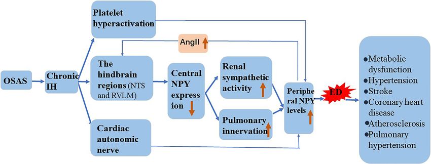
Li et al.                                                                                                                  NPY on CIH-Induced ED

energy metabolism (Kulkarni et al., 2000; Lee et al., 2003;              platelet activation, and thrombus formation (Hekman and
Wier et al., 2009; Zhou et al., 2013). Studies clarifying the            Loskutoff, 1987). The endothelium promotes angiogenesis by
effect of NPY on the pathophysiological mechanism underlying             secreting VE growth factor (VEGF) and fibroblast growth factor
chronic IH-induced vascular ED identify NPY as a potential               (Graupera and Claret, 2018). Endothelial cells allow the initiation
target for the treatment of OSAS-related cerebrovascular diseases.       of adaptive immunity and local recruitment of antigen-specific
This review highlights the relationship between ED and                   lymphocytes by sensing pathogen components in the blood
multisystem diseases, including the relationship between ED              (Taflin et al., 2011). The abnormal function of the vascular
and chronic IH-related vascular diseases.                                endothelium in response to a series of risk factors (hypoxia,
                                                                         smoking, obesity, infection, etc.) causes pathological events,
                                                                         such as the progression of cardiovascular and cerebrovascular
                                                                         diseases, which can lead to systemic consequences.
CHRONIC IH AND VASCULAR ED
Chronic IH                                                               Relationship Between ED and Chronic
Common clinical hypoxia describes the inability of the body              IH-Related Vascular Disease
to provide sufficient oxygenation to meet the needs of tissues           OSA is an independent risk factor for cardiovascular and
when exposed to a hypoxic environment. Hypoxia can                       cerebrovascular diseases. Furthermore, chronic IH is considered
be classified as acute or chronic depending on the duration              the main mediator of OSAS, and IH duration and intensity
of exposure and as continuous or intermittent depending on               depend on the severity of OSAS. Recently, Namtvedt et al.
the exposure pattern. The physiological and pathological reactions       (2013) reported that the severity of OSAS positively correlated
of hypoxia vary according to type (Nanduri and Nanduri,                  with impaired endothelial function, and that it had an adverse
2007). In humans, chronic IH often occurs during sleep apnea.            effect on vascular health. Additionally, previous studies confirmed
Repeated hypoxia-reoxygenation in OSAS leads to ED through               that correcting hypoxia using continuous positive airway pressure
oxidative stress, inflammation, and sympathetic nerve activation         (CPAP) treatment can improve endothelial function (Wilcox
among many other pathological reactions involved in disease              et al., 2011; Xu et al., 2015). There is currently no evidence
occurrence (Prabhakar et al., 2020). Recently, a new condition           that mild OSA has any beneficial effect on endothelial function;
associated with high altitude (i.e., long-term chronic IH) was           however, there are reports that short-term, low-intensity IH
reported in South America (Pena et al., 2020). Long-term                 or accompanying old age can to a certain extent protect against
chronic IH afflicts people that commute to work at high altitudes        ischemic events, such as myocardial infarction and stroke
but live and rest at sea level. Hypoxic constriction of pulmonary        (Beaudin et al., 2017). Therefore, as the initial factor of OSAS-
vessels is the first response of pulmonary circulation to alveolar       induced atherosclerosis, ED induces vascular diseases caused
hypoxia (Hoeper et al., 2016). Among the responses, endothelial          by IH, which has great significance for OSAS treatment.
production of nitric oxide (NO), prostacyclin (PGI), and                     Vascular diseases caused by OSAS include systemic
endothelin-1 (ET-1) among other vasoactive mediators are also            hypertension, transient ischemic attack, stroke, pulmonary
involved in the process of pulmonary hypoxic vascular contraction        hypertension, coronary heart disease, and pulmonary embolism
(Grimmer and Kuebler, 2017).                                             (Bauters et al., 2016). The mechanisms underlying the increased
                                                                         risk of vascular disease in OSAS are thought to include
                                                                         sympathetic nerve activation, ED, and changes in peripheral
Vascular ED                                                              and cerebrovascular regulation (Somers et al., 2008). Sympathetic
Endothelial cells originate from the mesoderm, are arranged
                                                                         nerve excitement, inflammation, and oxidative stress triggered
in a single-cell layer in the vascular cavity, and are connected
                                                                         by chronic IH can lead to ED and atherosclerosis.
via adhesive, tight, and gap junctions (Rodrigues and Granger,
2015). The endothelium is also covered by proteoglycans,
glycosaminoglycans, glycoproteins, and glycolipids, which are
referred to as the endothelial glycocalyx (GCX; Reiterer and
                                                                         MECHANISMS UNDERLYING VE INJURY
Branco, 2020). Vascular integrity is achieved by the actions             CAUSED BY CHRONIC IH
of the GCX, endothelial cells, and intercellular junctions. The
endothelium plays a key role in regulating vascular permeability,        Vascular Barrier Dysfunction
vascular tension, anticoagulation, inflammation, angiogenesis,           The function of the VE barrier mainly depends on the integrity
immune-response initiation, and endocrine processes.                     of the cytoskeleton and connexin (Dudek and Garcia, 2001).
Intercellular connections regulate passive diffusion and allow           Actin and cortactin are the main endothelial cytoskeletal proteins.
the endodermis to form selective osmotic layers to maintain              Additionally, IH can cause changes in the structure and function
homeostasis (Sukriti et al., 2014). Local regulation of vascular         of blood vessels (Figure 1). Table 1 summarizes the mechanisms
tension by endothelial cells depends on the release of a variety         underlying VE damage caused by chronic IH.
of vasoactive substances, including NO, prostaglandin, ET-1,
and other endothelium-derived hyperpolarization factors (Lai             Endothelial Cytoskeleton Rearrangement
and Kan, 2015). Endothelial cells release tissue plasminogen             Arnaud et al. (2018) found that in the monolayer of cultured
activator (tPA) to prevent the development of atherosclerosis,           endothelial cells, 8-h of IH significantly reduced transendothelial

Frontiers in Physiology | www.frontiersin.org                        2                                     August 2021 | Volume 12 | Article 712281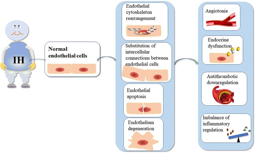
Li et al.                                                                                                                                        NPY on CIH-Induced ED

  FIGURE 1 | The mechanism underlying VE injury caused by chronic IH. The duration and intensity of IH are positively correlated with ED. Chronic IH-induced
  sympathetic nerve excitation, inflammation, and oxidative stress can lead to ED. IH can cause endothelial barrier dysfunction and endothelial cell dysfunction.
  ED is dependent on the integrity of the endothelial barrier.

resistance by 28%, indicating that IH leads to endothelial barrier                      Substitution of Intercellular Connections Between
dysfunction. Makarenko et al. (2014) found that reactive oxygen                         Endothelial Cells
species (ROS) play a central role in promoting IH-related                               The intercellular junctions of endothelial cells are an important
endothelial barrier dysfunction in pulmonary microvascular                              barrier in blood vessels and regulate the transport of water,
endothelial cell culture. Additionally, they reported that interval                     ions, and molecules through paracellular pathways in order
hypoxia disrupts endothelial barrier dysfunction through                                to maintain blood-vessel homeostasis. Zonula occludens-1 (ZO-1)
ROS-dependent extracellular signal-regulated kinase (ERK)1/2                            and VE-cadherin represent tight junctions and adhesion
activation and c-Jun N-terminal kinase (JNK)-mediated                                   junctions, respectively, that regulate endothelial cell permeability
cytoskeleton and junction protein recombination (Makarenko                              by regulating membrane adhesion of adjacent cells (Usatyuk
et al., 2014). A previous study showed that ROS exposure in                             et al., 2006, 2012). Obesity is an important risk factor for
endothelial cells induces the redistribution of actin stress fibers                     OSAS (Brant et al., 2017), and obesity hypoventilation syndrome
and VE-cadherin to the surrounding cells, resulting in abnormal                         (OHS) affects 10–20% of obese patients with OSAS. Multiple
endothelial barrier function (Prabhakar et al., 2007). Moreover,                        studies suggest that OHS initially interferes with endothelial
non-muscle myosin light chain kinase (nmMLCK) is the only                               homeostasis (Wang et al., 2015; Selthofer-Relatić et al., 2016;
MLCK subtype expressed in endothelial cells (Verin et al., 1998),                       Virdis, 2016). In vitro, plasma exosomes from patients with
and nmMLCK phosphorylates the myosin light chain, leading                               severe OSAS/OHS induce ED, and isolation of exosomes from
to changes in cytoskeletal structure and resulting in cell retraction                   the same patient after PAP treatment and addition to brain
(Baguet et al., 2012). In lipopolysaccharide-induced sepsis models,                     endothelioma 3 endothelial cells significantly increased the
nmMLCK induces oxidative stress and plays a key role in the                             circum-membrane-restrictive staining of VE-cadherin and ZO-1,
destruction of the vascular barrier (Ralay Ranaivo et al., 2007).                       indicating improved endothelial barrier integrity and confirming
Furthermore, animal experiments by Arnaud et al. (2018) revealed                        the improvement in membrane-barrier resistance observed on
that nmMLCK deletion prevented all IH-induced functional                                the ECIS assay (Bhattacharjee et al., 2018). Additionally, VEGF
and structural changes, including correction of the integrity of                        directly alters the intercellular junctions and actin cytoskeleton
the endothelial barrier. However, whether nmMLCK relies on                              of rat endothelial cells following induction by hypoxia/
ROS to play a role in IH-induced endothelial barrier dysfunction                        reoxygenation and promotes further destruction of the blood–
remains to be determined.                                                               brain barrier (BBB), ultimately leading to neurological

Frontiers in Physiology | www.frontiersin.org                                       3                                            August 2021 | Volume 12 | Article 712281
Li et al.                                                                                                                                                    NPY on CIH-Induced ED

TABLE 1 | Mechanism of VE injury caused by chronic IH.

Vascular                Pathophysiology                    Mechanism                                                                    References
endothelial injury

Vascular barrier        Endothelial cytoskeleton           ① IH → ROS↑ → ERK1/2 and JNK phosphorylation → reorganization of             Prabhakar et al., 2007; Makarenko et al.,
dysfunction             rearrangement                      cortactin and actin, stress fiber formation                                  2014
                                                           ② IH → nmMLCK↑ → myosin light chain phosphorylation → cell                   Verin et al., 1998; Ralay Ranaivo et al.,
                                                           retraction                                                                   2007; Baguet et al., 2012; Arnaud et al.,
                                                                                                                                        2018
                        Substitution of intercellular  ① IH → influx of Ca2+ into endothelial cells → VEGF↑→ replacement of             Usatyuk et al., 2006, 2012
                        connections between            ZO-1 and VE-cadherins
                        endothelial cells              ② IH → ROS↑ → ERK1/2 and JNK phosphorylation → VE-cadherin                       Dalal et al., 2020
                                                       redistributes
                        Endothelial apoptosis          IH → oxidative stress → ICAM-1 and VCAM-1↑ → leukocyte                           Zhao et al., 2018
                                                       adhesion → endothelial apoptosis↑
                        Endothelium degeneration       IH → TGF-β1↑ → EndMT                                                             Zhang et al., 2017; Kovacic et al., 2019
Vascular endothelial    Dysregulation of vascular tone IH → carotid body sympathetic tone↑/the expression ratio of                      Krause et al., 2015
dysfunction                                            arginase-1 to eNOS is out of balance → increased shrinkage capacity
                                                       and vascular remodeling
                        Endocrine dysfunction          ① IH → NO, PGI, H2S↓/ET, TXA2↑ → vasoconstriction↑                               Browatzki et al., 2000
                                                       ② IH → ET-1↑ → NF-κB IL-6↑ → CRP↑ → inflammation↑                                Ivey et al., 2008
                                                       ③ IH → E-selectin, P-selectin, VCAM-1↑ → AS↑                                     Uyar and Davutoglu, 2016;
                        The antithrombotic effect was ① IH → NO↓vasodilation↓ → platelet aggregation↑/leukocyte-                        Uyar and Davutoglu, 2016; García-
                        downregulated                  endothelial adhesion↑                                                            Ortega et al., 2019; Shahidi et al., 2020
                        Imbalance of inflammatory      ② IH → degradation of HMW hyaluronic acid↑ → TLR4/NF-κB↑                         Campo et al., 2011; Zhang et al., 2019
                        regulation

IH, intermittent hypoxia; ROS, reactive oxygen species; JNK, c-Jun N-terminal kinase; nmMLCK, non-muscle myosin light chain kinase; VEGF, vascular endothelial growth factor;
ZO-1, Zonula occludens-1; VE-cadherin, vascular endothelial cadherin; NO, nitric oxide; PGI, prostacyclin; H2S, hydrogen sulfide; ICAM-1, intercellular cell adhesion molecule 1;
VCAM-1, vascular cell adhesion molecule 1; TGF-β1, transforming growth factor-β1; EndMT, endothelial cell to mesenchymal transition; eNOS, endothelial nitric oxide synthase;
CRP, C-reactive protein; HMW, high-molecular-weight; TLR4, Toll like receptor 4; ET, endothelin; TXA2, thrombatin A2; NF-κB, nuclear factor kappa B; and IL-6, interleukin 6.

dysfunction (Restin et al., 2017). Moreover, Ca2+ is an                                      endothelial cells in OSAS patients was higher than that in
indispensable second messenger in endothelial cells and plays                                non-OSAS patients. The level of apoptotic endothelial cells is
a key role in regulating cell migration, angiogenesis, barrier                               associated with abnormal endothelial vasodilation. CPAP can
function, inflammation, and other physiological processes.                                   improve hypoxia and reduce endothelial cell apoptosis; however,
Inflammatory mediators (thrombin, histamine, and bradykinin)                                 these studies have not yet evaluated the specific mechanism
promote Ca2+ influx into endothelial cells, leading to the                                   of endothelial cell apoptosis caused by chronic IH. It is of
replacement of adhesion junctions and cytoskeletal                                           great significance to clarify the pathway of endothelial cell
rearrangement, thereby promoting endothelial cell permeability                               apoptosis for the establishment of future intervention targets.
and contractibility (Dalal et al., 2020). Whether ROS changes                                Zhang et al. (2019) used animal experiments to reveal that
the replacement of adhesion junctions and cytoskeletal                                       Toll-like receptor (TLR)4-dependent MyD88/nuclear factor kappa
rearrangement through Ca2+-mediated VEGF and nmMLCK                                          B (NF-κB; p65) activation plays a crucial role in the pathogenesis
expression remains to be elucidated.                                                         of hypoxia/reoxygenation-induced renal tubular dermal cell
                                                                                             apoptosis. TLR4 regulates the expression of a large number
Endothelial Apoptosis                                                                        of proinflammatory genes by controlling the fixed downstream
Endothelial cell apoptosis rarely occurs in normal physiological                             signaling factor MyD88, which translocates and activates
vessels. A previous study reported that circulating endothelial                              downstream NF-κB (p65). Based on this demonstrated
cell apoptosis is an indicator of vascular injury in vitro (Feng                             relationship between chronic IH and TLR4 expression,
et al., 2012), and an increasing number of studies show that                                 we speculate that chronic IH also follows a conserved TLR4-
chronic IH caused by oxidative stress can induce endothelial                                 dependent MyD88/NF-κB (p65)-activation pathway to induce
cell apoptosis, lead to loss of endothelial integrity, aggravate                             VE cell apoptosis. This needs to be further verified by additional
VE damage, promote the expression of redox-sensitive genes                                   animal experiments and clinical translational studies.
and adhesion molecules, and lead to the progression of hypoxia-
induced cardiovascular diseases (Liu et al., 2018; Xiao et al.,                              Endothelial Degeneration
2019). Zhao et al. (2018) found that IH-and cigarette smoke-                                 The endothelial-to-mesenchymal transition (EndMT) was first
induced emphysema can synergistically produce a greater                                      observed using electron microscopy in 1975. Increasing evidence
inflammatory response and endothelial cell apoptosis in an                                   shows that a portion of mesenchymal fibroblasts are derived
animal model of OSAS and chronic obstructive pulmonary                                       from the endothelium, and that transforming growth factor-β1
disease. They also showed that use of the antioxidant Tempol                                 might induce endothelial cells to proliferate into fibroblast-like
could antagonize these effects, as the density of apoptotic                                  cells (Kovacic et al., 2019). Endothelial cells undergo EndMT

Frontiers in Physiology | www.frontiersin.org                                            4                                                August 2021 | Volume 12 | Article 712281
Li et al. NPY on CIH-Induced ED
under continuous IH, which is associated with cardiovascular In human VSMCs, ET-1 activates the proinflammatory transcription
fibrosis. Zhang et al. (2017) found that IH can increase EndMT factor NF-κB, which in turn induces the release of interleukin
and the expression of prolyl 4-hydroxylase domain protein 3 (IL)-6 and other cytokines (Browatzki et al., 2000). Furthermore,
(PHD3), and that IH accelerates cardiac dysfunction and Verma et al. (2002) confirmed that the antagonistic effect of ET
increases collagen deposition via EndMT. Additionally, PHD3 and the inhibitory effect of IL-6 can reduce the atherosclerotic
overexpression improves cardiac dysfunction and excessive effect of C-reactive protein. Moreover, ET-1 stimulates VSMC
collagen deposition. Moreover, IH induces EndMT in vitro, proliferation and migration and promotes extracellular matrix
causing human umbilical vein endothelial cells to appear fusiform synthesis and matrix remodeling, thereby supporting its role in
with enhanced migration and collagen-secretion abilities. vascular remodeling and atherosclerosis (Ivey et al., 2008). To
Furthermore, OSA-induced perivascular fibrosis is related to date, most studies have focused on the increased IH-induced
EndMT, and PHD3 overexpression might be prevented by vasoconstrictive response to ET-1 (Kanagy et al., 2001; Belaidi
inhibiting EndMT. These results suggest that PHD3 et al., 2009); however, the underlying mechanism by which ET-1
overexpression has therapeutic potential for disease treatment. is involved in IH-associated vascular inflammation and remodeling
remains unclear. Furthermore, endothelial cells secrete E-selectin,
P-selectin, intercellular adhesion molecule 1, and vascular cell-
Vascular ED adhesion molecule 1 (VCAM-1), which are involved in
Dysregulation of Vascular Tone
atherosclerotic lesion formation (Uyar and Davutoglu, 2016).
The vascular endothelium regulates vascular tension by balancing
the degree of vasodilation and contraction. Endothelial cells
regulate vasodilation function by secreting vasoactive substances, Downregulation of Antithrombosis
such as NO, hydrogen sulfide, and PGI, and regulate NO is a major endodermal-derived vasodilator with important
vasoconstriction function by secreting substances, such as ET-1 vasculoprotective effects, such as the inhibition of platelet
and thrombatin A2 (TXA2; Krüger-Genge et al., 2019). Rats aggregation, adhesion-molecule expression, leukocyte-endothelial
exposed to IH show elevated resting vascular tone (Phillips adhesion, and smooth muscle cell proliferation (Cohen, 1995).
et al., 2006). Additionally, they observed resistance vessel The vascular endothelium can also maintain thrombus balance
remodeling in rats exposed to IH for a long period of time by secreting tissue factor (TF), Von Willebrand factor (vWF),
as evidenced by a reduced response to norepinephrine-induced coagulation factor, and fibrinolytic components (Shahidi et al.,
constriction and acetylcholine-induced diastolic response (Phillips 2020). Additionally, sleep apnea is associated with hypercoagulability,
et al., 2004). NO plays a key role in controlling vasomotor which might result from reduced NO levels and impaired
tension, which depends on L-arginine levels. Krause et al. vasodilation. Data from pathophysiological studies indicate that
(2015) found that the ED in chronic IH-induced hypertension IH and sleep disruption are related to increased blood clots,
might result from an imbalance in the expression ratio of ED, and venous stasis (García-Ortega et al., 2019). Moreover, a
arginase-1 to endothelial NO synthase (eNOS), which results growing body of evidence suggests that OSAS is a risk factor
in increased vascular remodeling and contractile capacity. ET-1 for pulmonary embolism (Epstein et al., 2010; Arzt et al., 2012;
plays a role by activating different receptors. ET receptor (ETR) Alonso-Fernández et al., 2013). Furthermore, studies suggest that
A is mainly expressed in vascular smooth muscle cells (VSMCs) CPAP can improve hypercoagulability and normalize the circadian
and mediates vasoconstriction, whereas ETRB is mainly expressed rhythm of certain coagulation molecules.
in endothelial myocytes and mediates vasodilation through the
release of NO. Mentek et al. (2018) found that chronic IH
Imbalance of Inflammatory Regulation
exposure impaired the vasodilation of rat ophthalmic artery
High-molecular-weight (HMW) hyaluronic acid is an important
stimulated by ETRB, and that IH-induced oxidative stress
component of the endothelial wall and has anti-inflammatory
alters the bioavailability of NO and endothelium-derived
and antioxidant properties (Campo et al., 2011). In the case
hyperpolarizing factor (EDHF), thereby changing ocular arterial
of inflammation or hypoxia, HMW hyaluronic acid is degraded
reactivity. However, the importance of the contribution of EDHF
by hyaluronidases, such as HYAL-1, to produce proinflammatory
to endothelium-dependent relaxation as a feature of healthy
low-molecular-weight fragments. In a clinical study of 68 OSA
endothelium is being actively discussed.
patients and 40 control volunteers, Meszaros et al. (2020) found
that chronic hypoxia is associated with increased plasma
ED hyaluronic acid-1 concentrations and accelerated degradation
VE cells are also important metabolic secretory organs in the of HMW hyaluronic acid. These findings suggest that changes
human body. VE cells regulate vasodilation by secreting vasoactive in hyaluronic acid metabolism are involved in the inflammatory
substances, such as NO, hydrogen sulfide, and PGI, and regulate cascade and promote ED in OSAS. In endothelial cells, hypoxia/
vasoconstriction by secreting substances, such as ET and TXA2. reoxygenation exposure promotes TLR4 expression and activation
ET is an active peptide secreted by VE cells and plays a major of proinflammatory TLR4/NF-κB signaling, whereas TLR4
role in cardiovascular disease development. Although interference inhibits these effects (Zhang et al., 2017).
vasoconstriction was the first characterized effect of ET-1, there Furthermore, IH accelerates the growth and vulnerability of
is growing evidence that ET-1 is also a potent proinflammatory atherosclerotic plaques, which might play a role in triggering
cytokine involved in vascular inflammation and atherosclerosis. the activation of proinflammatory TLR4/NF-κB signaling.
Frontiers in Physiology | www.frontiersin.org 5 August 2021 | Volume 12 | Article 712281Li et al. NPY on CIH-Induced ED
These findings suggest IH as a possible risk factor for vulnerable vasoconstriction and regulate pancreatic secretion and energy
plaques, thereby providing new insights for the treatment of metabolism. A recent study showed that Y1R, Y2R, and Y5R
atherosclerotic progression caused by OSA. are distributed on endothelial cells and mainly associated with
hypertension, atherosclerosis, myocardial ischemia, heart failure,
cardiac remodeling, and arrhythmia (Figure 2; Wier et al., 2009).
POSSIBLE MECHANISM OF
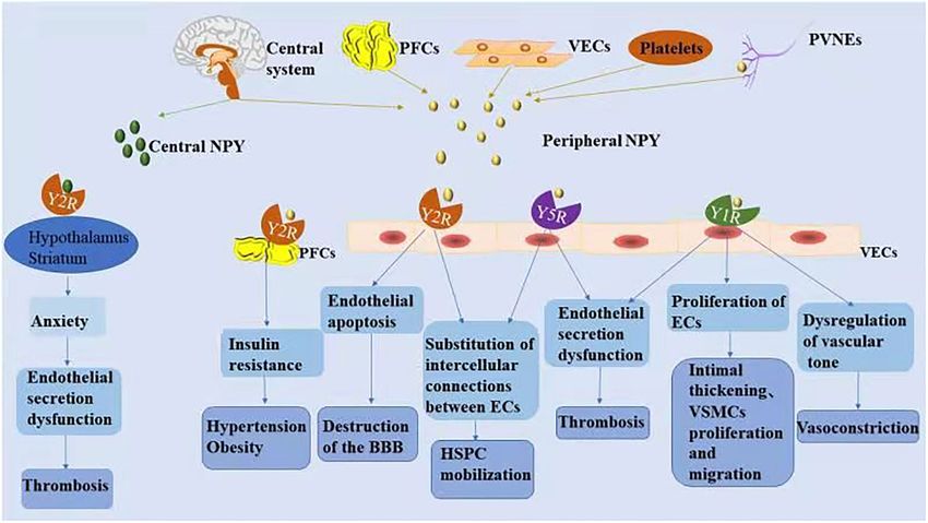
NPY-INDUCED ED NPY and ED
NPY activates different receptors to perform various physiological
NPY and Receptors
functions. Under various stress conditions, increases in NPY
NPY is a small protein-like molecule composed of 36 amino
level in the central and/or peripheral nervous system is directly
acids, widely expressed in the central and peripheral nervous
and indirectly involved in the process of vascular ED, thus
systems, and participates in a variety of physiological processes
participating in the pathophysiological process of vascular
by binding to receptors (Robinson and Thiele, 2017). Currently,
diseases (Zhu et al., 2016). The relationships between NPY
six main subtypes of NPY receptors are found in mammals
and vascular ED are summarized in Table 2.
(Y1R–Y6R). Except for Y3R, these molecules are G protein-
coupled receptors with different affinities and selectivity and
exert different biological effects (Zhu et al., 2016). The NPY and Substitution of Intercellular Connections
hypothalamus is closely related to cardiovascular regulation, Between Endothelial Cells
and its descending fibers directly reach the mediolateral column Endothelial cells regulate the homeostasis of hematopoietic stem
of the spinal cord to control the activity of sympathetic cells and progenitor cells (HSPCs) in the peripheral blood and
preganglionic neurons. NPY is secreted in the preganglionic are an integral part of the hematopoietic microenvironment.
and postganglionic sympathetic nerves and can be released NPY signaling in endothelial cells modulates vascular pathways
into the peripheral circulation together with norepinephrine in HSPCs (Singh et al., 2017). Dipeptidyl peptidase 4/CD26
(NE) to exert a cardiovascular neuromodulatory effect (Schütz (DPP4/CD26), an enzyme that truncates NPY, induces decreased
et al., 1998). Peripheral NPY is mainly secreted by peripheral expression of VE-cadherin and CD31 at the junction of endothelial
vascular nerve endings, peripheral fat cells, platelets, and VE cells by altering NPY signaling, leading to increased vascular
cells. Different NPY receptors on peripheral tissue cells can permeability and HSPC transport to peripheral blood (Itkin
promote endothelial cell proliferation, thrombosis, and et al., 2017). Additionally, selective Y2R and Y5R antagonists
FIGURE 2 | The mechanism associated with NPY-induced endothelial injury. Increased NPY level in the central and/or peripheral nervous systems is directly and
indirectly involved in the process of vascular ED. NPY activates different receptors to perform various physiological functions.
Frontiers in Physiology | www.frontiersin.org 6 August 2021 | Volume 12 | Article 712281Li et al. NPY on CIH-Induced ED
TABLE 2 | The direct role of NPY in ED.
Origin of endothelial Pathophysiology Mechanism References
cells
Vascular endothelial cells Substitution of intercellular DPP4 / CD26 cleaves NPY through CD26 signal transduction Y2R Itkin et al., 2017; Singh et al., 2017
connections between and Y5R → VE-cadherin and CD31↓ → vascular permeability↑HSPC
endothelial cells transport↑
Brain microvascular Endothelial apoptosis METH→Y2R↑ → NPY is activated by Y2R → methamphetamine- Enman et al., 2015; Ventura et al., 2020
endothelial cells induced apoptosis and ROS formation↓
Vascular endothelial cells Dysregulation of vascular NPY binds to the Y1R on endothelial cells to enhance the effect of Li et al., 2003; Wier et al., 2009
tone NE → vasoconstriction↑ → vascular stenosis and spasm↑
NPY binds to the Y1R → endothelial cell proliferation↑ → intimal Abdel-Samad et al., 2012
thickening NPY acts directly on ET-1 → constricts blood
vessels→ED↑
EECs Enhanced excitatory NPY binds to the Y1R → cytoplasmic, nuclear calcium in Abdel-Samad et al., 2012
secretory coupling in EECS EECS↑ → intracellular Ca2+ ↑ → NPY is released from cells↑
EECs Vascular Downregulation of Y1R antagonist reduces the vWF extrusion of EECs NPY binds to Mourik et al., 2013; Kleber et al., 2015;
endothelial cells antithrombosis the Y1R,Y5R → TXA2↑ → Thrombosis NPY binds to the Y2R → tPA, Reichmann and Holzer, 2016; Tyrrell
clotting factor, and fibrinogen↑ → Thrombosis et al., 2017
EECs Endothelial secretion NPY binds to the Y5R (hLEECs)/ the Y2R,Y5R (hREECs) → ET- Abdel-Samad et al., 2012
dysfunction 1↑ → arrhythmia, heart failure and cardiac hypertrophy
NPY, neuropeptide Y; DPP4/CD26, dipeptidyl peptidase 4/CD26; Y1R, NPY1 receptor; Y2R, NPY2 receptor; Y5R, NPY5 receptor; HSPC, hematopoietic stem cells and progenitor
cells; METH, metamphetamine; ROS, reactive oxygen species; NE, norepinephrine; ET-1, endothelin-1; ED, endothelial dysfunction; EECS, endocardial endothelial cells; vWF,
vascular hemophilia factors; TXA2, thrombatin A2; and tPA, tissue plasminogen activator.
restore vascular integrity and restrict HSPC mobilization, The presence of the cardiac active factor NPY and its receptor
suggesting that the enzyme-controlled vascular pathway specifically Y1R in endocardial endothelial cells (EECs) was confirmed
opens through CD26 signaling involving Y2R and Y5R to cleave using three-dimensional confocal microscopy. By binding to
NPY. Mice lacking CD26 or NPY show impaired HSPC transport, its receptors, NPY can induce an increase in cytoplasmic and
which is restored with truncated NPY treatment (Singh et al., nuclear Ca2+ in EECs, after which NPY is released from these
2017); therefore, these results indicate that the CD26-mediated cells to regulate the excitation-secretion coupling of EECs and
NPY axis might be a potential drug target for various immune the excitation-contraction coupling ability of cardiomyocytes
stressors regulating barrier integrity on VE cells. and VSMCs, thereby indirectly regulating cardiac function and
remodeling (Abdel-Samad et al., 2012).
NPY and Endothelial Apoptosis
Endothelial cells are the anatomical basis of the BBB. Human NPY and Downregulation of Antithrombosis
microvascular endothelial cells express both Y1R and Y2R EECs are more prone to thrombosis with the progression of
receptors (Enman et al., 2015). Ventura et al. (2020) used a heart failure (Saric et al., 2016). EECs are attached to the
human brain microvascular endothelial cell line (HCMEC/D3) sympathetic nerve of the heart, and neuropeptides, such as
to simulate an in vitro BBB model and found that only Y2R galanin and NPY, are released jointly by sympathetic nerve
was upregulated during metamphetamine (METH) exposure. endings (Herring et al., 2012). Thrombin (FIIa) at the surface
NPY is activated by Y2R and plays a protective role in METH- of the endothelial tube cavity converts fibrinogen to fibrin and
induced apoptosis and ROS formation in endothelial cells, promotes the release of vascular hemophilia factors (vWF)
suggesting the Y2R subtype as a promising target for the from endothelial cells (Mourik et al., 2013; Kleber et al., 2015).
prevention of METH-induced neurovascular dysfunction. Clusters of vWF gather on the endothelial surface to help
platelets adhere. Tyrrell et al. (2017) showed that glycerin and
NPY and Dysregulation of Vascular Tone a Y1R antagonist reduced vWF-mediated extrusion of EECs.
As the main peripheral vasoconstrictive neurotransmitter, NPY Additionally, nicotine exposure experiments showed that nicotine
binds to Y1R on endothelial cells under pathological conditions promotes NPY expression and induce ED (Hiremagalur and
to enhance the effect of NE, plays a role in vasoconstriction, Sabban, 1995; Huang and Winzer-Serhan, 2007). Nicotine
causes local vascular stenosis and spasm, and then promotes increases the expression of TF and TXA2 and decreases the
endothelial cell retraction and interruption (Wier et al., 2009). expression of prostaglandin I-2, leading to platelet adhesion
Additionally, NPY binds to Y1R on endothelial cells, promotes and aggregation by affecting the secretory function of endothelial
endothelial cell proliferation, and plays a key role in intimal cells (Amadio et al., 2015). Moreover, Csordasa and Bernhard
thickening (Li et al., 2003). NPY constricts blood vessels directly (Csordas and Bernhard, 2013) observed that NPY promotes
or through molecules, such as ET-1, causes vascular ED, the release of TXA2 from endothelial cells by activating Y1R
promotes VSMC proliferation and migration, and converts the and Y5R, thereby inducing thrombus formation. Furthermore,
contractile phenotype of VSMCs into a synthetic phenotype nicotine and NPY combine to promote pathological thrombosis.
with proliferative function (Abdel-Samad et al., 2012). NPY also induces anxiety by activating Y2R in the hypothalamus
Frontiers in Physiology | www.frontiersin.org 7 August 2021 | Volume 12 | Article 712281Li et al. NPY on CIH-Induced ED
and striatum (Reichmann and Holzer, 2016), with anxiety into the circulation of perivascular neurons from postganglionic
increasing the risk of thrombosis, which is promoted by inducing sympathetic nerves. Chronic IH causes overactivation of the
endothelial cells to release tPA, clotting factor, and fibrinogen. hindbrain regions that control sympathetic outflow, such as
the nucleus solitudes and ventrolateral medulla oblongata, and
NPY and Dysfunctional Endothelial Secretion promotes increased sympathetic outflow (Shell et al., 2016).
In cultured human EECs, NPY treatment promotes the release Moreover, increased renal sympathetic activity activates the
of ET-1 from left and right (LEECs and REECs, respectively); RAS system, leading to elevated circulating angiotensin II
however, the type of NPY receptor involved might be different (AngII), and an increase in circulating AngII reportedly further
(Abdel-Samad et al., 2012). The effect of NPY on ET-1 secretion increases central sympathetic outflow (Kim et al., 2018). The
of human (h) LEECs resulted only from Y5R activation, whereas pulmonary artery is innervated, and NPY/Y1R mediates the
the effect of NPY on ET-1 secretion of hREECs mainly resulted vasoconstriction and proliferation of pulmonary hypertension.
from the activation of Y2R and part of Y5R. EECs play an Furthermore, in a mouse model of chronic hypoxia, the
important role in cardiac pathological processes, such as expression of NPY and Y1R was upregulated in lung tissue
excitation-contraction coupling, arrhythmia, cardiac hypertrophy, (Crnkovic et al., 2014). Platelet hyperactivation during chronic
and heart failure, by secreting cardiac active factors, such as IH and activated platelets might further increase the circulating
NPY and ET-1 (Jacques et al., 2017). concentration of NPY (Gabryelska et al., 2018).
Indirect Effects of NPY Possible Mechanism of NPY Involvement
Increased NPY level in the central and/or peripheral nervous in IH-Induced ED
system is associated with dyslipidemia, hypertension, obesity, NPY participates in the pathogenesis of atherosclerosis by
diabetes, and impaired glucose tolerance, all risk factors for aggravating ED, VSMC growth, foam cell formation, and platelet
atherosclerotic cardiovascular disease (Sun et al., 2017). During aggregation, which are major pathogenic processes in
stress, NPY released from sympathetic nerves participates in cardiovascular disease (Zhu et al., 2016). However, whether
adipose tissue insulin resistance through Y2R in visceral adipose vasoconstriction, vascular remodeling and vascular ED caused
tissue, promotes food intake and fat storage, and enhances by chronic IH are mainly caused by the release of NPY has
macrophage-mediated inflammatory responses (Kuo et al., 2007). not yet been determined. Although OSAS causes VE damage,
Additionally, NPY increases the dependence on nicotine, which platelet activation increases in patients and promotes platelet
indirectly aggravates ED (Robinson and Thiele, 2017). These aggregation in the damaged intima (Kontos et al., 2020).
pathological processes promote oxidative stress, inflammatory Additionally, NPY expression is significantly elevated in activated
response, blood-flow disorders, and reduce NO bioavailability, platelets and associated with a cycle of endothelial injury
all of which eventually induce ED. (Ruohonen et al., 2009). Moreover, ED reportedly causes increased
NPY expression, further increasing the inflammatory/immune
response associated with NPY in the endothelium (Choi et al.,
INVOLVEMENT OF NPY IN THE 2019). Whether NPY-mediated endothelial function forms a
positive feedback loop after sympathetic excitation and ultimately
MECHANISM UNDERLYING IH-INDUCED
leads to the deterioration of atherosclerosis remains to be clarified.
ED
Chronic IH promotes NPY expression, and the induction of Effect of NPY on the Pathogenesis of
ED by NPY is similar to that of chronic IH. In the following Chronic IH-Related Vascular Diseases
sections, we attempt to elaborate on the role of NPY in chronic OSAS is associated with cardiovascular and cerebrovascular
IH-induced ED and its possible mechanism (Figure 3). diseases, including atherosclerosis, hypertension, stroke, coronary
heart disease, and metabolic dysfunction, contributing to an
NPY and CIH overall increase in cardiovascular mortality. ED is the primary
Hypoxia induces the release of NPY in mammals (Ali and cause of atherosclerosis through complex interactions among
Bhargava, 2017), and chronic IH affects central NPY expression lipoprotein accumulation, inflammatory infiltration, foam cell
and peripheral NPY levels. Shobatake et al. (2018) demonstrated formation, and smooth muscle cell changes (Jiang et al., 2017).
that IH reduces NPY expression in the central nervous system Decreased NO bioavailability leads to ED. Experiments in eNOS-
and weakens the inhibitory effect of sympathetic excitement. deficient mice confirmed that ED induces upregulation of NPY
NPY is mainly distributed in peripheral sympathetic nerve expression (Choi et al., 2019). Additionally, inflammatory protein
fibers, especially those in and around the cardiovascular system. 3 amplifies vascular inflammation by increasing macrophage
Animal experiments confirmed that hypoxia directly stimulates chemotaxis via the NPY/NPY receptor axis, thereby increasing
the release of large amounts of NPY from the autonomic the inflammatory/immune response in the endothelium.
nervous system (McDermott and Bell, 2007). We speculate that Furthermore, NPY induces lipid uptake of VSMCs and triggers
in response to chronic IH, central NPY expression decreases, the formation of smooth muscle foam cells (Tan et al., 2018).
and peripheral sympathetic nerve activity increases. Additionally, OSAS is the main cause of secondary hypertension, with
hypoxia causes sympathetic excitation and promotes the release a previous study confirming a close and independent relationship
Frontiers in Physiology | www.frontiersin.org 8 August 2021 | Volume 12 | Article 712281Li et al. NPY on CIH-Induced ED FIGURE 3 | Involvement of NPY in the mechanism underlying IH-induced ED. Chronic IH affects central NPY expression and peripheral NPY levels. NPY is involved in the pathogenesis of atherosclerosis, hypertension, stroke, coronary heart disease, and metabolic dysfunction by increasing ED. between secondary hypertension and nocturnal blood pressure and require further identification. The effects of IH on endothelial instability (Parati et al., 2013). Sympathetic nerve excitation function are limited by the severity of OSAS, and the animal is the main pathophysiological mechanism underlying this models that mimic OSAS are diverse. Animal experiments and relationship. When the apnea period is prolonged, the partial clinical studies are indispensable to investigating the contributions pressure of oxygen in the arteries decreases and carbon dioxide of NPY in OSAS-induced ED. Genetic studies have supported gradually increases in a process that stimulates chemoreceptors the association between NPY polymorphisms and an increased and causes sympathetic nerve excitement and release of NE. risk of cardiovascular diseases (Masoudi-Kazemabad et al., NPY is an important sympathetic neurotransmitter in peripheral 2013; de Luis et al., 2017). Taken together, the application of blood vessels and released together with NE. The adrenal NPY genomics to the study of associated vascular diseases medulla is a component and effector organ of the sympathetic may provide new ideas for the prevention and treatment of nervous system. NPY and NE are synthesized and stored in OSAS and its complications in the future. large, dense core vesicles of the adrenal medulla. Animal studies show that IH increases NPY synthesis in the adrenal medulla of rats, thereby inducing an increase in blood pressure AUTHOR CONTRIBUTIONS (Raghuraman et al., 2011). Moreover, an impaired baroreflex, activation of RAS, dysfunctional NO metabolism in the vascular M-mL, Y-lZ, W-dW, H-lL, and SL were involved in the genesis endothelium, and related downstream consequences leading of the review topic and reviewed, edited, and approved the to arterial vasoconstriction also play a role in OSAS-induced manuscript. M-mL drafted the manuscript. All authors hypertension (Ryan et al., 2007; Zalucky et al., 2015; Crinion contributed to the article and approved the submitted version. et al., 2017). However, the full mechanism of ED-induced OSAS-mediated hypertension remains unclear. Moreover, whether NPY is directly involved in the increase in blood pressure FUNDING induced by IH-induced ED has not yet been determined. Thus, NPY and its receptors might be potential therapeutic targets This work was supported by Science and Technology Bureau for the treatment of OSAS-related hypertension in the future. of Quanzhou (grant number 2020CT003). CONCLUSION ACKNOWLEDGMENTS The nervous system plays an important role in vascular function M-mL acknowledges SL and H-lL for their advice and assistance and participates in its regulation. Although the effect of NPY with this project. M-mL also thanks Y-lZ and W-dW for on vascular ED has recently attracted attention, its specific their assistance with systematic literature searching. M-mL role in ED pathogenesis remains unclear. In particular, whether was supported by the Second Affiliated Hospital of Fujian NPY and its receptor are involved in OSAS-induced ED has Medical University to pursue her doctor’s degree. We would not yet been determined. Furthermore, the factors that increase like to thank Editage (www.editage.cn) for English the risk of OSAS-induced vascular disease are complicated language editing. Frontiers in Physiology | www.frontiersin.org 9 August 2021 | Volume 12 | Article 712281
Li et al.                                                                                                                                                       NPY on CIH-Induced ED

REFERENCES                                                                                      Csordas, A., and Bernhard, D. (2013). The biology behind the atherothrombotic
                                                                                                    effects of cigarette smoke. Nat. Rev. Cardiol. 10, 219–230. doi: 10.1038/
Abdel-Samad, D., Perreault, C., Ahmarani, L., Avedanian, L., Bkaily, G., Magder, S.,                nrcardio.2013.8
    et al. (2012). Differences in neuropeptide Y-induced secretion of endothelin-1 in           Dalal, P., Muller, W., and Sullivan, D. (2020). Endothelial cell calcium signaling
    left and right human endocardial endothelial cells. Neuropeptides 46,                           during barrier function and inflammation. Am. J. Pathol. 190, 535–542. doi:
    373–382. doi: 10.1016/j.npep.2012.09.003                                                        10.1016/j.ajpath.2019.11.004
Ali, I., and Bhargava, S. (2017). Neuropeptide Y in the brain of Euphlyctis                     de Luis, D., Izaola, O., de la Fuente, B., Primo, D., and Aller, R. (2017).
    cyanophlyctis tadpoles responds to hypoxic stress. Gen. Comp. Endocrinol.                       Polymorphism of neuropeptide Y gene rs16147 modifies the response to a
    251, 38–45. doi: 10.1016/j.ygcen.2016.09.011                                                    hypocaloric diet on cardiovascular risk biomarkers and adipokines. J. Hum.
Alonso-Fernández, A., de la Peña, M., Romero, D., Piérola, J., Carrera, M., Barceló, A.,            Nutr. Diet. 30, 159–165. doi: 10.1111/jhn.12406
    et al. (2013). Association between obstructive sleep apnea and pulmonary                    Dudek, S., and Garcia, J. (2001). Cytoskeletal regulation of pulmonary vascular
    embolism. Mayo Clin. Proc. 88, 579–587. doi: 10.1016/j.mayocp.2013.02.005                       permeability. J. Appl. Physiol. 91, 1487–1500. doi: 10.1152/jappl.2001.91.4.1487
Amadio, P., Baldassarre, D., Tarantino, E., Zacchi, E., Gianellini, S., Squellerio, I.,         Enman, N., Sabban, E., McGonigle, P., and Van Bockstaele, E. (2015). Targeting
    et al. (2015). Production of prostaglandin E2 induced by cigarette smoke                        the neuropeptide Y system in stress-related psychiatric disorders. Neurobiol.
    modulates tissue factor expression and activity in endothelial cells. FASEB                     Stress 1, 33–43. doi: 10.1016/j.ynstr.2014.09.007
    J. 29, 4001–4010. doi: 10.1096/fj.14-268383                                                 Epstein, M., Segal, L., Ibrahim, S., Friedman, N., and Bustami, R. (2010).
Arnaud, C., Bouyon, S., Recoquillon, S., Brasseur, S., Lemarié, E.,                                 Snoring and the risk of obstructive sleep apnea in patients with pulmonary
    Briançon-Marjollet, A., et al. (2018). Nonmuscle myosin light chain kinase:                     embolism. Sleep 33, 1069–1074. doi: 10.1093/sleep/33.8.1069
    a key player in intermittent hypoxia-induced vascular alterations. J. Am.                   Félétou, M., and Vanhoutte, P. (2006). Endothelial dysfunction: a multifaceted
    Heart Assoc. 7:e007893. doi: 10.1161/JAHA.117.007893                                            disorder (The Wiggers Award Lecture). Am. J. Physiol. Heart Circ. Physiol.
Arzt, M., Luigart, R., Schum, C., Lüthje, L., Stein, A., Koper, I., et al. (2012).                  291, H985–H1002. doi: 10.1152/ajpheart.00292.2006
    Sleep-disordered breathing in deep vein thrombosis and acute pulmonary                      Feng, J., Chen, B., Cui, L., Wang, B., Liu, C., Chen, P., et al. (2009). Inflammation
    embolism. Eur. Respir. J. 40, 919–924. doi: 10.1183/09031936.00176711                           status of rabbit carotid artery model endothelium during intermittent hypoxia
Baguet, J., Barone-Rochette, G., Tamisier, R., Levy, P., and Pépin, J. (2012).                      exposure and its relationship with leptin. Sleep Breath. 13, 277–283. doi:
    Mechanisms of cardiac dysfunction in obstructive sleep apnea. Nat. Rev.                         10.1007/s11325-009-0246-6
    Cardiol. 9, 679–688. doi: 10.1038/nrcardio.2012.141                                         Feng, J., Zhang, D., and Chen, B. (2012). Endothelial mechanisms of endothelial
Bauters, F., Rietzschel, E., Hertegonne, K., and Chirinos, J. (2016). The link                      dysfunction in patients with obstructive sleep apnea. Sleep Breath. 16,
    Between obstructive sleep Apnea and cardiovascular disease. Curr. Atheroscler.                  283–294. doi: 10.1007/s11325-011-0519-8
    Rep. 18:1. doi: 10.1007/s11883-015-0556-z                                                   Gabryelska, A., Łukasik, Z., Makowska, J., and Białasiewicz, P. (2018). Obstructive
Beaudin, A., Waltz, X., Hanly, P., and Poulin, M. (2017). Impact of obstructive                     sleep Apnea: from intermittent hypoxia to cardiovascular complications via
    sleep apnoea and intermittent hypoxia on cardiovascular and cerebrovascular                     blood platelets. Front. Neurol. 9:635. doi: 10.3389/fneur.2018.00635
    regulation. Exp. Physiol. 102, 743–763. doi: 10.1113/EP086051                               García-Ortega, A., Mañas, E., López-Reyes, R., Selma, M., García-Sánchez, A.,
Belaidi, E., Joyeux-Faure, M., Ribuot, C., Launois, S., Levy, P., and Godin-Ribuot, D.              Oscullo, G., et al. (2019). Obstructive sleep apnoea and venous
    (2009). Major role for hypoxia inducible factor-1 and the endothelin system                     thromboembolism: pathophysiological links and clinical implications. Eur.
    in promoting myocardial infarction and hypertension in an animal model                          Respir. J. 53:1800893. doi: 10.1183/13993003.00893-2018
    of obstructive sleep apnea. J. Am. Coll. Cardiol. 53, 1309–1317. doi: 10.1016/j.            Goligorsky, M. (2005). Endothelial cell dysfunction: can't live with it, how to
    jacc.2008.12.050                                                                                live without it. Am. J. Physiol. Ren. Physiol. 288, F871–F880. doi: 10.1152/
Bhattacharjee, R., Khalyfa, A., Khalyfa, A., Mokhlesi, B., Kheirandish-Gozal, L.,                   ajprenal.00333.2004
    Almendros, I., et al. (2018). Exosomal cargo properties, endothelial function               Graupera, M., and Claret, M. (2018). Endothelial cells: new players in obesity
    and treatment of obesity hypoventilation syndrome: a proof of concept                           and related metabolic disorders. Trends Endocrinol. Metab. 29, 781–794.
    study. J. Clin. Sleep. Med. 14, 797–807. doi: 10.5664/jcsm.7110                                 doi: 10.1016/j.tem.2018.09.003
Brant, L., Wang, N., Ojeda, F., LaValley, M., Barreto, S., Benjamin, E., et al.                 Grimmer, B., and Kuebler, W. (2017). The endothelium in hypoxic pulmonary
    (2017). Relations of metabolically healthy and unhealthy obesity to digital                     vasoconstriction. J. Appl. Physiol. 123, 1635–1646. doi: 10.1152/japplphysiol.00120.2017
    vascular function in three community-based cohorts: a meta-analysis. J.                     Hekman, C., and Loskutoff, D. (1987). Fibrinolytic pathways and the endothelium.
    Am. Heart Assoc. 6:e004199. doi: 10.1161/JAHA.116.004199                                        Semin. Thromb. Hemost. 13, 514–527. doi: 10.1055/s-2007-1003527
Browatzki, M., Schmidt, J., Kübler, W., and Kranzhöfer, R. (2000). Endothelin-1                 Herring, N., Cranley, J., Lokale, M., Li, D., Shanks, J., Alston, E., et al. (2012).
    induces interleukin-6 release via activation of the transcription factor NF-                    The cardiac sympathetic co-transmitter galanin reduces acetylcholine release
    kappaB in human vascular smooth muscle cells. Basic Res. Cardiol. 95,                           and vagal bradycardia: implications for neural control of cardiac excitability.
    98–105. doi: 10.1007/s003950050170                                                              J. Mol. Cell. Cardiol. 52, 667–676. doi: 10.1016/j.yjmcc.2011.11.016
Campo, G., Avenoso, A., Nastasi, G., Micali, A., Prestipino, V., Vaccaro, M.,                   Hiremagalur, B., and Sabban, E. (1995). Nicotine elicits changes in expression
    et al. (2011). Hyaluronan reduces inflammation in experimental arthritis                        of adrenal catecholamine biosynthetic enzymes, neuropeptide Y and immediate
    by modulating TLR-2 and TLR-4 cartilage expression. Biochim. Biophys.                           early genes by injection but not continuous administration. Brain Res. Mol.
    Acta 1812, 1170–1181. doi: 10.1016/j.bbadis.2011.06.006                                         Brain Res. 32, 109–115. doi: 10.1016/0169-328X(95)00068-4
Choi, B., Shin, M., Kim, E., Park, J., Lee, H., Kim, S., et al. (2019). Elevated                Hoeper, M., Humbert, M., Souza, R., Idrees, M., Kawut, S., Sliwa-Hahnle, K.,
    neuropeptide Y in endothelial dysfunction promotes macrophage infiltration                      et al. (2016). A global view of pulmonary hypertension. Lancet Respir. Med.
    and smooth muscle foam cell formation. Front. Immunol. 10:1701. doi:                            4, 306–322. doi: 10.1016/S2213-2600(15)00543-3
    10.3389/fimmu.2019.01701                                                                    Huang, L., and Winzer-Serhan, U. (2007). Nicotine regulates mRNA expression
Cohen, R. (1995). The role of nitric oxide and other endothelium-derived                            of feeding peptides in the arcuate nucleus in neonatal rat pups. Dev. Neurobiol.
    vasoactive substances in vascular disease. Prog. Cardiovasc. Dis. 38, 105–128.                  67, 363–377. doi: 10.1002/dneu.20348
    doi: 10.1016/S0033-0620(05)80002-7                                                          Hung, M., Kravtsov, G., Lau, C., Poon, A., Tipoe, G., and Fung, M. (2013).
Crinion, S., Ryan, S., and McNicholas, W. (2017). Obstructive sleep apnoea as                       Melatonin ameliorates endothelial dysfunction, vascular inflammation, and
    a cause of nocturnal nondipping blood pressure: recent evidence regarding                       systemic hypertension in rats with chronic intermittent hypoxia. J. Pineal
    clinical importance and underlying mechanisms. Eur. Respir. J. 49:1601818.                      Res. 55, 247–256. doi: 10.1111/jpi.12067
    doi: 10.1183/13993003.01818-2016                                                            Itkin, T., Gómez-Salinero, J., and Rafii, S. (2017). Open the gates: vascular
Crnkovic, S., Egemnazarov, B., Jain, P., Seay, U., Gattinger, N., Marsh, L., et al.                 neurocrine signaling mobilizes hematopoietic stem and progenitor cells. J.
    (2014). NPY/Y₁ receptor-mediated vasoconstrictory and proliferative effects                     Clin. Invest. 127, 4231–4234. doi: 10.1172/JCI98323
    in pulmonary hypertension. Br. J. Pharmacol. 171, 3895–3907. doi: 10.1111/                  Ivey, M., Osman, N., and Little, P. (2008). Endothelin-1 signalling in vascular
    bph.12751                                                                                       smooth muscle: pathways controlling cellular functions associated with

Frontiers in Physiology | www.frontiersin.org                                              10                                                 August 2021 | Volume 12 | Article 712281
Li et al. NPY on CIH-Induced ED
atherosclerosis. Atherosclerosis 199, 237–247. doi: 10.1016/j. McNicholas, W. (2019). Obstructive sleep apnoea and comorbidity - an overview
atherosclerosis.2008.03.006 of the association and impact of continuous positive airway pressure
Jacques, D., D'Orléans-Juste, P., Magder, S., and Bkaily, G. (2017). Neuropeptide therapy. Expert Rev. Respir. Med. 13, 251–261. doi: 10.1080/17476348.
Y and its receptors in ventricular endocardial endothelial cells. Can. J. Physiol. 2019.1575204
Pharmacol. 95, 1224–1229. doi: 10.1139/cjpp-2017-0290 Mentek, M., Morand, J., Baldazza, M., Faury, G., Aptel, F., Pepin, J., et al.
Jiang, Z., Zhou, Y., Chen, X., Li, L., Liang, S., Lin, S., et al. (2017). Different (2018). Chronic intermittent hypoxia alters rat ophthalmic artery reactivity
effects of neuropeptide Y on proliferation of vascular smooth muscle cells through oxidative stress, endothelin and endothelium-derived hyperpolarizing
via regulation of Geminin. Mol. Cell. Biochem. 433, 205–211. doi: 10.1007/ pathways. Investig. Ophthalmol. Vis. Sci. 59, 5256–5265. doi: 10.1167/
s11010-017-3028-7 iovs.18-25151
Kanagy, N., Walker, B., and Nelin, L. (2001). Role of endothelin in intermittent Meszaros, M., Kis, A., Kunos, L., Tarnoki, A., Tarnoki, D., Lazar, Z., et al.
hypoxia-induced hypertension. Hypertension 37, 511–515. doi: 10.1161/01. (2020). The role of hyaluronic acid and hyaluronidase-1 in obstructive sleep
HYP.37.2.511 apnoea. Sci. Rep. 10:19484. doi: 10.1038/s41598-020-74769-4
Kim, S., Fong, A., Pilowsky, P., and Abbott, S. (2018). Sympathoexcitation Mourik, M., Valentijn, J., Voorberg, J., Koster, A., Valentijn, K., and Eikenboom, J.
following intermittent hypoxia in rat is mediated by circulating angiotensin (2013). von Willebrand factor remodeling during exocytosis from vascular
II acting at the carotid body and subfornical organ. J. Physiol. 596, endothelial cells. J. Thromb. Haemost. 11, 2009–2019. doi: 10.1111/jth.12401
3217–3232. doi: 10.1113/JP275804 Namtvedt, S., Hisdal, J., Randby, A., Agewall, S., Stranden, E., Somers, V.,
Kleber, M., Koller, L., Goliasch, G., Sulzgruber, P., Scharnagl, H., Silbernagel, G., et al. (2013). Impaired endothelial function in persons with obstructive
et al. (2015). Von Willebrand factor improves risk prediction in addition to sleep apnoea: impact of obesity. Heart 99, 30–34. doi: 10.1136/
N-terminal pro-B-type natriuretic peptide in patients referred to coronary heartjnl-2012-303009
angiography and signs and symptoms of heart failure and preserved ejection Nanduri, J., and Nanduri, R. (2007). Cellular mechanisms associated with
fraction. Circ. Heart Fail. 8, 25–32. doi: 10.1161/CIRCHEARTFAILURE. intermittent hypoxia. Essays Biochem. 43, 91–104. doi: 10.1042/BSE0430091
114.001478 Parati, G., Lombardi, C., Hedner, J., Bonsignore, M., Grote, L., Tkacova, R.,
Kontos, A., Willoughby, S., Lushington, K., Martin, J., Wabnitz, D., Dorrian, J., et al. (2013). Recommendations for the management of patients with
et al. (2020). Increased platelet aggregation in children and adolescents with obstructive sleep apnoea and hypertension. Eur. Respir. J. 41, 523–538. doi:
sleep-disordered breathing. Am. J. Respir. Crit. Care Med. 202, 1560–1566. 10.1183/09031936.00226711
doi: 10.1164/rccm.201911-2229OC Pena, E., Brito, J., El Alam, S., and Siques, P. (2020). Oxidative stress, kinase
Kovacic, J., Dimmeler, S., Harvey, R., Finkel, T., Aikawa, E., Krenning, G., activity and inflammatory implications in right ventricular hypertrophy and
et al. (2019). Endothelial to mesenchymal transition in cardiovascular disease: heart failure under hypobaric hypoxia. Int. J. Mol. Sci. 21:6421. doi: 10.3390/
JACC state-of-the-art review. J. Am. Coll. Cardiol. 73, 190–209. doi: 10.1016/j. ijms21176421
jacc.2018.09.089 Phillips, S., Olson, E., Lombard, J., and Morgan, B. (2006). Chronic intermittent
Krause, B., Del Rio, R., Moya, E., Marquez-Gutierrez, M., Casanello, P., and hypoxia alters NE reactivity and mechanics of skeletal muscle resistance
Iturriaga, R. (2015). Arginase-endothelial nitric oxide synthase imbalance arteries. J. Appl. Physiol. 100, 1117–1123. doi: 10.1152/japplphysiol.00994.2005
contributes to endothelial dysfunction during chronic intermittent hypoxia. Phillips, S., Olson, E., Morgan, B., and Lombard, J. (2004). Chronic intermittent
J. Hypertens. 33, 515–524. doi: 10.1097/HJH.0000000000000453 hypoxia impairs endothelium-dependent dilation in rat cerebral and skeletal
Krüger-Genge, A., Blocki, A., Franke, R., and Jung, F. (2019). Vascular muscle resistance arteries. Am. J. Physiol. Heart Circ. Physiol. 286, H388–H393.
endothelial cell biology: An update. Int. J. Mol. Sci. 20:4411. doi: 10.3390/ doi: 10.1152/ajpheart.00683.2003
ijms20184411 Prabhakar, N., Kumar, G., Nanduri, J., and Semenza, G. (2007). ROS signaling
Kulkarni, R., Wang, Z., Wang, R., Smith, D., Ghatei, M., and Bloom, S. (2000). in systemic and cellular responses to chronic intermittent hypoxia. Antioxid.
Glibenclamide but not other sulphonylureas stimulates release of neuropeptide Redox Signal. 9, 1397–1403. doi: 10.1089/ars.2007.1732
Y from perifused rat islets and hamster insulinoma cells. J. Endocrinol. 165, Prabhakar, N., Peng, Y., and Nanduri, J. (2020). Hypoxia-inducible factors and
509–518. doi: 10.1677/joe.0.1650509 obstructive sleep apnea. J. Clin. Invest. 130, 5042–5051. doi: 10.1172/JCI137560
Kuo, L., Kitlinska, J., Tilan, J., Li, L., Baker, S., Johnson, M., et al. (2007). Raghuraman, G., Kalari, A., Dhingra, R., Prabhakar, N., and Kumar, G. (2011).
Neuropeptide Y acts directly in the periphery on fat tissue and mediates Enhanced neuropeptide Y synthesis during intermittent hypoxia in the rat
stress-induced obesity and metabolic syndrome. Nat. Med. 13, 803–811. doi: adrenal medulla: role of reactive oxygen species-dependent alterations in
10.1038/nm1611 precursor peptide processing. Antioxid. Redox Signal. 14, 1179–1190. doi:
Lai, W., and Kan, M. (2015). Homocysteine-induced endothelial dysfunction. 10.1089/ars.2010.3353
Ann. Nutr. Metab. 67, 1–12. doi: 10.1159/000437098 Ralay Ranaivo, H., Carusio, N., Wangensteen, R., Ohlmann, P., Loichot, C.,
Lee, E., Michalkiewicz, M., Kitlinska, J., Kalezic, I., Switalska, H., Yoo, P., et al. Tesse, A., et al. (2007). Protection against endotoxic shock as a consequence
(2003). Neuropeptide Y induces ischemic angiogenesis and restores function of reduced nitrosative stress in MLCK210-null mice. Am. J. Pathol. 170,
of ischemic skeletal muscles. J. Clin. Invest. 111, 1853–1862. doi: 10.1172/ 439–446. doi: 10.2353/ajpath.2007.060219
JCI16929 Reichmann, F., and Holzer, P. (2016). Neuropeptide Y: a stressful review.
Li, L., Lee, E., Ji, H., and Zukowska, Z. (2003). Neuropeptide Y-induced Neuropeptides 55, 99–109. doi: 10.1016/j.npep.2015.09.008
acceleration of postangioplasty occlusion of rat carotid artery. Arterioscler. Reiterer, M., and Branco, C. (2020). Endothelial cells and organ function:
Thromb. Vasc. Biol. 23, 1204–1210. doi: 10.1161/01.Atv.0000071349.30914.25 applications and implications of understanding unique and reciprocal
Liu, K., Chen, G., Lin, P., Huang, J., Lin, X., Qi, J., et al. (2018). Detection remodelling. FEBS J. 287, 1088–1100. doi: 10.1111/febs.15143
and analysis of apoptosis- and autophagy-related miRNAs of mouse vascular Reitsma, S., Slaaf, D., Vink, H., van Zandvoort, M., and oude Egbrink, M.,
endothelial cells in chronic intermittent hypoxia model. Life Sci. 193, (2007). The endothelial glycocalyx: composition, functions, and visualization.
194–199. doi: 10.1016/j.lfs.2017.11.001 Pflugers Arch. - Eur. J. Physiol. 454, 345–359. doi: 10.1007/s00424-007-0212-8
Makarenko, V., Usatyuk, P., Yuan, G., Lee, M., Nanduri, J., Natarajan, V., et al. Restin, T., Kajdi, M., Schläpfer, M., Roth Z'graggen, B., Booy, C., Dumrese, C.,
(2014). Intermittent hypoxia-induced endothelial barrier dysfunction requires et al. (2017). Sevoflurane protects rat brain endothelial barrier structure
ROS-dependent MAP kinase activation. Am. J. Phys. Cell Phys. 306, and function after hypoxia-reoxygenation injury. PLoS One 12:e0184973.
C745–C752. doi: 10.1152/ajpcell.00313.2013 doi: 10.1371/journal.pone.0184973
Masoudi-Kazemabad, A., Jamialahmadi, K., Moohebati, M., Mojarrad, M., Robinson, S., and Thiele, T. (2017). The role of neuropeptide Y (NPY) in
Manshadi, R., Akhlaghi, S., et al. (2013). Neuropeptide Y Leu7Pro alcohol and drug abuse disorders. Int. Rev. Neurobiol. 136, 177–197. doi:
polymorphism associated with the metabolic syndrome and its features in 10.1016/bs.irn.2017.06.005
patients with coronary artery disease. Angiology 64, 40–45. doi: Rodrigues, S., and Granger, D. (2015). Blood cells and endothelial barrier
10.1177/0003319711435149 function. Tissue Barriers 3:e978720. doi: 10.4161/21688370.2014.978720
McDermott, B., and Bell, D. (2007). NPY and cardiac diseases. Curr. Top. Med. Ruohonen, S., Abe, K., Kero, M., Toukola, L., Ruohonen, S., Röyttä, M., et al.
Chem. 7, 1692–1703. doi: 10.2174/156802607782340939 (2009). Sympathetic nervous system-targeted neuropeptide Y overexpression
Frontiers in Physiology | www.frontiersin.org 11 August 2021 | Volume 12 | Article 712281You can also read