New Anti SARS-Cov-2 Targets for Quinoline Derivatives Chloroquine and Hydroxychloroquine - MDPI
←
→
Page content transcription
If your browser does not render page correctly, please read the page content below
International Journal of
            Molecular Sciences

Article
New Anti SARS-Cov-2 Targets for Quinoline
Derivatives Chloroquine and Hydroxychloroquine †
Davide Gentile 1 , Virginia Fuochi 2 , Antonio Rescifina 1, *          and Pio Maria Furneri 2, *
 1    Dipartimento di Scienze del Farmaco, University of Catania, 95125 Catania, Italy; davide.gentile@unict.it
 2    Dipartimento di Scienze Biomediche e Biotecnologiche, University of Catania, 95125 Catania, Italy;
      vfuochi@unict.it
 *    Correspondence: arescifina@unict.it (A.R.); furneri@unict.it (P.M.F.)
 †    In memory of Professor Carmela Spatafora, a friend, colleague and distinguished scientist, on the fourth
      anniversary of her premature death.

 Received: 8 July 2020; Accepted: 12 August 2020; Published: 14 August 2020

 Abstract: The rapid spread of severe acute respiratory syndrome coronavirus 2 (SARS-CoV-2) has
 created a severe global health crisis. In this paper, we used docking and simulation methods to
 identify potential targets and the mechanism of action of chloroquine (CQ) and hydroxychloroquine
 (HCQ) against SARS-CoV-2. Our results showed that both CQ and HCQ influenced the functionality
 of the envelope (E) protein, necessary in the maturation processes of the virus, due to interactions
 that modify the flexibility of the protein structure. Furthermore, CQ and HCQ also influenced the
 proofreading and capping of viral RNA in SARS-CoV-2, performed by nsp10/nsp14 and nsp10/nsp16.
 In particular, HCQ demonstrated a better energy binding with the examined targets compared to CQ,
 probably due to the hydrogen bonding of the hydroxyl group of HCQ with polar amino acid residues.

 Keywords: SARS-CoV-2; chloroquine; hydroxychloroquine

1. Introduction
      In recent years, two β-coronaviruses (CoVs) have caused outbreaks of pneumonia, the severe acute
respiratory syndrome (SARS), and the Middle Eastern respiratory syndrome (MERS) [1]. COVID-19 is
a disease caused by a new coronavirus known as severe acute respiratory syndrome coronavirus 2
(SARS-CoV-2), which leads to respiratory failures such as the previous SARS-CoV and MERS-CoV [2,3].
On 11 March, 2020, the World Health Organization characterized COVID-19 as a pandemic [4].
The outbreak started in mid-December 2019 in Wuhan, China [2,5]. Today, it is not possible to define
with certainty the route taken by the virus to reach humans; but numerous assumptions agree with the
animal origin [6–8]. Most likely, SARS-CoV-2 might have cryptically circulated within humans for
years before being discovered [9].
      Subsequently, due to the high contagiousness, this coronavirus spread worldwide very quickly [10].
CoVs belong to the subfamily Orthocoronavirinae of the Coronaviridae family, whose genome is a
single-stranded, positive-sense RNA.
      The reproductive cycle of coronaviruses is particularly elaborate and complex [11], and can be
divided into two different translational and transcriptional moments, which are strongly linked to
the action capacity of many antiviral drugs. Although numerous papers have been published on the
activity and molecular mechanisms of different antiviral molecules, no molecules having a marked
and specific activity have yet been found. Among the most discussed drugs of the last five months,
CQ and its HCQ derivative have been the subject of numerous in vitro and clinical studies [12–16].
      CQ was synthesized for the first time by H. Andersag in Bayer laboratories in Elberfeld in 1934 [17].
Initially studied also in vivo for its antimalarial activity, was abandoned in favor of quinacrine

Int. J. Mol. Sci. 2020, 21, 5856; doi:10.3390/ijms21165856                             www.mdpi.com/journal/ijms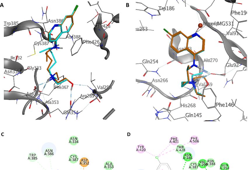
Int. J. Mol. Sci. 2020, 21, 5856                                                                      2 of 16

hydrochloride, a less toxic molecule [17,18]. Rediscovered in America during the Second World War,
it was marketed in 1947 [17,19–23]. Its structural formula derives from a molecule of natural origin,
quinine, from which it differs for the substituents at position 6 and the position linked to chain 4 [17,18].
Although CQ was used for many years as both antimalarial prophylaxis and therapy, its use has
been significantly reduced due to the onset of resistance, especially in Plasmodium falciparum [24].
Its antimetabolic activity has been reported since the first studies with particular reference to the
inhibition of numerous enzymatic pathways in plasmodia [24], and inhibition of DNA polymerase and
RNA polymerase in bacteria [25–27], as well as in Tetrahymena pyriformis, a ciliated protozoa [28].
Although CQ targets plasmodia, unfortunately, the same inhibitory effects have been demonstrated
in a model developed in vivo on rats [29]. However, the antiprotozoal mechanism was subsequently
associated with its lysosomotropic characteristics and its interaction with hemozoin [30]. The antiviral
activity, described in the early 1960s, was initially deduced from a clinical study on the therapy
of viral hepatitis [31], and then described in Mouse Hepatitis Virus (MHV, murine Beta Coronavirus)
experimentally [32]. In this case, the six-hour treatment with CQ of mouse peritoneal macrophages
infected with MHV reduced the viral load compared to those of the untreated cells. However, after 72 h
from the treatment, an increase in the viral load was noted.
     Consequently, the author concluded that this phenomenon was due in some way to a higher
permeability of lysosomes. The antiviral activity was later demonstrated in DNA phages [33]. CQ was
also reported to inhibit other viruses such as Encephalomyocarditis virus (Cardiovirus), Sindbis virus,
Influenza A2, Newcastle disease virus, Herpes simplex, and Vaccinia [34]. A study on the antiviral activity
against myxoviruses allowed the authors to argue that probably the mechanism of action was to
inhibit stripping through the so-called stabilization of lysosomal membranes [35]. In 1974, the first
in vivo study on CQ activity was conducted in a model of infection with Moloney Murine Sarcoma
virus (a murine retrovirus), which has shown that CQ inhibited the development of the tumor when
preventively inoculated at a concentration of 50 mg/kg in the newborn mouse [36]. Numerous other
reports were published regarding other animal and human viruses that attributed its antiviral activity
to lysosomotropy [37–40]. However, this feature and its real antiviral activity has not been more
confirmed in MHV [41]. Furthermore, some viruses, such as poliovirus, seem to be insensitive up to
the concentration of 150 µM [42,43].
     Particular interest has aroused the ability of CQ to inhibit both HIV replication and glycosylation
of viral particles and, at the same time, to act in synergy with the viral protease [44]. Furthermore,
impairment of ACE2 terminal glycosylation has been reported. In fact, at 25 µM CQ, although ACE2 is
expressed in similar quantities at the cell surface, the variations in its glycosylation status might render
the ACE2/SARS-CoV interaction less efficient and inhibit virus entry when the cells are treated with
the drug [45]. The same authors have also shown inhibition of endosome-mediated viral entry.
     Another quinoline derivative has been considered capable of inhibiting viral replication: In fact,
the hydroxychloroquine, a well-known antimalarial drug recognized as a life-saving drug from the
WHO (https://www.who.int/medicines/publications/essentialmedicines/en/), has also been used in the
treatment of arthritic complications during some viral infections [46,47]. HCQ has also been shown to
inhibit the replication of HIV by a mechanism similar to that of chloroquine [48,49]. A hypothetical
anti-HPV activity of HCQ has also been proposed [50].
     HCQ has recently been the subject of renewed interest both as a therapy for the disease caused
by SARS-CoV-2 [51–53], and prophylaxis [54]. Interestingly, HCQ can intervene as a competitor for
absorption by binding to the cellular receptor [55], or onto iron homeostasis [56].
     Looking for possible antiviral molecules, many new targets have been found in numerous
nonstructural proteins [57–61], but insufficient attention has been paid to the role of the nsp10/nsp14
complex [62], and the envelope (E) protein [63].
     This study aimed to investigate the molecular interaction of CQ or HCQ with three nonstructural
proteins of SARS-CoV-2 that could represent a new and more useful target of the two drugs. In fact,
it has been recently suggested that nsp10 and nsp16 could be an innovative target for antiviral drug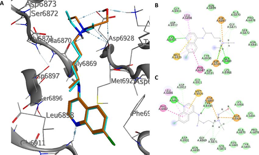
Int. J. Mol. Sci. 2020, 21, 5856                                                                     3 of 16

discovery [64]. These features could be an innovative and exciting explanation by which the mechanism
of quinoline derivatives might be directed more to the late replicative process rather than to uncoating.

2. Results and Discussion

2.1. E Protein
      E protein is a short peptide of 75 amino acids in SARS-CoV-2 [63]. It is organized in three different
domains: N-terminal, transmembrane (with most hydrophobic amino acids), and C-terminal [63,65].
E protein is one of the major structural proteins of the coronavirus [66], and is essential for infection
as well as for bending membranes, and playing a role in virus maturation processes [67]; in fact,
in CoV assembly, the formation of the envelope requires the only expression of M and E and not N.
Moreover, the ability of E and M to drive the formation of a virus-like particle clearly shows that E
is essential for assembly [68]. Moreover, E protein interacts with the host proteins [69], as well as
influences host cell membrane permeability [63,70], and it has been associated with different membrane
topologies [63,68,71,72]. In fact, it has been described, with a low frequency, a version with an
N-glycosylation [71] of asparagine in position 66 [72]. Its functions as an ion-channeling viroporin [73],
shared with two other proteins [68,74,75], have been associated with inflammation observed in acute
respiratory distress [76] as well as to endoplasmic reticulum stress. At the same time, its role in
cellular apoptosis remains questionable [63]. In addition, the Ca2+ passage to E protein channel has
been associated with the activation of NLSP3 inflammasomes [76,77]. Although not crucial for virus
multiplication, the absence of E protein showed an accumulation of probably aberrant virions [78].
Its interaction with PALS1, a particular class of tight junction-associated protein, has been associated
with damage to lung epithelium [79]. E protein may represent a novel strategy used by SARS-CoV to
increase its virulence [80], and, although it is not involved in the innate immune response to SARS-CoV,
it might have an important role in developing a vaccine strategy as well as might be an important
target for a chemotherapeutic approach [66,80,81].
      The great difficulty of crystallization for a hydrophobic protein did not allow the study and design
of selective inhibitors for E protein. Given the low reliability of the homology model obtained and
of the models present on the web, in addition to the lack of data in the literature on the design of
inhibitors, we decided to investigate the entire pentameric structure using the NMR structure of E
protein from SARS-CoV-1 [82]. A comparative sequence analysis via Multalin reveals that SARS-CoV-1
and SARS-CoV-2 E protein sequences share 94.7% identity amongst themselves. The results of the
molecular docking (Figures 1 and 2, Figures S1 and S2) showed that HCQ has better binding energy
(−8.6 kcal/mol) than CQ (−8.3 kcal/mol). The docking pose highlights that the interactions between
HCQ and the E Protein are due to the hydrogen bond with the Phe23 residue, while the drug quinolinic
region is stabilized by the hydrophobic bonds with the five-phenyl residues Phe26 in the central part
of the ion channel (Figure 2 and Figure S2). Other hydrophobic interactions concern the aliphatic area
of the drug with the residues Leu19, Ala22, Val25, and Val29. To obtain detailed information on the
structural characteristics of the protein-drug complex, molecular dynamics (MD) simulations were
performed using the YASARA software; the analysis of the MD trajectories showed excellent stability
of the complex as highlighted in the graphs of Figure S2. It is interesting to note that, at medium
HCQ laying, it establishes a new hydrogen bond between the amino nitrogen and the Phe26 residue,
with an average distance of 1.82 Å, while the hydroxyl group establishes a hydrogen bond with Leu65,
with an average distance of 1.74 Å. Both hydrogen bonds are stable throughout the MD simulations.
The re-docking (−10.6 kcal/mol) performed on the average laying confirmed the excellent stability
of the complex favored both by the hydrogen bonds and by the hydrophobic interactions inside the
central cavity with the Phe26 residue of the pentamer.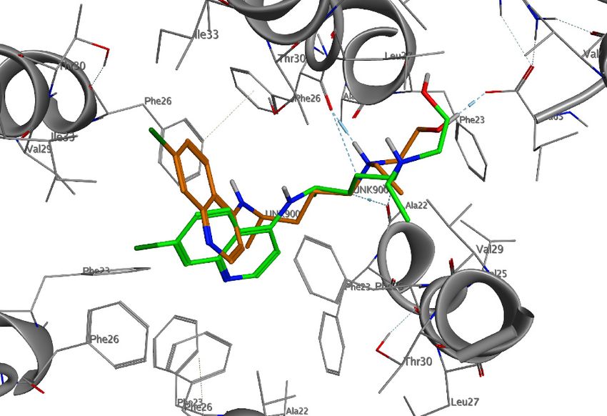
Int. J. Mol. Sci. 2020, 21, 5856 4 of 16
Int. J. Mol. Sci. 2020, 21, x FOR PEER REVIEW 4 of 16
Figure 1. Docked
Docked poses
poses of chloroquine (CQ) (A) and hydroxychloroquine (HCQ) (B) in the central
cavity of the envelope (E) protein.
Figure 2.
Figure 2. Three-dimensional
Three-dimensional (3D)-interaction diagram of
(3D)-interaction diagram of HCQ
HCQ in
in the
the docked
docked pose
pose (green)
(green) and
and average
average
pose during
pose during molecular
molecular dynamics
dynamics (MD)
(MD) simulation
simulation (orange)
(orange) with
with the
the EE protein.
protein.
CQ adopts a position exactly opposite to that of HCQ (Figure 1). The quinolinic ring is inserted
α-helices of the pentameric
between two α-helices pentameric structure
structure establishing
establishing hydrophobic
hydrophobic interactions
interactions with
with Val25,
Val25,
Phe26, Leu27, and Val29, while the small aliphatic chain is oriented oriented within
within thethe channel
channel (Figure
(Figure S1).
S1).
Among
Among other
other things,
things, in the tertiary amino group, it does not establish any interaction with the protein
residues. During
residues. During the the MD
MDsimulation,
simulation,the theCQCQmoves
movesalongalongthethe central
central axis
axis of ofthethe
ionion channel.
channel. After
After 5 ns5
of
ns MD simulation,
of MD simulation,the drug stabilizes,
the drug establishing
stabilizes, both hydrophobic
establishing both hydrophobicinteractions with Phe20,
interactions Ala22,
with Phe20,
Phe23,
Ala22, Val25,
Phe23,Phe26,
Val25,and a hydrogen
Phe26, bond withbond
and a hydrogen Leu19, while
with the re-docking
Leu19, while the value (−9.2 kcal/mol)
re-docking of
value (−9.2
the average
kcal/mol) of poses confirms
the average the confirms
poses excellent the
stability of the
excellent complex.
stability of the complex.
The ionic
ionicchannels
channels undergo
undergosignificant conformational
significant conformational changes so that so
changes they perform
that they their biological
perform their
biological
role. These role. These proteins
membrane membranehaveproteins have high
high plasticity, plasticity,
and some aminoand
acidssome amino
perform acids functions
essential perform
for the passage
essential functions from
forclosed to open
the passage state.
from During
closed the state.
to open MD simulation,
During the we MDnoticed a change
simulation, in the
we noticed
volume
a changeofinthe thecentral
volume tunnel
of the(the CQ led
central to an(the
tunnel increase
CQ led in to
theanvolume
increaseof in
thethecentral
volumelumen of 31.8%),
of the central
lumenHCQ
while of 31.8%),
reduced while HCQ reduced
the volume the volume
of the cavity of the
by 28.14%. cavity
Both by refer
values 28.14%. Both
to the valuesofrefer
volume to the
the central
cavity
volumeofofthe theprotein
centralwithout
cavity ofany
theligand inside
protein afterany
without 20 ns of MD
ligand simulation.
inside after 20 We hypothesized
ns of that
MD simulation.
We hypothesized that the two drugs interact differently with some central lumen residues,
influencing its ability to change its conformation during its functionality. Probably, CQ could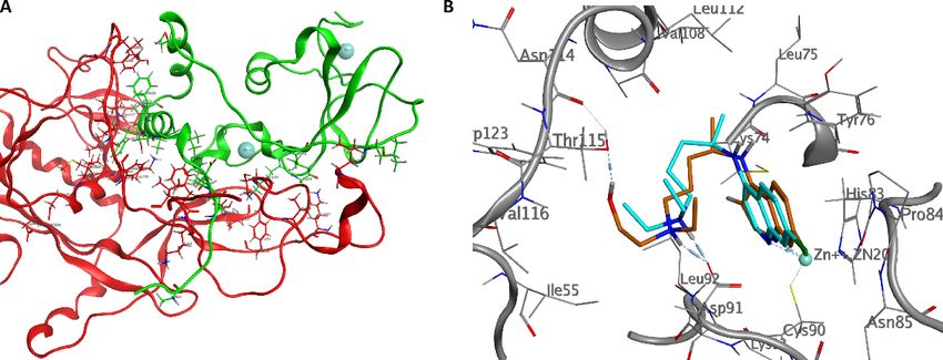
Int. J. Mol. Sci. 2020, 21, 5856 5 of 16
the two drugs interact differently with some central lumen residues, influencing its ability to change
its conformation during its functionality. Probably, CQ could destabilize the ion channel structure
by removing Phe26 residues from the center of the cavity, causing an expansion of the ion channel;
conversely, the hydrophobic interactions of HCQ with the Phe26 (A–E) residues increase the stability
of the central pore by bringing the chains of the pentameric structure closer, causing a reduction in the
internal volume.
2.2. NSP10/NSP14
Nsp14 is a nonstructural protein of coronaviruses. It is known that nsp14 had two different
roles showing both a proofreading activity [83] and methylase activity [84]. Nsp14 is a nonstructural
CoV-2 protein involved in virus replication fidelity by binding its proofreading subunit to the CoV-2
RNA polymerase [85]. Moreover, it has been shown that the nsp14, through its exonuclease domain,
reduced mismatch during replication [86–88]. Notably, this mismatch repair activity has been related
to the low mutation rate of SARS-CoV [62]. Furthermore, an accurate exonuclease activity represents
an essential factor that could affect the activity and use of nucleoside analogs in the treatment of
Coronavirus infections [89].
Nsp10, a nonstructural protein of 139 amino acids, is a most conserved protein of the replicative
machine of SARS-CoV [90] and has been considered as a crucial multifunctional cofactor in their
replication [91]. Although nsp10 is showing no enzyme activity, its role seems to be central to two
distinct regulatory mechanisms: In fact, as reported by Bouvet et al., nsp10 interacts with both nsp14
and nsp16 triggering 30 ,50 -exoribonuclease and 20 -O-methyltransferase activities, respectively [92–95].
Therefore, an interaction with nsp1, nsp7, nsp13, and itself has been also reported [91,96]. Probably,
such interaction with itself may explain why peptides derived from the interaction domain of nsp10
with nsp16 have been shown to inhibit the activity of 20 -O-methyltransferase complex nsp10/nsp16
SARS-CoV [96].
The N-terminal exoribonuclease (ExoN) domain plays a proofreading role in the prevention of
mutagenesis, while the C-terminal domain functions as a (guanine-N7) methyltransferase (N7-MTase)
for mRNA capping. The nsp10 protein interacts with nsp14 ExoN to stabilize it and stimulate
its activity [62]. The cap-precursor guanosine-P3-adenosine-50 ,50 -triphosphate, and S-adenosyl
methionine bind in proximity in a very tight pocket between two β-sheets to accomplish methyl
transfer. Assembly of a cap1 structure at the 50 end of viral mRNA assists in translation and evading
the host defense [97–99]. In the absence of nsp10, nsp14 cannot catalyze nucleotide excision efficiently.
The structure of the nsp10/nsp14 complex explains this requirement of nsp10 for the enzymatic activity
of nsp14. The extensive interaction of nsp10 with nsp14 suggests that nsp10 might be necessary to
maintain the structural stability of the ExoN domain and fully unleash the ExoN activity of nsp14 [62].
We speculated on both domains to find a possible inhibitory activity of CQ and HCQ. Table 1
summarizes the binding energies of the two drugs in both the active sites. HCQ is more active than CQ
in both sites, with a binding energy value of −7.0 kcal/mol for the N7-MTase domain and −7.3 kcal/mol
for the ExoN domain. The binding energy values for CQ are −6.2 and −6.0 kcal/mol, respectively.
Table 1. Calculated free binding energies (∆GB , in kcal/mol) of CQ and HCQ in N7-MTase and
ExoN domains.
Compound N7-MTase (kcal/mol) ExoN (kcal/mol)
CQ −6.2 −6.0
HCQ −7.0 −7.3
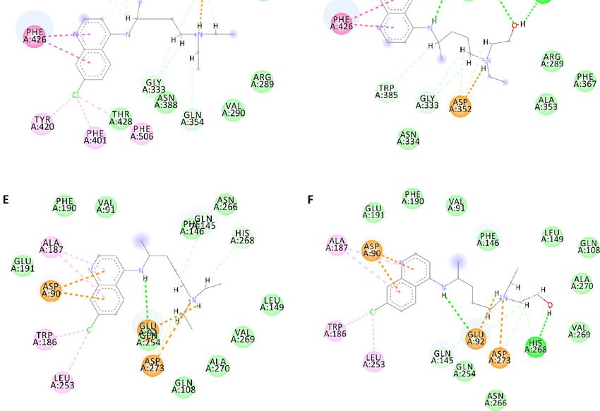
As shown in Figure 3, both drugs have an overlapping pose in the N7-MTase domain.
The hydrogens of the tertiary amino group form a hydrogen bond with the carboxyl group of
Asp352, while the hydroxyl group in the HCQ forms hydrogen bonds with the carbonyl group of
Gln354 and the amide hydrogen of Val296 at 1.7 and 2.2 Å, respectively, giving a higher energy binding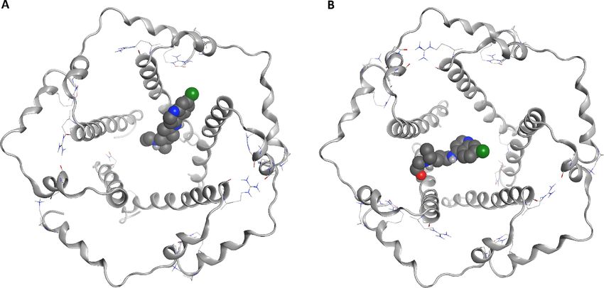
Int. J. Mol. Sci. 2020, 21, 5856                                                                                    6 of 16

value. The quinolinic ring, inside the pocket, is stabilized by the π-π interaction with Phe426, and the
chlorine
     Int. J.atom    establishes
             Mol. Sci.               hydrophobic
                       2020, 21, x FOR PEER REVIEW interactions with The401, Tyr401, and Phe506. Moreover,          for the
                                                                                                               6 of 16
ExoN domain, the poses of CQ and HCQ are very similar. In both cases, the secondary and tertiary
aminothenitrogen
           ExoN domain, form the    poses of CQ
                                 a bidentate     and HCQ bond
                                              hydrogen    are very similar.
                                                                 with Glu92,In both cases,
                                                                                while   thethe secondary ring
                                                                                             quinolinic  and tertiary
                                                                                                              establishes
cation-π interactions with the magnesium atom and Asp90. The hydrophobic and establishes
     amino     nitrogen      form   a bidentate hydrogen  bond   with Glu92,   while the  quinolinic ring Van der Waals
     cation-π interactions with the magnesium atom and Asp90. The hydrophobic and Van der Waals
interactions with Val91, Trp186, Ala187, Phe190, Glu191, and Leu253 further stabilize the aromatic
     interactions with Val91, Trp186, Ala187, Phe190, Glu191, and Leu253 further stabilize the aromatic
region of the ligands.
      region of the ligands.

           Figure
      Figure       3. Interaction
              3. Interaction      profile
                              profile     of the
                                      of the     best-docked
                                             best-docked     poses
                                                          poses forfor
                                                                    CQCQ  (blue)
                                                                       (blue) andand HCQ
                                                                                   HCQ   (orange)ininthe
                                                                                       (orange)        theN7-MTase
                                                                                                           N7-
           MTase (A) and ExoN (B) domain. Two-dimensional (2D) interaction diagram of CQ (C) and HCQ (D)
      (A) and ExoN (B) domain. Two-dimensional (2D) interaction diagram of CQ (C) and HCQ (D) in
           in N7-MTase domain, and CQ (E) and HCQ (F) in ExoN domain.
      N7-MTase domain, and CQ (E) and HCQ (F) in ExoN domain.
            MD simulations were performed to verify the stability of the complexes. At the N7-MTase site,
   MD simulations were performed to verify the stability of the complexes. At the N7-MTase site,
   HCQ showed better stability than CQ throughout the MD simulation period, with less square root
HCQ showed better stability than CQ throughout the MD simulation period, with less square root
Int. J. Mol. Sci. 2020, 21, 5856 7 of 16
Int. J. Mol. Sci. 2020, 21, x FOR PEER REVIEW 7 of 16
mean deviation (RMSD) of both the ligand and the protein system (Figures S3 and S4). The hydrogen
mean deviation (RMSD) of both the ligand and the protein system (Figures S3 and S4). The hydrogen
bond between the tertiary amine nitrogen atom and Asp352 was stable, with an average length of 1.78 Å,
bond between the tertiary amine nitrogen atom and Asp352 was stable, with an average length of
as well as the hydrogen bond between the oxygen of the hydroxyl group and Asn354, with an average
1.78 Å, as well as the hydrogen bond between the oxygen of the hydroxyl group and Asn354, with
length of 1.86 Å. The mean RMSD value of the protein backbone atoms is estimated 1.95 Å for the HCQ
an average length of 1.86 Å. The mean RMSD value of the protein backbone atoms is estimated 1.95
complex and 2.14 Å for the CQ one. Although the two systems showed good stability, the interaction
Å for the HCQ complex and 2.14 Å for the CQ one. Although the two systems showed good stability,
with HCQ was more efficient throughout the MD simulation.
the interaction with HCQ was more efficient throughout the MD simulation.
The study of the trajectories of the complexes in the ExoN domain allowed us to ascertain better
The study of the trajectories of the complexes in the ExoN domain allowed us to ascertain better
stability of HCQ compared to CQ. The former maintained a better RMDS with an average value
stability of HCQ compared to CQ. The former maintained a better RMDS with an average value of
of 1.58 Å compared to 1.71 Å of the CQ. Even the RMSD of the protein structure underwent fewer
1.58 Å compared to 1.71 Å of the CQ. Even the RMSD of the protein structure underwent fewer
fluctuations than the starting structure. It was interesting to note that HCQ maintained the hydrogen
fluctuations than the starting structure. It was interesting to note that HCQ maintained the hydrogen
bond bidentate throughout the MD simulation, unlike CQ, which preferred a more linear pose along
bond bidentate throughout the MD simulation, unlike CQ, which preferred a more linear pose along
with the active site (Figures S5 and S6).
with the active site (Figures S5 and S6).
In the absence of nsp10, nsp14 cannot catalyze nucleotide excision efficiently [62]. The structure
In the absence of nsp10, nsp14 cannot catalyze nucleotide excision efficiently [62]. The structure
of the nsp10/nsp14 complex explains this requirement of nsp10 for the enzymatic activity of nsp14.
of the nsp10/nsp14 complex explains this requirement of nsp10 for the enzymatic activity of nsp14.
The primary residues that contribute to the nsp14-nsp10 interaction belong to two specific regions
The primary residues that contribute to the nsp14-nsp10 interaction belong to two specific regions of
of the nsp10. The first contact area involves the N-terminal ring and the α1-helix (Pro1-Leu24) of
the nsp10. The first contact area involves the N-terminal ring and the α1-helix (Pro1-Leu24) of nsp10,
nsp10, which led to an electron density interpretable for the first nine residues of nsp10. The residues
which led to an electron density interpretable for the first nine residues of nsp10. The residues Ala1,
Ala1, Asn3, and Glu6 of nsp10 stabilize the N-terminal region of nsp14 forming hydrogen bonds
Asn3, and Glu6 of nsp10 stabilize the N-terminal region of nsp14 forming hydrogen bonds with Lys9,
with Lys9, Asp10, and Thr5, while Phe16, Phe19, and Val21 of nsp10 form Van der Waals interactions
Asp10, and Thr5, while Phe16, Phe19, and Val21 of nsp10 form Van der Waals interactions with
with Phe60, Met62, and Tyr64 of nsp14. The second area of intermolecular interactions is extensive
Phe60, Met62, and Tyr64 of nsp14. The second area of intermolecular interactions is extensive and
and includes residues from the ring region after the α2-helix and residues close to the zinc atom.
includes residues from the ring region after the α2-helix and residues close to the zinc atom.
Numerous complementary hydrogen bonds are observed here; Asn40, Lys43, Leu45, Thr58, Ser72,
Numerous complementary hydrogen bonds are observed here; Asn40, Lys43, Leu45, Thr58, Ser72,
Lys93, and Tyr96 of nsp10 interact with Thr25, His26, Cys39, Asp41, Ala23, Tyr51, and His19 of the
Lys93, and Tyr96 of nsp10 interact with Thr25, His26, Cys39, Asp41, Ala23, Tyr51, and His19 of the
nsp14 N-terminal domain. A salt bridge formed between His80 of nsp10 and Asp126 of nsp14 and a
nsp14 N-terminal domain. A salt bridge formed between His80 of nsp10 and Asp126 of nsp14 and a
hydrogen bond between Cys90 of nsp10 and Asn129 of nsp14 stabilize the structural elements between
hydrogen bond between Cys90 of nsp10 and Asn129 of nsp14 stabilize the structural elements
β5 and β6 of nsp14 (Figure 4A). The extensive interactions of the nsp10 with the nsp14 suggest that
between β5 and β6 of nsp14 (Figure 4A). The extensive interactions of the nsp10 with the nsp14
the nsp10 may be necessary to maintain the structural stability of the ExoN domain of the nsp14 and
suggest that the nsp10 may be necessary to maintain the structural stability of the ExoN domain of
simultaneously stimulate and maximize its activity [93].
the nsp14 and simultaneously stimulate and maximize its activity [93].
Figure 4.
Figure 4. (A)
(A) Intermolecular interactions between
Intermolecular interactions between nsp14
nsp14 (red)
(red) is
is stabilized
stabilized by
by nsp10
nsp10 (green).
(green). Zinc
Zinc ions
ions
are represented as blue spheres. (B) Docked poses of CQ (blue) and HCQ (orange) are shown
are represented as blue spheres. (B) Docked poses of CQ (blue) and HCQ (orange) are shown with the with
the residues
residues of binding
of the the binding pocket
pocket and and the residues
the residues interacting
interacting withwith the nsp10
the nsp10 cavity.
cavity.
We assumed
We assumed thatthat the
the drugs
drugs could
could interact
interact with
with the
the nsp10/nsp14
nsp10/nsp14 domain
domain by by destabilizing
destabilizing the
the
protein-protein complex. The docking results showed no significant interaction in the contact
protein-protein complex. The docking results showed no significant interaction in the contact region region
between the
between the two
twoproteins;
proteins;however,
however,interesting
interestingresults were
results wereobtained
obtainedin ainsmall pocket
a small nearnear
pocket the zinc
the
atom of nsp10. The two drugs had the following energy binding: −7.7 kcal/mol for
zinc atom of nsp10. The two drugs had the following energy binding: −7.7 kcal/mol for HCQ and HCQ and −6.6
kcal/mol
−6.6 for CQ.
kcal/mol for CQ.
The two drugs have a very similar pose. The nitrogen of the quinolinic ring establishes a
coordination bond with the zinc atom, Van der Waals interactions with Cys74, Asn85, and Thr111,
and π-π interactions with Tyr76. The amine forms a salt bridge with Asp91 at 2.06 Å, while theInt. J. Mol. Sci.
Sci. 2020,
2020, 21,
21, x5856
FOR PEER REVIEW 8 of 16
hydroxyl group of HCQ establishes a hydrogen bond with the carbonyl oxygen of Thr115 at 1.97 Å
The4B).
(Figure two drugs have a very similar pose. The nitrogen of the quinolinic ring establishes a
coordination bond with the zinc atom, Van der Waals interactions with Cys74, Asn85, and Thr111,
and π-π interactions with Tyr76. The amine forms a salt bridge with Asp91 at 2.06 Å, while the
2.3. NSP10/NSP16
hydroxyl group of HCQ establishes a hydrogen bond with the carbonyl oxygen of Thr115 at 1.97 Å
Nsp16 is a 298 amino acids protein that is involved in methylation of the 2′-hydroxy group of
(Figure 4B).
adenines using S-adenosylmethionine as a source of methyl [100,101]. However, the nsp16 protein is
an RNA
2.3. cap-modifying enzyme that is devoid of any enzymatic activity, but it is activated by nsp10,
NSP10/NSP16
which interacts with nsp16 and selectively confers upon its 2′-O-MTase activity on N7-methyl
Nsp16 is a 298 amino acids protein that is involved in methylation of the 20 -hydroxy group of
guanine RNA caps [102]. Nsp16 is also involved in the modulation of pathogenesis and type I
adenines using S-adenosylmethionine as a source of methyl [100,101]. However, the nsp16 protein is
Interferon susceptibility, as well as it has been suggested as a vaccine candidate [103].
an RNA cap-modifying enzyme that is devoid of any enzymatic activity, but it is activated by nsp10,
The crystal structure of the nsp10/nsp16 methyltransferase (PDB ID: 6W6L) of SARS-CoV-2 was
which interacts with nsp16 and selectively confers upon its 20 -O-MTase activity on N7-methyl guanine
used for docking purposes. The docking results we obtained show that the two drugs have a very
RNA caps [102]. Nsp16 is also involved in the modulation of pathogenesis and type I Interferon
similar pose inside the pocket that binds SAM, despite the hydroxyquinoline having a better energy
susceptibility, as well as it has been suggested as a vaccine candidate [103].
binding (−8.1 versus −7.6 kcal/mol). The quinolinic ring shows a hydrogen bond with the amide
The crystal structure of the nsp10/nsp16 methyltransferase (PDB ID: 6W6L) of SARS-CoV-2 was
residue Cys6913 and hydrophobic interactions with Leu6898, Phe6947, and Met6929. The tertiary
used for docking purposes. The docking results we obtained show that the two drugs have a very
amino nitrogen atom of CQ forms a salt bridge with Gly9869, while that of HCQ with the carbonyl
similar pose inside the pocket that binds SAM, despite the hydroxyquinoline having a better energy
oxygen of Asp6928; the alcoholic group of HCQ forms hydrogen bonds with the carboxylic residue
binding (−8.1 versus −7.6 kcal/mol). The quinolinic ring shows a hydrogen bond with the amide
of Asp6928 (1.87 Å) and the amino group of Lys6869 (1.98 Å) (Figure 5). Probably, the further
residue Cys6913 and hydrophobic interactions with Leu6898, Phe6947, and Met6929. The tertiary
interaction of the hydroxyl group in the HCQ, inside the enzyme pocket, increases the affinity of the
amino nitrogen atom of CQ forms a salt bridge with Gly9869, while that of HCQ with the carbonyl
drug.
oxygen of Asp6928; the alcoholic group of HCQ forms hydrogen bonds with the carboxylic residue of
The average pose in MD simulation showed that HCQ improves interactions with the active site,
Asp6928 (1.87 Å) and the amino group of Lys6869 (1.98 Å) (Figure 5). Probably, the further interaction
the hydrogen bonds with Asp6897, and Asp6928 whereas CQ maintains a stable pose demonstrating
of the hydroxyl group in the HCQ, inside the enzyme pocket, increases the affinity of the drug.
the low RMSD value (Figures S7 and S8).
Figure 5.
Figure 5. Interaction
Interaction profile
profile of
of the best-docked poses
the best-docked poses for
for CQ (blue) and
CQ (blue) and HCQ
HCQ (orange)
(orange) in
in the
the SAM
SAM
domain of nsp16 (A). 2D interaction diagram of CQ (B) and HCQ (C) in the SAM domain of
domain of nsp16 (A). 2D interaction diagram of CQ (B) and HCQ (C) in the SAM domain of nsp16.nsp16.
3. Materials and Methods
The average pose in MD simulation showed that HCQ improves interactions with the active site,
the hydrogen bonds with Asp6897, and Asp6928 whereas CQ maintains a stable pose demonstrating
3.1. low
the Structures
RMSDPreparation and Minimization
value (Figures S7 and S8).
The structures of all the molecules used in this study were built using Marvin Sketch (18.24,
ChemAxon Ltd., Budapest, Hungary, http://www.chemaxon.com). A first molecular mechanics
energy minimization was used for 3D structures created from the SMLES; the Merck molecular force
field (MMFF94) present in Marvin Sketch was used. The protonation states were calculated, assumingInt. J. Mol. Sci. 2020, 21, 5856 9 of 16
3. Materials and Methods
3.1. Structures Preparation and Minimization
The structures of all the molecules used in this study were built using Marvin Sketch (18.24,
ChemAxon Ltd., Budapest, Hungary, http://www.chemaxon.com). A first molecular mechanics energy
minimization was used for 3D structures created from the SMLES; the Merck molecular force field
(MMFF94) present in Marvin Sketch was used. The protonation states were calculated, assuming a
neutral pH. The PM3 Hamiltonian, as implemented in the MOPAC package (MOPAC2016 v. 18.151,
Stewart Computational Chemistry, Colorado Springs, CO, USA) [104], was then used to further
optimize the 3D structures before the alignment for the docking calculations.
3.2. Molecular Docking
Flexible ligand docking experiments were performed by employing the AutoDock 4.2.6 software
implemented in YASARA (v. 19.5.5, YASARA Biosciences GmbH, Vienna, Austria) [105,106], using the
three-dimensional crystal structure of the SARS coronavirus nsp10/nsp14 complex with functional
ligands SAH and GpppA (PDB ID: 5C8S), NMR structure of the SARS coronavirus E protein pentameric
ion channel (PDB ID: 5 × 29), and nsp10/nsp16 complex (PDB ID: 6W61) obtained from the Protein
Data Bank (PDB, http://www.rcsb.org/pdb), the Lamarckian genetic algorithm (LGA). The crystallized
ligand has been eliminated using the YASARA software. The maps were generated by the program
AutoGrid (4.2.6) with a spacing of 0.375 Å and dimensions that encompass all atoms extending 5 Å
from the surface of the structure of the crystallized ligand. All parameters were inserted at their
default settings, as previously reported [107,108]. In the docking tab, the macromolecule and ligand
were selected, and GA parameters were set as ga_runs = 100, ga_pop_size = 150, ga_num_evals =
25,000,000, ga_num_generations = 27,000, ga_elitism = 1, ga_mutation_rate = 0.02, ga_crossover_rate
= 0.8, ga_crossover_mode = two points, ga_cauchy_alpha = 0.0, ga_cauchy_beta = 1.0, number of
generations for picking worst individual = 10.
3.3. Molecular Dynamics Simulations
The molecular dynamics simulations of the nsp10/nsp14/ligand complexes and E protein/ligand
complexes were performed with the YASARA structure package. A periodic simulation cell with
boundaries extending 8 Å [109] from the surface of the complex was employed. The box was filled
with water, with a maximum sum of all water bumps of 1.0 Å, and a density of 0.997 g mL−1 .
The setup included an optimization of the hydrogen bonding network [110] to increase the solute
stability, and a pKa prediction to fine-tune the protonation states of protein residues at the chosen
pH of 7.4 [111]. NaCl ions were added with a physiological concentration of 0.9%, with an excess of
either Na or Cl to neutralize the cell. Water molecules were deleted to readjust the solvent density to
0.997 g/mL. The final system dimensions were approximately 80 × 80 × 130 Å3 for nsp10/nsp14/ligand
complexes and 94 × 78 × 94 Å3 for E protein/ligand complexes.
To best represent the biological environment, for E protein/ligand complexes, each of the best
pose ligand/receptor complex structure was immersed in a simulated bilayer membrane, in the
above reported physiological environment conditions, and subjected to a molecular dynamics (MD)
simulation. The simulation was set up automatically by first scanning the protein for exposed
transmembrane helices (i.e., helices longer than 16 residues, with more than seven hydrophobic
residues and more than three exposed ones (accessible side-chain surface area >30% of maximum)).
The major axis vectors of these helices (i.e., the direction vectors of the least-squares lines through the
Calpha atoms) were summed up to obtain the major axis of the protein, which was then oriented along
the Y-axis, generally with respect to the plane of the membrane and the XZ plane. The best shift of the
membrane along this major axis was obtained by scanning the protein for the region with the largest
number of exposed hydrophobic residues (see definition above) and a width of 28 Å (corresponding to
the membrane core).Int. J. Mol. Sci. 2020, 21, 5856 10 of 16
Having placed an equilibrated membrane structure (consisting of 50% of phosphatidylcholine
and 50% of phosphatidylethanolamine) at this location named ‘MemCenterY’, the system was enclosed
in a simulation cell of size (100 × 80 × 100) Å, and the protein was temporarily scaled by 0.9 along the
X–Z axes, and then, strongly clashing membrane lipids were deleted (lipids with an atom closer than
0.75 Å to a protein atom). The temporary protein scaling, which was needed to avoid the deletion of
too many lipids around the protein, was then slowly removed during a short simulation at 298 K in
vacuum. The protein (with all of the atoms kept fixed) was scaled by 1.02 along the X–Z axes every
200 fs, while the membrane was allowed to move, but was restrained to ideal geometry (by pulling
lipid residues with an atom further than 21.5 Å away from MemCenterY back into the membrane,
and by pushing phosphorus atoms closer than 14 Å to MemCenterY back outwards).
The simulations were run using the ff14SB force field [112] for the solute, with Lipid17, GAFF2 [113],
and AM1BCC [114] for non-standard residues, and TIP3P for water.
As soon as the protein had reached its original size again, the protein side-chain pKa s were
predicted, protonation states were assigned according to pH 7.4, and the simulation cell was filled
with water, 0.9% NaCl, and counter ions.
The main simulation was then run with a cutoff of 8 Å for Van der Waals forces (the default
used by AMBER) [115], and no cutoff was applied to electrostatic forces (using the Particle Mesh
Ewald algorithm) [116], a four fs time-step, constrained hydrogen atoms, and at constant pressure and
temperature (NPT ensemble) using algorithms described in detail previously [117]. The ligand force
field parameters were generated with the AutoSMILES utility [114], which employs semiempirical
AM1 geometry optimization and assignment of charges, followed by assignment of the AM1BCC atom
and bond types with refinement using the RESP charges, and finally the assignments of general AMBER
force field atom types. Optimization of the hydrogen bond network of the various enzyme-ligand
complexes was obtained using the method established by Hooft et al. [110], in order to address
ambiguities arising from multiple side-chain conformations and protonation states that are not well
resolved in the electron density [118]. During the initial 250 ps, the membrane was restrained to
avoid distortions while the simulation cell adapted to the pressure exerted by the membrane (see
above; additionally, water molecules that got closer than 14 Å to MemCenterY were pushed outside).
The source code of this simulation protocol and visualizations of the individual steps can be found at
www.yasara.org/membranemd.
After the membrane placement, a short MD was run on the solvent only. The entire system
was then energy minimized using first a steepest descent minimization to remove conformational
stress, followed by a simulated annealing minimization until convergence (Int. J. Mol. Sci. 2020, 21, 5856 11 of 16
nsp10/nsp16, could be influenced by CQ and HCQ. The energy binding for nsp16 in the SAM domain
was slightly higher than the viral regions of nsp14. MD simulation studies demonstrate the stability of
drug interactions with the protein regions analyzed. Although computational studies deserve more
attention for nsp10, we have shown that CQ and HQC have a good affinity with an area of the viral
protein that could influence the cofactor effect against nsp14 and nsp16.
Supplementary Materials: Supplementary materials can be found at http://www.mdpi.com/1422-0067/21/16/5856/s1.
Author Contributions: Conceptualization, A.R. and P.M.F.; methodology, D.G. and V.F.; software, A.R.; validation,
D.G., V.F., and A.R.; formal analysis, D.G., A.R., and P.M.F.; investigation, V.F. and P.M.F.; resources, D.G. and A.R.;
data curation, D.G., V.F., A.R., and P.M.F.; writing—original draft preparation, D.G. and V.F.; writing—review and
editing, A.R. and P.M.F.; supervision, A.R. and P.M.F.; project administration, A.R. All authors have read and
agreed to the published version of the manuscript.
Funding: This research received no external funding.
Acknowledgments: Free academic license from ChemAxon for their suite of programs is gratefully acknowledged.
Conflicts of Interest: The authors declare no conflict of interest.
References
1. de Wit, E.; van Doremalen, N.; Falzarano, D.; Munster, V.J. SARS and MERS: Recent insights into emerging
coronaviruses. Nat. Rev. Microbiol. 2016, 14, 523–534. [CrossRef] [PubMed]
2. Zhu, N.; Zhang, D.; Wang, W.; Li, X.; Yang, B.; Song, J.; Zhao, X.; Huang, B.; Shi, W.; Lu, R.; et al. A Novel
Coronavirus from Patients with Pneumonia in China, 2019. N. Engl. J. Med. 2020, 382, 727–733. [CrossRef]
[PubMed]
3. Hui, D.S.; Memish, Z.A.; Zumla, A. Severe acute respiratory syndrome vs. The Middle East respiratory
syndrome. Curr. Opin. Pulm. Med. 2014, 20, 233–241. [CrossRef] [PubMed]
4. WHO. WHO Characterizes COVID-19 as a Pandemic; WHO: Geneva, Switzerland, 2020.
5. Yu, W.B.; Tang, G.D.; Zhang, L.; Corlett, R.T. Decoding the evolution and transmissions of the novel
pneumonia coronavirus (SARS-CoV-2 / HCoV-19) using whole genomic data. Zool. Res. 2020, 41, 247–257.
[CrossRef] [PubMed]
6. Andersen, K.G.; Rambaut, A.; Lipkin, W.I.; Holmes, E.C.; Garry, R.F. The proximal origin of SARS-CoV-2.
Nat. Med. 2020, 26, 450–452. [CrossRef] [PubMed]
7. Malaiyan, J.; Arumugam, S.; Mohan, K.; Gomathi Radhakrishnan, G. An update on the origin of SARS-CoV-2:
Despite closest identity, bat (RaTG13) and pangolin derived coronaviruses varied in the critical binding site
and O-linked glycan residues. J. Med. Virol. 2020. [CrossRef]
8. Tang, X.L.; Wu, C.C.; Li, X.; Song, Y.H.; Yao, X.M.; Wu, X.K.; Duan, Y.G.; Zhang, H.; Wang, Y.R.; Qian, Z.H.; et al.
On the origin and continuing evolution of SARS-CoV-2. Natl. Sci. Rev. 2020, 7, 1012–1023. [CrossRef]
9. Chaw, S.M.; Tai, J.H.; Chen, S.L.; Hsieh, C.H.; Chang, S.Y.; Yeh, S.H.; Yang, W.S.; Chen, P.J.; Wang, H.Y.
The origin and underlying driving forces of the SARS-CoV-2 outbreak. J. Biomed. Sci. 2020, 27. [CrossRef]
10. Rothan, H.A.; Byrareddy, S.N. The epidemiology and pathogenesis of coronavirus disease (COVID-19)
outbreak. J. Autoimmun. 2020, 109, 102433. [CrossRef]
11. Robinson, C.; Loeffelholz, M.J.; Pinsky, B.A. Clinical Virology Manual. In Clinical Virology Manual, 5th ed.;
Loeffelholz, M.J., Ed.; ASM Press: Washington, DC, USA, 2016.
12. Carriere, F.; Longhi, S.; Record, M. The endosomal lipid bis(monoacylglycero) phosphate as a potential key
player in the mechanism of action of chloroquine against SARS-COV-2 and other enveloped viruses hijacking
the endocytic pathway. Biochimie 2020. [CrossRef]
13. Gao, Q. Pharmacological characteristics of chloroquine and suggestions for its use in treatment of coronavirus
disease 2019 (COVID-19). Zhongguo Xue Xi Chong Bing Fang Zhi Za Zhi 2020, 32, 119–122. [CrossRef]
[PubMed]
14. Beura, S.; Prabhakar, C. In-silico strategies for probing chloroquine based inhibitors against SARS-CoV-2.
J. Biomol. Struct. Dyn. 2020, 1–25. [CrossRef] [PubMed]
15. Rodriguez-Martinez, C.E.; Fernandes, R.M.; Hawcutt, D.B.; Sinha, I.P.; Pacheco, R.L. Efficacy, safety and
cost-effectiveness of hydroxychloroquine in children with COVID-19: A call for evidence. Acta Paediatr. 2020.
[CrossRef] [PubMed]Int. J. Mol. Sci. 2020, 21, 5856 12 of 16
16. D’Cruz, M. The ICMR bulletin on targeted hydroxychloroquine prophylaxis for Covid-19: Need to interpret
with caution. Indian J. Med. Ethics 2020, 5, 100–102. [CrossRef] [PubMed]
17. Coatney, G.R. Pitfalls in a discovery: The chronicle of chloroquine. Am. J. Trop. Med. Hyg. 1963, 12, 121–128.
[CrossRef] [PubMed]
18. Radl, S. From chloroquine to antineoplastic drugs? the story of antibacterial quinolones. Arch. Pharm. (Weinh.)
1996, 329, 115–119. [CrossRef] [PubMed]
19. Loeb, F.; Clark, W.M.; Coatney, G.R.; Coggeshall, L.T.; Dieuaide, F.R.; Dochez, A.R.; Marshall, E.K., Jr.;
Marvel, C.S.; McCay, O.R.; Sapero, J.J.; et al. ACTIVITY of a new antimalarial agent, chloroquine (SN 7618).
J. Am. Med. Assoc. 1946, 130, 1069. [CrossRef]
20. Halawani, A.; Baz, I.I.; Morcos, F. The antimalarial chloroquine-diphosphate (aralen). J. Egypt. Med. Assoc.
1947, 30, 128–136.
21. Anonimous, TRIAL use of the new antimalarial drug chloroquine. Bull. US Army. Med. Dep. 1946, 6, 9–10.
22. Anonimous, CHLOROQUINE-DIPHOSPHATE, a Newly-Standardized Antimalarial Drug. Bull. US Army.
Med. Dep. 1947, 7, 834.
23. Anonimous, CLINICAL evaluation of chloroquine. Bull U S Army. Med. Dep. 1947, 7, 835–837.
24. Cowman, A.F.; Foote, S.J. Chemotherapy and Drug-Resistance in Malaria. Int. J. Parasitol. 1990, 20, 503–513.
[CrossRef]
25. Ciak, J.; Hahn, F.E. Chloroquine: Mode of action. Science 1966, 151, 347–349. [CrossRef] [PubMed]
26. Whichard, L.P.; Washington, M.E.; Holbrook, D.J., Jr. The inhibition in vitro of bacterial DNA polymerases
and RNA polymerase by antimalarial 8-aminoquinolines and by chloroquine. Biochim. Biophys. Acta 1972,
287, 52–67. [CrossRef]
27. Marquez, V.E.; Cranston, J.W.; Ruddon, R.W.; Burckhalter, J.H. Binding to deoxyribonucleic acid and
inhibition of ribonucleic acid polymerase by analogs of chloroquine. J. Med. Chem. 1974, 17, 856–862.
[CrossRef] [PubMed]
28. Conklin, K.A.; Heu, P.; Chou, S.C. The effects of antimalarial drugs on nucleic acid synthesis in vitro in
Tetrahymena pyriformis. Mol. Pharm. 1973, 9, 304–310.
29. Field, R.C.; Gibson, B.R.; Holbrook, D.J., Jr.; McCall, B.M. Inhibition of precursor incorporation into nucleic
acids of mammalian tissues by antimalarial aminoquinolines. Br. J. Pharm. 1978, 62, 159–164. [CrossRef]
30. Egan, T.J.; Marques, H.M. The role of haem in the activity of chloroquine and related antimalarial drugs.
Coord. Chem. Rev. 1999, 190, 493–517. [CrossRef]
31. Pareja-Coronel, A. Treatment of viral hepatitis with chloroquine. Am. J. Gastroenterol. 1963, 39, 288–298.
32. Mallucci, L. Effect of chloroquine on lysosomes and on growth of mouse hepatitis virus (MHV-3). Virology
1966, 28, 355–362. [CrossRef]
33. Yielding, K.L. Inhibition of the replication of a bacterial DNA virus by chloroquine and other 4-aminoquinoline
drugs. Proc. Soc. Exp. Biol. Med. 1967, 125, 780–783. [CrossRef] [PubMed]
34. Inglot, A.D. Comparison of the antiviral activity in vitro of some non-steroidal anti-inflammatory drugs.
J. Gen. Virol. 1969, 4, 203–214. [CrossRef] [PubMed]
35. Durand, D.P.; Chalgren, S.D.; Franke, V. Effect of chloroquine on myxovirus replication. Antimicrob Agents
Chemother (Bethesda) 1970, 10, 105–108. [PubMed]
36. Pazmino, N.H.; Yuhas, J.M.; Tennant, R.W. Inhibition of murine RNA tumor virus replication and oncogenesis
by chloroquine. Int. J. Cancer 1974, 14, 379–385. [CrossRef]
37. Rolain, J.M.; Colson, P.; Raoult, D. Recycling of chloroquine and its hydroxyl analogue to face bacterial,
fungal and viral infections in the 21st century. Int. J. Antimicrob. Agents 2007, 30, 297–308. [CrossRef]
38. Al-Bari, M.A.A. Targeting endosomal acidification by chloroquine analogs as a promising strategy for the
treatment of emerging viral diseases. Pharmacol. Res. Perspect. 2017, 5. [CrossRef]
39. D’Alessandro, S.; Scaccabarozzi, D.; Signorini, L.; Perego, F.; Ilboudo, D.P.; Ferrante, P.; Delbue, S. The Use of
Antimalarial Drugs against Viral Infection. Microorganisms 2020, 8, 85. [CrossRef]
40. Devaux, C.A.; Rolain, J.M.; Colson, P.; Raoult, D. New insights on the antiviral effects of chloroquine against
coronavirus: What to expect for COVID-19? Int. J. Antimicrob. Agents 2020, 55, 105938. [CrossRef]
41. Kooi, C.; Cervin, M.; Anderson, R. Differentiation of acid-pH-dependent and -nondependent entry pathways
for mouse hepatitis virus. Virology 1991, 180, 108–119. [CrossRef]Int. J. Mol. Sci. 2020, 21, 5856 13 of 16
42. Zeichhardt, H.; Wetz, K.; Willingmann, P.; Habermehl, K.O. Entry of poliovirus type 1 and Mouse Elberfeld
(ME) virus into HEp-2 cells: Receptor-mediated endocytosis and endosomal or lysosomal uncoating.
J. Gen. Virol. 1985, 66, 483–492. [CrossRef]
43. Kronenberger, P.; Vrijsen, R.; Boeye, A. Chloroquine induces empty capsid formation during poliovirus
eclipse. J. Virol. 1991, 65, 7008–7011. [CrossRef] [PubMed]
44. Savarino, A.; Lucia, M.B.; Rastrelli, E.; Rutella, S.; Golotta, C.; Morra, E.; Tamburrini, E.; Perno, C.F.;
Boelaert, J.R.; Sperber, K.; et al. Anti-HIV effects of chloroquine: Inhibition of viral particle glycosylation and
synergism with protease inhibitors. J. Acquir. Immune Defic. Syndr. 2004, 35, 223–232. [CrossRef]
45. Vincent, M.J.; Bergeron, E.; Benjannet, S.; Erickson, B.R.; Rollin, P.E.; Ksiazek, T.G.; Seidah, N.G.; Nichol, S.T.
Chloroquine is a potent inhibitor of SARS coronavirus infection and spread. Virol. J. 2005, 2, 69. [CrossRef]
[PubMed]
46. Ferri, C.; Sebastiani, M.; Antonelli, A.; Colaci, M.; Manfredi, A.; Giuggioli, D. Current treatment of hepatitis
C-associated rheumatic diseases. Arthritis Res. 2012, 14. [CrossRef] [PubMed]
47. Marti-Carvajal, A.; Ramon-Pardo, P.; Javelle, E.; Simon, F.; Aldighieri, S.; Horvath, H.; Rodriguez-Abreu, J.;
Reveiz, L. Interventions for treating patients with chikungunya virus infection-related rheumatic and
musculoskeletal disorders: A systematic review. PLoS ONE 2017, 12. [CrossRef]
48. Savarino, A.; Gennero, L.; Sperber, K.; Boelaert, J.R. The anti-HIV-1 activity of chloroquine. J. Clin. Virol.
2001, 20, 131–135. [CrossRef]
49. Romanelli, F.; Smith, K.M.; Hoven, A.D. Chloroquine and hydroxychloroquine as inhibitors of human
immunodeficiency virus (HIV-1) activity. Curr. Pharm. Des. 2004, 10, 2643–2648. [CrossRef]
50. Bhushan, P.; Aggarwal, A.; Baliyan, V. Complete Clearance of Cutaneous Warts with Hydroxychloroquine:
Antiviral Action? Indian J. Derm. 2014, 59. [CrossRef]
51. Andreani, J.; Le Bideau, M.; Duflot, I.; Jardot, P.; Rolland, C.; Boxberger, M.; Wurtz, N.; Rolain, J.M.; Colson, P.;
La Scola, B.; et al. In vitro testing of combined hydroxychloroquine and azithromycin on SARS-CoV-2 shows
synergistic effect. Microb. Pathog. 2020, 145, 104228. [CrossRef]
52. Meo, S.A.; Klonoff, D.C.; Akram, J. Efficacy of chloroquine and hydroxychloroquine in the treatment of
COVID-19. Eur. Rev. Med. Pharm. Sci. 2020, 24, 4539–4547.
53. Sarma, P.; Kaur, H.; Kumar, H.; Mahendru, D.; Avti, P.; Bhattacharyya, A.; Prajapat, M.; Shekhar, N.; Kumar, S.;
Singh, R.; et al. Virological and clinical cure in COVID-19 patients treated with hydroxychloroquine:
A systematic review and meta-analysis. J. Med. Virol. 2020. [CrossRef]
54. Gentile, I.; Maraolo, A.E.; Piscitelli, P.; Colao, A. COVID-19: Time for Post-Exposure Prophylaxis? Int. J.
Environ. Res. Public. Health 2020, 17, 3997. [CrossRef] [PubMed]
55. Fantini, J.; Chahinian, H.; Yahi, N. Synergistic antiviral effect of hydroxychloroquine and azithromycin in
combination against SARS-CoV-2: What molecular dynamics studies of virus-host interactions reveal. Int. J.
Antimicrob. Agents 2020, 106020. [CrossRef] [PubMed]
56. Quiros Roldan, E.; Biasiotto, G.; Magro, P.; Zanella, I. The possible mechanisms of action of 4-aminoquinolines
(chloroquine/hydroxychloroquine) against Sars-Cov-2 infection (COVID-19): A role for iron homeostasis?
Pharm. Res. 2020, 158, 104904. [CrossRef] [PubMed]
57. Ostaszewski, M.; Mazein, A.; Gillespie, M.E.; Kuperstein, I.; Niarakis, A.; Hermjakob, H.; Pico, A.R.;
Willighagen, E.L.; Evelo, C.T.; Hasenauer, J.; et al. COVID-19 Disease Map, building a computational
repository of SARS-CoV-2 virus-host interaction mechanisms. Sci Data 2020, 7, 136. [CrossRef] [PubMed]
58. Ou, X.; Liu, Y.; Lei, X.; Li, P.; Mi, D.; Ren, L.; Guo, L.; Guo, R.; Chen, T.; Hu, J.; et al. Characterization of spike
glycoprotein of SARS-CoV-2 on virus entry and its immune cross-reactivity with SARS-CoV. Nat. Commun.
2020, 11, 1620. [CrossRef]
59. Hoffmann, M.; Kleine-Weber, H.; Schroeder, S.; Kruger, N.; Herrler, T.; Erichsen, S.; Schiergens, T.S.; Herrler, G.;
Wu, N.H.; Nitsche, A.; et al. SARS-CoV-2 Cell Entry Depends on ACE2 and TMPRSS2 and Is Blocked by a
Clinically Proven Protease Inhibitor. Cell 2020, 181, 271–280. [CrossRef]
60. Coutard, B.; Valle, C.; de Lamballerie, X.; Canard, B.; Seidah, N.G.; Decroly, E. The spike glycoprotein of the
new coronavirus 2019-nCoV contains a furin-like cleavage site absent in CoV of the same clade. Antivir. Res.
2020, 176, 104742. [CrossRef]
61. Zhang, L.; Lin, D.; Sun, X.; Curth, U.; Drosten, C.; Sauerhering, L.; Becker, S.; Rox, K.; Hilgenfeld, R.
Crystal structure of SARS-CoV-2 main protease provides a basis for design of improved alpha-ketoamide
inhibitors. Science 2020, 368, 409–412.Int. J. Mol. Sci. 2020, 21, 5856 14 of 16
62. Ma, Y.; Wu, L.; Shaw, N.; Gao, Y.; Wang, J.; Sun, Y.; Lou, Z.; Yan, L.; Zhang, R.; Rao, Z. Structural basis and
functional analysis of the SARS coronavirus nsp14-nsp10 complex. Proc. Natl. Acad. Sci. USA 2015, 112,
9436–9441. [CrossRef]
63. Schoeman, D.; Fielding, B.C. Coronavirus envelope protein: Current knowledge. Virol. J. 2019, 16, 69.
[CrossRef] [PubMed]
64. Viswanathan, T.; Arya, S.; Chan, S.H.; Qi, S.; Dai, N.; Hromas, R.A.; Park, J.G.; Oladunni, F.;
Martinez-Sobrido, L.; Gupta, Y.K. Structural Basis of RNA Cap Modification by SARS-CoV-2 Coronavirus.
Nat. Commun. 2020, 11, 3718. [CrossRef] [PubMed]
65. Verdia-Baguena, C.; Nieto-Torres, J.L.; Alcaraz, A.; Dediego, M.L.; Enjuanes, L.; Aguilella, V.M. Analysis of
SARS-CoV E protein ion channel activity by tuning the protein and lipid charge. Biochim. Biophys. Acta 2013,
1828, 2026–2031. [CrossRef] [PubMed]
66. DeDiego, M.L.; Nieto-Torres, J.L.; Jimenez-Guardeno, J.M.; Regla-Nava, J.A.; Castano-Rodriguez, C.;
Fernandez-Delgado, R.; Usera, F.; Enjuanes, L. Coronavirus virulence genes with main focus on SARS-CoV
envelope gene. Virus Res. 2014, 194, 124–137. [CrossRef] [PubMed]
67. Cohen, J.R.; Lin, L.D.; Machamer, C.E. Identification of a Golgi complex-targeting signal in the cytoplasmic
tail of the severe acute respiratory syndrome coronavirus envelope protein. J. Virol. 2011, 85, 5794–5803.
[CrossRef]
68. EA, J.A.; Jones, I.M. Membrane binding proteins of coronaviruses. Future Virol. 2019, 14, 275–286.
69. Ruch, T.R.; Machamer, C.E. The coronavirus E protein: Assembly and beyond. Viruses 2012, 4, 363–382.
[CrossRef]
70. Liu, D.X.; Yuan, Q.; Liao, Y. Coronavirus envelope protein: A small membrane protein with multiple
functions. Cell. Mol. Life Sci. 2007, 64, 2043–2048. [CrossRef]
71. Fung, T.S.; Liu, D.X. Post-translational modifications of coronavirus proteins: Roles and function. Future Virol.
2018, 13, 405–430. [CrossRef]
72. Yuan, Q.; Liao, Y.; Torres, J.; Tam, J.P.; Liu, D.X. Biochemical evidence for the presence of mixed membrane
topologies of the severe acute respiratory syndrome coronavirus envelope protein expressed in mammalian
cells. Febs Lett. 2006, 580, 3192–3200. [CrossRef]
73. Nieva, J.L.; Madan, V.; Carrasco, L. Viroporins: Structure and biological functions. Nat. Rev. Microbiol. 2012,
10, 563–574. [CrossRef] [PubMed]
74. Castano-Rodriguez, C.; Honrubia, J.M.; Gutierrez-Alvarez, J.; DeDiego, M.L.; Nieto-Torres, J.L.;
Jimenez-Guardeno, J.M.; Regla-Nava, J.A.; Fernandez-Delgado, R.; Verdia-Baguena, C.; Queralt-Martin, M.; et al.
Role of Severe Acute Respiratory Syndrome Coronavirus Viroporins E, 3a, and 8a in Replication and
Pathogenesis. mBio 2018, 9, e02325-17. [CrossRef] [PubMed]
75. Ye, Y.; Hogue, B.G. Role of the coronavirus E viroporin protein transmembrane domain in virus assembly.
J. Virol. 2007, 81, 3597–3607. [CrossRef]
76. Nieto-Torres, J.L.; Verdia-Baguena, C.; Jimenez-Guardeno, J.M.; Regla-Nava, J.A.; Castano-Rodriguez, C.;
Fernandez-Delgado, R.; Torres, J.; Aguilella, V.M.; Enjuanes, L. Severe acute respiratory syndrome coronavirus
E protein transports calcium ions and activates the NLRP3 inflammasome. Virology 2015, 485, 330–339.
[CrossRef] [PubMed]
77. Lo, Y.H.; Huang, Y.W.; Wu, Y.H.; Tsai, C.S.; Lin, Y.C.; Mo, S.T.; Kuo, W.C.; Chuang, Y.T.; Jiang, S.T.;
Shih, H.M.; et al. Selective inhibition of the NLRP3 inflammasome by targeting to promyelocytic leukemia
protein in mouse and human. Blood 2013, 121, 3185–3194. [CrossRef] [PubMed]
78. DeDiego, M.L.; Alvarez, E.; Almazan, F.; Rejas, M.T.; Lamirande, E.; Roberts, A.; Shieh, W.J.; Zaki, S.R.;
Subbarao, K.; Enjuanes, L. A severe acute respiratory syndrome coronavirus that lacks the E gene is attenuated
in vitro and in vivo. J. Virol. 2007, 81, 1701–1713. [CrossRef]
79. Teoh, K.T.; Siu, Y.L.; Chan, W.L.; Schluter, M.A.; Liu, C.J.; Peiris, J.S.; Bruzzone, R.; Margolis, B.; Nal, B.
The SARS coronavirus E protein interacts with PALS1 and alters tight junction formation and epithelial
morphogenesis. Mol. Biol. Cell 2010, 21, 3838–3852. [CrossRef]
80. DeDiego, M.L.; Nieto-Torres, J.L.; Jimenez-Guardeno, J.M.; Regla-Nava, J.A.; Alvarez, E.; Oliveros, J.C.;
Zhao, J.; Fett, C.; Perlman, S.; Enjuanes, L. Severe acute respiratory syndrome coronavirus envelope protein
regulates cell stress response and apoptosis. PLoS Pathog. 2011, 7, e1002315. [CrossRef]
81. Scott, C.; Griffin, S. Viroporins: Structure, function and potential as antiviral targets. J. Gen. Virol. 2015, 96,
2000–2027. [CrossRef]You can also read