New response evaluation criteria in solid tumours: Revised RECIST guideline (version 1.1)
←
→
Page content transcription
If your browser does not render page correctly, please read the page content below
EUROPEAN JOURNAL OF CANCER            4 5 ( 2 0 0 9 ) 2 2 8 –2 4 7

                                             available at www.sciencedirect.com

                                            journal homepage: www.ejconline.com

New response evaluation criteria in solid tumours:
Revised RECIST guideline (version 1.1)

E.A. Eisenhauera,*, P. Therasseb, J. Bogaertsc, L.H. Schwartzd, D. Sargente, R. Fordf,
J. Danceyg, S. Arbuckh, S. Gwytheri, M. Mooneyg, L. Rubinsteing, L. Shankarg, L. Doddg,
R. Kaplanj, D. Lacombec, J. Verweijk
a
  National Cancer Institute of Canada – Clinical Trials Group, 10 Stuart Street, Queen’s University, Kingston, ON, Canada
b
  GlaxoSmithKline Biologicals, Rixensart, Belgium
c
  European Organisation for Research and Treatment of Cancer, Data Centre, Brussels, Belgium
d
  Memorial Sloan Kettering Cancer Center, New York, NY, USA
e
  Mayo Clinic, Rochester, MN, USA
f
  RadPharm, Princeton, NJ, USA
g
  Division of Cancer Treatment and Diagnosis, National Cancer Institute, Bethesda, MD, USA
h
  Schering-Plough, Kenilworth, NJ, USA
i
  East Surrey Hospital, Redhill, Surrey, UK
j
  National Cancer Research Network, Leeds, UK
k
  Erasmus University Medical Center, Rotterdam, The Netherlands

A R T I C L E I N F O                         A B S T R A C T

Article history:                              Background: Assessment of the change in tumour burden is an important feature of the
Received 17 October 2008                      clinical evaluation of cancer therapeutics: both tumour shrinkage (objective response)
Accepted 29 October 2008                      and disease progression are useful endpoints in clinical trials. Since RECIST was published
                                              in 2000, many investigators, cooperative groups, industry and government authorities have
                                              adopted these criteria in the assessment of treatment outcomes. However, a number of
Keywords:                                     questions and issues have arisen which have led to the development of a revised RECIST
Response criteria                             guideline (version 1.1). Evidence for changes, summarised in separate papers in this special
Solid tumours                                 issue, has come from assessment of a large data warehouse (>6500 patients), simulation
Guidelines                                    studies and literature reviews.
                                             Highlights of revised RECIST 1.1: Major changes include: Number of lesions to be assessed: based
                                              on evidence from numerous trial databases merged into a data warehouse for analysis pur-
                                              poses, the number of lesions required to assess tumour burden for response determination
                                              has been reduced from a maximum of 10 to a maximum of five total (and from five to two
                                              per organ, maximum). Assessment of pathological lymph nodes is now incorporated: nodes
                                              with a short axis of P15 mm are considered measurable and assessable as target lesions.
                                              The short axis measurement should be included in the sum of lesions in calculation of
                                              tumour response. Nodes that shrink to
EUROPEAN JOURNAL OF CANCER         4 5 ( 2 0 0 9 ) 2 2 8 –2 4 7                                   229

                                           small. Furthermore, there is guidance offered on what constitutes ‘unequivocal progres-
                                           sion’ of non-measurable/non-target disease, a source of confusion in the original RECIST
                                           guideline. Finally, a section on detection of new lesions, including the interpretation of
                                           FDG-PET scan assessment is included. Imaging guidance: the revised RECIST includes a
                                           new imaging appendix with updated recommendations on the optimal anatomical assess-
                                           ment of lesions.
                                           Future work: A key question considered by the RECIST Working Group in developing RECIST
                                           1.1 was whether it was appropriate to move from anatomic unidimensional assessment of
                                           tumour burden to either volumetric anatomical assessment or to functional assessment
                                           with PET or MRI. It was concluded that, at present, there is not sufficient standardisation
                                           or evidence to abandon anatomical assessment of tumour burden. The only exception to
                                           this is in the use of FDG-PET imaging as an adjunct to determination of progression. As
                                           is detailed in the final paper in this special issue, the use of these promising newer
                                           approaches requires appropriate clinical validation studies.
                                                                                                    2008 Elsevier Ltd. All rights reserved.

1.      Background                                                  to confusion in interpretation of trial results6 and in fact,
                                                                    the application of varying response criteria was shown to lead
1.1.    History of RECIST criteria                                  to very different conclusions about the efficacy of the same
                                                                    regimen.7 In response to these problems, an International
Assessment of the change in tumour burden is an important           Working Party was formed in the mid 1990s to standardise
feature of the clinical evaluation of cancer therapeutics. Both     and simplify response criteria. New criteria, known as RECIST
tumour shrinkage (objective response) and time to the devel-        (Response Evaluation Criteria in Solid Tumours), were pub-
opment of disease progression are important endpoints in            lished in 2000.8 Key features of the original RECIST include
cancer clinical trials. The use of tumour regression as the         definitions of minimum size of measurable lesions, instruc-
endpoint for phase II trials screening new agents for evi-          tions on how many lesions to follow (up to 10; a maximum
dence of anti-tumour effect is supported by years of evi-           five per organ site), and the use of unidimensional, rather
dence suggesting that, for many solid tumours, agents               than bidimensional, measures for overall evaluation of tu-
which produce tumour shrinkage in a proportion of patients          mour burden. These criteria have subsequently been widely
have a reasonable (albeit imperfect) chance of subsequently         adopted by academic institutions, cooperative groups, and
demonstrating an improvement in overall survival or other           industry for trials where the primary endpoints are objective
time to event measures in randomised phase III studies (re-         response or progression. In addition, regulatory authorities
viewed in [1–4]). At the current time objective response car-       accept RECIST as an appropriate guideline for these
ries with it a body of evidence greater than for any other          assessments.
biomarker supporting its utility as a measure of promising
treatment effect in phase II screening trials. Furthermore,         1.2.     Why update RECIST?
at both the phase II and phase III stage of drug development,
clinical trials in advanced disease settings are increasingly       Since RECIST was published in 2000, many investigators have
utilising time to progression (or progression-free survival)        confirmed in prospective analyses the validity of substituting
as an endpoint upon which efficacy conclusions are drawn,           unidimensional for bidimensional (and even three-dimen-
which is also based on anatomical measurement of tumour             sional)-based criteria (reviewed in [9]). With rare exceptions
size.                                                               (e.g. mesothelioma), the use of unidimensional criteria seems
    However, both of these tumour endpoints, objective re-          to perform well in solid tumour phase II studies.
sponse and time to disease progression, are useful only if              However, a number of questions and issues have arisen
based on widely accepted and readily applied standard crite-        which merit answers and further clarity. Amongst these
ria based on anatomical tumour burden. In 1981 the World            are whether fewer than 10 lesions can be assessed without
Health Organisation (WHO) first published tumour response           affecting the overall assigned response for patients (or the
criteria, mainly for use in trials where tumour response was        conclusion about activity in trials); how to apply RECIST in
the primary endpoint. The WHO criteria introduced the con-          randomised phase III trials where progression, not response,
cept of an overall assessment of tumour burden by summing           is the primary endpoint particularly if not all patients have
the products of bidimensional lesion measurements and               measurable disease; whether or how to utilise newer imag-
determined response to therapy by evaluation of change from         ing technologies such as FDG-PET and MRI; how to handle
baseline while on treatment.5 However, in the decades that          assessment of lymph nodes; whether response confirmation
followed their publication, cooperative groups and pharma-          is truly needed; and, not least, the applicability of RECIST in
ceutical companies that used the WHO criteria often ‘modi-          trials of targeted non-cytotoxic drugs. This revision of the
fied’ them to accommodate new technologies or to address            RECIST guidelines includes updates that touch on all these
areas that were unclear in the original document. This led          points.
230 EUROPEAN JOURNAL OF CANCER 4 5 ( 2 0 0 9 ) 2 2 8 –2 4 7
1.3. Process of RECIST 1.1 development to such data emerging in the next few years to allow the
appropriate changes to the next iteration of the RECIST
The RECIST Working Group, consisting of clinicians with criteria.
expertise in early drug development from academic research
organisations, government and industry, together with imag-
ing specialists and statisticians, has met regularly to set the 2. Purpose of this guideline
agenda for an update to RECIST, determine the evidence
This guideline describes a standard approach to solid tumour
needed to justify the various changes made, and to review
measurement and definitions for objective assessment of
emerging evidence. A critical aspect of the revision process
change in tumour size for use in adult and paediatric cancer
was to create a database of prospectively documented solid
clinical trials. It is expected these criteria will be useful in all
tumour measurement data obtained from industry and aca-
trials where objective response is the primary study endpoint,
demic group trials. This database, assembled at the EORTC
as well as in trials where assessment of stable disease, tu-
Data Centre under the leadership of Jan Bogaerts and Patrick
mour progression or time to progression analyses are under-
Therasse (co-authors of this guideline), consists of >6500 pa-
taken, since all of these outcome measures are based on an
tients with >18,000 target lesions and was utilised to investi-
assessment of anatomical tumour burden and its change on
gate the impact of a variety of questions (e.g. number of
study. There are no assumptions in this paper about the pro-
target lesions required, the need for response confirmation,
portion of patients meeting the criteria for any of these end-
and lymph node measurement rules) on response and pro-
points which will signal that an agent or treatment regimen is
gression-free survival outcomes. The results of this work,
active: those definitions are dependent on type of cancer in
which after evaluation by the RECIST Working Group led to
which a trial is being undertaken and the specific agent(s) un-
most of the changes in this revised guideline, are reported
der study. Protocols must include appropriate statistical sec-
in detail in a separate paper in this special issue.10 Larry Sch-
tions which define the efficacy parameters upon which the
wartz and Robert Ford (also co-authors of this guideline) also
trial sample size and decision criteria are based. In addition
provided key databases from which inferences have been
to providing definitions and criteria for assessment of tumour
made that inform these revisions.11
response, this guideline also makes recommendations
The publication of this revised guideline is believed to be
regarding standard reporting of the results of trials that utilise
timely since it incorporates changes to simplify, optimise
tumour response as an endpoint.
and standardise the assessment of tumour burden in clinical
While these guidelines may be applied in malignant brain
trials. A summary of key changes is found in Appendix I. Be-
tumour studies, there are also separate criteria published for
cause the fundamental approach to assessment remains
response assessment in that setting.13 This guideline is not in-
grounded in the anatomical, rather than functional, assess-
tended for use for studies of malignant lymphoma since
ment of disease, we have elected to name this version RECIST
international guidelines for response assessment in lym-
1.1, rather than 2.0.
phoma are published separately.14
Finally, many oncologists in their daily clinical practice fol-
1.4. What about volumetric or functional assessment?
low their patients’ malignant disease by means of repeated
imaging studies and make decisions about continued therapy
This raises the question, frequently posed, about whether it is
on the basis of both objective and symptomatic criteria. It is
‘time’ to move from anatomic unidimensional assessment of
not intended that these RECIST guidelines play a role in that
tumour burden to either volumetric anatomical assessment
decision making, except if determined appropriate by the
or to functional assessment (e.g. dynamic contrast enhanced
treating oncologist.
MRI or CT or (18)F-fluorodeoxyglucose positron emission
tomographic (FDG-PET) techniques assessing tumour metab-
olism). As can be seen, the Working Group and particularly 3. Measurability of tumour at baseline
those involved in imaging research, did not believe that there
is at present sufficient standardisation and widespread avail- 3.1. Definitions
ability to recommend adoption of these alternative assess-
ment methods. The only exception to this is in the use of At baseline, tumour lesions/lymph nodes will be categorised
FDG-PET imaging as an adjunct to determination of progres- measurable or non-measurable as follows:
sion, as described later in this guideline. As detailed in paper
in this special issue12, we believe that the use of these prom- 3.1.1. Measurable
ising newer approaches (which could either add to or substitute Tumour lesions: Must be accurately measured in at least one
for anatomical assessment as described in RECIST) requires dimension (longest diameter in the plane of measurement is
appropriate and rigorous clinical validation studies. This pa- to be recorded) with a minimum size of:
per by Sargent et al. illustrates the type of data that will be
needed to be able to define ‘endpoints’ for these modalities • 10 mm by CT scan (CT scan slice thickness no greater than
and how to determine where and when such criteria/modal- 5 mm; see Appendix II on imaging guidance).
ities can be used to improve the reliability with which truly • 10 mm caliper measurement by clinical exam (lesions
active new agents are identified and truly inactive new agents which cannot be accurately measured with calipers should
are discarded in comparison to RECIST criteria in phase II be recorded as non-measurable).
screening trials. The RECIST Working Group looks forward • 20 mm by chest X-ray.EUROPEAN JOURNAL OF CANCER 4 5 ( 2 0 0 9 ) 2 2 8 –2 4 7 231
Malignant lymph nodes: To be considered pathologically en- should be performed as close as possible to the treatment
larged and measurable, a lymph node must be P15 mm in start and never more than 4 weeks before the beginning of
short axis when assessed by CT scan (CT scan slice thickness the treatment.
recommended to be no greater than 5 mm). At baseline and in
follow-up, only the short axis will be measured and followed 3.2.2. Method of assessment
(see Schwartz et al. in this Special Issue15). See also notes be- The same method of assessment and the same technique
low on ‘Baseline documentation of target and non-target le- should be used to characterise each identified and reported
sions’ for information on lymph node measurement. lesion at baseline and during follow-up. Imaging based evalu-
ation should always be done rather than clinical examination
3.1.2. Non-measurable unless the lesion(s) being followed cannot be imaged but are
All other lesions, including small lesions (longest diameter assessable by clinical exam.232 EUROPEAN JOURNAL OF CANCER 4 5 ( 2 0 0 9 ) 2 2 8 –2 4 7
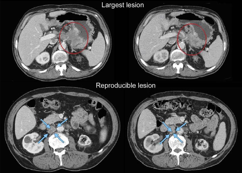
the upper normal limit, however, they must normalise for a volved organs, but in addition should be those that lend
patient to be considered in complete response. Because themselves to reproducible repeated measurements. It may be
tumour markers are disease specific, instructions for their the case that, on occasion, the largest lesion does not lend it-
measurement should be incorporated into protocols on a self to reproducible measurement in which circumstance the
disease specific basis. Specific guidelines for both CA-125 next largest lesion which can be measured reproducibly
response (in recurrent ovarian cancer) and PSA response (in should be selected. To illustrate this point see the example
recurrent prostate cancer), have been published.16–18 In addi- in Fig. 3 of Appendix II.
tion, the Gynecologic Cancer Intergroup has developed CA125 Lymph nodes merit special mention since they are normal
progression criteria which are to be integrated with objective anatomical structures which may be visible by imaging even
tumour assessment for use in first-line trials in ovarian if not involved by tumour. As noted in Section 3, pathological
cancer.19 nodes which are defined as measurable and may be identi-
fied as target lesions must meet the criterion of a short axis
Cytology, histology: These techniques can be used to differenti- of P15 mm by CT scan. Only the short axis of these nodes
ate between PR and CR in rare cases if required by protocol will contribute to the baseline sum. The short axis of the
(for example, residual lesions in tumour types such as germ node is the diameter normally used by radiologists to judge
cell tumours, where known residual benign tumours can re- if a node is involved by solid tumour. Nodal size is normally
main). When effusions are known to be a potential adverse reported as two dimensions in the plane in which the image
effect of treatment (e.g. with certain taxane compounds or is obtained (for CT scan this is almost always the axial plane;
angiogenesis inhibitors), the cytological confirmation of the for MRI the plane of acquisition may be axial, saggital or
neoplastic origin of any effusion that appears or worsens dur- coronal). The smaller of these measures is the short axis.
ing treatment can be considered if the measurable tumour For example, an abdominal node which is reported as being
has met criteria for response or stable disease in order to dif- 20 mm · 30 mm has a short axis of 20 mm and qualifies as a
ferentiate between response (or stable disease) and progres- malignant, measurable node. In this example, 20 mm should
sive disease. be recorded as the node measurement (See also the example
in Fig. 4 in Appendix II). All other pathological nodes (those
with short axis P10 mm butEUROPEAN JOURNAL OF CANCER 4 5 ( 2 0 0 9 ) 2 2 8 –2 4 7 233
Progressive Disease (PD): At least a 20% increase in the sum obtaining maximal diameter measurements of each individ-
of diameters of target lesions, taking as reference ual lesion. If the lesions have truly coalesced such that they
the smallest sum on study (this includes the baseline are no longer separable, the vector of the longest diameter
sum if that is the smallest on study). In addition to in this instance should be the maximal longest diameter for
the relative increase of 20%, the sum must also dem- the ‘coalesced lesion’.
onstrate an absolute increase of at least 5 mm. (Note:
the appearance of one or more new lesions is also 4.3.3. Evaluation of non-target lesions
considered progression). This section provides the definitions of the criteria used to deter-
Stable Disease (SD): Neither sufficient shrinkage to qualify for mine the tumour response for the group of non-target lesions.
PR nor sufficient increase to qualify for PD, taking as While some non-target lesions may actually be measurable,
reference the smallest sum diameters while on study. they need not be measured and instead should be assessed only
qualitatively at the time points specified in the protocol.
4.3.2. Special notes on the assessment of target lesions
Lymph nodes. Lymph nodes identified as target lesions should Complete Response (CR): Disappearance of all non-target le-
always have the actual short axis measurement recorded (mea- sions and normalisation of tumour marker level. All
sured in the same anatomical plane as the baseline examina- lymph nodes must be non-pathological in size
tion), even if the nodes regress to below 10 mm on study. This (234 EUROPEAN JOURNAL OF CANCER 4 5 ( 2 0 0 9 ) 2 2 8 –2 4 7
disease from localised to widespread, or may be described in 4.4. Evaluation of best overall response
protocols as ‘sufficient to require a change in therapy’. Some
illustrative examples are shown in Figs. 5 and 6 in Appendix II. The best overall response is the best response recorded from
If ‘unequivocal progression’ is seen, the patient should be con- the start of the study treatment until the end of treatment
sidered to have had overall PD at that point. While it would be taking into account any requirement for confirmation. On oc-
ideal to have objective criteria to apply to non-measurable dis- casion a response may not be documented until after the end
ease, the very nature of that disease makes it impossible to do of therapy so protocols should be clear if post-treatment
so, therefore the increase must be substantial. assessments are to be considered in determination of best
overall response. Protocols must specify how any new therapy
4.3.5. New lesions introduced before progression will affect best response desig-
The appearance of new malignant lesions denotes disease nation. The patient’s best overall response assignment will
progression; therefore, some comments on detection of new depend on the findings of both target and non-target disease
lesions are important. There are no specific criteria for the and will also take into consideration the appearance of new
identification of new radiographic lesions; however, the find- lesions. Furthermore, depending on the nature of the study
ing of a new lesion should be unequivocal: i.e. not attributable and the protocol requirements, it may also require confirma-
to differences in scanning technique, change in imaging tory measurement (see Section 4.6). Specifically, in non-ran-
modality or findings thought to represent something other domised trials where response is the primary endpoint,
than tumour (for example, some ‘new’ bone lesions may be confirmation of PR or CR is needed to deem either one the
simply healing or flare of pre-existing lesions). This is partic- ‘best overall response’. This is described further below.
ularly important when the patient’s baseline lesions show
partial or complete response. For example, necrosis of a liver 4.4.1. Time point response
lesion may be reported on a CT scan report as a ‘new’ cystic It is assumed that at each protocol specified time point, a re-
lesion, which it is not. sponse assessment occurs. Table 1 on the next page provides
A lesion identified on a follow-up study in an anatomical a summary of the overall response status calculation at each
location that was not scanned at baseline is considered a new time point for patients who have measurable disease at
lesion and will indicate disease progression. An example of this baseline.
is the patient who has visceral disease at baseline and while on When patients have non-measurable (therefore non-tar-
study has a CT or MRI brain ordered which reveals metastases. get) disease only, Table 2 is to be used.
The patient’s brain metastases are considered to be evidence of
PD even if he/she did not have brain imaging at baseline. 4.4.2. Missing assessments and inevaluable designation
If a new lesion is equivocal, for example because of its When no imaging/measurement is done at all at a particular
small size, continued therapy and follow-up evaluation will time point, the patient is not evaluable (NE) at that time point.
clarify if it represents truly new disease. If repeat scans con- If only a subset of lesion measurements are made at an
firm there is definitely a new lesion, then progression should assessment, usually the case is also considered NE at that
be declared using the date of the initial scan. time point, unless a convincing argument can be made that
While FDG-PET response assessments need additional the contribution of the individual missing lesion(s) would
study, it is sometimes reasonable to incorporate the use of not change the assigned time point response. This would be
FDG-PET scanning to complement CT scanning in assessment most likely to happen in the case of PD. For example, if a pa-
of progression (particularly possible ‘new’ disease). New le- tient had a baseline sum of 50 mm with three measured le-
sions on the basis of FDG-PET imaging can be identified sions and at follow-up only two lesions were assessed, but
according to the following algorithm: those gave a sum of 80 mm, the patient will have achieved
PD status, regardless of the contribution of the missing lesion.
a. Negative FDG-PET at baseline, with a positivel FDG-PET
at follow-up is a sign of PD based on a new lesion. 4.4.3. Best overall response: all time points
b. No FDG-PET at baseline and a positive FDG-PET at fol- The best overall response is determined once all the data for the
low-up: patient is known.
If the positive FDG-PET at follow-up corresponds to a Best response determination in trials where confirmation of com-
new site of disease confirmed by CT, this is PD. plete or partial response IS NOT required: Best response in these
If the positive FDG-PET at follow-up is not confirmed as trials is defined as the best response across all time points (for
a new site of disease on CT, additional follow-up CT example, a patient who has SD at first assessment, PR at sec-
scans are needed to determine if there is truly progres- ond assessment, and PD on last assessment has a best overall
sion occurring at that site (if so, the date of PD will be response of PR). When SD is believed to be best response, it
the date of the initial abnormal FDG-PET scan). must also meet the protocol specified minimum time from
If the positive FDG-PET at follow-up corresponds to a baseline. If the minimum time is not met when SD is other-
pre-existing site of disease on CT that is not progress- wise the best time point response, the patient’s best response
ing on the basis of the anatomic images, this is not PD. depends on the subsequent assessments. For example, a pa-
tient who has SD at first assessment, PD at second and does
l
A ‘positive’ FDG-PET scan lesion means one which is FDG avid not meet minimum duration for SD, will have a best response
with an uptake greater than twice that of the surrounding tissue of PD. The same patient lost to follow-up after the first SD
on the attenuation corrected image. assessment would be considered inevaluable.EUROPEAN JOURNAL OF CANCER 4 5 ( 2 0 0 9 ) 2 2 8 –2 4 7 235
at a subsequent time point as specified in the protocol (gener-
Table 1 – Time point response: patients with target (+/–
non-target) disease. ally 4 weeks later). In this circumstance, the best overall re-
sponse can be interpreted as in Table 3.
Target lesions Non-target lesions New Overall
lesions response
4.4.4. Special notes on response assessment
CR CR No CR When nodal disease is included in the sum of target lesions
CR Non-CR/non-PD No PR
and the nodes decrease to ‘normal’ size (236 EUROPEAN JOURNAL OF CANCER 4 5 ( 2 0 0 9 ) 2 2 8 –2 4 7
needle aspirate/biopsy) before assigning a status of complete stances, i.e. in randomised trials (phase II or III) or studies
response. FDG-PET may be used to upgrade a response to a CR where stable disease or progression are the primary endpoints,
in a manner similar to a biopsy in cases where a residual confirmation of response is not required since it will not add va-
radiographic abnormality is thought to represent fibrosis or lue to the interpretation of trial results. However, elimination of
scarring. The use of FDG-PET in this circumstance should be the requirement for response confirmation may increase the
prospectively described in the protocol and supported by dis- importance of central review to protect against bias, in partic-
ease specific medical literature for the indication. However, it ular in studies which are not blinded.
must be acknowledged that both approaches may lead to In the case of SD, measurements must have met the SD
false positive CR due to limitations of FDG-PET and biopsy res- criteria at least once after study entry at a minimum interval
olution/sensitivity. (in general not less than 6–8 weeks) that is defined in the
For equivocal findings of progression (e.g. very small and study protocol.
uncertain new lesions; cystic changes or necrosis in existing
lesions), treatment may continue until the next scheduled 4.6.2. Duration of overall response
assessment. If at the next scheduled assessment, progression The duration of overall response is measured from the time
is confirmed, the date of progression should be the earlier measurement criteria are first met for CR/PR (whichever is first
date when progression was suspected. recorded) until the first date that recurrent or progressive dis-
ease is objectively documented (taking as reference for progres-
4.5. Frequency of tumour re-evaluation sive disease the smallest measurements recorded on study).
The duration of overall complete response is measured
Frequency of tumour re-evaluation while on treatment from the time measurement criteria are first met for CR until
should be protocol specific and adapted to the type and sche- the first date that recurrent disease is objectively documented.
dule of treatment. However, in the context of phase II studies
where the beneficial effect of therapy is not known, follow-up 4.6.3. Duration of stable disease
every 6–8 weeks (timed to coincide with the end of a cycle) is Stable disease is measured from the start of the treatment (in
reasonable. Smaller or greater time intervals than these could randomised trials, from date of randomisation) until the crite-
be justified in specific regimens or circumstances. The proto- ria for progression are met, taking as reference the smallest
col should specify which organ sites are to be evaluated at sum on study (if the baseline sum is the smallest, this is the
baseline (usually those most likely to be involved with meta- reference for calculation of PD).
static disease for the tumour type under study) and how often The clinical relevance of the duration of stable disease var-
evaluations are repeated. Normally, all target and non-target ies in different studies and diseases. If the proportion of pa-
sites are evaluated at each assessment. In selected circum- tients achieving stable disease for a minimum period of time
stances certain non-target organs may be evaluated less fre- is an endpoint of importance in a particular trial, the protocol
quently. For example, bone scans may need to be repeated should specify the minimal time interval required between
only when complete response is identified in target disease two measurements for determination of stable disease.
or when progression in bone is suspected. Note: The duration of response and stable disease as well as
After the end of the treatment, the need for repetitive tu- the progression-free survival are influenced by the frequency of
mour evaluations depends on whether the trial has as a goal follow-up after baseline evaluation. It is not in the scope of this
the response rate or the time to an event (progression/death). guideline to define a standard follow-up frequency. The fre-
If ‘time to an event’ (e.g. time to progression, disease-free quency should take into account many parameters including
survival, progression-free survival) is the main endpoint of disease types and stages, treatment periodicity and standard
the study, then routine scheduled re-evaluation of protocol practice. However, these limitations of the precision of the
specified sites of disease is warranted. In randomised com- measured endpoint should be taken into account if compari-
parative trials in particular, the scheduled assessments sons between trials are to be made.
should be performed as identified on a calendar schedule
(for example: every 6–8 weeks on treatment or every 3–4 4.7. Progression-free survival/proportion progression-free
months after treatment) and should not be affected by delays
in therapy, drug holidays or any other events that might lead 4.7.1. Phase II trials
to imbalance in a treatment arm in the timing of disease This guideline is focused primarily on the use of objective re-
assessment. sponse endpoints for phase II trials. In some circumstances, ‘re-
sponse rate’ may not be the optimal method to assess the
4.6. Confirmatory measurement/duration of response potential anticancer activity of new agents/regimens. In such
cases ‘progression-free survival’ (PFS) or the ‘proportion pro-
4.6.1. Confirmation gression-free’ at landmark time points, might be considered
In non-randomised trials where response is the primary end- appropriate alternatives to provide an initial signal of biologic
point, confirmation of PR and CR is required to ensure re- effect of new agents. It is clear, however, that in an uncontrolled
sponses identified are not the result of measurement error. trial, these measures are subject to criticism since an appar-
This will also permit appropriate interpretation of results in ently promising observation may be related to biological factors
the context of historical data where response has traditionally such as patient selection and not the impact of the intervention.
required confirmation in such trials (see the paper by Bogaerts Thus, phase II screening trials utilising these endpoints are best
et al. in this Special Issue10). However, in all other circum- designed with a randomised control. Exceptions may existEUROPEAN JOURNAL OF CANCER 4 5 ( 2 0 0 9 ) 2 2 8 –2 4 7 237
where the behaviour patterns of certain cancers are so consis- 4.9. Reporting best response results
tent (and usually consistently poor), that a non-randomised
trial is justifiable (see for example van Glabbeke et al.20). How- 4.9.1. Phase II trials
ever, in these cases it will be essential to document with care When response is the primary endpoint, and thus all patients
the basis for estimating the expected PFS or proportion progres- must have measurable disease to enter the trial, all patients
sion-free in the absence of a treatment effect. included in the study must be accounted for in the report of
the results, even if there are major protocol treatment devia-
4.7.2. Phase III trials tions or if they are not evaluable. Each patient will be assigned
Phase III trials in advanced cancers are increasingly designed one of the following categories:
to evaluate progression-free survival or time to progression as
the primary outcome of interest. Assessment of progression 1. Complete response
is relatively straightforward if the protocol requires all pa- 2. Partial response
tients to have measurable disease. However, restricting entry 3. Stable disease
to this subset of patients is subject to criticism: it may result 4. Progression
in a trial where the results are less likely to be generalisable if, 5. Inevaluable for response: specify reasons (for example: early
in the disease under study, a substantial proportion of pa- death, malignant disease; early death, toxicity; tumour
tients would be excluded. Moreover, the restriction to entry assessments not repeated/incomplete; other (specify)).
will slow recruitment to the study. Increasingly, therefore, tri-
als allow entry of both patients with measurable disease as Normally, all eligible patients should be included in the
well as those with non-measurable disease only. In this cir- denominator for the calculation of the response rate for phase
cumstance, care must be taken to explicitly describe the find- II trials (in some protocols it will be appropriate to include all
ings which would qualify for progressive disease for those treated patients). It is generally preferred that 95% two-sided
patients without measurable lesions. Furthermore, in this set- confidence limits are given for the calculated response rate.
ting, protocols must indicate if the maximum number of re- Trial conclusions should be based on the response rate for
corded target lesions for those patients with measurable all eligible (or all treated) patients and should not be based
disease may be relaxed from five to three (based on the data on a selected ‘evaluable’ subset.
found in Bogaerts et al.10 and Moskowitz et al.11). As found in
the ‘special notes on assessment of progression’, these guide-
lines offer recommendations for assessment of progression 4.9.2. Phase III trials
in this setting. Furthermore, if available, validated tumour mar- Response evaluation in phase III trials may be an indicator
ker measures of progression (as has been proposed for ovarian of the relative anti-tumour activity of the treatments eval-
cancer) may be useful to integrate into the definition of pro- uated and is almost always a secondary endpoint. Ob-
gression. Centralised blinded review of imaging studies or of served differences in response rate may not predict the
source imaging reports to verify ‘unequivocal progression’ clinically relevant therapeutic benefit for the population
may be needed if important drug development or drug ap- studied. If objective response is selected as a primary end-
proval decisions are to be based on the study outcome. Finally, point for a phase III study (only in circumstances where a
as noted earlier, because the date of progression is subject to direct relationship between objective tumour response and
ascertainment bias, timing of investigations in study arms a clinically relevant therapeutic benefit can be unambigu-
should be the same. The article by Dancey et al. in this special ously demonstrated for the population studied), the same
issue21 provides a more detailed discussion of the assessment criteria as those applying to phase II trials should be used
of progression in randomised trials. and all patients entered should have at least one measur-
able lesion.
4.8. Independent review of response and progression In those many cases where response is a secondary end-
point and not all trial patients have measurable disease, the
For trials where objective response (CR + PR) is the primary end- method for reporting overall best response rates must be
point, and in particular where key drug development deci- pre-specified in the protocol. In practice, response rate may
sions are based on the observation of a minimum number of be reported using either an ‘intent to treat’ analysis (all ran-
responders, it is recommended that all claimed responses be domised patients in the denominator) or an analysis where
reviewed by an expert(s) independent of the study. If the study only the subset of patients with measurable disease at
is a randomised trial, ideally reviewers should be blinded to baseline are included. The protocol should clearly specify
treatment assignment. Simultaneous review of the patients’ how response results will be reported, including any subset
files and radiological images is the best approach. analyses that are planned.
Independent review of progression presents some more The original version of RECIST suggested that in phase III
complex issues: for example, there are statistical problems trials one could write protocols using a ‘relaxed’ interpreta-
with the use of central-review-based progression time in tion of the RECIST guidelines (for example, reducing the num-
place of investigator-based progression time due to the poten- ber of lesions measured) but this should no longer be done
tial introduction of informative censoring when the former since these revised guidelines have been amended in such a
precedes the latter. An overview of these factors and other way that it is clear how these criteria should be applied for
lessons learned from independent review is provided in an all trials in which anatomical assessment of tumour response
article by Ford et al. in this special issue.22 or progression are endpoints.238
Appendix I. Summary of major changes RECIST 1.0 to RECIST 1.1
RECIST 1.0 RECIST 1.1 Rationale Reference in special issue
(if applicable)
Minimum size measurable CT: 10 mm spiral CT 10 mm; delete reference to Most scans used have 5 mm or less slice
lesions 20 mm non-spiral spiral scan thickness Clearer to give instruction based on
slice interval if it is greater than 5 mm
Clinical: 20 mm Clinical: 10 mm (must be Caliper measurement will make this reliable
measurable with calipers)
Lymph node: not mentioned CT: Since nodes are normal structure need to define Schwartz et al.15
EUROPEAN JOURNAL OF CANCER
P15 mm short axis for target pathological enlargement. Short axis is most
P10–RECIST 1.0 RECIST 1.1 Rationale Reference in special issue
(if applicable)
Special notes: Frequently asked questions on these topics
How to assess and measure
lymph nodes
CR in face of residual tissue
Discussion of ‘equivocal’
progression
Confirmatory measure For CR and PR: criteria Retain this requirement ONLY Data warehouse shows that response rates Bogaerts et al.10
must be met again 4 for rise when confirmation is eliminated, but
weeks after initial non-randomised trials with the only circumstance where this is
documentation primary endpoint of response important is in trials where there is no
concurrent comparative control and where
this measure is the primary endpoint
Progression-free survival General comments only More specific comments on Increasing use of PFS in phase III trials Dancey et al.21
EUROPEAN JOURNAL OF CANCER
use of PFS (or proportion requires guidance on assessment of PD in
progression-free) as patients with non-measurable disease
phase II endpoint
Greater detail on PFS
assessment in phase III trials
Reporting of response 9 categories suggested for Divided into phase II and phase Simplifies reporting and clarifies how to
results reporting phase II results III report phase II and III data consistently
9 categories collapsed into 5
In phase III, guidance given
about reporting response
Response in phase III More relaxed guidelines This section removed and Simplification of response assessment by
trials possible if protocol specified referenced in section reducing number of lesions and eliminating
4 5 ( 2 0 0 9 ) 2 2 8 –2 4 7
above: no need to have need for confirmation in randomised
different criteria for phase II studies where response is not the primary
and III endpoint makes separate ‘rules’
unnecessary
Imaging appendix Appendix I Appendix II: updated with Evolving use of newer modalities addressed.
detailed guidance on Enhanced guidance in response to frequent
use of MRI, PET/CT questions and from radiology review
Other practical guidance experience
included
New appendices Appendix I: comparison of
RECIST 1.0 and 1.1
Appendix III: frequently asked
questions
239240 EUROPEAN JOURNAL OF CANCER 4 5 ( 2 0 0 9 ) 2 2 8 –2 4 7
Conflict of interest statement mendations intended for patients on clinical trials where
RECIST assessment will be performed. Standardisation of
None declared. imaging requirements and image acquisition parameters is
ideal to allow for optimal comparability of subjects within a
study and results between studies. These recommendations
Acknowledgements are designed to balance optimised image acquisition proto-
cols with techniques that should be feasible to perform glob-
The RECIST Working Group would like to thank the following ally at imaging facilities in all types of radiology practices.
organisations which made data bases available to us in order These guidelines are not applicable to functional imaging
to perform the analyses which informed decisions about techniques or volumetric assessment of tumour size.
changes to this version of the criteria: Amgen; AstraZeneca; Scanner quality control is highly recommended and should
Breast Cancer International Research Group (BCIRG); Bristol- follow standard manufacturer and facility maintenance
Myers Squibb; European Organisation for Research and schedules using commercial phantoms. It is likely that for RE-
Treatment of Cancer (EORTC) Breast Cancer Group and Gas- CIST unidimensional measurements this will be adequate to
trointestinal Group; Erasmus University Medical Center, produce reproducible measurements. Imaging quality control
Rotterdam, The Netherlands; Genentech; Pfizer; RadPharm; for CT includes an analysis of image noise and uniformity and
Roche; Sanofi Aventis. CT number as well as spatial resolution. The frequency of
We would also like to thank the following individuals from quality control analysis is also variable and should focus on
academic, government, and pharmaceutical organisations for clinically relevant scanning parameters. Dose analysis is al-
providing helpful comments on an earlier draft of these revised ways important and the use of imaging should follow the
guidelines: Ohad Amit, Phil Murphy, Teri Crofts and Janet Be- ALARA principle, ‘As Low As Reasonably Achievable’, which
gun, GlaxoSmithKline, USA; Laurence H. Baker, Southwest refers to making every reasonable effort to maintain radiation
Oncology Group, USA; Karla Ballman, Mayo Clinic, USA; exposures as far below the dose limits as possible.
Charles Baum, Darrel Cohen, and Mary Ashford Collier, Pfizer,
USA; Gary J. Becker, American Board of Radiology, Tucson, Specific.notes
USA; Jean-Yves Blay, University Claude Pertrand, Lyon France;
Renzo Canetta, Bristol-Myers Squibb, USA; David Chang, Am- Chest X-ray measurement of lesions surrounded by pulmon-
gen Inc., USA; Sandra Chica, Perceptive Informations Inc. (PAR- ary parenchyma is feasible, but not preferable as the
EXEL), USA; Martin Edelman, University of Maryland measurement represents a summation of densities. Further-
Greenbaum Cancer Centre, USA; Gwendolyn Fyfe, Genentech, more, there is poor identification of new lesions within the
USA; Bruce Giantonio, Eastern Cooperative Oncology Group, chest on X-ray as compared with CT. Therefore, measure-
USA; Gary Gordon, Abbott Pharmaceuticals, USA; Ronald Gott- ments of pulmonary parenchymal lesions as well as medias-
lieb, Roswell Park Cancer Institute, USA; Simon Kao, University tinal disease are optimally performed with CT of the chest.
of Iowa College of Medicine, USA; Wasaburo Koizumi, Kitasato MRI of the chest should only be performed in extenuating cir-
University, Japan; Alessandro Riva, Novartis Pharmaceuticals, cumstances. Even if IV contrast cannot be administered (for
USA; Wayne Rackhoff, Ortho Biotech Oncology Research and example, in the situation of allergy to contrast), a non-con-
Development, USA; Nagahiro Saijo, President Japanese Society trast CT of the chest is still preferred over MRI or chest X-ray.
of Medical Oncology, Japan; Mitchell Schnall American College
of Radiology Imaging Network, USA; Yoshik Shimamura, PAR- CT scans: CT scans of the chest, abdomen, and pelvis should
EXEL International Inc., Japan; Rajeshwari Sridhara, Centre be contiguous throughout all the anatomic region of interest.
for Drug Evaluation and Research, Food and Drug Administra- As a general rule, the minimum size of a measurable lesion at
tion, USA; Andrew Stone, Alan Barge, AstraZeneca, United baseline should be no less than double the slice thickness and
Kingdom; Orhan Suleiman, Centre for Drug Evaluation and Re- also have a minimum size of 10 mm (see below for minimum
search, Food and Drug Administration, USA; Daniel C. Sullivan, size when scanners have a slice thickness more than 5 mm).
Duke University Medical Centre, USA; Masakazu Toi, Kyoto While the precise physics of lesion size and partial volume
University, Japan; Cindy Welsh, Centre for Drug Evaluation averaging is complex, lesions smaller than 10 mm may be dif-
and Research, Food and Drug Administration, USA. ficult to accurately and reproducibly measure. While this rule
Finally, the RECIST Working Group would like to thank indi- is applicable to baseline scans, as lesions potentially decrease
viduals who were not permanent members of the group (which in size at follow-up CT studies, they should still be measured.
are all acknowledged as co-authors) but who attended working Lesions which are reported as ‘too small to measure’ should
group meetings from time to time and made contributions to be assigned a default measurement of 5 mm if they are still
the total process over the past 7 years: Richard Pazdur, Food visible.
and Drug Administration, USA; Francesco Pignatti, European The most critical CT image acquisition parameters for opti-
Medicines Agency, London, UK. mal tumour evaluation using RECIST are anatomic coverage,
contrast administration, slice thickness, and reconstruction interval.
Appendix II. Specifications for standard
anatomical radiological imaging a. Anatomic coverage: Optimal anatomic coverage for most
solid tumours is the chest, abdomen and pelvis. Cover-
These protocols for image acquisition of computed tomogra- age should encompass all areas of known predilection
phy (CT) and magnetic resonance imaging (MRI) are recom- for metastases in the disease under evaluation andEUROPEAN JOURNAL OF CANCER 4 5 ( 2 0 0 9 ) 2 2 8 –2 4 7 241
should additionally investigate areas that may be low-up examinations for a given patient. This will
involved based on signs and symptoms of individual greatly enhance the reproducibility of the tumour mea-
patients. Because a lesion later identified in a body part surements. If prior to enrolment it is known a patient is
not scanned at baseline would be considered as a new not able to undergo CT scans with IV contrast due to
lesion representing disease progression, careful consid- allergy or renal insufficiency, the decision as to
eration should be given to the extent of imaging coverage whether a non-contrast CT or MRI (with or without IV
at baseline and at subsequent follow-up time points. contrast) should be used to evaluate the subject at
This will enable better consistency not only of tumour baseline and follow-up should be guided by the tumour
measurements but also identification of new disease. type under investigation and the anatomic location of
b. IV contrast administration: Optimal visualisation and the disease. For patients who develop contraindica-
measurement of metastases in solid tumours requires tions to contrast after baseline contrast CT is done,
consistent administration (dose and rate) of IV contrast the decision as to whether non-contrast CT or MRI
as well as timing of scanning. Typically, most abdomi- (enhanced or non-enhanced) should be performed
nal imaging is performed during the portal venous should also be based on the tumour type, anatomic
phase and (optimally) about the same time frame after location of the disease and should be optimised to
injection on each examination (see Fig. 1 for impact of allow for comparison to the prior studies if possible.
different phase of IV contrast on lesion measurement). Each case should be discussed with the radiologist to
Most solid tumours may be scanned with a single determine if substitution of these other approaches is
phase after administration of contrast. While triphasic possible and, if not, the patient should be considered
CT scans are sometimes performed on other types of not evaluable from that point forward. Care must be
vascular tumours to improve lesion conspicuity, for taken in measurement of target lesions on a different
consistency and uniformity, we would recommend tri- modality and interpretation of non-target disease or
phasic CT for hepatocellular and neuroendocrine new lesions, since the same lesion may appear to have
tumours for which this scanning protocol is generally a different size using a new modality (see Fig. 2 for a
standard of care, and the improved temporal resolution comparison of CT and MRI of the same lesion). Oral
of the triphasic scan will enhance the radiologists’ abil- contrast is recommended to help visualise and differ-
ity to consistently and reproducibly measure these entiate structures in the abdomen.
lesions. The precise dose and rate of IV contrast is c. Slice thickness and reconstruction interval: RECIST measure-
dependent upon the CT scanning equipment, CT acqui- ments may be performed at most clinically obtained
sition protocol, the type of contrast used, the available slice thicknesses. It is recommended that CT scans be
venous access and the medical condition of the performed at 5 mm contiguous slice thickness or less
patient. Therefore, the method of administration of and indeed this guideline presumes a minimum 5 mm
intravenous contrast agents is variable. Rather than thickness in recommendations for measurable lesion
try to institute rigid rules regarding methods for definition. Indeed, variations in slice thickness can have
administering contrast agents and the volume injected, an impact on lesion measurement and on detection of
it is appropriate to suggest that an adequate volume of new lesions. However, consideration should also be
a suitable contrast agent should be given so that the given for minimising radiation exposure. With these
metastases are demonstrated to best effect and a con- parameters, a minimum 10 mm lesion is considered
sistent method is used on subsequent examinations for measurable at baseline. Occasionally, institutions may
any given patient (ideally, this would be specified in perform medically acceptable scans at slice thicknesses
the protocol or for an institution). It is very important greater than 5 mm. If this occurs, the minimum size of
that the same technique be used at baseline and on fol- measurable lesions at baseline should be twice the slice
Fig. 1 – Difference in measurement/visualisation with different phases of IV contrast administration. Hypervascular
metastases imaged in the arterial phase (left) and the portal venous phase (right). Note that the number of lesions visible
differs greatly between the two phases of contrast administration as does any potential lesion measurement. Consistent CT
scan acquisition, including phase of contrast administration, is important for optimal and reproducible tumour242 EUROPEAN JOURNAL OF CANCER 4 5 ( 2 0 0 9 ) 2 2 8 –2 4 7
Fig. 2 – CT versus MRI of same lesions showing apparent ‘progression’ due only to differing method of measurement.
thickness of the baseline scans. Most contemporary CT performed as part of a PET–CT is of identical diagnostic qual-
scanners are multidetector which have many imaging ity to a diagnostic CT (with IV and oral contrast) then the CT
options for these acquisition parameters.23 The equip- portion of the PET–CT can be used for RECIST measurements.
ment vendor and scanning manual should be reviewed Note, however, that the PET portion of the CT introduces addi-
if there are any specific system questions. tional data which may bias an investigator if it is not routinely
d. Alternative contrast agents: There are a number of other, or serially performed.
new contrast agents, some organ specific.24 They may Ultrasound examinations should not be used in clinical trials
be used as part of patient care for instance, in liver to measure tumour regression or progression of lesions be-
lesion assessment, or lymph node characterisation25, cause the examination is necessarily subjective and operator
but should not as yet be used in clinical trials. dependent. The reasons for this are several: Entire examina-
tions cannot be reproduced for independent review at a later
FDG-PET has gained acceptance as a valuable tool for date, and it must be assumed, whether or not it is the case,
detecting, staging and restaging several malignancies. Criteria that the hard-copy films available represent a true and accu-
for incorporating (or substituting) FDG-PET into anatomical rate reflection of events. Furthermore, if, for example, the
assessment of tumour response in phase II trials are not yet only measurable lesion is in the para-aortic region of the
available, though much research is ongoing. Nevertheless, abdomen and if gas in the bowel overlies the lesion, the lesion
FDG-PET is being used in many drug development trials both will not be detected because the ultrasound beam cannot
as a tool to assess therapeutic efficacy and also in assessment penetrate the gas. Accordingly, the disease staging (or restag-
of progression. If FDG-PET scans are included in a protocol, by ing for treatment evaluation) for this patient will not be
consensus, an FDG uptake period of 60 min prior to imaging accurate.
has been decided as the most appropriate for imaging of pa- While evaluation of lesions by physical examination is also
tients with malignancy.26 Whole-body acquisition is impor- of limited reproducibility, it is permitted when lesions are
tant since this allows for sampling of all areas of interest superficial, at least 10 mm size, and can be assessed using
and can assess if new lesions have appeared thus determining calipers. In general, it is preferred if patients on clinical trials
the possibility of interval progression of disease. Images from have at least one lesion that is measurable by CT. Other skin
the base of the skull to the level of the mid-thigh should be ob- or palpable lesions may be measured on physical examina-
tained 60 min post injection. PET camera specifications are tion and be considered target lesions.
variable and manufacturer specific, so every attempt should Use of MRI remains a complex issue. MRI has excellent
be made to use the same scanner, or the same model scanner, contrast, spatial and temporal resolution; however, there
for serial scans on the same patient. Whole-body acquisitions are many image acquisition variables involved in MRI, which
can be performed in either 2- or 3-dimensional mode with greatly impact image quality, lesion conspicuity and mea-
attenuation correction, but the method chosen should be con- surement. Furthermore, the availability of MRI is variable
sistent across all patients and serial scans in the clinical trial. globally. As with CT, if an MRI is performed, the technical
PET/CT scans: Combined modality scanning such as with specifications of the scanning sequences used should be
PET–CT is increasingly used in clinical care, and is a modal- optimised for the evaluation of the type and site of disease.
ity/technology that is in rapid evolution; therefore, the recom- Furthermore, as with CT, the modality used at follow-up
mendations in this paper may change rather quickly with should be the same as was used at baseline and the lesions
time. At present, low dose or attenuation correction CT por- should be measured/assessed on the same pulse sequence.
tions of a combined PET–CT are of limited use in anatomically Generally, axial imaging of the abdomen and pelvis with T1
based efficacy assessments and it is therefore suggested that and T2 weighted imaging along with gadolinium enhanced
they should not be substituted for dedicated diagnostic con- imaging should be performed. The field of view, matrix,
trast enhanced CT scans for anatomically based RECIST mea- number of excitations, phase encode steps, use of fat sup-
surements. However, if a site can document that the CT pression and fast sequences should be optimised for the spe-You can also read