Nicotinamide combined with gemcitabine is an immunomodulatory therapy that restrains pancreatic cancer in mice
←
→
Page content transcription
If your browser does not render page correctly, please read the page content below
Open access Original research
J Immunother Cancer: first published as 10.1136/jitc-2020-001250 on 5 November 2020. Downloaded from http://jitc.bmj.com/ on November 30, 2021 by guest. Protected by copyright.
Nicotinamide combined with
gemcitabine is an immunomodulatory
therapy that restrains pancreatic cancer
in mice
Benson Chellakkan Selvanesan ,1 Kiran Meena,1 Amanda Beck,2
Lydie Meheus,3 Olaya Lara ,4 Ilse Rooman,3,4 Claudia Gravekamp
1
To cite: Selvanesan BC, ABSTRACT BACKGROUND
Meena K, Beck A, et al. Background Treatments for pancreatic ductal Patients with pancreatic ductal adenocarci-
Nicotinamide combined
adenocarcinoma are poorly effective, at least partly due to noma (PDAC) have a 5-year survival below
with gemcitabine is an
immunomodulatory therapy
the tumor’s immune-suppressive stromal compartment. 10%. Part of this poor outcome has been
that restrains pancreatic New evidence of positive effects on immune responses attributed to the fact that PDAC is charac-
cancer in mice. Journal for in the tumor microenvironment (TME), compelled us to terized by a dense desmoplastic stroma that
ImmunoTherapy of Cancer test the combination of gemcitabine (GEM), a standard prevents drugs and immune cells from pene-
2020;8:e001250. doi:10.1136/ chemotherapeutic for pancreatic cancer, with nicotinamide
jitc-2020-001250 trating the pancreatic tumors.1 2 This stroma
(NAM), the amide form of niacin (vitamin B3), in mice with
consists of different types of cancer activated
pancreatic cancer.
►► Additional material is fibroblasts (CAF)3 that not only produce
Methods Various mouse tumor models of pancreatic
published online only. To view
cancer, that is, orthotopic Panc-02 and KPC (KrasG12D, extracellular matrix (ECM) components
please visit the journal online such as collagen but also crosstalk to immune
(http://dx.doi.org/10.1136/jitc-
p53R172H, Pdx1-Cre) grafts, were treated alternately with
NAM and GEM for 2 weeks, and the effects on efficacy, cells.4 5 Among these inflammatory cells are
2020-001250).
survival, stromal architecture and tumor-infiltrating tumor- associated macrophages (TAM) and
Accepted 29 September 2020 immune cells was examined by immunohistochemistry myeloid- derived suppressor cells (MDSC)
(IHC), flow cytometry, Enzyme-linked immunospot that promote immune suppression, tumor
(ELISPOT), T cell depletions in vivo, Nanostring analysis progression, angiogenesis, invasion and
and RNAscope. extravasation of tumor cells resulting in the
Results A significant reduction in tumor weight development of metastases.6 Immunomodu-
and number of metastases was found, as well as latory therapies to counteract tumor progres-
© Author(s) (or their a significant improved survival of the NAM+GEM sion are highly sought after but with limited
employer(s)) 2020. Re-use group compared with all control groups. IHC and
permitted under CC BY-NC. No success.
flow cytometry showed a significant decrease in Gemcitabine (GEM) is an Food and Drug
commercial re-use. See rights
tumor-associated macrophages and myeloid-derived
and permissions. Published by Administration (FDA)- approved drug for
BMJ. suppressor cells in the tumors of NAM+GEM-treated
advanced pancreatic cancer (PDAC) that
1
Department of Microbiology mice. This correlated with a significant increase in the
in combination with nab- paclitaxel,7 only
and Immunology, Albert Einstein number of CD4 and CD8 T cells of NAM+GEM-treated
College of Medicine, Bronx, New tumors, and CD4 and CD8 T cell responses to tumor- modestly improves patient survival.8 9 Hence,
York, USA associated antigen survivin, most likely through epitope more effective therapies are warranted. GEM
2
Michael F. Price Center, Albert spreading. In vivo depletions of T cells demonstrated is a deoxycytidine analog that inhibits DNA
Einstein College of Medicine, synthesis. New evidence indicates that anti-
the involvement of CD4 T cells in the eradication of the
Bronx, New York, USA tumor activities of chemotherapy such as GEM
3
AntiCancer Fund, Boechoutlaan,
tumor by NAM+GEM treatment. In addition, remodeling
Strombeek-Bever, Belgium of the tumor stroma was observed with decreased also rely on several off-target effects, espe-
4
Laboratory of Medical collagen I and lower expression of hyaluronic acid cially directed to the host’s immune system
and Molecular Oncology, binding protein, reorganization of the immune cells into that contribute to tumor eradication. So far,
Vrije Universiteit Brussel, lymph node like structures and CD31 positive vessels. studies that have been focusing on evaluating
Laarbeeklaan, Brussels, Belgium Expression profiling for a panel of immuno-oncology the effect of GEM on T cell responses to the
genes revealed significant changes in genes involved in tumors are limited. A few reports describe
Correspondence to
Dr Claudia Gravekamp; migration and activation of T cells, attraction of dendritic that GEM improves T cell activation in mice
claudia.gravekamp@ cells and epitope spreading. and humans with pancreatic cancer.10–14 GEM
einsteinmed.o rg Conclusion This study highlights the potential of
is known for its ability to reduce the MDSC
NAM+GEM as immunotherapy for advanced pancreatic
Dr Ilse Rooman; population in tumor- bearing humans and
cancer.
irooman@vub.be mice.15 Finding drugs to combine with GEM
Selvanesan BC, et al. J Immunother Cancer 2020;8:e001250. doi:10.1136/jitc-2020-001250 1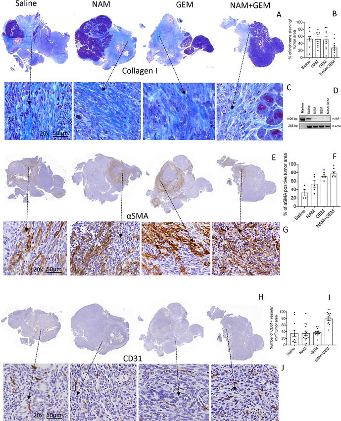
Open access
J Immunother Cancer: first published as 10.1136/jitc-2020-001250 on 5 November 2020. Downloaded from http://jitc.bmj.com/ on November 30, 2021 by guest. Protected by copyright.
that can strengthen the immune response would be a Cell lines
significant step forward. The Panc-02 cell line was derived from a
Nicotinamide (NAM), the amide form of niacin methylcholanthrene- induced ductal adenocarcinoma
(vitamin B3), is a nutrient provided by dietary source growing in a C57BL/6 female mouse.26 Panc-02 cells were
and supplement, and has little side effects.16 NAM is a cultured in McCoy’s5A medium supplemented with 10%
precursor of the coenzyme NAM adenine dinucleotide fetal bovine serum, 2 mM glutamine, non-essential amino
and participates in the cellular energy metabolism in the acids, 1 mM sodium pyruvate and 100 U/mL Pen/Strep.
mitochondrial electron transport chain.17 It has been The KPC tumor cell line was derived from a pancreatic
shown that NAM kills pancreatic tumor cells through KPC tumor (LSLKrasG12D+/- LSL-p53R172-/-; Pdx1-Cre) in our
down regulation of SIRT-1, K-ras and Akt-1 expression,18 lab, which was kindly provided by Jacco van Rheenen,
and that NAM sensitizes tumor cells to chemotherapy Cancer Genomics, Utrecht, The Netherlands.27
and radiotherapy through inhibition of poly (ADP-
ribose) polymerase (PARP).19 New evidence has been Mouse models
reported that NAM affects immune responses positively. Orthotopic Panc-02 model
For instance, NAM increased the expansion of human T Orthotopic Panc-02 tumors were generated in immune
cells through mitochondrial activation20 and acted immu- competent C57BL/6 mice as we described previously.14
noprophylactic as well as immunotherapeutic in preclin- Briefly, mice were anesthetized with ketamine (Mylan
ical mouse models with hormone receptor-positive breast Institutional LLC)/xylazine (Akorn Animal Health)
cancer.21 Also, peritumoral and infiltrating CD4 and CD8 (respectively, 100 mg and 10 mg/kg, ip), the hair was
T cells were significantly increased in melanomas on removed at the location of the spleen, and the skin was
NAM treatment compared with a placebo.22 Together this
sterilized with betadine, followed by 70% alcohol. The
suggests that NAM may qualify as an effective therapeutic
animal was covered with gauze sponge surrounding the
add on in PDAC. Moreover, it is safe to use and showed
incision site. A 1 cm incision was made in the abdominal
cancer preventative actions in a phase three clinical trial
skin and muscle just lateral to the midline and directly
of non-melanoma skin cancer.23
above the spleen/pancreas to allow visualization. The
Based on this information we evaluated the effect of
spleen/pancreas was gently retracted and positioned
combining GEM with NAM on pancreatic cancer and
to allow injection of Panc-02 tumor cells (106/50 µL
focused on the immune system. We found a significant
phosphate- buffered saline (PBS)) directly into the
decrease in the growth of primary tumors and metastases,
pancreas, from the tail all the way to the head of the
as well as an increase in the survival time of NAM+GEM
pancreas. To prevent leakage of injected cell suspension,
treated mice, in correlation with a significant influx of
the injection site was tied off after tumor cell injections
immune cells into the pancreatic tumors, and with a
with dissolvable suture. The spleen/pancreas were then
significant increase in CD31-positive blood vessels. The
replaced within the abdominal cavity, and both muscle
immune infiltrate was characterized by T cells that formed
and skin layers closed with sutures. Following recovery
peritumoral lymph node-like structures (LNS) predomi-
from surgery, mice were monitored and weighed daily. A
nantly in the NAM+GEM group, and intratumoral LNS
palpable tumor appeared within 10 days in the pancreas.
sometimes in the NAM or GEM groups. CD4 and CD8 T
cells were activated by NAM+GEM to the tumor-associated In this model, tumor cells migrate via the blood stream
antigen (TAA) survivin, which is highly expressed by to the liver and grow into small metastases visible by the
tumors and metastases in the Panc-02 model,24 one of the naked eye. Sometimes, some tumor cells leaked into the
experimental models used here. In addition, the number peritoneal cavity. Matrigel was not used here since it may
of TAM and MDSC in the tumor decreased. Potential prevent dissemination of tumor cells and the develop-
immunoregulatory mechanisms of NAM+GEM based on ment of metastases. The tumor weight was measured, and
Nanostring analysis will be discussed. the number of metastases was counted after euthanizing
the mice.
METHODS Orthotopic KPC model
Animals Orthotopic KPC tumors were generated similarly, but now
C57BL/6 female mice aged 3 months were obtained the KPC tumor cells (105/50 µL PBS) were injected into
from Charles River. These mice were used to generate the pancreas of immune competent ‘KPC-wild type’ mice.
the Panc-02 model. Male and female KPC-wild type mice KPC-wild type mice have the same genetic background
were maintained at Einstein, which have the same genetic as the KPC transgenic mice (C57BL/6xFVBxJ129) but
background as KPC mice (LSLKrasG12D+/- LSL-p53R17+/- lack the expression of Kras and p53 mutations and the
Pdx1 Cre)25 but are negative for KrasG12D and Tp53R172H Pdx-Cre, and allow the generation of pancreatic tumors
mutations and for Cre recombinase. These mice were and metastases in the liver. In this orthotopic model, a
used to generate the orthotopic KPC mice. In addi- palpable tumor appeared within 10 days, and the metas-
tion, a nude mouse model was used, that is, Fox n1nu/J tases are blood born, like in the orthotopic Panc-02
(cat#000819), Jackson Laboratories, lacking T cells. model.
2 Selvanesan BC, et al. J Immunother Cancer 2020;8:e001250. doi:10.1136/jitc-2020-001250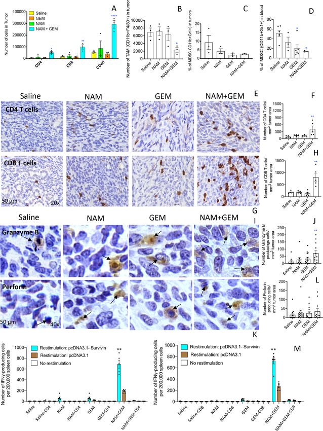
Open access
J Immunother Cancer: first published as 10.1136/jitc-2020-001250 on 5 November 2020. Downloaded from http://jitc.bmj.com/ on November 30, 2021 by guest. Protected by copyright.
Orthotopic Panc-02 model in nude mice order (Beckton and Dickinson), and analyzed using
Nude mice (Fox n1nu/J (000819) Jackson Laboratories) FlowJo V.7.6 software. Cell debris and dead cells were
with a C57BL/6 background were used. These mice are excluded from the analysis based on scatter signals and
immune incompetent because they lack T cells. Tumors use of Fixable Blue or Green Live/Dead Cell Stain Kit
were generated orthotopically in the pancreas by injec- (BD Bioscience, cat # 564406).
tion with Panc-02 cells as described above. A palpable
tumor appeared within 10 days, and the metastases are
blood born, like in the orthotopic Panc-02 model. ELISPOT
Immune cells from spleens or tumors were isolated
Protocol for treatment of mice with NAM+GEM from treated and control Panc-02 mice for ELISPOT
The optimal dose of NAM (Green Labs Nutrition, Poland) (BD Biosciences, cat# 551083) analysis, as described
was determined by testing different concentrations of previously.29 30 To detect T cell responses to survivin, 105
NAM (100, 50 and 25, 12.5 and 6.25 mg/200 µL/dose). spleen cells were transfected with pcDNA-3.1-survivin or
The highest dose without any physiological side effects pCDNA3.1 alone (negative control) or nothing (nega-
appeared to be 25 mg/200 µL. Based on these results, we tive control). The frequency of IFNγ-producing spleen
decided to use 20 mg/200 µL as the optimal dose used in cells was measured 72 hours later using an ELISPOT
all experiments. First, tumors and metastases were gener- reader (CTL Immunospot S4 analyzer). To determine
ated as described above. NAM (20 mg/200 µL/dose) was the frequency of IFNγ-producing CD4 and CD8 T cells,
administered orally and GEM (1.2 mg/200 µL/dose) was spleen cells were depleted for CD4 and CD8 T cells using
administered ip, alternately, starting 10 days after tumor magnetic bead depletion techniques according to the
cell injection and continued for 2 weeks as outlined for all manufacturer’s instructions (Miltenyi Biotec).
three pancreatic cancer models in online supplemental
figure S1A–C. Preparation of NAM: 1 gram of NAM was Immunohistochemistry
dissolved in 10 mls of apple juice. Black pepper of 16 mg Tumors were dissected from pancreas and immediately
was added to the solution to prevent glucuronidation. fixed with buffered formalin, and the tissue was embedded
One dose consisted of 20 mg of NAM in 200 µL apple juice. in paraffin. Sections (5 µm) were sliced and placed on
The NAM solution was aliquoted and stored at −20°C slides, then deparaffinized at 60°C for 1 hour, followed by
until use. Preparation of GEM (Fresensius Kabi; Gemita): xylene, an ethanol gradient (100%–70%), water, and PBS.
1 g of GEM was dissolved in 100 mls of endotoxin-free Slides were then incubated for 30 min in 3% hydrogen
saline, diluted until 6 mg/mL, and then aliquoted and peroxide, followed by boiling in citrate buffer for 20 min.
stored at −20°C until use. One dose consisted of 1.2 mg Once the slides were cooled, washed, and blocked with
in 200 µL saline. 5% goat serum, the sections were incubated with primary
antibodies such as anti-CD4 (Cell Signaling Technology,
Survival study cat # 25229) (1:100 dilution), anti-CD8α (1:400 dilution)
Orthotopic Panc-02 mice were treated with NAM+GEM (Cell Signaling Technology, cat # 98941), anti-Perforin
as outlined in online supplemental figure S1A. At the end (Cell Signaling Technology, cat # 31647)(1:300 dilution),
of treatments, mice were monitored without any further anti-Granzyme (Cell Signaling Technology, cat # 44153)
treatment until they succumbed spontaneously, or were (1:200 dilution), anti-CD31 (Cell Signaling Technology,
terminated on appearance of severe premorbid symp- cat # 77699) (1:100 dilution), or anti-α-Smooth muscle
toms requiring euthanasia as specified by our approved actin (αSMA) antibodies (Cell Signaling Technology, cat
animal use protocol. # 19245)(1:400), followed by incubation with secondary
antibody (mouse anti- goat IgG-HRP) (Cell Signaling
Flow cytometry Technology, cat# 8114S), and SignalStain Boost immuno-
Immune cells from spleens of mice were isolated histochemistry (IHC) Detection Reagent (Cell Signaling
as described previously.28 Immune cells were also Technology, cat# 8114S). Subsequently, the slides were
isolated from pancreatic tumors using Dispase (Roche incubated with 3,3'-diaminobenzidine (DAB) (Vector
cat#049422078001) and Collagenase (Sigma- Aldrich Laboratories, cat# SK-4100), counterstained with hema-
cat #C0130) as we described previously.29 Anti-CD3 (BD toxylin, dehydrated through an ethanol gradient (70%–
Bioscience, cat # 560527) and anti-CD8 antibodies (BD 100%) and xylene, and mounted with Permount. The
Bioscience, cat # 552877) were used to identify CD8 T cells, slides were scanned with a 3D Histech P250 High Capacity
anti-CD3 and anti-CD4 (BD Bioscience, cat # 552051) to Slide Scanner to acquire images and quantification data.
identify CD4 T cells, anti-CD11b (BD Bioscience, cat # Secondary antibodies without primary antibodies were
553312) and anti-Gr1 (BD Bioscience, cat # 553127) to used as negative control.
identify MDSC, and anti-CD11b and anti-F4/80 (eBio-
science, cat # 17-4801-82) to identify TAM. Appropriate CD4 and CD8 T cell depletions in vivo in orthotopic Panc-02
isotype controls were included for each sample. A total model
of 50 000–1 00 000 cells were acquired by scanning using CD4 and CD8 T cells were depleted in C57BL/6 mice with
a LSR-II Fluorescence Activated Cell Sorter system special orthotopic Panc-02 tumors during GEM+NAM treatment.
Selvanesan BC, et al. J Immunother Cancer 2020;8:e001250. doi:10.1136/jitc-2020-001250 3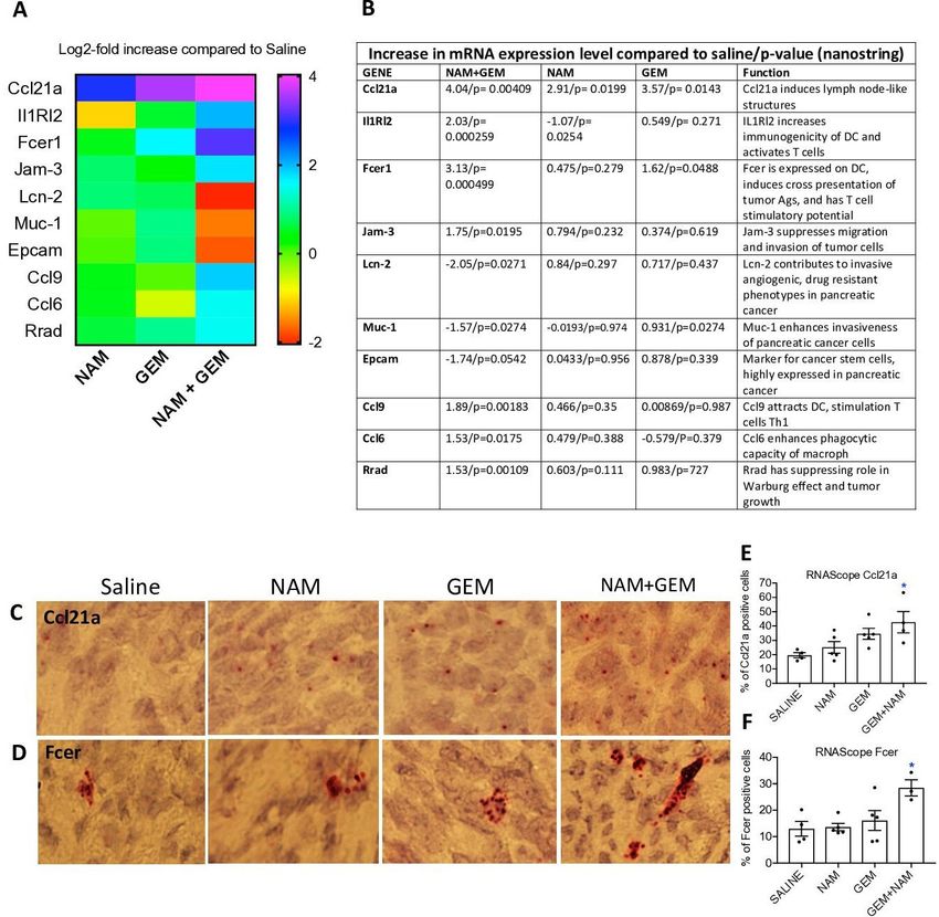
Open access
J Immunother Cancer: first published as 10.1136/jitc-2020-001250 on 5 November 2020. Downloaded from http://jitc.bmj.com/ on November 30, 2021 by guest. Protected by copyright.
Briefly, 10 days after tumor cell injection (when tumors Differentially expressed genes were compared with
were approximately 5 mm2), mice were treated with Student’s t-test, and adjusted p values of less than 0.01
NAM+GEM and with 300 µg of anti-CD4 (Clone GK1.5; and fold changes of greater than 2 were considered to
Cat#BE0003-1: BioXCell) or anti-CD8 (Clone YTS169.4; indicate significant dysregulation. p values were adjusted
Cat$BE0117: BioXCell) antibodies (five injections every by the Benjamini-Hochberg (BH) method. Adjusted p
third day), as outlined in online supplemental figure S1D. values are also called q values. Data were analyzed using
All mice were euthanized 2 days after the last anti-CD4 R (V.3.5.1).
or anti-CD8 treatment and analyzed for tumor weight
and number of metastases. As control, isotype-matched Statistical analysis tumors, metastases and immunological
rat antibodies against HRPN were used (Clone LTF-2; responses
Cat#BE0090: BioXCell). To statistically compare the effects NAM+GEM on
the growth of metastases and tumors or on immune
Nanostring technology responses in the mouse models, the Mann-Whitney U test
Pieces of 3 mm3 tumors were submerged in 5 vol of RNAl- was applied using Prism. *p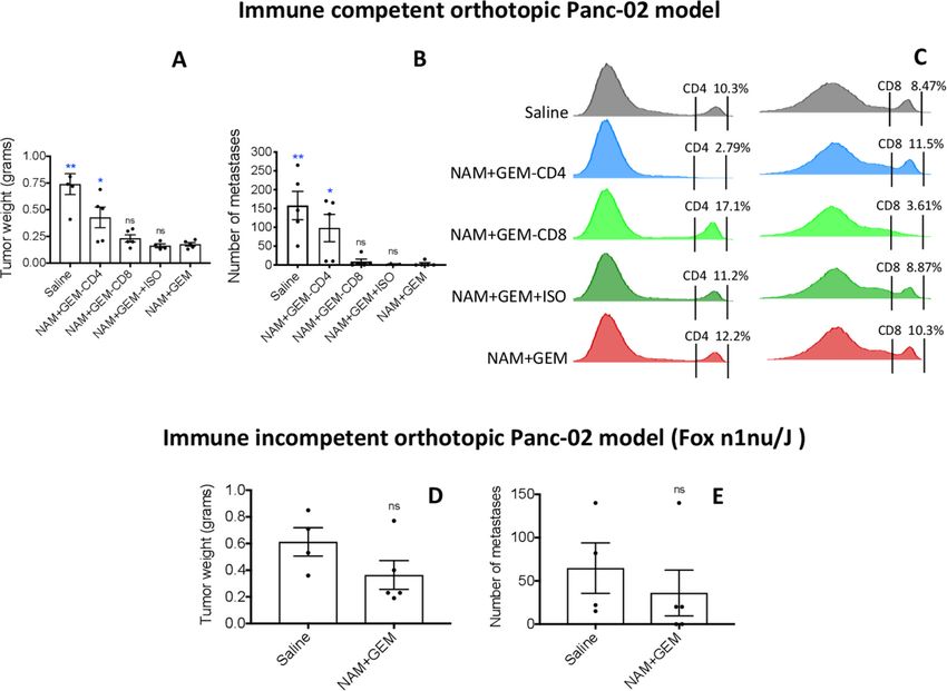
Open access
J Immunother Cancer: first published as 10.1136/jitc-2020-001250 on 5 November 2020. Downloaded from http://jitc.bmj.com/ on November 30, 2021 by guest. Protected by copyright.
Figure 1 NAM+GEM significantly improved clinical parameters, including survival, in mouse PDAC. Orthotopic Panc-02 model.
Tumors and metastases were generated and treatments with NAM+GEM and controls were performed as outlined in online
supplemental figure S1A. At the conclusion of the experiment tumor weight (A) and number of metastases (B) was measured,
as well as the grade of ascites (T0–5) (C). Average of two experiments with n=10 mice per group. Orthotopic KPC model.
Tumors and metastases were generated and treatments with NAM+GEM and controls were performed as outlined in online
supplemental figure S1B. At the conclusion of the experiment tumor weight (D) and number of metastases (E) was measured,
as well as the grade of ascites (T0–5) (F). Average of two experiments with n=10 mice per group. Significant differences were
determined by Mann-Whitney U test. *P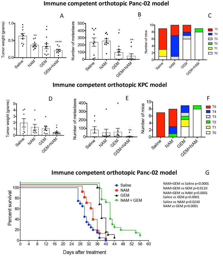
Open access
J Immunother Cancer: first published as 10.1136/jitc-2020-001250 on 5 November 2020. Downloaded from http://jitc.bmj.com/ on November 30, 2021 by guest. Protected by copyright.
NAM+GEM treatment was started and continued for 14 CD4 T cells eradicate tumors and metastases in orthotopic
days as described above. Mice were monitored for the Panc-02 mice
following weeks without any further treatment. Here, we Since the number and the activity of both CD4 and CD8 T
show that NAM+GEM significantly improved the survival cells was significantly increased in the orthotopic Panc-02
time of the orthotopic Panc-02 mice not only compared tumors of NAM+GEM-treated mice in correlation with
with the saline group but also compared with all other a significant reduction of tumors and metastases, we
control groups (figure 1G). analyzed in further detail whether this NAM+GEM effect
could be reduced by CD4 or CD8 T cells depletions in
NAM+GEM increases the influx of immune cells and activates vivo, as outlined in online supplemental figure S1D. As
T cells while reducing TAM and MDSC shown in figure 3A, the average tumor weight in mice
In skin cancer prevention, it has been shown that NAM treated with NAM+GEM plus CD4 antibodies was signifi-
significantly increased peritumoral and infiltrating CD4 cantly increased by 58% compared with the NAM+GEM
and CD8 T cells compared with a placebo.22 Here, we alone group (p=0.0397, Mann-Whitney U test), while the
tested the effect of the combination of GEM+NAM on tumors in mice depleted for CD8 T cells were increased
immune cells in the orthotopic Panc-02 tumors. At the by 22% compared with the NAM+GEM alone group
end of treatment, whole tumors were digested with colla- but this was not significant (p=0.3016, Mann- Whitney
genase and dispase to generate single cell suspensions as U test). As expected, the tumors in mice treated with
we described previously.14 Subsequently, the cell popu- NAM+GEM plus the isotype control (negative control)
lation was analyzed by flow cytometry. We found that were not significantly different from the tumor in mice
the CD45+ cells (leucocytes) (pOpen access
J Immunother Cancer: first published as 10.1136/jitc-2020-001250 on 5 November 2020. Downloaded from http://jitc.bmj.com/ on November 30, 2021 by guest. Protected by copyright.
Figure 2 NAM+GEM increased the influx of immune cells and activates T cells while reducing TAM and MDSC. Immune cells
(CD45+) were isolated from whole tumors and analyzed by flow cytometry for the presence of CD4 and CD8 T cells (A), TAM
(B) and MDSC (C). In addition, blood was also analyzed for MDSC (D). Subsequently, tumor tissues were analyzed by IHC for
the presence of CD4 and CD8 T cells (E, G), and quantified (F, H). 5 fields in each group were analyzed and the number of CD4
and CD8 T cells was calculated per mm2. n=5 mice per group. The results were averaged and analyzed by Mann-Whitney U
test. *pOpen access
J Immunother Cancer: first published as 10.1136/jitc-2020-001250 on 5 November 2020. Downloaded from http://jitc.bmj.com/ on November 30, 2021 by guest. Protected by copyright.
Figure 3 CD4 T cells significantly reduced pancreatic cancer by NAM+GEM in immune competent mice, while little effect
of NAM+GEM was observed in nude mice. Orthotopic Panc-02 tumors were generated in immune competent C57BL/6 mice
and depleted for CD4 and CD8 T cells during NAM+GEM treatment as outlined in online supplemental figure S1D. Antibodies
to CD4 T cells (300 µg/200 µL) or to CD8 T cells (300 µg/200 µL) or isotype control were administered IP every 3rd day for 2
weeks. At the conclusion of the experiment the average tumor weight (A) and number of metastases (B) was determined in the
NAM+GEM-treated/T cell-depleted compared with NAM+GEM-alone treated mice, with n=5 mice per group. Mann-Whitney
U test. *POpen access
J Immunother Cancer: first published as 10.1136/jitc-2020-001250 on 5 November 2020. Downloaded from http://jitc.bmj.com/ on November 30, 2021 by guest. Protected by copyright.
Figure 4 NAM+GEM decreased the production of collagen I and HABP, and increased αSMA and CD31, in pancreatic
tumors of orthopic Panc-02 model. Tumors were analyzed for collagen I by trichrome staining (A) and quantified (B) and shown
in more detail (C). n=5 mice per group and the results of 5 fields were averaged. HABP fibrils was analyzed by RT-PCR (D).
αSMA protein was by analyzed by IHC (E), and quantified (F), and shown in more detail (G). For both trichrome and αSMA the
percentage of positive areas were determined and the results of n=5 mice per group was averaged. The presence of blood
vessels in the pancreatic tumors was analyzed by IHC using anti-CD31 antibodies (H) and quantified (I) and shown in more
detail (J). n=5 mice per group and the results of 10 fields were averaged. Mann-Whitney U test. *POpen access
J Immunother Cancer: first published as 10.1136/jitc-2020-001250 on 5 November 2020. Downloaded from http://jitc.bmj.com/ on November 30, 2021 by guest. Protected by copyright.
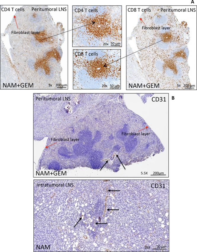
Figure 5 Peritumoral and intratumoral LNS in pancreatic tumors of orthotopic Panc-02 model. Detail of a peritumoral LNS
(A). Peritumoral LNS, characterized by well-developed follicular structures with occasional germinal centers. CD4+ and CD8+ T
cells are concentrated in the deep cortical zone/paracortex, where they were arranged in dense sheets, and are scattered within
follicles and surrounding tissue. The peritumoral LNS is surrounded by a fibroblast layer (red arrows). For more detail about
peritumoral and intratumoral LNS (see online supplemental figure S9). Immunostaining for CD31 highlights the presence of
vessels within peritumoral and intratumoral LNS in pancreatic tumors (black arrows) (B), which may allow the T cells to migrate
to the tumor areas. LNS, lymph node-like structures.
which is involved in T cell migration and recruitment of activating T cells35. Chemokine (C-C) ligand 21a (Ccl21a)
LNS, a fourfold increase in Interleukin-1 Receptor-Like was also increased in the NAM or GEM only groups but
2 (IL1RL2), which is involved in activating T cells and less robust than in NAM+GEM combination. In another
increasing DC immunogenicity33, a 10-fold increase in study, we also found that GEM improved the migration of
Fcer-1 which is involved in epitope spreading34, a fourfold T cells to pancreatic tumors.14 Finally, we found a four-
increase in Ccl9 which is involved in attracting DC and fold reduction in Lipocalin-2 (Lcn-2)36 and a less robust
10 Selvanesan BC, et al. J Immunother Cancer 2020;8:e001250. doi:10.1136/jitc-2020-001250Open access
J Immunother Cancer: first published as 10.1136/jitc-2020-001250 on 5 November 2020. Downloaded from http://jitc.bmj.com/ on November 30, 2021 by guest. Protected by copyright.
Figure 6 NAM+GEM increased the expression of genes involved in T cell migration and activation, and reduced the expression
of genes involved in invasion of tumor cells. Orthotopic Panc-02 tumors of the different treatment groups were analyzed by
Nanostring through gene expression profiles involved in T cell migration, activation, epitope spreading and invasion of tumor
cells. A heatmap of relevant genes is shown (A), and the function of each gene and p values (B). Gene expression levels in
the NAM+GEM group were compared with the saline group and analyzed by ANOVA pOpen access
J Immunother Cancer: first published as 10.1136/jitc-2020-001250 on 5 November 2020. Downloaded from http://jitc.bmj.com/ on November 30, 2021 by guest. Protected by copyright.
tumor, strong immune suppression at multiple levels, cells from penetrating into the pancreatic tumor.1 50
and inefficient activation of T cells in the TME.8 9 11–14 Cleaving fibronectin leads to degradation of Collagen I
Here, we demonstrate that a novel combination of density and may lead to a better infiltration of immune
NAM+GEM in mice with pancreatic cancer not only cells. We observed a significant decrease in Collagen I
reduced the pancreatic cancer (tumors and metas- density and in HABP in the NAM+GEM-treated group,
tases) but also significantly improved the survival time in correlation with an influx of immune cells into the
compared with all control groups. We found suggestive pancreatic tumors, and increase in CD31 positive blood
evidence that tumor cells were killed by T cells through vessels in the tumor areas.
epitope spreading. This is based on the strong CD4 and LNSs are often found in cancer although their func-
CD8 T cell responses to survivin, which is expressed by tion is not well understood. Tertiary lymphoid struc-
the Panc-02 tumor cells,24 and the 10-fold increase in tures in close proximity to pancreatic tumors have been
Fcer mRNA in the tumors of NAM+GEM treated mice, correlated with increased patient survival in PDAC.31
which is responsible for epitope spreading, and specif- Conversely, tumor draining lymph nodes (intratumoral
ically the significant eradication of pancreatic cancer LNS) were found to correlate with immune suppres-
by CD4 T cells through NAM+GEM treatment in the sion, a more metastatic character, and poor outcome in
immune competent orthotopic Panc-02 model. CD4 PDAC patients.32 Our study showed both peritumoral and
T cells are known to kill tumor cells through different intratumoral LNS in the TME of the orthotopic Panc-02
mechanisms, that is, through perforin and Granzyme model. The peritumoral LNS contain well- developed
B,43 or indirectly through IFNγ-activated M1 macro- follicular structures, germinal centers and T cells, all
phages,44 or by helping B cell activation producing surrounded by a fibroblast layer, at the periphery of the
antigen-specific antibodies resulting in antibody- tumor, and the intratumoral LNS are located inside the
dependent cytotoxicity.45 Such mechanisms need to be tumor with an accumulation of T cells in the tumor areas.
further analyzed in detail. CD8 T cells also eradicated While in NAM+GEM-treated mice a positive correlation
the tumors and metastases but considerably less robust was found between the peritumoral LNS and improved
than CD4 T cells. Also, a reduction in the pancreatic T cells responses to TAA, efficacy, and survival, no such
tumors was observed in nude immune incompetent correlation was observed for intratumoral LNS. None of
mice lacking T cells, but this was statistically not signifi- these effects were found in the NAM or GEM group only.
cant. Most likely tumor cell kill here was caused directly We found peritumoral LNS in all tumors of mice treated
by NAM or GEM through down regulation of SIRT-1, with NAM+GEM, while in the NAM or GEM groups intra-
K-ras and Akt-1 expression,18 inhibition of PARP,19 or tumoral LNS only were observed. The T cell responses
through inhibition of DNA synthesis, respectively. A in the tumors of NAM or GEM treated mice were not
schematic view of potential immune mechanisms of significantly different from the saline group. It has been
NAM+GEM in pancreatic cancer is shown in online suggested that peritumoral LNS protects T cells from
supplemental figure S10. immune suppression.51 Based on these and our results
Immune suppression is one of the hallmarks of PDAC. we speculate that the peritumoral LNS are a place where
Unfortunately, checkpoint inhibitors do not improve T cells can be activated without inhibition by immune
the outcome of therapy in PDAC patients, which we suppression, while T cells in the intratumoral LNS are
also observed when adding anti-PD-L1 antibodies to the exposed to the same immune suppression as T cells in
NAM+GEM treatment (data not shown). However, the the TME. However, this hypothesis needs to be tested in
TAM and MDSC populations were significantly reduced studies specifically designed for it.
in the pancreatic tumors and blood, respectively, of High αSMA expression is characteristic of myofibro-
the NAM+GEM-treated mice compared with the saline blastic CAFs.49 However, different correlations of αSMA
group. This most likely has contributed to the improved expression with survival have been reported. One study
T cell responses to survivin in the NAM+GEM group as in PDAC patients reported that high expression levels of
well. αSMA in tumor stroma promoted invasion and cellular
An interesting observation was the increased number migration or was associated with a worse outcome,52 while
of perforin and particularly granzyme B-producing cells others found that high expression of αSMA is associated
in the tumors of the NAM+GEM group in IHC analysis. with improved outcome.53 Also, deletion of αSMA+ fibro-
Granzyme B is not only known to be produced by T cells blasts in transgenic mice led to invasive undifferentiated
and NK cells, but also by B cells and macrophages.46 47 tumors and reduced survival.54 In our study, we found
More detailed studies are required to identify the cell that αSMA increased in the NAM+GEM group compared
type(s) that produce granzyme B here. Other studies with the saline group and this correlated with significant
have shown that the production of granzyme B plays improved survival of the orthotopic Panc-02 mice. This
an important role in ECM remodeling.48 Granzyme B heterogeneity and plasticity of αSMA in the reported
cleaves fibronectin, which is involved in the formation studies may have been the result of different types of
of collagen I fibrils.48 Collagen I is produced by CAFs.49 therapy. Therefore, more detailed studies are required
These fibrils form cross- links between the collagen to obtain better insight in the role of αSMA in PDAC in
fibers, which in turn may prevent drugs and immune relation to different therapies. It is yet unclear how these
12 Selvanesan BC, et al. J Immunother Cancer 2020;8:e001250. doi:10.1136/jitc-2020-001250Open access
J Immunother Cancer: first published as 10.1136/jitc-2020-001250 on 5 November 2020. Downloaded from http://jitc.bmj.com/ on November 30, 2021 by guest. Protected by copyright.
data, which are snapshots in time, need to be interpreted ORCID iDs
in a context of CAF heterogeneity and plasticity.5 Benson Chellakkan Selvanesan http://orcid.org/0000-0001-8013-1294
Olaya Lara http://orcid.org/0000-0002-7466-3072
Claudia Gravekamp http://orcid.org/0000-0002-1713-3008
CONCLUSIONS
In conclusion, we have demonstrated that NAM+GEM
REFERENCES
significantly reduced pancreatic cancer in different 1 Olive KP, Jacobetz MA, Davidson CJ, et al. Inhibition of Hedgehog
mouse tumor models, and significantly improved the signaling enhances delivery of chemotherapy in a mouse model of
pancreatic cancer. Science 2009;324:1457–61.
survival time compared with all control groups. This 2 Ene-Obong A, Clear AJ, Watt J, et al. Activated pancreatic stellate
correlated with an altered tumor architecture and cells sequester CD8+ T cells to reduce their infiltration of the
immune responses in the TME. Collagen I and HABP juxtatumoral compartment of pancreatic ductal adenocarcinoma.
Gastroenterology 2013;145:1121–32.
was significantly reduced by NAM+GEM, while more T 3 Apte M, Pirola RC, Wilson JS. Pancreatic stellate cell: physiologic
cells infiltrated the pancreatic tumors, and CD4 T cells role, role in fibrosis and cancer. Curr Opin Gastroenterol
2015;31:416–23.
significantly eradicated the pancreatic tumors and metas- 4 Elyada E, Bolisetty M, Laise P, et al. Cross-Species single-cell
tases in vivo. Also improved T cell responses to survivin analysis of pancreatic ductal adenocarcinoma reveals antigen-
were observed, in correlation with reduction in TAM and presenting cancer-associated fibroblasts. Cancer Discov
2019;9:1102–23.
MDSC in the tumor microenvironment. The results of 5 Sahai E, Astsaturov I, Cukierman E, et al. A framework for advancing
this study, and its promising effect in a phase 3 clinical our understanding of cancer-associated fibroblasts. Nat Rev Cancer
2020;20:174–86.
trial to prevent skin cancer,23 as well as the immunopro- 6 Lunardi S, Muschel RJ, Brunner TB. The stromal compartments in
phylactic and immunotherapeutic success of NAM in pancreatic cancer: are there any therapeutic targets? Cancer Lett
2014;343:147–55.
the preclinical hormone receptor-positive breast cancer 7 Von Hoff DD, Ramanathan RK, Borad MJ, et al. Gemcitabine plus
models21 suggest that NAM as addition to GEM, which is nab-paclitaxel is an active regimen in patients with advanced
the backbone of standard care for PDAC patients, could pancreatic cancer: a phase I/II trial. J Clin Oncol 2011;29:4548–54.
8 Kulke MH, Blaszkowsky LS, Ryan DP, et al. Capecitabine plus
be a promising new lead for the treatment of pancreatic erlotinib in gemcitabine-refractory advanced pancreatic cancer. J
cancer. Clin Oncol 2007;25:4787–92.
9 Maitra A, Hruban RH. Pancreatic cancer. Annu Rev Pathol
2008;3:157–88.
Acknowledgements We greatly thank Ms Hong Zhang, Department of Pathology, 10 Nowak AK, Robinson BWS, Lake RA. Synergy between
and Dr Vera DeMarais, Director of Light Microscopy and Image Analysis, chemotherapy and immunotherapy in the treatment of established
Department of Structural and Cell Biology, Einstein for providing outstanding murine solid tumors. Cancer Res 2003;63:4490–6.
training and support regarding the IHC and image analysis. 11 Plate JMD, Plate AE, Shott S, et al. Effect of gemcitabine on immune
cells in subjects with adenocarcinoma of the pancreas. Cancer
Contributors BCS performed and designed most of the experiments and analyzed Immunol Immunother 2005;54:915–25.
the data. KM, OL and AB presented in figures 1-6 and contributed experimentally to 12 Zitvogel L, Apetoh L, Ghiringhelli F, et al. Immunological aspects of
the data and online supplemental figures. AB contributed to the IHC analysis. BCS, cancer chemotherapy. Nat Rev Immunol 2008;8:59–73.
KM, OL, LM and IR contributed with critical discussions and design of experiments. 13 Bracci L, Schiavoni G, Sistigu A, et al. Immune-Based mechanisms
CG conceived, designed and supervised the study and wrote the manuscript. All of cytotoxic chemotherapy: implications for the design of novel and
authors reviewed the manuscript. rationale-based combined treatments against cancer. Cell Death
Differ 2014;21:15–25.
Funding This work was supported by the Anticancer Fund, a private donation of 14 Selvanesan BC CD, Quispe-Tintaya Q, Jahangir PA, et al. Tumor-
Janet and Marty Spatz, NCI cancer center support P30CA013330 (Flow Cytometry Targeted delivery of childhood vaccine recall antigens by attenuated
Core, Pathology Core), and IR was supported by the FWO Odysseus Program Listeria reduces pancreatic cancer. under revision with science
(Research Foundation Flanders), and OL is recipient of a Fellowship (FWOTM904, translational medicine. BioRchiv 2020.
Research Foundation Flanders). 15 Suzuki E, Kapoor V, Jassar AS, et al. Gemcitabine selectively
eliminates splenic Gr-1+/CD11b+ myeloid suppressor cells in
Competing interests None declared. tumor-bearing animals and enhances antitumor immune activity. Clin
Cancer Res 2005;11:6713–21.
Patient consent for publication Not required. 16 Maiese K, Chong ZZ, Hou J, et al. The vitamin nicotinamide:
Provenance and peer review Not commissioned; externally peer reviewed. translating nutrition into clinical care. Molecules 2009;14:3446–85.
17 Maiese K, Chong ZZ. Nicotinamide: necessary nutrient emerges
Data availability statement Data sharing not applicable as no datasets as a novel cytoprotectant for the brain. Trends Pharmacol Sci
generated and/or analyzed for this study. We do not have datasets generated in this 2003;24:228–32.
manuscript. 18 Zhang J-gang, Zhao G, Qin Q, et al. Nicotinamide prohibits
proliferation and enhances chemosensitivity of pancreatic
This content has been supplied by the author(s). It has not been vetted by BMJ cancer cells through deregulating SIRT1 and Ras/Akt pathways.
Publishing Group Limited (BMJ) and may not have been peer-reviewed. Any Pancreatology 2013;13:140–6.
opinions or recommendations discussed are solely those of the author(s) and are 19 Domínguez-Gómez G, Díaz-Chávez J, Chávez-Blanco A, et al.
not endorsed by BMJ. BMJ disclaims all liability and responsibility arising from any Nicotinamide sensitizes human breast cancer cells to the cytotoxic
reliance placed on the content. Where the content includes any translated material, effects of radiation and cisplatin. Oncol Rep 2015;33:721–8.
BMJ does not warrant the accuracy and reliability of the translations (including but 20 Choi HJ, Jang S-Y, Hwang ES. High-Dose Nicotinamide Suppresses
ROS Generation and Augments Population Expansion during CD8(+)
not limited to local regulations, clinical guidelines, terminology, drug names and
T Cell Activation. Mol Cells 2015;38:918–24.
drug dosages), and is not responsible for any error and/or omissions arising from 21 Buqué A, Bloy N, Perez-Lanzón M, et al. Immunoprophylactic and
translation and adaptation or otherwise. immunotherapeutic control of hormone receptor-positive breast
Open access This is an open access article distributed in accordance with the cancer. Nat Commun 2020;11:3819.
22 Malesu R, Martin AJ, Lyons JG, et al. Nicotinamide for skin cancer
Creative Commons Attribution Non Commercial (CC BY-NC 4.0) license, which
chemoprevention: effects of nicotinamide on melanoma in vitro and
permits others to distribute, remix, adapt, build upon this work non-commercially, in vivo. Photochem Photobiol Sci 2020;19:171–9.
and license their derivative works on different terms, provided the original work is 23 Chen AC, Martin AJ, Choy B, et al. A phase 3 randomized trial
properly cited, appropriate credit is given, any changes made indicated, and the use of nicotinamide for skin-cancer chemoprevention. N Engl J Med
is non-commercial. See http://c reativecommons.org/licenses/by-nc/4.0 /. 2015;373:1618–26.
Selvanesan BC, et al. J Immunother Cancer 2020;8:e001250. doi:10.1136/jitc-2020-001250 13Open access
J Immunother Cancer: first published as 10.1136/jitc-2020-001250 on 5 November 2020. Downloaded from http://jitc.bmj.com/ on November 30, 2021 by guest. Protected by copyright.
24 Ishizaki H, Manuel ER, Song G-Y, et al. Modified vaccinia Ankara 40 Zhou D, Tang W, Zhang Y, et al. Jam3 functions as a novel tumor
expressing survivin combined with gemcitabine generates specific suppressor and is inactivated by DNA methylation in colorectal
antitumor effects in a murine pancreatic carcinoma model. Cancer cancer. Cancer Manag Res 2019;11:2457–70.
Immunol Immunother 2011;60:99–109. 41 Coelho AL, Schaller MA, Benjamim CF, et al. The chemokine
25 Hingorani SR, Wang L, Multani AS, et al. Trp53R172H and CCL6 promotes innate immunity via immune cell activation and
KrasG12D cooperate to promote chromosomal instability and widely recruitment. J Immunol 2007;179:5474–82.
metastatic pancreatic ductal adenocarcinoma in mice. Cancer Cell 42 Yan Y, Xu H, Zhang L, et al. Rrad suppresses the Warburg effect by
2005;7:469–83. downregulating ACTG1 in hepatocellular carcinoma. Onco Targets
26 Corbett TH, Roberts BJ, Leopold WR, et al. Induction and Ther 2019;12:1691–703.
chemotherapeutic response of two transplantable ductal 43 Couturier J, Hutchison AT, Medina MA, et al. HIV replication in
adenocarcinomas of the pancreas in C57BL/6 mice. Cancer Res conjunction with granzyme B production by CCR5+ memory CD4 T
1984;44:717–26. cells: implications for bystander cell and tissue pathologies. Virology
27 Beerling E, Oosterom I, Voest E, et al. Intravital characterization of 2014;462-463:175–88.
tumor cell migration in pancreatic cancer. Intravital 2016;5:e1261773. 44 Haabeth OAW, Tveita AA, Fauskanger M, et al. How Do CD4(+) T
28 Castro F, Leal B, Denny A, et al. Vaccination with Mage-b DNA Cells Detect and Eliminate Tumor Cells That Either Lack or Express
induces CD8 T-cell responses at young but not old age in mice with MHC Class II Molecules? Front Immunol 2014;5:174.
metastatic breast cancer. Br J Cancer 2009;101:1329–37. 45 Largeot A, Pagano G, Gonder S, et al. The B-side of cancer
29 Jahangir A, Chandra D, Quispe-Tintaya W, et al. Immunotherapy immunity: the underrated tune. Cells 2019;8:449–3.
with Listeria reduces metastatic breast cancer in young and old mice 46 Arabpour M, Rasolmali R, Talei A-R, et al. Granzyme B production by
through different mechanisms. Oncoimmunology 2017;6:e1342025. activated B cells derived from breast cancer-draining lymph nodes.
30 Kim SH, Castro F, Gonzalez D, et al. Mage-B vaccine delivered by Mol Immunol 2019;114:172–8.
recombinant Listeria monocytogenes is highly effective against 47 Elavazhagan S, Fatehchand K, Santhanam V, et al. Granzyme B
breast cancer metastases. Br J Cancer 2008;99:741–9. expression is enhanced in human monocytes by TLR8 agonists and
31 Stromnes IM, Hulbert A, Pierce RH, et al. T-Cell localization,
contributes to antibody-dependent cellular cytotoxicity. J Immunol
activation, and clonal expansion in human pancreatic ductal
2015;194:2786–95.
adenocarcinoma. Cancer Immunol Res 2017;5:978–91.
48 Parkinson LG, Toro A, Zhao H, et al. Granzyme B mediates
32 Fink DM, Steele MM, Hollingsworth MA. The lymphatic system and
both direct and indirect cleavage of extracellular matrix in skin
pancreatic cancer. Cancer Lett 2016;381:217–36.
after chronic low-dose ultraviolet light irradiation. Aging Cell
33 Chen S-C, Vassileva G, Kinsley D, et al. Ectopic expression of the
2015;14:67–77.
murine chemokines CCL21a and CCL21b induces the formation of
lymph node-like structures in pancreas, but not skin, of transgenic 49 Öhlund D, Handly-Santana A, Biffi G, et al. Distinct populations of
mice. J Immunol 2002;168:1001–8. inflammatory fibroblasts and myofibroblasts in pancreatic cancer. J
34 Nasi A, Bollampalli VP, Sun M, et al. Immunogenicity is preferentially Exp Med 2017;214:579–96.
induced in sparse dendritic cell cultures. Sci Rep 2017;7:43989. 50 Provenzano PP, Cuevas C, Chang AE, et al. Enzymatic targeting of
35 Zhao X, Sato A, Dela Cruz CS, et al. CCL9 is secreted by the follicle- the stroma ablates physical barriers to treatment of pancreatic ductal
associated epithelium and recruits dome region Peyer's patch adenocarcinoma. Cancer Cell 2012;21:418–29.
CD11b+ dendritic cells. J Immunol 2003;171:2797–803. 51 Goc J, Fridman W-H, Sautès-Fridman C, et al. Characteristics of
36 Leung L, Radulovich N, Zhu C-Q, et al. Lipocalin2 promotes invasion, tertiary lymphoid structures in primary cancers. Oncoimmunology
tumorigenicity and gemcitabine resistance in pancreatic ductal 2013;2:e26836.
adenocarcinoma. PLoS One 2012;7:e46677. 52 Sinn M, Denkert C, Striefler JK, et al. α-Smooth muscle actin
37 Roy LD, Sahraei M, Subramani DB, et al. Muc1 enhances expression and desmoplastic stromal reaction in pancreatic
invasiveness of pancreatic cancer cells by inducing epithelial to cancer: results from the CONKO-001 study. Br J Cancer
mesenchymal transition. Oncogene 2011;30:1449–59. 2014;111:1917–23.
38 Salnikov AV, Groth A, Apel A, et al. Targeting of cancer stem 53 Wang LM, Silva MA, D'Costa Z, et al. The prognostic role of
cell marker EpCAM by bispecific antibody EpCAMxCD3 inhibits desmoplastic stroma in pancreatic ductal adenocarcinoma.
pancreatic carcinoma. J Cell Mol Med 2009;13:4023–33. Oncotarget 2016;7:4183–94.
39 Bernard V, Semaan A, Huang J, et al. Single-Cell transcriptomics 54 Özdemir BC, Pentcheva-Hoang T, Carstens JL, et al. Depletion
of pancreatic cancer precursors demonstrates epithelial and of carcinoma-associated fibroblasts and fibrosis induces
microenvironmental heterogeneity as an early event in neoplastic immunosuppression and accelerates pancreas cancer with reduced
progression. Clin Cancer Res 2019;25:2194–205. survival. Cancer Cell 2014;25:719–34.
14 Selvanesan BC, et al. J Immunother Cancer 2020;8:e001250. doi:10.1136/jitc-2020-001250You can also read


