No Time to Age: Uncoupling Aging from Chronological Time - Preprints.org
←
→
Page content transcription
If your browser does not render page correctly, please read the page content below
Preprints (www.preprints.org) | NOT PEER-REVIEWED | Posted: 14 April 2021                        doi:10.20944/preprints202104.0384.v1

      Review

      No Time to Age: Uncoupling Aging from Chronological Time
      Dana Larocca 1, *, Jieun Lee 2, Michael D. West 2, Ivan Labat 2 and Hal Sternberg 2

                                    1   DC Biotechnology, Alameda, CA 94502, USA
                                    2   AgeX Therapeutics Inc. Alameda, CA 94501, USA
                                    * Correspondence: dclarocca@danaclairebio.com; Tel.: 510-761-8442

                                    Abstract: Multicellular life evolved from simple unicellular organisms that could replicate indefi-
                                    nitely being essentially ageless. At this point, life split into 2 fundamentally different cell types: the
                                    immortal germline representing an unbroken lineage of cell division with no intrinsic endpoint and
                                    the mortal soma which ages and dies. In this review, we describe the germline as clock-free and the
                                    soma as clock-bound and discuss aging with respect to 3 DNA-based cellular clocks (telomeric,
                                    DNA methylation, and transposable element). The ticking of these clocks corresponds to the step-
                                    wise progressive limitation of growth and regeneration of somatic cells that we term, somatic re-
                                    striction. Somatic restriction acts in opposition to strategies that ensure continued germline repli-
                                    cation and regeneration. We thus consider the plasticity of aging as a process not fixed to the pace
                                    of chronological time but one that can speed up or slow down depending on the rate of intrinsic
                                    cellular clocks. We further describe how germline factor reprogramming might be used to slow
                                    the rate of aging and potentially reverse it by causing the clocks to tick backwards. Therefore, re-
                                    programming may eventually lead to therapeutic strategies to treat degenerative diseases by alter-
                                    ing aging itself, the one condition common to us all.

                                    Keywords: Aging; cellular clocks; reprogramming; development; epigenetics; DNA methylation;
                                    telomeres; transposable elements; longevity; regeneration

                                   1.   Introduction
                                         Whether or not aging is programmed or is the result of accumulated damage has
                                    been the subject of debate for many decades. Early twentieth century theories favoring
                                    programming proposed that aging was a consequence of differentiation as put forth by
                                    Minot (1) or factors leading to cessation of growth as proposed by Bidder (2). Evolution
                                    of a genetic program for aging seemed unlikely because there is little selective pressure
                                    for gene variants once reproductive maturity is reached. However, Williams proposed
                                    that aging is the result of antagonistic pleiotropy where mutations that are adaptive for
                                    fitness in early life but deleterious after reproduction would accumulate in the gene pool
                                    because there is little selective pressure to eliminate them (3). In contrast, aging theories
                                    of the late twentieth century focused largely on “wear and tear”, suggesting aging results
                                    from stochastic accumulation of damage, such as cellular waste, oxidative damage, DNA
                                    mutations, and misfolded proteins (4, 5). However, the vast range of adult lifespans
                                    among animal species from hours (mayfly), days (fruit fly) or weeks (dwarf pygmy goby)
                                    to as long as centuries (Aldabra giant tortoise, bowhead whale, Greenland shark) as well
                                    as examples of indefinite lifespans with no apparent intrinsic aging (hydra, planaria,
                                    sponge, mussel) suggests that underlying genetic programs must play a pivotal role (6, 7).
                                    Indeed, the wide variation in lifespan even between closely related species underscores
                                    the notion that aging is plastic and depends upon a balance between intrinsic cellular
                                    strategies that maintain genetic and epigenetic integrity and the effects of “wear and tear”.
                                    We have previously described the stepwise progressive loss of regenerative germline

            © 2021 by the author(s). Distributed under a Creative Commons CC BY license.
Preprints (www.preprints.org) | NOT PEER-REVIEWED | Posted: 14 April 2021 doi:10.20944/preprints202104.0384.v1
strategies in mortal somatic cells (somatic restriction) as an underlying cause of organis-
mic decline associated with aging (8). In contrast, we see in the regeneration of the im-
mortal germline, the potential for life to be uncoupled from time, and therefore free from
aging. The fact that there can be an uncoupling of life from the fixed progression of
chronological time as seen in the germline hints at the basis by which aging can be accel-
erated, decelerated, and perhaps most astonishingly reversed altogether.
In his germ plasma theory, the 19th century biologist, August Weismann, described a
division of labor in cellular life that enabled the evolution of complex multicellular life-
forms that make up the tremendous diversity of life we see today (9). To make the leap
from simple unicellular organisms to complex metazoans, life evolved into 2 fundamen-
tally different types of cells consisting of the mortal cells of the soma or body, which even-
tually ages and dies, and the immortal self-renewing germline cells that carry the heredi-
tary information forward to the next generation. Well over half a century before the dis-
covery of cellular senescence, DNA, telomeres and telomerase, Weismann predicted the
limited cellular lifespan of somatic cells in contrast to cells of the germline, which have no
intrinsic end to their replicative lifespan. He further asserted that “there is nothing in-
herent in life that implies death (9).” Aristotle wrote, “things which are always are not,
as such, in time nor is there being measured by time… none of them is affected by time,
which indicates they are not in time.” In this sense, Weismann understood that life is not
bound by time. To illustrate the evolutionary split of the soma and germline, Weismann
described two genera of volvox, one of the simplest multicellular organisms (Figure 1).
Pandorina morum consists of a simple ball of identical cells (Figure 1A) that reproduces by
budding off smaller clusters of cells and Volvox minor consists of a hollow sphere of cells
(Figure 1B) that contains the simpler balls of germline cells within it. As the germline clus-
ters grow larger, they trigger the sphere to wither and die and thus release the next gen-
eration of volvox. Accordingly, we see in the evolution of volvox, cells that exhibit a di-
vision of labor into immortal germline (embryonic balls of cells) and mortal soma (hollow
sphere of cells). The germline can forever renew itself, giving birth to young organisms
at each generation and thus we refer to germline cells as clock-free and somatic cells as
clock-bound. Indeed, we can trace an unbroken chain of cell division from all life on
earth going back 4 billion years to converge at the base of the evolutionary tree to a single
cell, LUCA, the “last universal common ancestor” (10). In contrast, somatic cells lose
their robustness with time exhibiting the hallmarks of aging (11), which eventually lead
to organismal degeneration and an exponential increase in the probability of death with
the passage of time (12). In this review, we will explore the nature of 3 somatic cellular
clocks (telomeric, DNA methylation (DNAm), and transposable element (TE)) and the
germ line strategies used to become clock-free or unbound from chronological time. We
also consider the relationship of aging to development and describe remarkable examples
of phenotypic plasticity seen in lower multicellular organisms that appear to reverse de-
velopment. Finally, we discuss how this plasticity can be revived in higher animals using
cell reprogramming technology aimed at conferring germline qualities onto the soma and
how these methods might be applied to reverse aging and extend healthy lifespan.Preprints (www.preprints.org) | NOT PEER-REVIEWED | Posted: 14 April 2021 doi:10.20944/preprints202104.0384.v1
Figure 1. The appearance of germline and somatic cells in the evolution of metazoans. Two Vol-
vocinian genera that were used by Weismann to illustrate the difference between A.) a homoplas-
tid organism (Pandorina morum) consisting of a single cell type and the evolution of B.) a hetero-
plastid (Volvox minor) which has evolved to consist of two primary cell types: the germline cells
(Kz) that form the internal embryos and somatic cells (Sz) which are subject to death upon which
the developing embryos are released.
Nature of somatic cellular clocks – Telomeric clock
The telomeric clock that limits the number of cell divisions of somatic cells was ac-
curately predicted by Weismann. He noted that “death takes place because a worn-out
tissue cannot forever renew itself, and because a capacity for increase by means of cell
division is not everlasting but finite” (9). However, it would be a half century before
Leonard Hayflick performed definitive experiments that proved the limited lifespan of
somatic cells (13). Previously, it had been widely accepted that somatic cells were im-
mortal. In 1912, Carrel had demonstrated the continuous passage of chick heart cells for
over three months (14) and indeed the cells were passaged for more than three decades
outliving Carrel himself (14). Carrel’s influence in the press and Nobel laureate status
was so strong that the lack of reproducibility in other laboratories was not enough to keep
his theory of cellular immortality from becoming the prevailing view. However, in 1961,
Hayflick reported conclusive evidence that human fibroblasts were limited to about 50
population doublings before reaching replicative senescence (13). This limited replica-
tion capacity of somatic cells is referred to as the Hayflick limit. He elegantly proved that
the cessation of replication was intrinsic to the cells and not due to external factors by
coculture of late passage male cells with early passage female cells and demonstrating that
the young female cells continued to grow well after the male cells reached senescence (15).
It is now well established that virtually all normal human cells have a finite replicative
capacity (16). There is much speculation as to how Carrel was able to maintain continuous
replication of his cultures for decades including inadvertent or deliberate spiking with
new cells. One source of external cells may have been the chick embryo extract that was
used early on to maintain the health of the cultures. Alternatively given what we now
know about cellular reprogramming using germline factors, it is interesting to speculate
that secreted factors in Carrel’s embryo extracts may have immortalized his cell cultures.
Hayflick’s experiment confirmed Weismann’s prior conjecture about the limited rep-
licative lifespan of somatic cells and demonstrated the phenomenon of cellular senescence
which left cells in a seemingly irreversible suspension of the cell cycle. The Hayflick
clock can be suspended temporarily by inducing quiescence or by storage at low temper-
ature because it is a measure of replication cycles (15, 17). The clock mechanism was
independently proposed in the early seventies by Olovnikov and Watson, who eachPreprints (www.preprints.org) | NOT PEER-REVIEWED | Posted: 14 April 2021 doi:10.20944/preprints202104.0384.v1
theorized that the ends of DNA would necessarily shorten each time it replicated due to
the end replication problem (18, 19). Olovnikov further hypothesized that enzymatic re-
pair could preserve or elongate the ends of telomeres to prevent the shortening of chro-
mosomal telomere ends at each cell division (18, 20). This hypothesis was validated in
the 1990’s with cloning of human telomerase reverse transcriptase (hTERT), the gene en-
coding the catalytic component of the enzyme responsible for telomere maintenance and
by showing that forced expression of hTERT prevented senescence thus immortalizing the
cells (21). Remarkably, for both the germline and pluripotent embryonic stem cells, the
telomeric clock is suspended, but for somatic cells, the clock begins to tick not long after
the initiation of embryonic development (22, 23). We have previously described the loss
of immortal regeneration and replication as the pluripotency transition (PT) (8). It is the
first step in a series of life history transitions, termed “somatic restriction”, that lead to
the loss of regenerative capacity characteristic of aging (8). Another type of immortal
cell, the cancer cell, also expresses telomerase as shown using a telomerase activity assay
(23). Indeed, the evolutionary advantage of shutting off telomerase may be as a protec-
tion against cancer resulting in greater reproductive fitness in early life. However, pro-
gressive shortening of telomeres eventually triggers cell senescence which in later life con-
tributes to lack of regeneration, immune system failure and chronic inflammation due to
senescence associated secretory products (SASP) (24). Thus, the progressive restriction
of germline strategies for immortal regeneration is consistent with the antagonistic pleiot-
ropy theory of the evolution of aging.
Evidence for the connection between the telomeric clock and aging comes from the
correlation of telomere shortening with age in humans and experimental animals, simu-
lation of human-like aging in mice lacking telomerase, and from conditions such as pro-
geria and HIV infection that result in rapid telomere shortening and accelerated aging (25,
26). Although there is some controversy arising from the variability of telomere meas-
urement techniques and cell type chosen for analysis, many studies demonstrate a corre-
lation of telomere attrition with aging in humans (27-29). Moreover, data from a variety
of vertebrate model animals demonstrate that telomere shortening is associated with hu-
man-like age-related disease and lifespan. For example, the short-lived African killifish
has served as a useful model telomere dysfunction and aging (30). Mice normally have
much longer telomeres than humans, however, successive generations of single Terc
(mouse catalytic component of telomerase) knockout or double knockout of Wrn (the hel-
icase gene that is mutated in Werner syndrome) and Terc show telomere attrition and age-
related conditions that more closely model human aging (31, 32). Importantly, reactiva-
tion of telomerase in this model restores degenerative damage in multiple systems includ-
ing neurodegeneration (33). Furthermore, an accelerated telomeric clock resulting from
defects in telomere maintenance is associated with several human diseases that mimic
rapid aging including Hutchinson-Gilford progeria syndrome (HGPS), Werner syn-
drome, and dyskeratosis congenita (DKC) (25). Additional association of the telomeric
clock with age-associated disease comes from combined mouse models that demonstrate
the contributing influence of telomere shortening on diabetes and cardiomyopathy in Du-
chenne muscular dystrophy (34, 35). In addition, immune deficiencies in the elderly and
HIV patients have been linked to the Hayflick limit of T cells (36). Indeed, relatively
young HIV patients show a similar degree of T cell telomere attrition as centenarians (26).
Although long-lived post-mitotic cells in non-proliferative tissues like brain, skeletal mus-
cle, fat and heart were not initially thought to be effected by the telomere clock, telomere
shortening has been identified more recently in these tissues, which may be triggered by
SASP from other cells (37). Telomere attrition has also been proposed to contribute to both
the timing of parturition and death through onset of inflammation (38). The telomeric
clock can also be slowed, for example, by experimental overexpression of telomerase
which has been shown to extend lifespan even when given late in life (39). Interestingly,
while the telomere length at birth is highly variable and has not been found to correlate
with lifespan, the rate of telomere attrition correlates well with lifespan in a wide variety
of bird and mammal species (40). Moreover, mice generated from ES cells with hyper-Preprints (www.preprints.org) | NOT PEER-REVIEWED | Posted: 14 April 2021 doi:10.20944/preprints202104.0384.v1
long telomeres show an increase in lifespan, are leaner, and have a decrease in metabolic
aging (41). Importantly, environmental interventions such as calorie restriction and ex-
ercise have been shown to decelerate the telomeric clock in humans (42, 43). Taken to-
gether, these data support the notion that the loss of telomerase near the pluripotency to
embryonic transition (PT) correlates with onset of mortality of the soma and a clock-
bound process that contributes to the eventual aging and death of the body. We will next
discuss two additional clocks, the epigenetic or DNA methylation (DNAm) clock and the
transposable element (TE) clock.
Nature of somatic cellular clocks – Epigenetic (DNAm) clock
The epigenetic clock represents changes in the methylation pattern of CpG sites in
the genome that occur in somatic cells over time in a predictable manner. Methylome
changes also occur in the germline with age but are reset upon fertilization (44). Other
epigenetic changes such as reduced global heterochromatin, increase in senescence asso-
ciated heterochromatin foci (SAHF), changes in histone marks and relocation of chroma-
tin-modifying factors are recently reviewed elsewhere (45). The DNAm clock, which
likely is influenced by and influences other epigenetic changes, is currently the most ad-
vanced in its ability to predict chronological age using data from a variety of tissues (46,
47). Initially, global DNA methylation was found to decrease with age (48). However,
microarray technology enabled the assessment of site specific CpG methylation which led
to the first methylation clocks based on large data sets of blood samples (46). Although,
the development of DNAm clocks has included clocks based on only one or a few loci (48-
50), the more widely used multi-tissue clock developed by Horvath measures DNAm at
353 CpG sites has greater accuracy with a median error of chronological age prediction of
less than 4 years (47). Like the telomeric clock, the DNAm clock starts at the beginning
of development, soon after the differentiation of pluripotent embryonic stem cells when
cells transition from immortal germline to mortal somatic cells. Although the DNAm
clock is linked to development, it is not necessarily in synchrony with differentiation (51).
The developing retina, for example, contains cells representing various stages of differen-
tiation but all have the same DNAm age (51). Several placental and cord blood DNAm
clocks have been developed that estimate gestational age at birth (52-54) to various de-
grees of accuracy depending on the study design (55) They may be useful for correlation
of gestational age acceleration with health outcomes . Distinct DNAm patterns have also
shown value in delineating the fetal from the adult state (56). Importantly, hematopoietic
stem cell transplantation patients have shown that the recipient’s blood cells continue to
reflect the DNAm age of the donor despite large differences between donor and recipient
ages, indicating that DNAm clock age is a cell-intrinsic property (57). Remarkably, a
lack of age acceleration in human hematopoietic cells that are transplanted into mice pro-
vides further evidence for a cell intrinsic DNAm clock (58) Many other DNAm clocks
have been developed since the initial multi-tissue clock including a clock for mouse tissues
(59) as well as for cells in culture (60). The availability of a non-biased measurement of
biological aging in experimental models will undoubtably increase our understanding of
aging and our ability to screen for compounds that can slow or reverse the process.
Many questions remain about the nature of DNAm clocks, their underlying molecu-
lar mechanism, and their relationship as a cause of aging or effect of other biological pro-
cesses that bind somatic cells to time. So far, we have seen that two clocks, telomeric and
DNAm, both start at the beginning of development when pluripotent stem cells differen-
tiate to cells with specialized form and function. These data suggest that aging involves
developmental processes that start in early development and continue through the
lifespan. Indeed, many aspects of development such as cell division, differentiation, and
cell senescence do not end at the onset of adulthood. Various degrees of turnover occur
in most systems ranging from high in blood, skin, intestine, and bone for example com-
pared to low turnover in brain and heart. These processes are necessarily slowed down
with the onset of adulthood as they now serve to maintain and repair rather than construct
new organs. However, eventually the repair processes cannot keep up with ongoingPreprints (www.preprints.org) | NOT PEER-REVIEWED | Posted: 14 April 2021 doi:10.20944/preprints202104.0384.v1
damage leading to loss of homeostasis and tissue integrity associated with aging. The
DNAm clock starts ticking and is accelerated during development and may follow a log-
arithmic function (61, 62). It seems reasonable to assume that the rate at which the
DNAm clock ticks through adulthood would correlate with the relative rate of aging
within and among various species. For example, recently mouse DNAm clocks have
been shown to tick faster than the human clock reflecting their shorter lifespan (63-65).
There is indeed a wide variation in the rate of aging among and within a species (6). In
addition, there are wide variations in life history with some organisms having an ex-
tended juvenile period and short adulthood and others having the opposite life history.
Thus, it would also be interesting to measure DNAm aging across species with varying
lifespan and life histories. Toward this end, Horvath et al. have measured DNAm across
128 mammalian species in multiple tissues at various ages in an effort to develop a uni-
versal mammalian DNAm clock (66). Epigenetic programming may also impact the ex-
tremely wide variation in lifespan within a species, for example, in social insects such as
bees and termites (67).
The DNAm clock appears to correlate well with the plasticity of lifespan. Multiple
DNAm clocks show accelerated aging in many disease conditions including cancer and
they can successfully predict all-cause mortality and frailty (47, 68, 69). Accelerated epi-
genetic aging in cancer cells seems contradictory to their escape from senescence. How-
ever, hTERT immortalized cells also continue to age in culture by the Horvath DNAm
clock (70) indicating that although epigenetic aging and replicative senescence are both
linked to the rate of aging, they can become uncoupled under certain conditions (71). In
this way, abnormal cancer cells differ from the germ line which escapes both replicative
and epigenetic aging. The Horvath DNAm clock shows age acceleration in diseases which
are associated with premature aging and accelerated telomere attrition such as Werner’s
syndrome and chronic HIV infection (72, 73). The newer blood and skin clock developed
by Horvath measures accelerated aging in samples from Hutchinson Gilford Progeria
(60). DNAm clocks are able to measure age deceleration as well, for example, in humans
undergoing treatments such exercise, nutritional interventions and other lifestyle factors
associated with increased life expectancy (74, 75). Moreover, decelerated DNAm aging
is observed in calorie restricted and long-lived dwarf mice (63, 64). DNAm age deceler-
ation is also seen in extremely long-lived humans who also have extended healthspans
(76). Thus, DNAm clocks are useful for measuring both accelerated and decelerated ag-
ing within a species. Finally, the third type of aging clock we will discuss involves repeti-
tive DNA elements including ribosomal DNA (rDNA), transposable elements (TE) and
retroviral transposable elements (RTE) that are dispersed throughout the genome.
Nature of somatic cellular clocks – Transposable element clock
The TE clock measures changes in expression and mobilization of transposable ele-
ments, a type of repetitive DNA sequence in the genome. One of the earliest observations
of a connection between DNA repetitive elements and aging was made in yeast where
accumulation of extrachromosomal ribosomal (rDNA) circles (ERC) was identified as a
cause of aging (77). Yeast undergoes aging even though they are single cell organisms
because the yeast cell acts as both soma and germline. The mother cell gives birth to
daughter cells by a budding process that partitions the aging factors (e.g., rDNA circles)
asymmetrically such that they remain in the mother cells which age while the daughter
cells are rejuvenated. The rDNA locus is a large family of gene repeats associated with
homologous recombination and ERC. The chromatin modifier, Sir2 (an NAD+ depend-
ent class III histone deacetylase), which promotes chromatin silencing at yeast mating-
type loci and telomeres was found to stabilize the rDNA loci (78). Moreover, the loss of
Sir2 accelerates replicative aging in yeast and its overexpression leads to extended lifespan
(79). These observations led Oberdoerffer and Sinclair to propose the relocation of chro-
matin modifiers (RCM) hypothesis, which proposes that chromatin modifying factors,
whose silencing activity normally maintains cellular identity, are relocated to sites of
DNA damage for repair. The RCM then return to maintain silencing in young organismsPreprints (www.preprints.org) | NOT PEER-REVIEWED | Posted: 14 April 2021 doi:10.20944/preprints202104.0384.v1
but as the organism ages the RCM cannot keep up with DNA damage leading to loss of
cellular identity and dysfunction associated with aging (80). They speculate that RCM
is an ancient survival mechanism that coordinates DNA repair and activation of survival
genes in order to, for example, prevent mating while DNA damage is present (45, 80). A
similar mechanism may be present in mammals as related surtuins, SIRT1 and SIRT6, re-
locate from diverse gene promoters and repetitive DNA to sites of DNA damage to facil-
itate repair (81-83). As a consequence, transcription of repetitive sequences and age-re-
lated genes occurs but can be prevented by over-expression of SIRT1 or SIRT6 (81, 83).
Further evidence for conservation of RCM in mammals includes the observation that
SIRT7 stabilizes rDNA in mice via recruitment of SIRT1 and DNMT1 (84). In addition,
Peredes et al. have demonstrated that sirtuins prevent senescence in human cells by sta-
bilizing rDNA (85). Taken together, these studies point to a critical role of repetitive
DNA sequence silencing in the regulation of aging.
Although there is evidence for the association of changes in rDNA as well as nucleo-
lar size and activity with aging and senescence (86, 87), it was only recently that an rDNA
methylation clock has been proposed. This is likely due to the exclusion of rDNA repet-
itive sequences in genomic builds and in methylation arrays used in public databases.
Wang and Lemos have developed an rDNA aging clock based on whole genome bisulfite
sequencing data that revealed sites of age-related CpG hypermethylation in rDNA rela-
tive to the remaining genome (88). The clock accurately estimates age of individuals
within a species and because of the ultra-high conservation of the rDNA sequence and
CpG methylation sites, it can be used across species as diverse as human, mouse and dog
(88). Importantly, the ribosomal DNA methylation (rDNAm) clock age is low in human
embryonic stem cells (hESC) which is consistent with a resetting of all aging clocks at each
generation in the immortal germline. Moreover, the flexibility of the clock is demon-
strated by its deceleration in response to well-established aging interventions such as ca-
loric restriction and mutation of growth hormone receptor (GhR) in mice (88). Thus, the
rDNAm clock may act as a universal age marker for higher organisms and could be useful
for estimating age of animal species in the wild for which chronological age data is not
available. Both site specific hypermethylation in the form of the rDNAm clock and hy-
pomethylation which activates rDNA transcription and double stranded breaks (DSB) ap-
pear to play a role in aging in higher organisms. However, more studies are needed to
connect the rDNAm clock with observations of the role of rDNA in aging in lower animals
such as C. elegans and D. melanogaster where very little or no CpG methylation is detected.
The observation of decreased heterochromatinization of repetitive elements includ-
ing TE and increased TE transcription and mobilization with aging supports the hypoth-
esis of a TE clock (89-91). Heterochromatinization is promoted by DNAm via methyl
CpG binding protein (MeCP2) which recruits histone deacetylases and other chromatin
regulators. Overall DNA methylation at repetitive sequences decreases with aging sug-
gesting a relationship with the DNAm clock (89). TE are ubiquitous and exist in the ge-
nomes of virtually all organisms, playing an important role in genome regulation and
evolution (92, 93). Remarkably, about half of mammalian genomes are made up of these
descendants of ancestral viruses (94). The universality of TE suggests that they may be
fundamental to common biological processes like development, aging and evolution. In-
deed, the transient activation of certain TE in early development may have profound ef-
fects on gene expression patterns (95). TE are classified as DNA transposons, that use a
transposase dependent cut and paste transposition mechanism, and retrotransposons
(RTEs) that use a reverse transcriptase based copy and paste mechanism (96). RTE make
up about 40% of mammalian genomes. They consist of endogenous retroviruses (ERVs)
that contain long terminal repeats (LTRs) and non-LTR RTE consisting of the long-inter-
spersed elements (LINEs) and short-interspersed elements (SINEs) (97). ERV and LINE
transpositions are autonomous, but SINEs can be activated in trans by LINE reverse tran-
scriptase. The vast majority of RTE are inactive except for evolutionarily recent RTE such
as LINE 1 (L1) and the HML-2 group, which are activated in early embryogenesis, neuro-
genesis and certain cancers (98-101). The HML-2 provirus, a subtype of humanPreprints (www.preprints.org) | NOT PEER-REVIEWED | Posted: 14 April 2021 doi:10.20944/preprints202104.0384.v1
endogenous retrovirus-K (HERV-K), can even form viral like particles in teratocarcinoma
cells, melanoma cells, blastocysts and hESC (98, 102). In early embryos, activation of
HERV-K may be an evolutionary defense against viral infection via the interferon induced
transmembrane protein-1 response (102). TE mobilization is associated with DSB which
are in turn associated with aging (103-105). Indeed, Wood et al. identified an age-de-
pendent increase in RTE mobilization in Drosophila as well as a correlation of a reduced
rate of mobilization with lifespan extension by caloric restriction (106). Furthermore, in-
terventions that increase maintenance of heterochromatic repression were also found to
suppress TE mobilization and increase lifespan (106). The effects of increased RTE mo-
bilization were suppressible using the reverse transcriptase inhibitor, lamivudine (106).
Similar associations of TE activation/mobilization and aging have been found in mammals
(91). L1 copy number increases with the onset of senescence in cultured fibroblasts and an
increase in mobilized RTE has been found in late-stage senescent cells both in vitro and in
vivo (90, 107, 108). Moreover, somatic L1 elements are derepressed in SIRT6 knockout
mice which exhibit a progeroid rapid aging phenotype (109). The accumulation of L1
cDNA was found to activate an interferon response resulting in sterile inflammation, a
hallmark of aging, in these mice suggesting a mechanism by which RTE activation can
contribute to organismal aging (109). Reverse transcriptase inhibitors improved health-
span and extended lifespan of the SIRT6 KO mice providing further evidence of the role
of TE activation in aging and potential treatments for age-related disease (109). Further
evidence for the role of TE as an aging clock in somatic tissues comes from a recent stud-
ying revealing that repetitive element transcription can be used as an age predictor in
diverse species including mice and humans (110). Although further studies are needed
to elucidate, TE transcription and mobilization leading to DNA damage and inflammation
clearly play an important role in the aging of time-bound somatic tissues.
Non-coding RNA (ncRNA) including microRNA and P-element-induced wimpy tes-
tes (PIWI) interacting RNA (piRNA) may play an important role in regulating the rate of
TE associated aging by participating in the suppression of TE by gene silencing and tran-
script/translational regulation (111, 112). Accordingly, the PIWI-piRNA pathway has
been proposed as a mechanism for immortal cell regeneration as well as regulating the
rate of aging (113, 114). The piRNA is processed from long ncRNAs that are enriched in
TE sequences and are used to guide PIWI proteins to complementary RNAs to silence the
TE (115). The PIWI-piRNA pathway also plays a role in histone modification and chro-
matin silencing (115, 116). Accordingly, the clock-free immortal germline largely escapes
TE mobilization due to active suppression of these elements by the PIWI-piRNA pathway,
although limited transposition may play a role as a driver of genetic diversity and species
evolution (115, 117). The PIWI-piRNA pathway is also active in somatic stem cells with
high proliferative potential and in cells that maintain and regenerate tissue in highly re-
generative lower metazoans that often display negligible senescence. For example,
PIWI-pathway is found in the archeocytes of sponges (118), neoblasts in planarians (119),
and somatic stem cells of hydra (120). Recently, a direct association of PIWI-piRNA and
TE repression was identified in hydra (121). Studies have also identified piRNA in the
regenerating limb of the axolotl, presumably acting as a silencing response to activation
of L1 and other RTE during a reprogramming-like activation of regenerative cells of the
blastema (122). PIWI protein expression in somatic cells occurs primarily in stem/pro-
genitor cells and is species-specific (113). In humans, PIWI proteins are expressed in
CD34+ hematopoietic progenitor cells (123). A dramatic example of the influence of TE
activation and PIWI-piRNA on regulation of lifespan is seen in the termite species Macro-
termis bellicosus, where the reproductives (king and queen) have maximum lifespans of at
least 20 years compared to several weeks for non-reproductive workers (124). This
study identified silencing of TE in both young and old long-lived reproductives while
down-regulation of PIWI-piRNA pathway correlated with active TE transcription in old
but not young short-lived worker termites (124). The termite colony can be viewed as
one superorganism where the reproductives represent the germline and the workers thePreprints (www.preprints.org) | NOT PEER-REVIEWED | Posted: 14 April 2021 doi:10.20944/preprints202104.0384.v1
soma. Thus, the TE clock provides an intrinsic mechanism that may account for an adap-
tive stretching or contraction of lifespan.
Nature of somatic cellular clocks – Aging and development
The 3 clocks we have described that bind the soma to time can be thought of as part
of a larger developmental clock. We have previously described, in our somatic re-
striction hypothesis, a series of developmental steps whereby there is a progressive loss
of the replicative and regenerative potential of somatic cells and tissues that eventually
leads to age-associated disease and tissue degeneration (Figure 2) (8). In accordance with
Williams’ antagonistic pleiotropy hypothesis, this restriction is adaptive in early life be-
cause it prevents unregulated growth such as malignant cancer cells but is deleterious in
later life leading to fibrotic healing and aging. As we have discussed, the first step in
somatic restriction is the loss of immortal replicative potential at the pluripotency transi-
tion (PT) after which the telomeric clock (loss of telomerase) and DNAm clock begin to
tick. The next developmental phase is the embryonic period where all major body struc-
tures are formed, and regenerative capacity is maximal. This stage ends with the embry-
onic to fetal transition (EFT) followed by a period of rapid growth with some residual
regenerative capacity that is further diminished following the neonatal transition (NT).
Finally, there is the period from birth to adulthood ending the growth stage with the adult
transition (AT). Each transition is defined by a characteristic shift in gene expression pat-
tern. For example, we identified expression of certain genes such as the protocadherin,
PCDHB2, as a pre-EFT markers and other genes such as the mitochondrial complex IV
gene, COX7A1, as post-EFT markers (125). Indeed, we have observed a shift in expres-
sion of the protocadherin cluster from the α and β forms during embryogenesis to the ɤ
forms in the fetal/adult state with this pattern reverting to the embryonic state in cancer
cells (126). We propose a model where topological changes in chromatin mediated by the
shift from LMNB1 to LMNA expression drives the shift in protocadherin gene expression
at the EFT (126). The rapid growth of the embryo and fetus is markedly curtailed and
there is a loss of residual regeneration such as in the heart shortly after the NT which is
marked by change in gene expression at many imprinted loci such as loss of insulin-like
growth factor-2 expression. Following the cessation of growth at the AT, the somatic
clocks continue and there is a progressive loss of tissue integrity owing to decreased abil-
ity to accurately regenerate cells and tissues leading to the increased senescence (de-
creased survival rate) we associate with aging.Preprints (www.preprints.org) | NOT PEER-REVIEWED | Posted: 14 April 2021 doi:10.20944/preprints202104.0384.v1
Figure 2. Somatic restriction model of regenerative capacity in germline versus somatic cells. The
immortal germline is capable of indefinite replication (clock-free). In contrast, the cells of the
body are clock-bound and therefore lose regenerative capacity as the cellular clocks (telomere,
DNAm and transposable element) tick forward soon after development starts and continue
through a series of distinct transitions marked by changes in gene expression and epigenetic con-
figuration of chromatin. PT, pluripotency transition; EFT, embryonic to fetal transition; NT, neona-
tal transition; AT, adult transition.
Interestingly, the DNAm clock ticks faster following a logarithmic relationship up to
the AT and linear relationship to chronological age through adulthood (47). The telomere
clock also ticks more rapidly in early development with half of telomere length lost before
birth. Data from leukocyte telomere length indicate rapid telomere attrition up to age 3,
with the rate reduced through childhood and slowing down again after the AT (127).
Horvath and Raj have speculated on the link between epigenetic aging and development
noting an overrepresentation of clock methylation sites near genes that are regulated by
the Polycomb repressive complex (PRC) which plays an important role in embryonic de-
velopment, stem cell differentiation and tissue homeostasis (61). The proposal that aging
is a consequence of development was put forth in 1907 by Minot who wrote that, “it is
during the embryonic period that the loss of power of growth is greatest” and that “the
condition of old age is merely a culmination of changes which have been going on from
the first stage of the germ up to the adult” (1). If we substitute “regenerative capacity”
for “power of growth”, Minot’s words are consistent with an accelerated DNAm clock
during development and with our somatic restriction hypothesis (8). More recent models
and data supporting aging as a consequence of development have been reviewed by
Magalhaes and Church (128). Indeed, there appears to be an impact of the early devel-
opmental environment and aging rate/longevity as indicated by studies in mice and hu-
mans (129).
Heterochronic genes such as LIN28 (a pre-EFT marker and reprogramming factor)
alter the timing of developmental transitions and thus potentially impact regenerative ca-
pacity and lifespan. Metamorphosis is an example of developmental processes occurring
at various times during the life history as it always involves extensive remodeling of or-
gans and tissues at the histological level including growth of new organs and limbs (130).
Perhaps one of the most remarkable examples of heterochrony are the variation in timing
of metamorphosis at various stages of the life history or not at all in the case of paedomor-
phism where an organism retains juvenile characteristics throughout adulthood (130).
The paedomorphic axolotl, for example, rarely undergoes metamorphosis thus retaining
its juvenile aquatic form throughout adulthood. When it does metamorphose under dry
conditions, the resulting salamander lifespan is greatly reduced (131). The heterochronic
gene, Lin28, is expressed in the regenerating limb of the axolotl and mice engineered to
express Lin28 in adulthood have enhanced regenerative capacity (132). Mammals such
as the naked mole rat and humans, similarly may exhibit neoteny, where embryonic and
juvenile physiological, metabolic and gene expression characteristics of closely related
species are retained through adulthood (131). Retention of these early developmental
features may account for their relatively long lifespans. Indeed, the naked mole rat
which lives about 10 fold longer than a mouse, may be the first mammal reported to ex-
hibit negligible senescence (133). Humans have about a 4-fold greater maximum lifespan
than chimpanzees. In humans, fetal gene expression patterns in the dorsolateral prefrontal
cortex that drop rapidly after birth in the chimpanzee are delayed, peaking in childhood
and persisting through adolescence (131). Accordingly, the DNAm clock has recently
been shown to tick faster in chimpanzee than in humans which is consistent with their
shorter lifespan (134). Thus, naked mole rats and humans are examples of delayed de-
velopment being associated with lengthened lifespan. The recently developed universal
mammalian DNAm clock also correlates the rate of development to sexual maturity to
maximal lifespan across mammalian species with lifespans varying from 3 to over 100
years (66). Further studies are needed to determine how the rate of the DNAm clock
during development and timing of developmental transitions impact longevity and aging.Preprints (www.preprints.org) | NOT PEER-REVIEWED | Posted: 14 April 2021 doi:10.20944/preprints202104.0384.v1
Uncoupling biological time from chronological time
In the late 19th century, Weismann accurately described the dual nature of life, sepa-
rating it into the immortal germline and mortal soma. Here we have described 3 cellular
clocks and their relation to the limited lifespan of somatic cells. Certainly, many more
likely exist such as transcriptomic, proteomic and metabolomic clocks (135, 136) but the 3
discussed here are likely primary because they directly affect the genome either by telo-
mere shortening, DSB, or chemical modification. The molecular basis of the immortal
germline extending from the first unicellular organisms through to the current multitude
of lifeforms would not be understood until the mid-20th century with the discovery of
DNA. Although the information stored in DNA changes through mutation and reas-
sortment of genetic alleles as a driver of evolution, the DNA nevertheless retains all the
information needed to make a new body, so each generation is born young. As Weis-
mann expressed in a letter to Nature, “An immortal unalterable living substance does not
exist, but only “immortal forms of activity” of organized matter (137). We can now up-
date this to “immortal information encoded in DNA” of organized matter. As we have
described, the germline either escapes or resets the aging clocks so that the genomic infor-
mation is preserved. Thus, the germline preserves the information needed perpetuate
itself indefinitely and to make a new body, even though the body that carries it ages.
Unlike the immortal germline, the aging soma follows a predictable course that is
coupled to time yet flexible enough to accelerate or decelerate depending on environmen-
tal and evolutionary pressure. Remarkably, it is now possible to reverse the cellular
clocks of development and aging winding them back from an aged somatic cell to the
earliest stage of life. We have evidence from cloning (somatic cell nuclear transfer
(SCNT)) experiments first performed in frogs and later in mammals including humans
(138-140) that while somatic cells age and accumulate the key hallmarks of aging with
time (11)), the information to make a young cell is preserved in the DNA. During SCNT,
exposure of a somatic nucleus to germline factors in the egg resets the aging clock to zero,
such that cloned animals are born young as if they were conceived from 2 germ cells (141-
143). Moreover, Yamanaka demonstrated over a decade ago that forced expression of
as few as 4 germline factors, OCT4, KLF4, SOX2 and c-MYC (OKSM), alone could be used
to reprogram development in-vitro, reverting a differentiated cell back to an induced plu-
ripotent stem cell (iPSC)(144). However, it was initially unclear whether the reprogram-
ming of a differentiated cell would also result in reverting the hallmarks of aging includ-
ing telomere shortening and DNAm age. Initial studies suggested that iPSC lines were
potentially limited in their differentiation capacity because of short telomere (145, 146).
Determination of telomere resetting in iPSC lines is complicated by wide variation in orig-
inal embryonic telomere length. We overcame this issue by studying telomere resetting
in an hESC-derived fibroblast cell line such that the original hESC telomere length could
be easily established (147). The results showed that telomere length can be restored to
the length of the parental hESC line by reprogramming (147). Additional studies have
demonstrated that reprogramming resets telomere length in cultured senescent fibro-
blasts and even in fibroblasts obtained from centenarian donors (148, 149). Indeed, we
recently demonstrated that reprogramming could reset telomere length even in cells from
a donor near the current limit of human aging (114 years) (150). As mentioned, the telo-
mere clock ticks rapidly in patients with premature aging syndromes but we and others
have shown that telomeres in these cells can also be reset to embryonic length by repro-
gramming (150-152). In addition, epigenetic markers of aging such as senescence-asso-
ciated heterochromatin foci (SAHF) and heterochromatin protein-1α (HP1α) are restored
to young levels by reprogramming cells from prematurely aged as well as naturally aged
donors (148, 151). Other hallmarks of aging including mitochondrial fitness have been
shown to be reversed by reprogramming (148, 153). Derivatives of reprogrammed iPSC
have also been shown to retain a young phenotype. Accordingly, differentiation of iPSC
lines to their respective cell type of origin such as mesenchymal stromal cells, hematopoi-
etic stem cells, and fibroblasts results in a rejuvenated phenotype as assessed by transcrip-
tome and epigenome analysis (150, 154, 155). Moreover, iPSC from old mice can bePreprints (www.preprints.org) | NOT PEER-REVIEWED | Posted: 14 April 2021 doi:10.20944/preprints202104.0384.v1
differentiated, in vivo, into heart cells that closely resemble their younger counterparts
(156). Taken together, these studies strongly support the reversal of both developmental
stage and aging by germline factor reprogramming to iPSCs.
Interestingly, examples of reversal of developmental stage can be found in nature as
an adaptation to environmental conditions as observed in the expression of embryonic
pathways in the regenerating limbs of urodeles or in oncogenic transformation. One of
the most profound natural reversals, noted by Weismann in 1883, is seen in the hydro-
zoan, Torritopsis dohrnii, which normally develops into a medusa from a benthic polyp
form but undergoes a reversal from medusa to polyp via transformation to a cyst inter-
mediate under adverse conditions such as injury, starvation or senescence (157). Compar-
ative transcriptome analysis of the cyst versus medusa and polyp forms shows an enrich-
ment in gene pathways such as DNA integration, transposition, repair, and telomere
maintenance suggesting germline-like maintenance of genome integrity in the intermedi-
ate cyst stage (158). Although it remains to be determined whether there is suppression
of TE at the cyst stage, as in the hydra and long-lived termite reproductives. In contrast,
somatic cell associated pathways such as cell signaling, aging, and differentiation were
downregulated in the cyst stage (158). Another example of developmental reversal oc-
curs in the process of sex transition in a marine fish, the bluehead wrasse, which remark-
ably involves some of the same germline factors used for cell reprogramming. The fe-
male wrasse undergoes rapid behavioral and complete physical transformation to sperm
producing terminal phase (TP) male in 8-10 days in response to loss of the dominant TP
male and the subsequent increase in cortisol levels. Mutually antagonistic male and fe-
male gene networks determine and maintain gonadal fate in fishes and thus accounts for
the retention of a feature of embryonic development, bipotentiality of sex, into adulthood.
The mechanism of transformation in the bluehead wrasse has been recently elucidated at
the molecular level (159). Transcriptomic and methylomic analysis of gonads at various
stages from female through TP male reveal that the gonads pass through an undifferenti-
ated midway stage that resembles mammalian pluripotent stem cells (PSC) and primor-
dial germ cells (PGC) rather than transdifferentiation from the female to male state (159).
For example, Polycomb group members of PRC2, which are responsible for tri-methyla-
tion of lysine 27 on histone H3 (H3K27me3) and are downregulated in PSC, are also down-
regulated during the midway stage of the female to male transition. The variant histone
H2A.2 which is also low in PSC shows a similar pattern. In addition, writers and erasers
of histone acetylation are expressed dynamically during the transition. Evidence of ex-
tensive reprogramming of DNA methylation was also observed with upregulation of ten-
eleven translocation demethylase (TET) (as in PSC and PGC) midway through transition
leading to a shift from female to male pattern of methyltransferases. Genome wide DNA
methylation changes from the female to male pattern were found to occur. Notably, like
the transcriptome, the midway methylome state, represents a developmental shift rather
than an intermediate differentiated state. Indeed, it would be interesting to determine
whether the DNAm age of the gonad regresses during the transition, if an epigenetic aging
clock can be created for marine fishes. These data illustrate remarkable plasticity in a nor-
mally committed developmental process of sex determination via epigenetic reprogram-
ming involving transition through an earlier developmental state. Remarkably mutually
antagonistic gene networks that both determine and maintain sex in adulthood have also
been found in mice indicating conservation of some degree of plasticity even in mammals
(160, 161). Given the relationship of aging to developmental processes and examples of
the maintenance of developmental plasticity into adulthood, could epigenetic reprogram-
ming uncover a latent phenotypic plasticity in mammals to return aging adults to a
younger epigenetic state and thereby reverse aging?
Given the astonishing degree of phenotypic plasticity observed in the forward and
reverse programming of development and aging in nature and in the laboratory, we may
perhaps view young and old organisms as 2 epigenetic states of the same genome with
the young state contained (as information) within the old and the old state contained in
the young as a potentiality that becomes actualized by the ticking of the cellular clocksPreprints (www.preprints.org) | NOT PEER-REVIEWED | Posted: 14 April 2021 doi:10.20944/preprints202104.0384.v1
with the passage of time (Figure 3). If aging is indeed a continuation of development as
suggested by the behavior of cellular aging clocks, then it is not surprising that repro-
gramming can reverse the forward programming of both development and aging. But it
also raises the intriguing possibility of reversing aging without loss of developmental sta-
tus if reprogramming is capable turning back development and aging in the reverse order
in which they occurred. A study of transcriptomic and methylomic data from a time
course of fibroblasts undergoing reprogramming suggests that this may indeed be the
case and that reprogramming is a reverse programming of aging first followed by a re-
versal of developmental state (Figure 4) (162). The study shows a linear decrease in
DNAm age during the early phase of reprogramming (partial reprogramming; day 3-11)
when fibroblast identity has not been lost. At this early stage, the cells undergoing re-
programming have a high propensity for spontaneous reversion to their initial fully dif-
ferentiated state. Importantly, early and late markers of pluripotency (LIN28, DNMT3A,
ZIC3, TERT) are not induced and the fibroblast defining genes are not fully repressed
during the initial age reversal/partial reprogramming phase before the DNAm age has
reached zero (day 20). Similarly, genes we have identified as fetal/adult specific (post-
EFT), such as COX7A1 and ADIRF, are reduced but not to minimal levels during partial
reprogramming and embryonic specific genes (pre-EFT) such as PCDHB2 are not ex-
pressed until day 20 (Figure 4) (125). Another study has recently reported rejuvenation
of transcriptomic and DNAm age by as much as 30 years in transiently reprogrammed
fibroblasts from middle aged donors (163). It will be important to obtain single cell anal-
ysis to determine the dynamics of these changes within subpopulations of the reprogram-
ming factor treated cells. The data suggest, however, that reprogramming first runs the
epigenetic aging steps in the reverse order that they occurred during the life history fol-
lowed by a reversal of developmental state. Therefore, these data indicate the potential to
apply reprogramming methods therapeutically for rejuvenating aged cells, tissues, and
perhaps whole organisms without loss of cell identity.
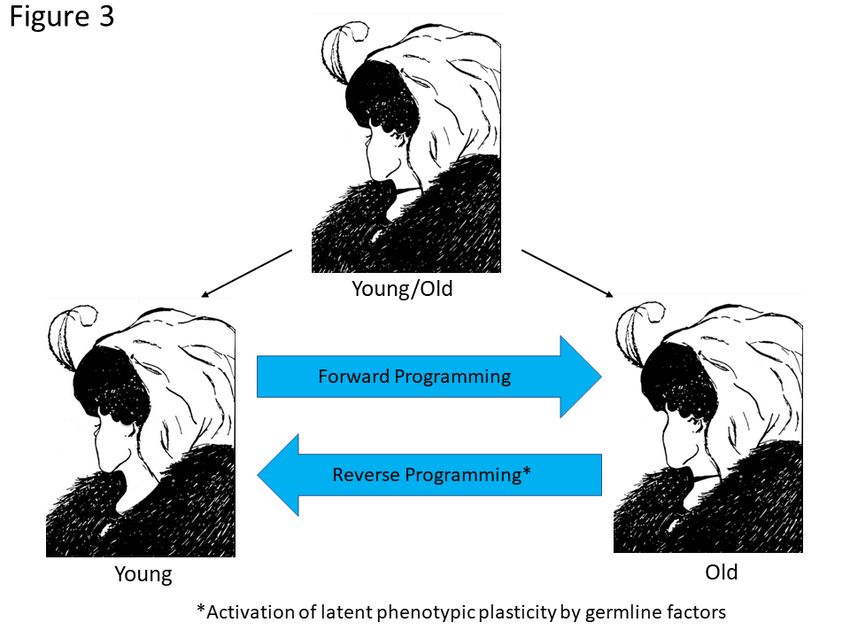
Figure 3. The two faces of epigenetic aging. The top illustration is an illusion designed so that the
viewer will see either a young woman or an old woman. However, both are present in the same
figure just as the young and old woman are present in the same person as the genome’s epigenetic
potential. Forward programming takes the young woman through a series of epigenetic states to
an old woman as the DNAm and other cellular clocks tick forward. Reverse programming can
potentially be achieved using germline factors to revert the genome to an earlier epigenetic age.Preprints (www.preprints.org) | NOT PEER-REVIEWED | Posted: 14 April 2021 doi:10.20944/preprints202104.0384.v1
Figure 4. Age reversal precedes reversion of developmental state during reprogramming of fibro-
blasts using OKSM germline factors. Post EFT genes (COX7A1, ADIRF) are rapidly reduced dur-
ing early (partial) reprogramming (day 3-20) while a linear decrease in DNAm age is occurring.
Pre-EFT genes are turned on at day 20 when DNAm age has gone to zero and their expression
increases as cells reverse their developmental state to full pluripotency (≥28 days). The data sug-
gest the possibility of age reprogramming without permanent loss of cell identity. Figure is
adapted from Olova et al. Figure 1 (162)
Initial attempts at in-vivo reprogramming using 4-factor (OKSM) reprogramming
were not encouraging because they resulted in extensive tumor formation (164, 165).
However, Ocampo et al. later reported reversal of epigenetic aging markers with no signs
of tumor development in transgenic OKSM mice when the factors are induced at a lower
dose and on a cyclic schedule of 2 days on and 5 days off (166). Instead of tumor for-
mation, cyclic OKSM induction led to amelioration of the premature aging phenotype and
extension of lifespan in a mouse model of HGPS. They further demonstrated that cyclic
OKSM induction resulted in greater resistance to metabolic disease following pancreatic
injury and increased repair of skeletal muscle damage in physiologically aged mice. Ad-
ditional in-vivo studies have shown reduced scar formation of skin wounds using viral
vector-mediated partial reprogramming and protection against liver damage using a
small molecule approach (167, 168). The question remained, however, as to whether par-
tial reprogramming in vivo reversed the DNAm clock. Two recent studies provide evi-
dence suggesting that a reversal of the DNAm clock during in vivo reprogramming may
be possible (169, 170). In one study, transient expression of 6 reprogramming factors,
OCT4, SOX2, KLF4, LIN28, c-MYC, and NANOG (OSKLMN), was obtained using mRNA
transfection which resulted in a reversion of fibroblasts and endothelial cells to a younger
phenotype as measured by epigenomic markers (tri-methylation of lysine 9 on histone 3
(H3K9me3), HP1γ, and lamina-associated polypeptide 2α (LAP2α)), mitochondrial fit-
ness, autophagy, transcriptome and DNAm clock (170). Transient expression of OSK-
LMN mitigated the inflammatory phenotype of osteoarthritic chondrocytes in culture and
implants of ex-vivo OSKLMN treated aged mouse or human skeletal muscle stem cells
resulted in rejuvenated muscle regenerative response following injury in physiologically
aged mice with no signs of tumor formation (170). In another in-vivo study, only 3 fac-
tors (OKS) were delivered to mice using an inducible adeno-associated virus vector
(AAV). The results indicate that induction of OKS resulted in protection of retinal gan-
glia cells and regeneration of axons in an optic nerve crush injury model, and the same
treatment restored vision in a mouse model of glaucoma (169). Importantly, the DNAm
age was reversed by the treatment in both models and the regenerative effects were Tet1You can also read