Non-invasive sampling in Itatiaia National Park, Brazil: wild mammal parasite detection - BMC Veterinary Research
←
→
Page content transcription
If your browser does not render page correctly, please read the page content below
Dib et al. BMC Veterinary Research    (2020) 16:295
https://doi.org/10.1186/s12917-020-02490-5

 RESEARCH ARTICLE                                                                                                                                    Open Access

Non-invasive sampling in Itatiaia National
Park, Brazil: wild mammal parasite
detection
Laís Verdan Dib1, João Pedro Siqueira Palmer1, Camila de Souza Carvalho Class1, Jessica Lima Pinheiro1,
Raissa Cristina Ferreira Ramos1, Claudijane Ramos dos Santos1, Ana Beatriz Monteiro Fonseca2,
Karen Gisele Rodríguez-Castro3, Camila Francisco Gonçalves3, Pedro Manoel Galetti Jr.3,
Otilio Machado Pereira Bastos1, Claudia Maria Antunes Uchôa1, Laís Lisboa Corrêa1,
Augusto Cezar Machado Pereira Bastos1, Maria Regina Reis Amendoeira4 and Alynne da Silva Barbosa1,4*

  Abstract
  Background: Non-invasive sampling through faecal collection is one of the most cost-effective alternatives for
  monitoring of free-living wild mammals, as it provides information on animal taxonomy as well as the dynamics of the
  gastrointestinal parasites that potentially infect these animals. In this context, this study aimed to perform an epidemiological
  survey of gastrointestinal parasites using non-invasive faecal samples from carnivores and artiodactyls identified by stool
  macroscopy, guard hair morphology and DNA sequencing in Itatiaia National Park. Between 2017 and 2018, faeces from
  carnivores and artiodactyls were collected along trails in the park. The host species were identified through macroscopic
  and trichological examinations and molecular biology. To investigate the parasites, the Faust, Lutz and modified Ritchie and
  Sheather techniques and enzyme immunoassays to detect Cryptosporidium sp. antigens were used.
  Results: A total of 244 stool samples were collected. The species identified were Chrysocyon brachyurus, Leopardus guttulus,
  Canis familiaris, Cerdocyon thous, Puma yagouaroundi, Leopardus pardalis, Puma concolor and Sus scrofa. There were 81.1%
  samples that were positive for parasites distributed mainly in the high part of the park. Helminths, especially eggs of the
  family Ascarididae, were more frequently detected in carnivore faeces (70.9%). Protozoa, especially Cryptosporidium sp.,
  represented the highest frequency of infection in artiodactyl faeces (87.1%). This zoonotic protozoon was detected in eight
  mammalian species, including in a wild boar. High values of structural richness and Shannon and Simpson diversity indices
  were observed for the parasites, especially in the faeces of C. brachyurus. Significant differences in parasite diversity were
  observed between wild and domestic animals, such as C. brachyurus and C. familiaris, respectively, and between
  taxonomically distant species, such as C. brachyurus and S. scrofa. The highest values for parasite similarity were found
  among the species that frequented similar areas of the park, such as C. brachyurus and L. guttulus.
  (Continued on next page)

* Correspondence: alynnedsb@gmail.com
1
 Department of Microbiology and Parasitology, Laboratory of Parasitology,
Federal Fluminense University, Biomedical Institute, Professor Hernani Mello
Street, São Domingos, Niterói, Rio de Janeiro 24210-130, Brazil
4
 Laboratory of Toxoplasmosis and Other Protozoan Diseases, Oswaldo Cruz
Foundation (Fiocruz, Rio de Janeiro), Oswaldo Cruz Institute, Avenue Brazil,
4365, Manguinhos, Rio de Janeiro 21040-360, Brazil
Full list of author information is available at the end of the article

                                         © The Author(s). 2020 Open Access This article is licensed under a Creative Commons Attribution 4.0 International License,
                                         which permits use, sharing, adaptation, distribution and reproduction in any medium or format, as long as you give
                                         appropriate credit to the original author(s) and the source, provide a link to the Creative Commons licence, and indicate if
                                         changes were made. The images or other third party material in this article are included in the article's Creative Commons
                                         licence, unless indicated otherwise in a credit line to the material. If material is not included in the article's Creative Commons
                                         licence and your intended use is not permitted by statutory regulation or exceeds the permitted use, you will need to obtain
                                         permission directly from the copyright holder. To view a copy of this licence, visit http://creativecommons.org/licenses/by/4.0/.
                                         The Creative Commons Public Domain Dedication waiver (http://creativecommons.org/publicdomain/zero/1.0/) applies to the
                                         data made available in this article, unless otherwise stated in a credit line to the data.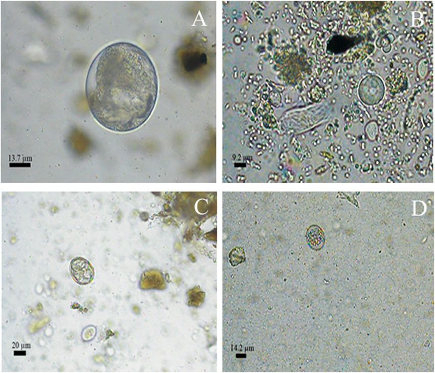
Dib et al. BMC Veterinary Research   (2020) 16:295                                                               Page 2 of 21

 (Continued from previous page)
 Conclusions: The animals and parasite infections were identified through the combination of three techniques. High
 frequency parasite structures were diagnosed. Zoonotic protozoa were found and mainly occurred in samples from
 introduced species.
 Keywords: Gastrointestinal parasites, Wild animals, Coproparasitologic, Trichology, DNA sequencing

Background                                                      from non-invasive samples and to correlate it to parasite
Over the years, wild mammalian fauna have been declin-          biodiversity, it is essential to precisely identify the host,
ing around the world for different reasons, including ve-       which is possible through the association of faecal macro-
hicle fatalities, agricultural frontier expansion, pasture      scopy, trichology of guard hairs (syn: overhairs), and DNA
formation, deforestation, environmental pollution and           sequencing.
fur trafficking [1, 2]. Another factor that may result in          The use of morphological analysis to study faeces from
declines in mammalian fauna is the parasite load of dif-        free-living wild animals is very important since it serves
ferent aetiological agents, such as gastrointestinal para-      as an initial screening of the samples to be collected,
sites. These agents infect a myriad of hosts and make           allowing reliable host taxonomic classification at the
important alterations to community structure, directly          order level [17]. On the other hand, the trichology of
impacting biodiversity and ecosystem dynamics [3]. The          mammalian guard hairs can provide specific information
susceptibility of hosts to these infections is related to       about the host species by associating the guard hair
their phylogenetic proximity, body morphology and dietary       colour pattern (macroscopic guard-hair morphology)
habits [4]. Parasitism may result in weight loss, metabolic     and the analysis of its cuticle and medullary designs
imbalance, reproductive disorders, anaemia, dehydration,        (microscopic guard-hair morphology). Moreover, DNA
foetal malformations, locomotor injuries, and even death        sequencing is widely used in studies of free-living wild
among wild animals [5, 6]. When parasitized, many wild          mammals; similar to trichology, it can also identify the
mammals end up presenting behavioural and functional            host species.
alterations in their niche [7–9]. Therefore, studies on the        To avoid possible ecological imbalances, it is import-
prevalence of parasites in both wild and sympatric domestic     ant to perform constant monitoring of the wildlife in
animals are important for better understanding the possible     conservation areas. In this context and while any para-
effects on wildlife, the parasitic distribution dynamics        sitological study was performed with mammals in the
among the hosts and possible parasitic sources for fauna.       first Brazilian national park, this study aimed to perform
   There are three main types of sampling for wild mam-         an epidemiological survey of gastrointestinal parasites in
mal studies: destructive sampling from dead animals;            non-invasive faecal samples from carnivores and artio-
non-destructive sampling from captured animals; and             dactyls identified by stool macroscopy, guard-hair trich-
non-invasive sampling, in which samples, such as traces         ology and DNA sequencing in Itatiaia National Park
in the environment, loose hair or feathers, faeces and          (PNI).
other remnants, are obtained without catching or hand-
ling the animal [10, 11]. It is worth mentioning that           Results
non-invasive sampling has become an alternative strat-          Host identification
egy for species monitoring and conservation, especially         Three identification techniques, macroscopy, trichology
for those species with low population density and elusive       and DNA sequencing, were used to analyse 244 faecal
nocturnal habits, such as carnivores [12]. Through non-         samples (S1, supplementary material and Table 1). It
invasive faecal sampling, it is possible to obtain data         was possible to confirm eight mammal species, which in-
about biodiversity, including identification of the species     cluded seven carnivores and one artiodactyl. The host
inhabiting a region, their diet composition and their role      species were identified for 180 of the samples. However,
in the ecosystem, as well as information about poten-           the three techniques did not achieve the same results in
tially infectious gastrointestinal parasites of these ani-      110 of the samples for which the species were identified.
mals [13].                                                        Therefore, Pearson’s correlation was performed and
   Brazil harbours high mammal biodiversity that includes       established a very strong relationship (ρ > 0.9) in most
different species of carnivores and artiodactyls [14], many     cases for the information associated with the C. bra-
of which are endangered [15]. However, very little is           chyurus and L. guttulus samples and a moderate rela-
known about the parasite distribution in Brazilian wild         tionship (0.5 ≤ ρ ≥ 0.7) for the P. yagouaroundi samples.
mammal fauna, especially in conservation areas [16]. To         The other samples in this group did not show significant
obtain reliable information about the mammalian fauna           degrees of correlation (ρ < 0.5). After associating the
Dib et al. BMC Veterinary Research     (2020) 16:295                                                                               Page 3 of 21

Table 1 Hosts classification based on the association of faecal macroscopy, guard hair trichology and DNA sequencing
Taxonomy                     Macroscopy           Trichology            DNA sequencing              Pearson’s correlation   Total fecal samples
                                                                                                    coeficiente (ρ)         (n = 244)
Order Carnivora                                                                                                             168 (68.8%)
  Family Canidae                                                                                                            112 (45.9%)
    Chrysocyon brachyurus                                                                                                   97 (39.7%)
                                                                                                    c
                             Order Carnivora      C. brachyurus         C. brachyurus                                       42
                             Order Carnivora      Family Mustelidae     C. brachyurus               0.95                    11
                             Order Carnivora      Family Mephitidae     C. brachyurus               0.95                    2
                             Order Carnivora      C. brachyurus         Low quality gene sequence   0.97                    6
                                                                                                    c
                             Order Carnivora      Family Canidae        C. brachyurus                                       1
                             Order Carnivora      Absent guard hair     C. brachyurus               0.95                    24
                                                                                                    c
                             Family Canidae       C. brachyurus         C. brachyurus                                       3
                             Family Felidae       C. brachyurus         C. brachyurus               0.99                    7
                             Family Felidae       Family Canidae        C. brachyurus               –                       1
    Canis familiaris                                                                                                        13 (5.3%)
                                                                                                    c
                             Order Carnivora      Family Canidae        C. familiaris                                       8
                             Order Carnivora      Procyon cancrivorus   C. familiaris               –                       1
                             Order Carnivora      Family Mephitidae     C. familiaris               –                       1
                             Order Carnivora      Absent guard hair     C. familiaris               –                       2
                                                                                                    c
                             Family Canidae       Family Canidae        C. familiaris                                       1
    Cerdocyon thous                                                                                                         2 (0.8%)
                                                                                                    c
                             Order Carnivora      C. thous              C. thous                                            1
                             Order Carnivora      Absent guard hair     C. thous                    –                       1
  Family Felidae                                                                                                            56 (22.9%)
    Leopardus guttulus                                                                                                      52 (21.3%)
                                                                                                    c
                             Family Felidae       L. guttulus           L. guttulus                                         3
                             Family Felidae       L. guttulus           Low quality gene sequence   0.97                    1
                             Family Felidae       Absent guard hair     L. guttulus                 0.94                    24
                             Family Felidae       Family Canidae        L. guttulus                 0.94                    6
                             Family Felidae       Procyon cancrivorus   L. guttulus                 0.94                    2
                             Family Felidae       Family Mustelidae     L. guttulus                 0.94                    2
                             Family Felidae       Family Mephitidae     L. guttulus                 0.94                    5
                             Order Carnivora      Absent guard hair     L. guttulus                 –                       7
                             Order Carnivora      L. pardalis           L. guttulus                 –                       1
                             Order Carnivora      P. yagouaroundi       L. guttulus                 –                       1
    Puma yagouaroundi                                                                                                       2 (0.8%)
                             Family Felidae       P. yagouaroundi       Low quality gene sequence   0.68                    2
    Leopardus pardalis                                                                                                      1 (0.4%)
                             Order Carnivora      L. pardalis           Low quality gene sequence   –                       1
    Puma concolor                                                                                                           1 (0.4%)
                             Order Carnivora      Família Mustelidae    P. concolor                 –                       1
Order Artiodactyla                                                                                                          31 (12.7%)
  Family Suidae                                                                                                             12 (4.9%)
    Sus scrofa                                                                                                              12 (4.9%)
                             Order Artiodactyla   Absent guard hair     S. scrofa                   –                       1
                                                                                                    c
                             Order Artiodactyla   Order Artiodactyla    S. scrofa                                           11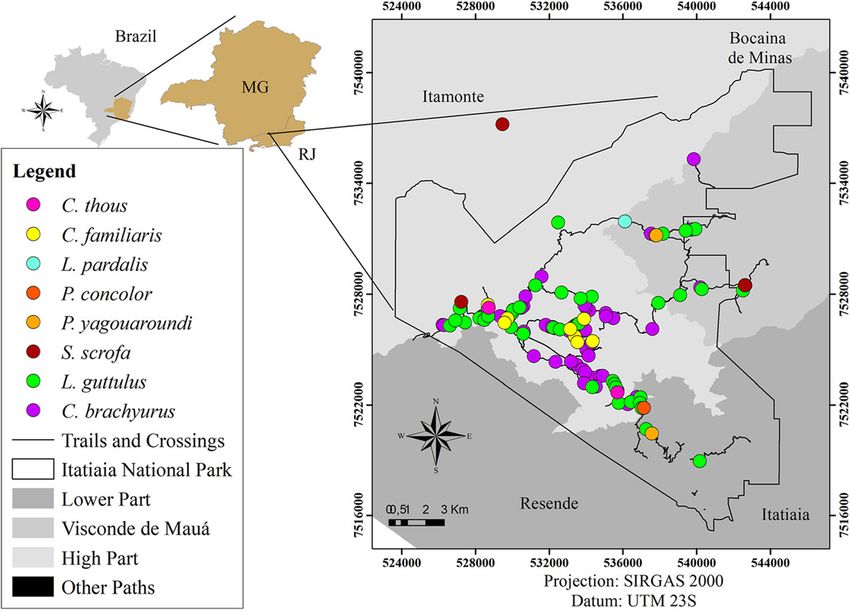
Dib et al. BMC Veterinary Research            (2020) 16:295                                                                                        Page 4 of 21

Table 1 Hosts classification based on the association of faecal macroscopy, guard hair trichology and DNA sequencing (Continued)
Taxonomy                          Macroscopy             Trichology              DNA sequencing                   Total fecal samples (n = 244)
Order Carnivora                                                                                                   168 (68.8%)
     Order Carnivora USa                                                                                          45 (18.4%)
                                  Order Carnivora        L. guttulus             C. familiaris                    1
                                  Order Carnivora        L. pardalis             C. brachyurus                    2
                                  Order Carnivora        P. yagouaroundi         C. brachyurus                    1
                                  Order Carnivora        Absent guard hair       Low quality gene sequence        9
                                  Order Carnivora        Family Mephitidae       Low quality gene sequence        1
                                  Order Carnivora        C. brachyurus           L. guttulus                      5
                                  Order Carnivora        Leopardus wiedii        C. thous                         1
                                  Order Carnivora        Nasua nasua             Low quality gene sequence        1
                                  Order Carnivora        Family Mustelidae       Low quality gene sequence        1
                                  Order Carnivora        Family Canidae          Low quality gene sequence        4
                                  Family Felidae         C. brachyurus           L. guttulus                      1
                                  Family Felidae         Family Canidae          Low quality gene sequence        3
                                  Family Felidae         Nasua nasua             Low quality gene sequence        1
                                  Family Felidae         Family Mustelidae       Low quality gene sequence        3
                                  Family Felidae         Absent guard hair       Low quality gene sequence        6
                                  Family Felidae         Family Mephitidae       Low quality gene sequence        4
                                  Family Felidae         C. brachyurus           Low quality gene sequence        1
Order Artiodactyla                                                                                                31 (12.7%)
     Order Artiodactyla USb                                                                                       19 (7.8%)
                                  Order Artiodactyla     Absent guard hair       Low quality gene sequence        6
                                  Order Artiodactyla     Order Artiodactyla      Low quality gene sequence        13
a
    Order Carnivora Unidentified Species; b Order Artiodactyla Unidentified Species. c Total agreement among the three identification techniques

three identification techniques, a final classification of                           gastrointestinal parasites was 27 samples (87.1%).
host species was conducted using the highlighted                                     Antigens of Cryptosporidium sp. were the most frequently
information (Table 1).                                                               detected structure, followed by cysts of Balantioides coli
                                                                                     and nematode larvae (Table 2).
Parasitological diagnosis                                                              Out of the 244 faecal samples retrieved from the park,
In the faeces of the animals, several parasitic taxa were                            most were identified as belonging to C. brachyurus (97)
identified, including the phyla Nematoda, Platyhelminthes                            and L. guttulus (52). Among these, structures of gastro-
and Protozoa. These were characterized into morphotypes                              intestinal parasites were detected in 79 (81.4%) samples
by their different sizes, colours and shapes. Out of the 244                         from C. brachyurus and in 43 (82.7%) from L. guttulus
faecal samples collected, structures of gastrointestinal                             (Figs. 1 and 2). Such parasites were also observed in other
parasites were revealed in 198 (81.1%) through the                                   faecal samples of carnivores and artiodactyls that had been
combined use of microscopic coproparasitological tech-                               collected along the park trails (Figs. 1 and 2).
niques and ELISA. In general, helminths were observed                                  In addition, a difference in the distribution of parasitic
more frequently than protozoa and were mainly seen in                                structures can be observed in the three areas of the park.
faeces from carnivores. The inverse was observed in                                  The High Part was the region where all parasitic taxa were
relation to the stool samples from artiodactyls (Table 2).                           detected, except for Eimeria sp. However, the Lower Part
  Among the 213 faecal samples from animals of the                                   was the region that presented the least diversity of
order Carnivora that were analysed, 171 (80.3%) showed                               parasitic structures, being detected 7 out of 15 taxa. In
structures from gastrointestinal parasites, among which                              Visconde de Mauá, 10 out of 15 taxa were detected,
eggs of the families Ascarididae and Diphyllobothriidae                              highlighting the presence of antigens of Cryptosporidium
and coproantigens of Cryptosporidium sp. can be                                      sp. which was quite evident (Fig. 3).
highlighted. Among the samples from the animals of the                                 Eggs of the family Ascarididae, especially those classified
order Artiodactyla, the total number positive for                                    as morphotype 1, that were similar to Toxocara sp. were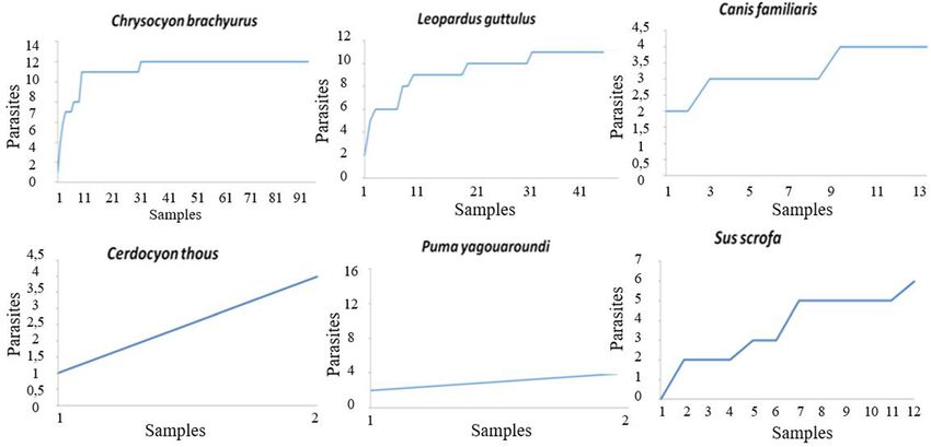
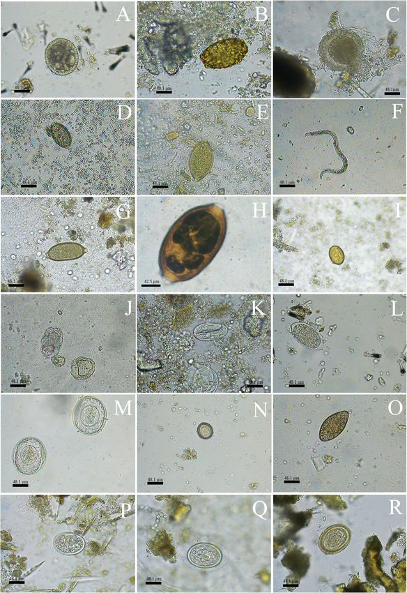
Dib et al. BMC Veterinary Research       (2020) 16:295                                                                          Page 5 of 21

Table 2 Frequency of gastrointestinal parasites in carnivorous and artiodactyls faecal samples surveyed in Itatiaia National Park, Brazil
Helminth and protozoan structures                        Order Carnivora (n = 213)           Order Artiodactyla (n = 31)       Total (n = 244)
Helminths
  Family Ascarididae                                     71 (33.3%)                          4 (12.9%)                         75 (30.8%)
  Trichuris sp.                                          31 (14.5%)                          –                                 31 (12.7%)
  Capillaria sp.                                         29 (13.6%)                          –                                 29 (11.9%)
  Nematode larvae                                        25 (11.7%)                          6 (19.4%)                         31 (12.7%)
  Thin-shelled nematode egg                              21 (9.8%)                           3 (9.7%)                          24 (9.8%)
  Physaloptera sp.                                       12 (5.6%)                           –                                 12 (4.9%)
  Family Diphyllobothriidae                              52 (24.1%)                          –                                 52 (21.3%)
  Order Cyclophyllidea                                   8 (3.7%)                            –                                 8 (3.3%)
  Family Dicrocoeliidae                                  10 (4.7%)                           –                                 10 (4.1%)
  Phylum Acanthocephala                                  1 (0.5%)                            –                                 1 (0.4%)
  Subtotal of helminths positive samples                 151 (70.9%)                         10 (32.2%)                        161 (66%)
Protozoan
  Non-sporulated coccidian                               10 (4.7%)                           –                                 10 (4.1%)
  Eimeria sp.                                            –                                   1 (3.2%)                          1 (0.4%)
  Balantioides coli                                      –                                   6 (19.4%)                         6 (2.4%)
  Amoebae                                                3 (1.4%)                            –                                 3 (1.2%)
  Coproantigens of Cryptosporidium sp.                   42 (19.7%)                          25 (80.6%)                        67 (27.4%)
  Subtotal of protozoan positive samples                 54 (25.3%)                          27 (87.1%)                        81 (33.2%)
Total of positive samples                                171 (80.3%)                         27 (87.1%)                        198 (81.1%)

detected mainly in faecal samples from carnivores, except                     faeces from C. brachyurus, C. familiaris and L. guttulus,
from L. pardalis and P. concolor. Parasite structures that                    and morphotype 2 eggs were detected in samples from L.
matched the typical morphology of infertile Ascaris eggs                      guttulus, C. thous and S. scrofa. Thin-shelled eggs with
(morphotype 2) were only observed in samples from S.                          tapered ends were diagnosed in the faeces of both C.
scrofa. A third morphotype of ascarid was detected in 11.9%                   familiaris and unidentified artiodactyls (Table 3 and Fig. 4).
of the samples, including the faeces from C. brachyurus, C.                     In the faeces from carnivores, eggs of other helminths,
familiaris and L. guttulus (Table 3 and Fig. 4).                              such as Physaloptera sp. and the family Dicrocoeliidae were
  Eggs of the family Diphyllobothriidae were the second                       also observed. Cestode eggs of the order Cyclophyllidea,
most frequent parasite among the helminths and were                           which were classified as morphotype 1, were detected in
detected only in the faecal samples from carnivores.                          2.9% of the faeces analysed. Eggs from the family Taeniidae,
Nematode larvae and Trichuris sp. eggs were observed in                       which were named morphotype 2, were detected in one
12.7% of the stool samples. Nematode larvae were                              sample from C. brachyurus. In a faecal sample that was
detected in faeces from both carnivores and artiodactyls                      positive for eggs of the phylum Acanthocephala, the host
and were not classified into different morphotypes. Eggs                      was only characterized down to the taxonomic group of the
of Trichuris sp. were only diagnosed in samples from                          order Carnivora (Table 3 and Fig. 4).
carnivores, and these were morphologically classified as                        Among the protozoa detected, Cryptosporidium sp. was
morphotypes 1 and 2. Capillaria sp. eggs were diagnosed                       diagnosed through antigens in the faeces of all animals that
in 11.9% of the faecal samples analysed. These were                           were identified to the species level except for P. concolor
classified into morphotypes 1 and 2 and were detected in                      (Fig. 2). Unsporulated coccidia oocysts and tetranucleated
faeces from C. brachyurus, L. guttulus, P. yagouaroundi                       amoeba cysts were detected in faeces that were identified as
and L. pardalis (Table 3 and Fig. 4).                                         from C. brachyurus, and the latter were also found in faeces
  Thin-shelled nematode eggs were observed in 9.8% of                         from L. guttulus. Sporulated coccidian oocysts with the typ-
the samples. Among these, the eggs were classified into                       ical morphological pattern of Eimeria sp. and Balantioides
morphotype 1, which were similar to those of the super-                       coli cysts were detected only in faeces from artiodactyls
family Strongyloidea, and morphotype 2, which were                            (Table 3 and Fig. 5). All different morphotypes of protozoa
similar to strongylids (superfamilies Trichostrongyloidea                     structures detected presented similar morphology and
and Strongyloidea). Morphotype 1 eggs were detected in                        varied only in size.
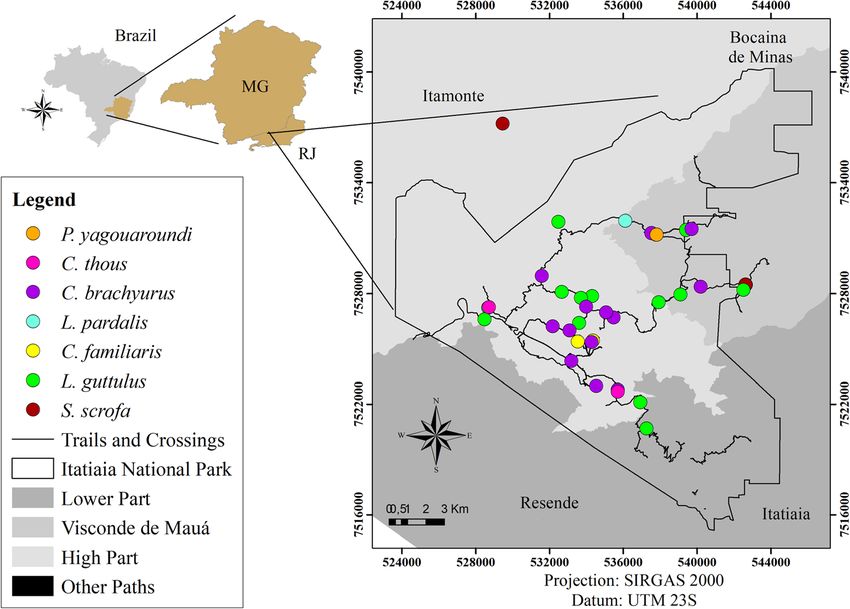
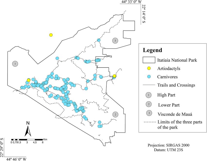
Dib et al. BMC Veterinary Research        (2020) 16:295                                                                                  Page 6 of 21

 Fig. 1 Collection points for faecal samples positive for parasites in Itatiaia National Park. (Arcgis version 10.5)

  Regarding parasitic associations, polyparasitism was                           families Ascarididae and Diphyllobothriidae and between
observed in 94 samples (38.5%), which had two to eight                           eggs of Ascarididae and Trichuris sp.; both combinations
parasite structures. Associations were found between                             were present in five samples from carnivores (2.3%). In
helminths alone, between helminths and protozoa and                              the faeces from artiodactyls, the most frequent association
between protozoa alone. The most frequently detected                             occurred between nematode larvae and Cryptosporidium
parasitic associations occurred between eggs of the                              sp. coproantigens in three samples (9.7%).

 Fig. 2 Collection points for faecal samples from carnivores and an artiodactyl positive for Cryptosporidium sp. (Arcgis version 10.5)
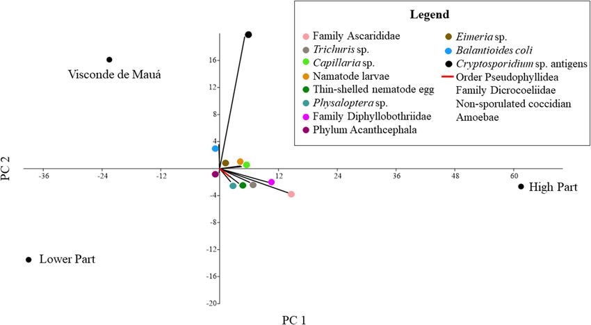
Dib et al. BMC Veterinary Research       (2020) 16:295                                                                                      Page 7 of 21

 Fig. 3 Distribution of parasitic taxa detected in fecal samples of carnivores and artiodactyl in the three parts of the park: High Part, Lower Part
 and Visconde de Mauá through Principal component analysis (Past version 3.2.2)

  To analyse the richness, diversity, and similarity indices,                 Discussion
the sample sufficiency for each host species (relationship                    From the association of the macroscopic, trichological and
between the faecal samples and the different parasitic taxa                   DNA sequencing results, a final identification of the host
detected) was analysed and plotted on accumulation                            species was obtained. Through macroscopic examination
curves. The accumulation curves of C. brachyurus, L.                          of the faeces, problems in identification occurred with
guttulus, and C. familiaris stabilized, which means that                      canid samples when seeds, which are common in the diet
the number of samples recovered for each of these hosts                       of neotropical canids such as C. brachyurus, were not
was enough to estimate the richness and diversity indices                     observed. In addition, there is no consensus among
of the parasitic fauna. It was not possible to establish                      authors regarding the shape of mammalian stool. Thus, to
accumulation curves for L. pardalis and P. concolor since                     minimize misidentification of the host by macroscopy, the
only one faecal sample was collected from each of these                       samples were classified only into high-level taxonomic
species (Fig. 6).                                                             categories, such as order, in most cases. Despite the low
  The faecal samples from C. brachyurus and L. guttulus                       resolution of host identification, this is a low-cost technique
presented the highest richness and were positive for a                        and can be used by any researcher during field work.
great number of different parasite taxa, and similarity, as                      In this study, host species were not identified through
demonstrated by the parasitic likeness between the hosts                      trichological analysis in most of the samples. As pointed
(Table 4 and Fig. 7). However, despite the detection of                       out at Serra dos Órgãos National Park, one of the limita-
common parasites between artiodactyls and carnivores,                         tions in mammalian identification through trichology is
many agents were detected in only one of these hosts,                         that faeces often presents many hairs, but not all are guard
which justified the low parasite similarity index between                     hairs [16]. In addition, some guard hair had deteriorated,
them (Fig. 7). In the pooled t test, the Shannon diversity                    making it impossible to assess their cuticles. In this case,
index (H′) of the parasites was significant (p < 0.05)                        since only the medullary patterns were observed, it was
between C. brachyurus and C. familiaris and between S.                        only possible to identify the animals down to the family
scrofa and C. brachyurus (Table 5). Thus, it was demon-                       level, such as the family Canidae. On the other hand, the
strated that the parasitic taxa and their distribution                        guard hairs recovered from artiodactyl samples did not
differed considerably among hosts.                                            have cuticles, and only the internal layer was composed of
Table 3 Frequency of gastrointestinal parasite morphotypes in mammal faecal samples from Itatiaia National Park, Brazil
Morphotypes of helminth and protozoan structures           Chrysocyon   Canis        Cerdocyon       Leopardus    Puma           Leopardus   Puma concolor   Order Carnivora   Sus         Order Artiodactyla   Total (n = 244)
                                                           brachyurus   familiaris   thous (n = 2)   guttulus     yagouaroundi   pardalis    (n = 1)         US (n = 45)       scrofa      US (n = 19)
                                                           (n = 97)     (n = 13)                     (n = 52)     (n = 2)        (n = 1)                                       (n = 12)
Helminths
  Family Ascarididae                                       25 (25.8%)   4 (30.8%)    1 (50%)         20 (38.5%)   1 (50%)        –           –               21 (46.7%)        4 (33.3%)   –                    75 (30.8%)
    Morphotype 1 - C: 92.5 ± 5.8 × 81.4 × 7.1;             20 (20.6%)   2 (15.4%)    1 (50%)         19 (36.5%)   1 (50%)        –           –               15 (33.3%)        3 (25%)     –                    61 (25%)
    F: 92.5 ± 6 × 81.4 × 6.2; S: 88.8 ± 8.7 × 85.1 × 7.6
    Morphotype 2 - C: 111 ± 18.5 × 107.3 ± 23.2;           –            –            –               –            –              –           –               –                 1 (8.3%)    –                    1 (0.4%)
    F: 107.3 ± 8.9 × 83.2 ± 12.9
                                                                                                                                                                                                                                  Dib et al. BMC Veterinary Research

    Morphotype 3 - S: 114.7 ± 4.8 × 74 ± 1.6               11 (11.3%)   3 (23.1%)    –               10 (19.2%)   –              –           –               4 (8.9%)          –           –                    29 (11.9%)
  Trichuris sp.                                            19 (19.6%)   –            –               4 (7.7%)     2 (100%)       –           –               6 (13.3%)         –           –                    31 (12.7%)
    Morphotype 1 - C: 88.8 ± 8 × 37 ± 2.8;                 12 (12.4%)   –            –               1 (1.9%)     2 (100%)       –           –               5 (11.1%)         –           –                    18 (7.4%)
    F: 81.4 ± 4.5 × 40.7 ± 4.2
    Morphotype 2 - C: 107.3 ± 7.3 × 40.7 ± 4.9;            10 (10.3%)   –            –               4 (7.7%)     –              –           –               2 (4.4%)          –           –                    18 (7.4%)
    F: 107.3 ± 7.6 × 42.5 ± 3.7
                                                                                                                                                                                                                                    (2020) 16:295

  Capillaria sp.                                           14 (14.4%)   –            –               11 (21.1%)   1 (50%)        1 (100%)    –               2 (4.4%)          –           –                    29 (11.9%)
    Morphotype 1 - C: 85.1 ± 5.1 × 44.4 ± 4.9;             8 (8.2%)     –            –               6 (11.5%)    1 (50%)        1 (100%)    –               –                 –           –                    15 (6.1%)
    F: 85.1 ± 6.4 × 44.4 ± 3.9
    Morphotype 2 - C:99.9 ± 8.1 × 44.4 ± 6.1;              9 (9.3%)     –            –               10 (19.2%)   –              1 (100%)    –               2 (4.4%)          –           –                    23 (9.4%)
    F: 101.7 ± 5.6 × 51.8 ± 6.5
  Nematode larvae                                          13 (13.4%)   –            –               5 (9.6%)     –              –           1 (100%)        6 (13.3%)         1 (8.3%)    5 (26.3%)            31 (12.7%)
  Thin-shelled nematode egg                                9 (9.3%)     3 (23.1%)    1 (50%)         4 (7.7%)     –              –           –               4 (8.9%)          2 (16.7%)   1 (5.3%)             24 (9.8%)
    Morphotype 1 - C: 85.1 ± 4.2 × 55.5 ± 3.7;             7 (7.2%)     1 (7.7%)     –               1 (1.9%)     –              –           –               3 (6.7%)          –           –                    12 (4.9%)
    F: 96.2 × 40.7
    Morphotype 2 - C: 107.3 ± 5.5 × 57.3 ± 4.8;            2 (2.1%)     1 (7.7%)     1 (50%)         3 (5.8%)     –              –           –               1 (2.2%)          2 (16.7%)   –                    10 (4.1%)
    F: 109.1 ± 2.6 × 85.1 ± 31.4; S: 172 ± 29.8
    × 79.5 ± 10.6
    Morphotype 3 - C: 92.5 ± 6.1 × 55.5 ± 5.6              –            1 (7.7%)     –               –            –              –           –               –                 –           1 (5.3%)             2 (0.8%)
  Physaloptera sp.- C: 70.3 ± 10 × 46.2 ± 9.7;             9 (9.3%)     –            –               1 (1.9%)     –              –           –               2 (4.4%)          –           –                    12 (4.9%)
  F: 70.3 × 51.8
  Family Diphyllobothriidae - C:88.8 ± 9.3 ×               31 (32%)     –            1 (50%)         10 (19.2%)   –              –           –               10 (22.2%)        –           –                    52 (21.3%)
  48.1 ± 6.3; F: 85.1 ± 7.1 × 48.1 ± 4
  Order Cyclophyllidea                                     4 (4.1%)     1 (7.7%)     –               1 (1.9%)     –              –           –               2 (4.4%)          –           –                    8 (3.3%)
    Morphotype 1 - C: 83.2 ± 8.1 × 77.7 ± 12.3;            3 (3.1%)     1 (7.7%)     –               1 (1.9%)     –              –           –               2 (4.4%)          –           –                    7 (2.9%)
    F: 81.4 ± 15.7 × 75.8 ± 7.8
    Morphotype 2 - C: 48.1 × 37                            1 (1%)       –            –               –            –              –           –               –                 –           –                    1 (0.4%)
  Family Dicrocoeliidae - C: 55.5 ± 11.3 × 33.3            5 (5.1%)     –            –               3 (5.8%)     –              –           –               2 (4.4%)          –           –                    10 (4.1%)
  ± 3.7; F: 51.8 ± 1.5 × 33.3 ± 3.3
  Phylum Acanthocephala                                    –            –            –               –            –              –           –               1 (2.2%)          –           –                    1 (0.4%)
    Morphotype 1–88.8 ± 9.5 × 74 ± 3.3                     –            –            –               –            –              –           –               1 (2.2%)          –           –                    1 (0.4%)
    Morphotype 2–85.1 × 66.7                               –            –            –               –            –              –           –               1 (2.2%)          –           –                    1 (0.4%)
  Helminths positive samples                               72 (74.2%)   7 (53.8%)    1 (50%)         35 (67.3%)   2 (100%)       1 (100%)    1 (100%)        31 (68.9%)        5 (41.7%)   5 (26.3%)            161 (66%)
                                                                                                                                                                                                                                     Page 8 of 21
Table 3 Frequency of gastrointestinal parasite morphotypes in mammal faecal samples from Itatiaia National Park, Brazil (Continued)
Morphotypes of helminth and protozoan structures Chrysocyon Canis Cerdocyon Leopardus Puma Leopardus Puma concolor Order Carnivora Sus Order Artiodactyla Total (n = 244)
brachyurus familiaris thous (n = 2) guttulus yagouaroundi pardalis (n = 1) US (n = 45) scrofa US (n = 19)
(n = 97) (n = 13) (n = 52) (n = 2) (n = 1) (n = 12)
Protozoa
Non-sporulated coccidian 9 (9.3%) – – – – – – 1 (2.2%) – – 10 (4.1%)
Morphotype 1 - C: 25.9 ± 2.4 × 22.2 ± 2.5 7 (7.2%) – – – – – – – – – 7 (2.9%)
Morphotype 2 - C: 37 ± 6.9 × 33.3 ± 5.6 5 (5.1%) – – – – – – – – – 5 (2%)
Eimeria sp. - S: 37 × 37 – – – – – – – – 1 (8.3%) – 1 (0.4%)
Dib et al. BMC Veterinary Research
Balantioides coli - S: 48.1 ± 38.8 × 44.4 ± 35.5 – – – – – – – – 2 (16.7%) 4 (21%) 6 (2.4%)
Amoebae - C: 18.5 ± 5.8 × 18.5 ± 3.6; F: 18.5 2 (2.1%) – – 1 (1.9%) – – – – – – 3 (1.2%)
± 3.2 × 18.5 ± 2
Cryptosporidium sp. coproantigens 13 (13.4%) 2 (15.4%) 2 (100%) 14 (26.9%) 1 (50%) 1 (100%) – 9 (20%) 10 (83.3%) 15 (78.9%) 67 (27.4%)
Protozoan positive samples 22 (22.7%) 2 (15.4%) 2 (100%) 15 (28.8%) 1 (50%) 1 (100%) – 10 (22.2%) 10 (83.3%) 17 (89.5%) 81 (33.2%)
Total positive samples 79 (81.4%) 8 (61.5%) 2 (100%) 43 (82.7%) 2 (200%) 1 (100%) 1 (100%) 35 (77.8%) 10 (83.3%) 17 (89.5%) 198 (81.1%)
(2020) 16:295
Page 9 of 21Dib et al. BMC Veterinary Research (2020) 16:295 Page 10 of 21 Fig. 4 Morphotypes of helminths eggs in 400 x (A to F; H to S) and 1000 x (G) detected in faecal samples of carnivores and artiodactyls form Itatiaia National Park, Brazil. a Ascarididae family 1. b Ascarididae family 2. c Ascarididae family 3. d Trichuris sp. 1. e Trichuris sp. 2. f Nematode larvae. g Capillaria sp. 1. h Capillaria sp. 2. i Dicrocoellidae family. j Thin-shelled nematode egg 1. k Thin-shelled nematode egg 2. l Thin-shelled nematode egg 3. m Cyclophyllidea order 1. n Cyclophyllidea order 2. o Diphyllobothriidae family. p Physaloptera sp. q Acanthocephala phylum 1. r Acanthocephala phylum 2 fibres. The loss of the cuticular layer may have oc- and allows a very specific taxonomic classification of curred during laboratory processing or by the passage the animal. of the guard hairs through the gastrointestinal tract. DNA sequencing was also used for host identification. It Nevertheless, the cuticle may have been rubbed off is important to highlight that genetic sequences do not when artiodactyls rubbed their bodies on natural ensure that it is the true host due to the possibility of substrates, such as trees. It is noteworthy that tricho- countermarking by other animals with territorial behav- logical processing is time-consuming, and the assess- iour. Nonetheless, the use of DNA sequencing may be the ment of guard hairs requires trained professionals. only alternative for the identification of S. scrofa, for which Moreover, it is important to make controls with faecal hair release in feces is not common. However, this guard hairs that were taken directly from animals to technique was the most expensive among those used in achieve greater accuracy in host identification from this study, and it does not always provide satisfactory re- the non-invasive samples. Nevertheless, this method- sults due to many faecal inhibitors. Thus, the association ology is also very advantageous since it is inexpensive among macroscopy, trichology, and DNA sequencing is
Dib et al. BMC Veterinary Research      (2020) 16:295                                                                                   Page 11 of 21

 Fig. 5 Morphotypes of protozoa cysts and oocysts in 1000 x detected in faecal samples of carnivores and artiodactyls form Itatiaia National Park,
 Brazil. a Balantioides coli. b Amoebae. c Eimeria sp. d Non-sporulated coccidia

essential to provide the most trustworthy identification                     been reported in other studies, varying from 58 to 74.7%
from non-invasive samples.                                                   for feline faeces from reserves and forests in Mexico
  The overall positive rate for gastrointestinal parasites                   [18–20], 75% for feline and artiodactyl faeces from a
was 81.1%. In this study, from all the 244 samples, the                      reserve in Bolivia [21], 53.3% for faeces from canids, felids,
positive rates were 36.5% for Canidae family, 19.3% for                      mustelids and procyonids collected in a reserve in Minas
Felidae and 4.1 for Suidae. Lower overall frequencies have                   Gerais, Brazil [22], and 70% for feline faeces found in a

 Fig. 6 Accumulation curves for gastrointestinal parasite structures detected in faecal samples from carnivores and an artiodactyl from Itatiaia
 National Park, Brazil
Dib et al. BMC Veterinary Research      (2020) 16:295                                                                    Page 12 of 21

Table 4 Richness and diversity of gastrointestinal parasites in          conditions that were favourable for maintenance of nema-
mammal faeces from Itatiaia National Park, Brazil                        tode, cestode, trematode and acanthocephalan life cycles.
Host                         Richness       Shannon (H′)       Simpson   Unlike in carnivore faecal samples, protozoa were the
Family Canidae                                                           most frequently detected parasites in the faeces of S.
  Chrysocyon brachyurus      12             2.2761             0.887     scrofa. Among the studies that analysed samples of free-
                                                                         living S. scrofa, Tayassu pecari and Pecari tajacu, only one
  Canis familiaris           4              1.2799             0.778
                                                                         conducted in Texas, USA, reported protozoa in faeces
  Cerdocyon thous            4              1.3297             0.867
                                                                         from pigs [30].
Family Felidae                                                              Eggs from the family Ascarididae were the most
  Leopardus guttulus         11             1.9662             0.850     frequently observed group in the faecal material of carniv-
  Puma yagouaroundi          4              1.3322             0.900     orous hosts. Most of the eggs detected were similar to
  Leopardus pardalis         2              0.6931             1         Toxocara sp. (morphotype 1) and were consistent with T.
                                                                         canis and T. cati. Brownish ascarid eggs with very thick
  Puma concolor              2              0.6931             1
                                                                         shells (morphotype 3) were also detected. These eggs may
Family Suidae
                                                                         have belonged to another species that has not yet been
  Sus scrofa                 6              1.4286             0.721     reported or may even have resulted from parasitic
                                                                         adaptation. In Itatiaia, the high positivity for Toxocara sp.
                                                                         may have been favoured due to the extreme resistance of
reserve in Espírito Santo, Brazil [23]. In contrast, the                 these ascarid eggs the park’s abundant edaphic environ-
frequency of parasites detected in faeces from Serra dos                 ment and humid tropical climate, which contribute
Órgãos National Park was 86.6%, which was slightly                       towards larval development within the egg. Moreover,
higher than that observed in Itatiaia National Park [16].                predation can be cited as a possible form of transmission
The high frequency of gastrointestinal parasites detected                due to paratenic hosts [24] and pseudoparasitism, in
may have occurred since these animals live in natural                    which the animal does not truly become infected.
environments that are rich in abiotic and biotic factors                    Eggs of the family Diphyllobothriidae were the second
that promote infection through contact with contami-                     most frequently detected parasite structures among
nated soil, water, food and infected prey.                               helminths in faeces from carnivores. Morphologically, the
  In general, helminths were more frequent than protozoa                 eggs were consistent with Spirometra sp. [31]. The high
in the faeces from carnivores. High frequencies of hel-                  frequency of these parasites seemed to be related to the
minths have also been observed in stool samples from car-                abundance of rivers and waterfalls in the park, where
nivores in Thailand, Mexico, Bolivia and Brazil [16, 18–                 copepods, fish, amphibians and snakes (intermediate
29]. In this study, the high frequency of helminths showed               hosts) can be found.
that the environment inhabited by the animals presented                     Nematode larvae were detected in the faeces of C.
                                                                         brachyurus, L. guttulus, P. concolor and S. scrofa. These
                                                                         species have also been reported in faeces from felids at a
                                                                         reserve in São Paulo and in the Serra dos Órgãos
                                                                         National Park in Rio de Janeiro, both of which are in
                                                                         Brazil [16]. Although no specific classification of the
                                                                         larvae was made, it was verified if the larvae detected
                                                                         belonged to the genus Aelurostrongylus and none
                                                                         presented a subterminal spine structure. Since the faecal
                                                                         samples were collected directly from the ground, the
                                                                         larvae observed may have been free-living nematodes. In
                                                                         addition, these larvae could have developed from patho-
                                                                         genic parasites such as hookworm eggs, Strongyloides
                                                                         eggs or even, in the case of artiodactyls, strongylid eggs
                                                                         if the samples were collected from the rectum of the
                                                                         animals or immediately after the they defecated.
                                                                            Thin-shelled nematode eggs were observed in faecal ma-
                                                                         terial from both the carnivores and the artiodactyls and
 Fig. 7 Dendrogram generated from cluster analysis (UPGMA) using         appeared similar to hookworm eggs (morphotype 1).
 the Sorensen similarity index for comparison of parasite structures     Strongylid-like eggs (morphotype 2) were specifically
 detected in faecal samples from carnivores and an artiodactyl
                                                                         detected in artiodactyl faeces. This finding was expected
 collected in Itatiaia National Park, Brazil
                                                                         and corroborated the host identification. Strongylid eggs
Dib et al. BMC Veterinary Research (2020) 16:295 Page 13 of 21
Table 5 Statistical significance of Poole t test of identified hosts from Itatiaia National Park, Brazil
pvalue Leopardus Chrysocyon Canis Cerdocyon thous Leopardus. pardalis Sus scrofa Puma yagouaroundi Puma concolor
guttulus brachyurus familiaris
Leopardus guttulus 0.05 0.12 0.34 0.46 0.1 0.43 0.46
Chrysocyon brachyurus 0.03* 0.17 0.38 0.01* 0.26 0.38
Canis familiaris 0.94 0.72 0.76 0.95 0.72
Cerdocyon thous 0.7 0.88 1 0.7
Leopardus pardalis 0.66 0.7 1
Sus scrofa 0.9 0.66
Puma yagouaroundi 0.6
Puma concolor
*p valueDib et al. BMC Veterinary Research (2020) 16:295 Page 14 of 21 The identification of protozoan oocysts and coproanti- Importantly, not all the parasites detected in faeces from gens in faeces from carnivores and artiodactyls is directly C. brachyurus were present in samples from C. familiaris, associated with the ingestion of sporulated oocysts, mainly and this may have given rise to significant parasite diver- through water consumption or through predation. In the sity in the pooled t test for canids. Although circulation of case of Cystoisospora sp., carnivores and artiodactyls domestic dogs is prohibited in the park, they are sporadic- become infected by ingesting cysts containing zoites ally seen on the trails, which increases the chances of this within the tissues of intermediate hosts, such as mammals, animal becoming infected with the same parasites as C. birds and rodents. Both forms of infection may be brachyurus and vice versa. Domestic animals have also occurring in Itatiaia National Park given the abundance of been seen in or near other Brazilian conservation areas possible prey for these hosts, as well as the richness of and are potential parasite dispersers [22, 25, 28, 35]. In water resources, such as lakes, rivers and waterfalls. this study, pets that were present in the park were associ- Protozoan cysts such as amoeboids that were similar to ated with dog owners properties within or near the park, Entamoeba sp. were detected in C. brachyurus and L. tourists or even abandonment; these are all situations guttulus faeces. At Emas National Park in Goiás, Brazil, which the park is not able to handle properly. Although amoeboid cysts were also detected in the faeces of C. the parasitic similarity between C. brachyurus and C. brachyurus [40]. Diagnosing these structures in faeces familiaris is low, it still denotes the possibility of parasite from free-living carnivores in a national park was an transmission between these canids, since all the structures unexpected finding. However, this diagnosis needs to be detected in dog faeces were also detected in faeces from reported, even if it might have resulted from contamin- C. brachyurus and some positive samples from both ation of the sample through contact with the soil or due canids were geographically proximate. to pseudoparasitism. The proximity of wild animals to It needs to be borne in mind that canids, especially C. humans, especially tourists visiting and camping in the brachyurus, and S. scrofa are omnivorous. This dietary park, may favour zoonotic transmission. In addition, habit expands the feeding options of these animals, and Balantioides coli cysts were detected in the faecal material this may have favoured infection by distinct parasitic of artiodactyls in Itatiaia National Park, especially at agents in the present study. The parasite diversity and Visconde de Mauá, where most samples from this group richness diagnosed in the felid faecal material, mainly in L. of animals were found. In addition to the macroscopic guttulus, may be directly related to carnivory. Thus, these evaluation, the diagnosis of B. coli cysts contributed to animals can ingest a large variety of prey and are consid- confirming that these samples belonged to the order ered excellent parasite accumulators [43]. Since both felids Artiodactyla. It is noteworthy that S. scrofa is considered and canids are territorial animals and travel long dis- the main reservoir for this protozoan, which has zoonotic tances, they could also participate in the dispersal of para- transmission potential. Moreover, further surveys in envi- sites in the park environment. ronments proximate to human use should also be made in It was also observed that the patterns of parasite struc- order to obtain information about the possibility of tures in the faeces from C. brachyurus and L. guttulus were dissemination of B. coli between artiodactyls and humans. similar, as corroborated by the high Sorensen index. This The stabilization of the parasite accumulation curve similarity implies the possibility of shared parasites, since demonstrated that the amount of samples collected from most of the samples from C. brachyurus were collected C. brachyurus, L. gutullus and C. familiaris was sufficient geographically proximate to the faeces of L. guttulus; both for all parasitological analyses, which is extremely import- were mainly found in the upper part of Itatiaia National ant since it is not known how many individuals of each of Park and in Visconde de Mauá (Figs. 1, 2 and 3). these mammals inhabit the park. In Serra da Calçada, In relation to S. scrofa, it was found that the Minas Gerais, Brazil, faeces from C. brachyurus were accumulation curve for parasite structures did not found to present a lower richness index (R = 6) than that stabilize, thus suggesting that the number of species observed for the same species in the present study [28]. could increase if more samples from these animals High parasite diversity was found for the faeces of C. were collected. In faecal samples from the Serra da brachyurus and L. guttulus, however few studies reported Capivara National Park, Piauí, Brazil, a similar picture their diversity indices [26]. It is important to highlight that to that in the present study was also seen with regard diversity indices, such as Shannon and Simpson’s indices, to both the richness and the non-stabilization of the depend on the estimation of parasite abundance. Therefore, S. scrofa graph maybe also because this species is om- one of the major limitations of parasitological surveys nivorous [25]. In addition, the different parasitism through faecal analysis is the lack of an appropriate method patterns observed between artiodactyls and carnivores for precisely quantifying the parasite abundancy since the seem to be associated with the specificity of the number of eggs found in non-invasive faecal sampling may parasites to their hosts, the natural resources available not reflect the actual parasite burden [41, 42]. in the park, the behaviour of the animal species
Dib et al. BMC Veterinary Research   (2020) 16:295                                                                Page 15 of 21

(including their feeding habits) and the dispersal of para-      Visconde de Mauá, which has predominantly Atlantic
site structures in different areas of Itatiaia National Park.    Forest vegetation and many waterfalls (Fig. 8).
   Importantly, some parasite taxa were shared among               Itatiaia National Park is located in the Atlantic Forest
native, domestic (C. familiaris) and introduced (S. scrofa)      biome and presents a wide range of abiotic factors, such
animals. It was also verified that many of the detected          as different types of soil, atmospheric pressure, and
parasites were zoonotic, including Cryptosporidium sp. and       temperatures. The relief is mainly mountainous, and the
Balantioides coli. These species were mainly detected in the     elevation ranges from 540 m in the southern part of the
introduced species, especially in omnivorous animals that        park to 2791.55 m at Pico das Agulhas Negras. The cli-
can move between the park and human dominated                    mate is moderate and humid, with temperatures ranging
landscapes. Both the prospect of similar parasitic taxa          from 10 °C to 18 °C [44]. The park also has a high diversity
between different hosts and that of zoonotic infectious          of biotic factors, including plants, animals, microorgan-
agents should be faced with concern. Therefore, the para-        isms and helminths. Regarding biodiversity, many animal
sites in introduced and domestic fauna should be frequently      species have been catalogued in this park, including
monitored, as they may have negative implications for wild-      reptiles, amphibians, mammals and birds. This includes
life conservation and even cause public health problems.         111 species of mammals that have been identified.

                                                                 Collection of faecal samples
Conclusions
                                                                 Between June 2017 and April 2018, faecal material was
Through the present study, it was possible to confirm the
                                                                 obtained through opportunistic sampling. This material
presence of mammalian species, such as carnivores and an
                                                                 was only collected if it was morphologically consistent
artiodactyl, as well as the high richness and diversity of
                                                                 with the faeces of carnivores or artiodactyls. Faecal sam-
parasite structures in the faeces of these animals. Within
                                                                 ples that were extremely dehydrated and/or deteriorated
this parasite richness, different helminth eggs, cysts,
                                                                 were not collected. A total of 352.2 km was surveyed,
oocysts and protozoan antigens were detected. Our results
                                                                 including 27 trails, 3 crossings and 6 roads. In addition, a
demonstrated that the park has the elements necessary for
                                                                 sample from a georeferenced artiodactyl outside the park
the maintenance of complex parasite life cycles that in-
                                                                 in the municipality of Campo Redondo, Minas Gerais, was
clude various hosts, such as intermediate and paratenic
                                                                 also collected. Since this specimen was caught on a trail
hosts. It is important to highlight that several parasites ob-
                                                                 leading to the park, its sample was also included in this
served in the present study have the potential for zoonotic
                                                                 study. During field collection, all samples were georefer-
transmission, given that they may have been transmitted
                                                                 enced, identified with the aid of identification keys, photo-
to animals due to their proximity to humans or due to
                                                                 graphed, and stored in plastic bags without chemical
some anthropogenic alterations. So, although it is a hard
                                                                 preservatives in non-refrigerated bags. In addition, the
work, preventing domestic companion and invasive
                                                                 identification number, date, time, and place of collection
animals from entering and colonize the park would
                                                                 were registered on each datasheet for each sample. All
contribute to the maintenance of the environment balance
                                                                 obtained faeces were sent to the Laboratory of Parasit-
in the park. Even so, the possibility that these parasites
                                                                 ology at the Biomedical Institute of Fluminense Federal
truly form part of the parasitic fauna of these animals can-
                                                                 University, where they were refrigerated for 2 days.
not be ruled out. This scenario emphasizes the importance
of constant surveillance of potentially infectious biological
                                                                 Host identification - macroscopic morphological analysis
agents in the park and reserve environments.
                                                                 The first step in identifying the host species was macro-
                                                                 scopic morphological analysis of the stool samples. First, the
Methods                                                          samples were weighed and then the material was deposited
Study site                                                       on a white sheet to register the coloration and presence of
The study was carried out in Itatiaia National Park, which       artefacts and dietary components and measured. After, all
is a Brazilian protected area covering 28,084,100 ha. It is      this information was compared with the faecal morphology
located in the Serra da Mantiqueira mountain range and           descriptions of the mammalian species of Brazil [12, 45].
encompasses parts of the states of Minas Gerais and Rio
de Janeiro. The park is divided into three areas: the lower      Host identification - guard hair trichology
part, the high part and Visconde de Mauá. The Lower part         To retrieve any hair present in the collected faecal
encompasses the southern area of the park, where vegeta-         samples, half of each sample was washed, dried and
tion of the Atlantic Forest biome predominates. The high         stored in plastic bags. The guard hairs were then
part, where the Maciço das Prateleiras and the Agulhas           selected and subjected to cuticular impression and
Negras are located, comprises rock formations and                medullary diaphanization [46]. The cuticular and
predominantly high-altitude grassland vegetation, and            medullary patterns of the guard hairs were examined,
Dib et al. BMC Veterinary Research (2020) 16:295 Page 16 of 21 Fig. 8 Sample collection points plotted on the map of Itatiaia National Park, Brazil (Arcgis version 10.5) photomicrographed using an Olympus® BX 41 optical transferred to another sterile tube, where 100 μL of microscope, and compared with descriptions in the chloroform was added. After another centrifugation, the literature [46–56]. In addition, reference slides were resulting faecal suspension was collected and aliquoted into made using guard hairs retrieved from mammal faeces 1.5 mL microtubes, which were stored at − 20 °C overnight. collected at the Rio de Janeiro Zoo, guar hairs depos- DNA extraction was then performed from 200 μl of the ited in collections in the Serra dos Órgãos National faecal suspension using the High Pure PCR Template Park and hairs from taxidermized animals from Preparation kit (Roche®) following the manufacturer’s Itatiaia National Park. recommendations. Host identification – DNA sequencing Polymerase chain reaction (PCR), sequencing and phylogenetic Faecal sample preprocessing and DNA extraction analysis The second half of the faecal sample was homogenized PCR was performed on the carnivore faecal samples in distilled water, and the resulting filtrate was aliquoted using the forward primer Car12Ss2 (5 ‘GGTTTGGTCC into 15 mL conical-bottom centrifuge tubes, which were TRGCCTT 3’) and the reverse primer Car12Ss2 (5 refrigerated for 3 to 4 days and subjected to faecal ‘AGCAAGGTGTTATGAGCTAC 3’), which amplify a suspensions and DNA extraction. 12S mitochondrial gene fragment [57]. Samples that pre- The faecal suspensions were then prepared in sterile sented low-quality electropherograms were also submit- tubes using 200 μl of faecal filtrate and 800 μl of 0.01 M ted to PCR using the forward primer ATP6-DF3 (5 Tris-Ca++ buffer (pH 7.2). After centrifugation at 1500 ‘AACGAAAATCTATTCGCCTCT 3’) and reverse pri- RPM for 10 min, the supernatant was collected and mer ATP6-DR1 (5 ‘CCAGTATTTGTTTTGATGTTAG
Table 6 Similarity, accession number, and publication of genic reference sequences used for classification of the hosts
Species                 12S                                          ATP6                                                                 COI
                                                                                                                                                                                   Dib et al. BMC Veterinary Research

                        % identity    References    References       % identity    References   References obtained                       % identity   Reference   References
                                                    obtained from                               from this study                                                    obtained from
                                                    this study                                                                                                     this study
Chrysocyon brachyurus   98.04–100%    KJ508409      MN509185         100%          KJ508409     https://doi.org/10.5061/dryad.djh9w0vvx   –            –           –
                                      MF802260      MN509186                       –
                                                    MN509187
                                                                                                                                                                                     (2020) 16:295

                                                    MN509188
                                                    MN509189
                                                    MN509190
                                                    MN509191
Cerdocyon. thous        100%          MN444854      MN509192         –             –            –                                         –            –           –
Canis familiaris        98.04–100%    MH746950      MN509193         –             –            –                                         –            –           –
                                      KT591870      MN509194
Leopardus guttulus      95.16–100%    MF802256      MN509195         96.3–100%     –            https://doi.org/10.5061/dryad.djh9w0vvx   –            –           –
                                                    MN509196
                                                    MN509197
                                                    MN509198
Puma concolor           –             –             –                100%          MH818222     https://doi.org/10.5061/dryad.djh9w0vvx   –            –           –
                                                                                   MH814705
Sus scrofa              –             –             –                –             –            –                                         96.43–100%   MG837550    MN608174
                                                                                                                                                                   MN608175
                                                                                                                                                                   MN608176
                                                                                                                                                                                      Page 17 of 21
Dib et al. BMC Veterinary Research (2020) 16:295 Page 18 of 21
TTG 3’), which amplify a fragment of the ATP6 mito- specific results of each method and the final classification
chondrial gene. To analyse the samples from the artiodac- of the host (Table 7). Pearson’s correlation coefficient (ρ)
tyls, the forward primer BC-F2 (5 ‘ATCACCACTATTGT was calculated to support the association of information
TAATATAAAACC 3’) and reverse primer HCO2198 (5 for the identification of host species. This method was not
‘TAAACTTCAGGGTGACCAAAAAATCA 3’) were used used for those samples for which all three identification
to amplify a fragment of the COI mitochondrial gene. techniques completely agreed.
Both PCRs were performed using validated protocols [58]. Through macroscopy, the samples were classified into a
All amplified products were confirmed through electro- taxonomic group of order or family. Using trichology, the
phoresis on a 1.5% agarose gel and were purified using the samples were classified into a family and species taxo-
ExoSAP-IT enzyme and sequenced in the forward direc- nomic group, and DNA sequencing provided taxonomic
tion by a 3730 × 1 DNA Analyser automated sequencer information about the host species.
(Applied Biosystems). Finally, the sequences were aligned Faecal samples were considered positive for gastro-
with the reference sequences, which were retrieved from intestinal parasite structures when at least one cyst,
GenBank, using BioEdit software, version 7.2.5. The DNA oocyst, egg or nematode larva was detected and/or anti-
sequences from the mammalian samples matched the gen of Cryptosporidium sp. was shown. The parasito-
DNA reference sequences at a level of 95% or higher logical results were presented descriptively at the lowest
(Table 6). possible taxonomic rank and into morphotypes of hel-
minth eggs and protozoan oocysts. These morphotypes
Parasitological techniques - microscopy were distinguished from each other by their taxonomic
The filtrate resulting from the second half of each sample rank, morphology (colour and shape) and size. The
was used for faecal sample preprocessing and DNA extrac- richness, diversity, and similarity indices and Principal
tion and for parasitological techniques. The filtrate was Component Analysis (PCA) were analysed only
aliquoted into 15 mL conical-bottom centrifuge tubes, according to the results obtained from the different taxa
which were subjected to centrifuge-sedimentation [59, 60], parasite structures. The morphotypes detected in the
centrifugal-flotation techniques using zinc at a density of same taxa were not considered in the index analysis.
1.180 g/cm3 [61] and centrifugal flotation with 1.300 g/cm3
sucrose solution [62, 63]. The remaining filtrate was trans- Table 7 Criteria used in the association of information obtained
posed to a conical-bottom glass for use with the spontan- from macroscopic analysis of faeces, trichology of guard-hair
eous sedimentation technique [64]. The microscopy slides and DNA sequencing for the final classification of hosts
obtained from each parasitological technique were read Host identification
and photomicrographed using an Olympus® BX 41 optical Host species
microscope; slides were initially examined at 100X magni-
1) Association of the 3 techniques when they completely agreed
fication and, when necessary, at 400X magnification. The with each other.
morphometry of the parasite structures was evaluated
2) Association of 2 techniques when one of the methods does not
using a 400X and 1000X magnification eyepiece under an provide taxonomic information about the host.
Olympus® BX 41 microscope. 3) Trichology + DNA sequencing when macroscopy provides
taxonomic information that does not agree with the obtained by
Parasitological techniques - ELISA for Cryptosporidium sp. other techniques.
The frozen samples in microtubes were subjected to 4) Macroscopy + DNA sequencing DNA when:
enzyme-linked immunosorbent assay (ELISA) using the - Trichology provided information on small carnivores (mustelids
and procionids), which are incompatible with the morphology of
“Cryptosporidium antigen detection microwell” kit (IVD samples with large fecal volume.
Research®). Prior to the enzyme immunoassay, a solution - Different species of small felids were identified by trichology and
was made using 60 μL of the sample and 60 μL of the sequencing.
diluent provided in the kit. After dilution, 100 μL of this Unindentified species of Carnivores / Artiodactyls
solution was transferred to the assay plate, and thus, the 1) Complete disagreement with all information obtained by the
technique was performed as recommended by the manu- techniques.
facturer. The plates were read in an ELISA reader (Thermo 2) Taxonomic information from the host obtained only by a single
plate® TP-reader LGC Biotechnology Ltda.). identification technique.
3) Identification of a feline and a canine by trichology and sequencing.
Analysis of results 4) Absence of information on gene sequencing and information on
The host identification was summarized with the parasito- small carnivores (mustelids and procionids) by trichology, which
are incompatible with the morphology of samples with large fecal
logical results. Since the techniques did not provide infor- volume.
mation with the same degree of precision for the host
5) Absence of taxonomic information on the host species.
taxonomy, an association was made between the moreYou can also read