Novel Opportunities for Cathepsin S Inhibitors in Cancer Immunotherapy by Nanocarrier-Mediated Delivery - MDPI
←
→
Page content transcription
If your browser does not render page correctly, please read the page content below
cells
Review
Novel Opportunities for Cathepsin S Inhibitors in
Cancer Immunotherapy by
Nanocarrier-Mediated Delivery
Natalie Fuchs 1 , Mergim Meta 1 , Detlef Schuppan 2,3, *, Lutz Nuhn 4, * and Tanja Schirmeister 1, *
 1    Institute of Pharmaceutical and Biomedical Sciences, Johannes Gutenberg University of Mainz,
      Staudingerweg 5, D, 55128 Mainz, Germany; nafuchs@uni-mainz.de (N.F.); meta@uni-mainz.de (M.M.)
 2    Institute of Translational Immunology and Research Center for Immunotherapy (FZI), University Medical
      Center of the Johannes Gutenberg-University Mainz, Obere Zahlbacher Str. 63, 55131 Mainz, Germany
 3    Division of Gastroenterology, Beth Israel Deaconess Medical Center, Harvard Medical School, 330 Brookline
      Avenue, Boston, MA 02215, USA
 4    Max Planck Institute for Polymer Research Ackermannweg 10, 55128 Mainz, Germany
 *    Correspondence: detlef.schuppan@unimedizin-mainz.de (D.S.); lutz.nuhn@mpip-mainz.mpg.de (L.N.);
      schirmei@uni-mainz.de (T.S.)

 Received: 11 August 2020; Accepted: 1 September 2020; Published: 2 September 2020

 Abstract: Cathepsin S (CatS) is a secreted cysteine protease that cleaves certain extracellular matrix
 proteins, regulates antigen presentation in antigen-presenting cells (APC), and promotes M2-type
 macrophage and dendritic cell polarization. CatS is overexpressed in many solid cancers, and overall,
 it appears to promote an immune-suppressive and tumor-promoting microenvironment. While most
 data suggest that CatS inhibition or knockdown promotes anti-cancer immunity, cell-specific inhibition,
 especially in myeloid cells, appears to be important for therapeutic efficacy. This makes the design of
 CatS selective inhibitors and their targeting to tumor-associated M2-type macrophages (TAM) and DC
 an attractive therapeutic strategy compared to the use of non-selective immunosuppressive compounds
 or untargeted approaches. The selective inhibition of CatS can be achieved through optimized small
 molecule inhibitors that show good pharmacokinetic profiles and are orally bioavailable. The targeting
 of these inhibitors to TAM is now more feasible using nanocarriers that are functionalized for a directed
 delivery. This review discusses the role of CatS in the immunological tumor microenvironment and
 upcoming possibilities for a nanocarrier-mediated delivery of potent and selective CatS inhibitors to
 TAM and related APC to promote anti-tumor immunity.

 Keywords: cysteine protease; cysteine cathepsin; nanoparticle; tumor microenvironment; immune
 suppression; therapy; targeting; tumor associated macrophage; dendritic cell; T cell; antigen
 presentation; antigen presenting cell; extracellular matrix (ECM); polarization; M2 macrophage;
 tumor-associated macrophage (TAM)

1. Introduction
     Intra- and extracellular protein degradation is central to the maintenance of homeostasis in health
and disease and therefore needs to be tightly controlled. Chronic diseases, especially autoimmunity,
organ fibrosis, and cancer are usually characterized by dysregulated proteolysis that contributes to
disease progression. The lysosomal proteases are involved in protein catabolism and some of them,
prominently several cysteine cathepsins, are overexpressed in tumors [1–3]. This makes them attractive
targets in the development of new anti-cancer drugs.
     Cysteine cathepsins comprise a family of 11 proteases, of which 5 have been repeatedly
implicated in the progression of solid cancers (cathepsins B, H, K, L, and S) [4]. Among these,

Cells 2020, 9, 2021; doi:10.3390/cells9092021                                        www.mdpi.com/journal/cells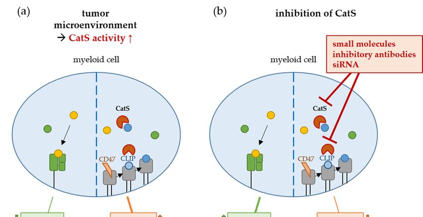
Cells 2020, 9, x                                                                                           2 of 17
  Cells 2020, 9, 2021                                                                                                2 of 17
      Cysteine cathepsins comprise a family of 11 proteases, of which 5 have been repeatedly
implicated in the progression of solid cancers (cathepsins B, H, K, L, and S) [4]. Among these,
   cathepsin
cathepsin    S S(CatS)
                 (CatS)has
                        hasemerged
                            emerged as
                                     as an attractive
                                             attractive potential
                                                          potentialtarget
                                                                      targetwhose
                                                                             whoseinhibition
                                                                                     inhibitionpromises
                                                                                                  promises  to toaddress
address   the immune-suppressive
   the immune-suppressive           milieu
                                milieu      of the
                                        of the      tumormicroenvironment
                                                 tumor      microenvironment (TME)
                                                                                 (TME) [1,3,4]
                                                                                          [1,3,4]due
                                                                                                  duetotoits its
                                                                                                              rolerole in
in the
    thepolarization
          polarization  of antigen-presenting
                     of antigen-presenting  cellscells
                                                  (APC)  (APC)   from
                                                           from an   M1 an   M1 atoward
                                                                         toward             a tumor-favoring
                                                                                   tumor-favoring      M2-phenotype.
M2-phenotype.        M2-typemyeloid-derived
   M2-type APC support         APC supportsuppressor
                                                   myeloid-derived       suppressor
                                                              cells (MDSC)              cells (MDSC)
                                                                             and tumor-associated             and
                                                                                                        macrophages
tumor-associated
  (TAM), favoringmacrophages
                      the expansion(TAM),   favoring
                                    of tolerogenic      the expansion
                                                      regulatory  T cells of tolerogenic
                                                                          (Treg)           regulatory
                                                                                 cells instead          T cells
                                                                                                of cytotoxic     CD8+ T
(Treg)   cells instead of cytotoxic CD8+  T  cells,  which   leads  to an  immune   suppression
   cells, which leads to an immune suppression in favor of tumor cells (Figure 1) [5].              in favor    of
tumor cells (Figure 1) [5].

      Figure 1. Role of cathepsin S (CatS) in the immune-suppressive milieu of the tumor
         Figure 1. Role of cathepsin S (CatS) in the immune-suppressive milieu of the tumor microenvironment
      microenvironment (TME). (a) CatS is overexpressed in many tumors and favors the MHC-II
         (TME). (a) CatS is overexpressed in many tumors and favors the MHC-II pathway, leading to an increase
      pathway, leading to an increase of CD4+ T cells instead of cytotoxic CD8+ T cells. (b) The inhibition
         of CD4+ T cells instead of cytotoxic CD8+ T cells. (b) The inhibition of CatS can enhance the anti-tumor
      of CatS can enhance the anti-tumor immune response by promoting cytotoxic CD8+ T cells instead of
         immune response by promoting cytotoxic CD8+ T cells instead of CD4+ T cells [1,3].
      CD4+ T cells [1,3].
        In this review, the mechanisms of tumor promotion by cysteine cathepsins, with a focus on CatS
  andInits
        this review,
           role in thethe mechanisms
                       TME,            of tumor Several
                            will be illustrated. promotion by cysteine
                                                        inhibitors     cathepsins,
                                                                   of CatS         with afor
                                                                           and strategies focus  on CatS
                                                                                             efficient delivery
and its role in the TME, will be illustrated. Several inhibitors of CatS and strategies for efficient
  into the TME, mediated by nanoparticles, will be presented.
delivery into the TME, mediated by nanoparticles, will be presented.
   2. Cysteine Cathepsins in Tumor Progression
2. Cysteine Cathepsins in Tumor Progression
        Cysteine cathepsins (Cat) are lysosomal proteases, usually with an activity optimum at an acidic
      Cysteine cathepsins (Cat) are lysosomal proteases, usually with an activity optimum at an
   pH, that play an important role in intracellular protein catabolism. The cysteine cathepsins B, H,
acidic pH, that play an important role in intracellular protein catabolism. The cysteine cathepsins B,
   K, L, and S are also excreted, partly tethered to the cell surface, where they also degrade certain
H, K, L, and S are also excreted, partly tethered to the cell surface, where they also degrade certain
   extracellular matrix (ECM) proteins. For CatS, these are several collagens, elastin, laminin-5, but
extracellular matrix (ECM) proteins. For CatS, these are several collagens, elastin, laminin-5, but also
   also cell surface receptors, such as protease-activated receptor-2, and cell adhesion molecules such
cell surface receptors, such as protease-activated receptor-2, and cell adhesion molecules such as
   as junctional adhesion molecule B (JAM-B) and E-cadherin, which is an activity that facilitates tissue
junctional adhesion molecule B (JAM-B) and E-cadherin, which is an activity that facilitates tissue
   remodeling, cancer cell growth, and spreading ([4] Table 1). The expression and activity of these
remodeling, cancer cell growth, and spreading ([4] Table 1). The expression and activity of these
   cathepsins is generally upregulated in (chronic) inflammation and in cancers. Consequently, they are
   overexpressed in tumors, prominently for CatB and CatS, including in follicular lymphoma, gastric,
   colon, brain, breast, and pancreatic cancer [1,3,6]. Overall, most but not all of these cathepsin-mediated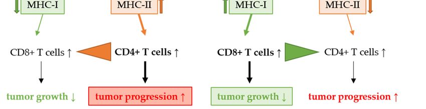
Cells 2020, 9, 2021                                                                                                   3 of 17

mechanisms result in enhanced ECM turnover and angiogenesis, clearing the way for tumor expansion,
securing the cancers’ nutrient supply, and, most notably, in suppressing the T-cell induced anti-cancer
immune response that is located in the TME. Here, cancer cells have developed mechanisms to escape
the surveillance of the immune system, both by limiting their (tumor) antigen presentation which
makes them “invisible” to the immune system and by their ability to actively condition their TME
by the secretion of factors that switch the non-tumor antigen-presenting cells (APC), mainly myeloid
cells and partly B cells, from an M1-type to an M2-type polarization. M1-type APC activate tumor
destroying CD8+ T cells, while M2-type APC induce tumor tolerogenic regulatory T cells (Treg) and
inactivate tumor destroying cytotoxic CD8+ T cells [3,7,8]. Here, the proteolytic activity of mainly
CatB and CatS, despite their complex regulation and diverse activities, has an overall disastrous effect
on anti-tumor immune responses by polarizing the myeloid APC in the TME from an M1-type toward
an M2-phenotype, favoring the expansion and suppressive function of myeloid-derived suppressor
cells (MDSC) and the related tumor-associated macrophages (TAM), resulting in the inactivation and
depletion of cytotoxic CD8+ T cells and the expansion of Treg [5]. CatS regulates antigen processing
and presentation, enhancing major histocompatibility class II (MHCII) expression and antigen loading
on myeloid endothelial and epithelial cells, including cancer epithelia. However, in contrast to antigen
presentation via MHC class I, prominently via (cancer) epithelial cells, that activate tumor cytotoxic
CD8+ T cells, MHCII presentation in the TME usually activates CD4+ Treg that actively suppress
cancer immunity [9–12]. Furthermore, a mouse model suggests that the CatS inhibition of Treg cells
may reduce the overall T-cell immunity under normal conditions, but it enhances the CD8+ T-cell
immunity in the presence of cancer cells [13]. Taken together, all these effects of CatS on cancer growth
make the inhibition of CatS an attractive strategy to limit tumor expansion and increase anti-tumor
immunogenicity [3,9–11].

                      Table 1. Cysteine cathepsins and their prominent roles in tumor progression.

                                 CatB             CatK                CatL                CatS                CatX
                                                                                    ubiquitous, more
                                                                                      prominent in
                                                ubiquitous;                                               predominantly
      Physiological                                                                     M2-type
                              ubiquitous      predominantly        ubiquitous,                           in immune cells
     occurrence [14]                                                                  macrophages
                                              in bone tissue                                                   [15]
                                                                                     than T cells, EC,
                                                                                        epithelia
                              cytoplasm
                                                                                    TAM > MDSC >
   Modified/additional      [16,17], plasma                          nucleus,
                                                                                     angiogenic EC,
      occurrence in           membrane,              -             secreted in                                   -
                                                                                      TM epithelia
    tumor tissue [14]         secreted in                           ECM [18]
                                                                                         [5,19]
                                 ECM
                                                                                     ECM turnover,
                                                                                    angiogenesis [22],
                                               angiogenesis,     metastasis, cell                        additive effects
     Mechanisms of           angiogenesis                                            suppression of
                                              bone metastasis     proliferation                          of CatB + CatX
   tumor progression             [20]                                                  anti-tumor
                                                   [21]                [18]                                    [15]
                                                                                        immune
                                                                                    responses ↓ [3,5]
                                                                                         antigen
                                                secretion of
                                                                                      presentation +
                              CD8+ cell           IL-6 [19],                                                enhanced
      Functions in                                                  antigen          processing [26],
                            apoptosis [23],      enhanced                                                 migration of T
    immune response                                              presentation +          M2-type
                               MDSC            expression of                                              lymphocytes
          [5]                                                    processing [12]       macrophage
                            promotion [24]     COX2 + CatB                                                    [28]
                                                                                       polarization
                                              (via CCL2) [25]
                                                                                    toward TAM [27]
      Influence on
       anti-tumor                 ↓                 ↓↑                  ↑                   ↓                   ↑
    immune response
      EC = endothelial cell; ECM = extracellular matrix; MDSC = myeloid-derived suppressor cells; IL-6 = interleukin 6;
      COX2 = cyclooxygenase 2, CCL2 = CC-chemokine ligand-2; TAM = tumor-associated macrophage.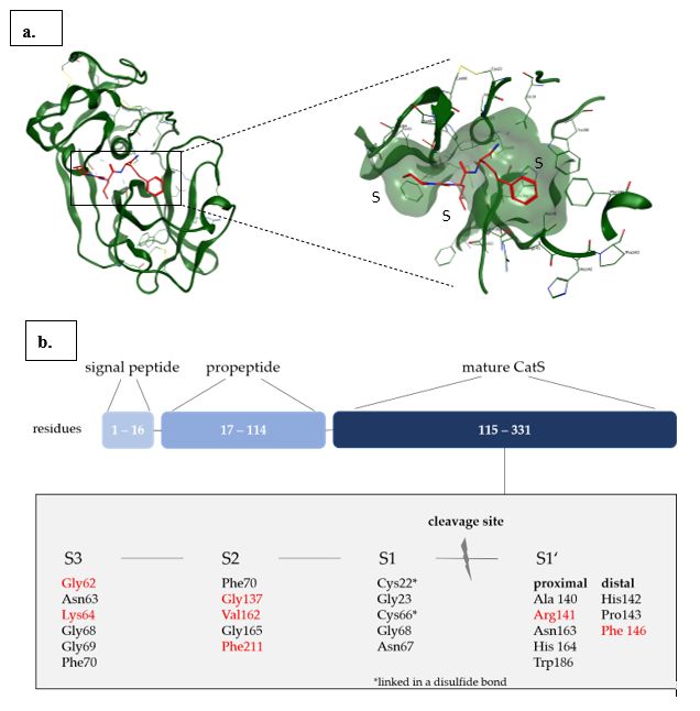
Cells 2020, 9, x                                                                                                            4 of 17

 CellsEC
      2020, 9, 2021
         = endothelial cell; ECM = extracellular matrix; MDSC = myeloid-derived suppressor cells; IL-6 =                              4 of 17
      interleukin 6; COX2 = cyclooxygenase 2, CCL2 = CC-chemokine ligand-2; TAM = tumor-associated
      macrophage.
 Cathepsin S
Cathepsin S
      Cathepsin S (CatS) is a papain-like two-domain protein that is synthesized in vivo as an inactive
 precursor.
     Cathepsin ItsSpropeptide     is essential
                   (CatS) is a papain-like      for the activation
                                             two-domain              of the
                                                           protein that      enzyme and
                                                                        is synthesized      proper
                                                                                        in vivo as anfolding
                                                                                                       inactive[29,30].
precursor.  Its propeptide  is  essential for the activation of the enzyme   and  proper  folding
 The mature enzyme contains 217 residues with a catalytic Cys25 located in the active site and    [29,30]. Theseveral
mature  enzyme
 residues  in the contains   217 residues
                   S1’–S3 pockets            with a catalytic
                                     that determine           Cys25specificity
                                                       the binding    located infor
                                                                                  the active
                                                                                    CatS     site and
                                                                                          among   otherseveral
                                                                                                         papain-like
residues
 cysteine in   the S1’–S3 pockets
           endopeptidases      (Figurethat   determine the binding specificity for CatS among other
                                        2) [31,32].
papain-like cysteine endopeptidases (Figure 2) [31,32].

       Figure2.2.(a)(a)Left:
      Figure            Left:Cartoon
                              Cartoon    model
                                       model    ofofthe
                                                      thecrystal
                                                           crystalstructure
                                                                    structureofofCatS
                                                                                  CatSiningreen
                                                                                           green(PDB(PDB1MS6)
                                                                                                            1MS6)inincomplex
                                                                                                                      complex with
       a covalent-reversible
      with a covalent-reversible  nitrile-based   inhibitor
                                       nitrile-based          in red
                                                        inhibitor   in bound   covalently
                                                                       red bound           to thetocatalytic
                                                                                   covalently                 Cys25Cys25
                                                                                                     the catalytic    of CatS.
                                                                                                                            of Right:
      CatS. Right: zoom into the active site of CatS with hydrogen bonds depicted in dashed lines. TheS2, and
       zoom   into   the active site  of CatS   with   hydrogen     bonds   depicted  in dashed    lines.  The subsites   S1,
       S3 are highlighted.
      subsites  S1, S2, and S3 (b)are
                                    Domain     structure
                                       highlighted.     (b) of CatS. The
                                                            Domain          residues
                                                                        structure     that define
                                                                                  of CatS.           the S1’–S3
                                                                                            The residues     thatpockets   are listed
                                                                                                                   define the
       in the  light  gray   box; residues    that are   the determinants      of CatS binding    specificity
      S1’–S3 pockets are listed in the light gray box; residues that are the determinants of CatS binding      among     papain-like
       cysteine endopeptidases
      specificity  among papain-like   are marked
                                             cysteineinendopeptidases
                                                          red [32]. Figureare  1a was  prepared
                                                                                  marked    in redwith [32].MOE  2019.0102.
                                                                                                             Figure   1a was
      prepared with MOE 2019.0102.
      Although the different cathepsin subtypes have a high sequence homology, CatS differs from other
 cysteine cathepsins by its stability at neutral pH and its limited tissue distribution with high levels found
 in spleen and lung macrophages of healthy organisms [33,34]. CatS degrades the invariant chain that
 occupies the MHCII binding pocket and promotes (tumor) antigen processing and their presentation
Cells 2020, 9, 2021 5 of 17
to T cells via MHCII on APC [9]. However, as discussed above, this activates immune-suppressive
CD4+ Treg instead of tumor-destructive CD8+ T cells.
Recently, the effect of CatS inhibition in several experimental cancers has been demonstrated.
For instance, Da Costa et al. explored the inhibition of CatS in gastric cancer, where it is expressed
not only in the lysosomes but also secreted into the ECM. CatS silencing via small interfering RNA
(siRNA) led to a reduction of tumor volume and invasion accompanied by increased apoptosis and
attenuated angiogenesis [35,36]. Sevenich et al. claimed that CatS specifically mediates the blood–brain
barrier transmigration of breast cancer cells through proteolytic processing of the junctional adhesion
molecule B (JAM-B) and therefore, it plays an important role in brain metastasis [37]. Additionally,
Yang et al. showed that the increased expression of CatS correlates with the aggressiveness of human
colon cancer due to its promotion of the M2-type macrophage polarization (TAM), which is favored by
CatS-induced autophagy [38]. Moreover, preclinical studies by Burden et al. illustrate the impact of
inhibitory antibodies against CatS, resulting in an increased efficacy of chemotherapeutic treatments,
leading to a significant reduction of tumor growth [39,40]. Notably, CatS appears to play an important
role in liver cancer that is otherwise resistant to most conventional tumor therapies [41–43].
Bararia et al. reported an overexpression of CatS and its hyperactive mutant Y132D in follicular
lymphoma. The Y132D mutation results in a higher autocatalytic conversion from the inactive proform
to active CatS. In a CatS Y132D transgenic mouse model of follicular lymphoma, they observed
increased cancer growth versus wild-type controls and an increase of the tumor-suppressive CD4+ Treg
over cytotoxic CD8+ T cells infiltrating the tumor [1]. In confirmation, Dheilly et al. showed that in
patients with non-Hodgkin lymphoma, the same activating CatS Y132D mutation in malignant B cells
is promoted, and its inhibition abrogated lymphoma growth via enhancing the cytotoxic CD8+ T-cell
response and attenuating the expansion of CD4+ Treg [3]. Hence, the directed inhibition of CatS with
small molecule inhibitors could enhance the anti-tumor immune response in cancer, especially when
targeting the TME and the relevant APC, i.e., primarily myeloid TAM and MDSC, and—depending on
the tumor type—also B cells.
3. Cathepsin S Inhibitors
The search for selective CatS inhibitors dates back to the early 2000s and continues until today.
Some early reviews [44–47] cover the inhibition of cysteine proteases in general, while the first specific
compilation of selective CatS inhibitors appeared in 2004 [48]. CatS has a high sequence homology
with other Cathepsins (K, L, B), with some distinct differences in the S2 and S3 pockets which can be
addressed for selective CatS inhibition [32]. The S2 pocket of CatS contains Phe70, Gly137, Val162, Gly
165, and a flexible Phe211 located at the bottom of the pocket, which can lead to an open conformation
making space between itself and Phe70 and providing a possibility for π-stacking interactions with
either or both phenylalanine residues [48]. In cathepsin K (CatK), Phe211 is replaced by Leu, making
the subsite tighter and more shallow. The two Gly residues in CatS are both Ala in Cat K and serve as
‘gatekeepers’. The missing methyl groups of the two Gly amino acids instead of Ala in CatS enable
the S2 pockets to be opened in width and in depth [32,49,50]. These differences make the search for
selective CatS inhibitors attractive, since the S2 pocket in CatS can be addressed by significantly lager
substituents compared to other cathepsins, e.g., CatK [49].
To date, there are over 1800 entries in the public access ZINC (ZINC is not commercial) database
regarding CatS inhibitors [51], most of them being non-selective and inhibiting other cathepsins as well.
The most selective and potent compounds developed for CatS inhibition comprise covalent as well as
non-covalent inhibitors [48,50,52]. Since the first investigated inhibitors for CatS, e.g., the pan-cathepsin
inhibitor LHVS (leucine homophenylalanine vinyl sulfone), contained an electrophilic warhead,
which bound to the catalytical cysteine residue in the active site of CatS in a covalent irreversible
mechanism, a wide variety of covalent reversible and interestingly also non-covalent inhibitors have
been developed [48]. This has opened up new possibilities in CatS inhibitor design. Most of the covalent
inhibitors contain a nitrile or an aldehyde as an electrophilic warhead, and the recognition sequenceCells 2020, 9, x 6 of 17
contained an electrophilic warhead, which bound to the catalytical cysteine residue in the active site
Cells CatS9,in
of2020, 2021 6 of 17
a covalent irreversible mechanism, a wide variety of covalent reversible and interestingly
also non-covalent inhibitors have been developed [48]. This has opened up new possibilities in CatS
inhibitor design. Most of the covalent inhibitors contain a nitrile or an aldehyde as an electrophilic
harbors either peptidic or non-peptidic residues. Both covalent and non-covalent inhibitors were
warhead, and the recognition sequence harbors either peptidic or non-peptidic residues. Both
successfully
covalent andco-crystallized
non-covalentwith CatS (Figure
inhibitors 3) and many
were successfully of those crystal
co-crystallized withstructures are published
CatS (Figure 3) and
in many
the protein database (PDB) [53–58]. Since the latest reviews of CatS inhibitors covered the state of
of those crystal structures are published in the protein database (PDB) [53–58]. Since the latest
research
reviews of CatS inhibitors covered the state of research in CatS inhibitor design for the time between on
in CatS inhibitor design for the time between early 2000 to 2010, this paragraph focuses
theearly
progress made
2000 to 2010,inthis
theparagraph
last decade [48,50,52].
focuses on the progress made in the last decade [48,50,52].
non-covalent inhibitors
O
O
O
O
N O N
H N N N
HN O N NH HN H
N HN O
O O NH
F O
2FZ (4P6G) GNF (2F1G) 2FZ (4P6E)
IC50: 7.7 nM Ki: 29 nM IC50: 1290 nM
covalent inhibitors
N O NC
N N O N
O HN
H
N HN
O N N N CN O
H
CN O O
N O
N CF3
BLN (1MS6) EF3 (3N4C) Y11 (2R9M)
IC50: 19 nM IC50: 9.5 nM IC50: 1.5 nM
O
CF3 O Cl
O O NC N CF3
O H
N N N
S N O N H
O H
O N
C1P (1NPZ) 93N (3N3G) O64 (3OVX)
Ki: 13 nM IC50: 58 nM IC50: 31 nM
Figure
Figure 3. 3.Overview
Overviewof of different
different non-covalent
non-covalent and
and covalent
covalent CatS-selective
CatS-selectiveinhibitors from
inhibitors crystal
from crystal
structures with compound ID and PDB (protein database) code of the corresponding
structures with compound ID and PDB (protein database) code of the corresponding crystal structure crystal
(instructure (in parenthesis).
parenthesis). The enzymatic
The enzymatic data are
data are given given IC
as either as either IC50 or Ki values [53–58.]
50 or Ki values [53–58].
One
One effortfor
effort foroptimizing
optimizingthe
theselectivity
selectivity toward
toward CatS
CatSvia
viaP2
P2substituents
substituentswas
was done
donebybyKerns
Kernset et
al. reporting azepanone-based inhibitors (Figure 4) with 175-fold and 286-fold selectivity
al. reporting azepanone-based inhibitors (Figure 4) with 175-fold and 286-fold selectivity for CatS for CatS
versus
versus CatKand
CatK andCatLCatLrespectively
respectively [59].
[59]. The
Thecrucial
crucialmodification
modification in in
P2 P2
with the the
with introduction of a of
introduction
1-methyl-cyclohexyl alanine lead to compound 1 with an overall balanced potency
a 1-methyl-cyclohexyl alanine lead to compound 1 with an overall balanced potency and selectivity and selectivity
profile. Another approach has been taken by Hilpert et al. through structure-based drug design
profile. Another approach has been taken by Hilpert et al. through structure-based drug design starting
from a weakly active dual CatS/CatK inhibitor discovering a series of CatS-selective inhibitors that
contain different cyclic central scaffolds [57]. Out of the different tested scaffolds, a proline derivative
was identified as the most promising with IC50 values in the low nM range in enzymatic as well as
cell-based assays and a good ligand-binding efficiency (LE) of 0.47 (Figure 4) [57]. Another set ofCells 2020, 9, x 7 of 17
starting from a weakly active dual CatS/CatK inhibitor discovering a series of CatS-selective
Cells 2020, 9,that
inhibitors 2021 contain different cyclic central scaffolds [57]. Out of the different tested scaffolds,
7 ofa17
proline derivative was identified as the most promising with IC50 values in the low nM range in
enzymatic as well as cell-based assays and a good ligand-binding efficiency (LE) of 0.47 (Figure 4)
proline-derived compounds was synthesized and evaluated as CatS inhibitors by Kim et al., with
[57]. Another set of proline-derived compounds was synthesized and evaluated as CatS inhibitors
compound 3 (Figure 3) showing promising in vitro/in vivo pharmacological activities [60]. A known
by Kim et al., with compound 3 (Figure 3) showing promising in vitro/in vivo pharmacological
bioavailable CatS nitrile inhibitor previously developed by Merck Frosst [49] was re-evaluated, and its
activities [60]. A known bioavailable CatS nitrile inhibitor previously developed by Merck Frosst
selective targeting of CatS over CatK/L/V and B was confirmed by an in vitro enzymatic assay. In vivo
[49] was re-evaluated, and its selective targeting of CatS over CatK/L/V and B was confirmed by an
experiments revealed that compound 4 could significantly reduce tumor volume in murine MC38
in vitro enzymatic assay. In vivo experiments revealed that compound 4 could significantly reduce
syngeneic and MCF7 xenograft models. Immunohistochemical analysis of MCF7 tumors revealed
tumor volume in murine MC38 syngeneic and MCF7 xenograft models. Immunohistochemical
that CatS inhibitor treatment with compound 4 significantly reduced proliferation and increased
analysis of MCF7 tumors revealed that CatS inhibitor treatment with compound 4 significantly
apoptosis [61].
reduced proliferation and increased apoptosis [61].
O
O
O O
H O N
N N N O
N H CN
H O
O O Cl S
N S HN O
O
O O O
O N
N
1 2 3
CN
SO2 CF3
CF3 N N
H CN
N
N N N N
H H
O O
F
4 5
Figure 4. Structures of five relevant CatS inhibitors discovered through SAR (structure–activity
Figure 4. Structures of five relevant CatS inhibitors discovered through SAR (structure–activity relation)
relation) studies of the last decade.
studies of the last decade.
Thus,
Thus,thisthisinhibitor
inhibitormaymayhold
holdtherapeutic
therapeuticpotential
potentialand
andcould
couldbe beused
usedtotofurther
furtheranalyze
analyzethetherole
role
ofofCatS
CatS in tumor development. One additional series of reversible CatS inhibitors consistingofof
in tumor development. One additional series of reversible CatS inhibitors consisting
2,4,6-trisubstituted
2,4,6-trisubstituted1,3,5-triazines
1,3,5-triazineswaswassynthesized
synthesizedand andbiologically
biologicallyevaluated
evaluatedbybyTberTberetetal.,
al.,with
with
compound 5 being the most potent and selective inhibitor among the
compound 5 being the most potent and selective inhibitor among the set, with an IC50 of 3 nMset, with an IC 50 of 3 nM andand a
Kai of
Ki 2ofnM,
2 nM,respectively
respectively [62].
[62].
Overall, there has been
Overall, there has been progressprogressininthe the field
field of CatS
of CatS inhibitor
inhibitor research
research resulting
resulting in several
in several potent
potent
and selective compounds that have been developed in the last decade and that are suitable for thefor
and selective compounds that have been developed in the last decade and that are suitable use
the use in investigations
in further further investigations
focusing onfocusing
the roleonthattheCatS
roleplays
that in CatS
cancerplays
andinalso
cancer
otherand also other
pathologies.
pathologies.
4. Nanocarrier-Mediated Delivery of Cathepsin Inhibitors
4. Nanocarrier-Mediated Delivery of Cathepsin Inhibitors
Despite pharmacodynamic target optimization, physicochemical parameters may sometimes
hamperDespite pharmacodynamic
the in vivo translatabilitytarget optimization,
of small physicochemical
molecular drugs, including some parameters may
of the novel sometimes
CatS inhibitors
hamper
discussed theabove.
in vivoFortranslatability
instance, poor of small molecular
solubility and reduced drugs, includingmight
bioavailability somealready
of the prevent
novel CatSthem
inhibitors
from enteringdiscussed above.
in vivo For instance,
testings. poor solubility
Here, nano-sized carrierand
systemsreduced
can bioavailability
provide uniquemight already
opportunities
prevent
to alter them from pharmacokinetic
the drugs’ entering in vivo testings.
profile and Here, nano-sized
enhance carrier systems
their delivery cansites
to targeted provide unique
of action. By
opportunities to alter the
preventing premature drugs’from
release pharmacokinetic
the carrier or profile
unwantedandearlyenhance their delivery
metabolic to targeted
degradation, sites
nano-sized
ofcarrier
action. By preventing
systems can support the premature
overall drugrelease from the
performance carrier
in vivo and ator the
unwanted
same timeearly
reducemetabolic
unwanted
degradation, nano-sized
side effects, which carrier enable
can further systems can support
higher dosing,thethusoverall drug the
increasing performance
therapeuticin window
vivo and[63].
at the
same time
Throughreduce theunwanted side effects,
last four decades, which can have
nanocarriers further enable higher
intensely dosing, thusfor
been investigated increasing the
the delivery
therapeutic
of classicalwindow [63]. chemotherapeutics [64,65]. This has so far mostly been stimulated by
anti-cancer
blood-circulating nanocarriers that can passively accumulate in highly vascularized tumors through
the enhanced permeability and retention (EPR) effect [66–68]. In parallel, several studies demonstrated
that nano-sized drug carriers also accumulate in lymphoid organs, such as the spleen, but also in the liverCells 2020, 9, 2021 8 of 17
and lungs, as major first pass organs, to be rapidly taken up and processed by immune cells, in analogy to
classical pathogens of similar sizes, i.e., “the immune system likes nanotechnology” [69,70]. These more
recent insights have increased the interest in nanocarriers to assist in cancer immunotherapy [71–73].
Here, nanocarrier-mediated delivery of CatS inhibitors to focus their delivery to certain immune cells
in the tumor microenvironment, especially to TAM and MDSC, is an attractive approach.
Key criteria for precise delivery to specific immune cell populations include the carriers’
chemical composition, which should guarantee biocompatibility, biostability, and biointegrity
under physiological conditions. Moreover, toward clinical translatability, further issues including
biodegradability and clearance from the body must be taken into account as well. Consequently, these
characteristics needed for in vivo application exclude several nanocarriers that have been designed
throughout the last decades and claimed useful for drug delivery purposes [74]. So far, only a few
formulations passed phase 1–3 clinical trials and led to successful products on the market. Most
prolific amongst them are lipid formulation e.g., liposomal doxorubicine (Doxil) for solid tumors (e.g.,
breast or ovarian cancer), or the lately approved lipid-encapsulated siRNA formulation Patisiran [75].
Additionally, such formulations can be combined with polymer-based strategies, e.g., PEGylation
(poly(ethylene glycol) decoration) of liposomes or therapeutic proteins [76,77], which enable tissue
mobility and prolonged blood circulation.
Combining biocompatible hydrophilic polymers such as PEG and hydrophobic, self-assembling
block copolymers affords lipid-analogous polymer micelles with superior particle stabilities and drug
encapsulation capabilities [78]. As an example, South Korea approved a paclitaxel formulation using
PEG-block-poly-lactic-co-glycolic acid (PLGA) block copolymers (Genexol) [79]. Such systems support
the solubility of poorly water-soluble drugs by physiochemically entrapping them into the hydrophobic
domains of resulting nano-sized block copolymer self-assemblies.
PLGA has been approved by the FDA as synthetic hydrophobic copolymer and therefore, it
is frequently used for encapsulating therapeutically active molecules [80]. Consequently, similar
attempts have been made lately to encapsulate also cathepsin inhibitors, as recently summarized by
Prunk et al. [14] and Cogo et al. [81]. For instance, Kos et al. have encapsulated the endogenous
pan-cathepsin inhibitor cystatin, a 13.3 kDa protein, into PLGA microspheres and nanospheres via
a double emulsion process [82,83]. They also studied a biological polymer, chitosan, for cystatin
encapsulation via a gelation process including polyphosphates [84]. Similarly, the small molecule
hydroxyquinoline derivative nitroxoline, which exhibits CatB inhibitory activity, has more recently
been encapsulated into nanoparticles via gel-forming chitosan/chondroitin sulfate mixtures that further
allowed co-encapsulation of the anti-cancer agent 5-fluoruracil. [85] Other small molecule-based
cathepsin inhibitors could be co-formulated into polymeric nanoparticles, too. For instance, the group
of Schindeler locally delivered the CatK inhibitor L006235 encapsulated into PLGA nanospheres
together with recombinant human bone morphogenetic protein-2 (rhBMP-2) [86], while the group
of Prud’homme studied co-formulations of the CatK inhibitor odanacatib with the antifungal drug
itraconazole, which are both encapsulated into amphiphilic PEG-block-poly(styrene) nanocarriers [87].
Cathepsin inhibitor delivery has also been studied in liposomal formulations. In analogy to
the natural pan-cathepsin inhibitor cystatin, Mikhaylov et al. loaded the small molecule broad spectrum
inhibitor JPM-565 into PEGylated liposomes together with ferrimagnetic iron oxide nanoparticles
yielding “ferri-liposomes”, which allowed tumor-directed MRI-based imaging, and resulted in local
cathepsin inhibition and finally reduced mammary tumor growth [88]. Alternatively, in another study,
Mikhaylov et al. covalently attached the small molecule CatB inhibitor NS-629 to a DSPE-PEG (2000)
lipid and thus anchored it into the bilayer of resulting liposomes [89]. More lately, the same group
also applied this approach for the conjugation of the natural CatS and CatL inhibitor stefin A onto
the liposome surface, too [90].
Tabish et al. recently summarized further CatL inhibitors and proposed their delivery by
nanocarriers to enhance anti-tumor and antimetastatic activities [91]. As a recent example, Junior et al.(2000) lipid and thus anchored it into the bilayer of resulting liposomes [89]. More lately, the same
group also applied this approach for the conjugation of the natural CatS and CatL inhibitor stefin A
onto the liposome surface, too [90].
Tabish et al. recently summarized further CatL inhibitors and proposed their delivery by
nanocarriers
Cells 2020, 9, 2021to enhance anti-tumor and antimetastatic activities [91]. As a recent example, Junior et
9 of 17
al. reported the encapsulation of the CatL inhibitors Neq0551, Neq0554, and Neq0568 into
protein-based hollow ferritin nanocages and demonstrated improved drug delivery into cancer cells
reported the encapsulation of the CatL inhibitors Neq0551, Neq0554, and Neq0568 into protein-based
in vitro [92].
hollow ferritin
Taken together,nanocages
suchand demonstrated improved
physicochemical encapsulationdrug delivery
methodsinto cancer
seem cellsstraightforward
to be in vitro [92].
Taken together,
regarding fabrication such physicochemical
processes; however, encapsulation methods challenges,
several important seem to be straightforward regarding
including reproducible
fabrication processes; however, several important challenges, including reproducible
loading capacities, avoiding premature drug release and controlled drug release at the target site loading capacities,
avoiding
need to be premature drug releasea and
shown. Especially, controlled
chemically drug
better releaseand
defined at the target site
carefully need to
designed be shown.
approach to
Especially, a chemically better defined and carefully designed approach to covalently
covalently link small molecule drugs to the carrier may be promising. Here, the group of Kopeček link small molecule
drugs to the carrier
synthesized may be promising.
well-defined conjugates Here,of thetheCatK
group of Kopeček
inhibitor synthesized
1,5-bis well-defined
(N-benzyloxy- conjugates
carbonylleucyl)
of the CatK inhibitor
carbohydrazide with1,5-bis (N-benzyloxy-hydrophilic
the water-soluble carbonylleucyl) carbohydrazide
polymers PEG and with the water-soluble
poly(2-hydroxypropyl
hydrophilic
methacrylamide) (HPMA) [93,94]. Yet, ligating water-insoluble small molecules to Yet,
polymers PEG and poly(2-hydroxypropyl methacrylamide) (HPMA) [93,94]. ligating
hydrophilic
water-insoluble
polymers can stimulate less controlled inter- and intramolecular self-assemblies that affect and
small molecules to hydrophilic polymers can stimulate less controlled inter- the
intramolecular self-assemblies that affect the nanocarriers’ integrity, especially
nanocarriers’ integrity, especially under biologically relevant conditions, e.g., by a less predictable under biologically
relevant
adsorption conditions,
of serume.g., by a less
proteins predictable adsorption of serum proteins [95,96].
[95,96].
To address these challenges,
To address these challenges, block block copolymer
copolymer micelles havehave
micelles been been
demonstrated
demonstratedto provide higher
to provide
drug-loading
higher drug-loading stabilitystability
and carrier
andintegrity when interchain
carrier integrity cross-linkscross-links
when interchain are introduced, especially
are introduced,
into the hydrophobic core of the drug nanocarrier [97]. We have recently
especially into the hydrophobic core of the drug nanocarrier [97]. We have recently been following been following this
strategy [98] [98]
this strategy andandoptimized
optimized thethe
nanocarriers
nanocarriers forfor
immunotherapeutic
immunotherapeuticdelivery delivery(Figure
(Figure5)5)[99].
[99]. Via
Via
covalent drug conjugation and hydrophilic core-crosslinking of self-assembled
covalent drug conjugation and hydrophilic core-crosslinking of self-assembled reactive ester block reactive ester block
copolymers,
copolymers, drug-loaded nanogels can
drug-loaded nanogels can bebe obtained
obtained withwith pH-
pH- oror reductive-responsive
reductive-responsive degradation
degradation
profiles
profiles [100,101]. Interestingly, these carriers nicely facilitated the antiviral and antitumor immune
[100,101]. Interestingly, these carriers nicely facilitated the antiviral and antitumor immune
responses
responses ofofhighlyhighlypotent
potent small molecular
small molecular Toll-like receptor
Toll-like 7/8 agonists
receptor [102–104].
7/8 agonists Moreover,
[102–104]. surface
Moreover,
chemistry modification
surface chemistry can also can
modification be applied
also betoapplied
furthertopromote
further an active delivery
promote an activeofdelivery
these carriers
of these to
certain target immune cell subpopulations [105].
carriers to certain target immune cell subpopulations [105].
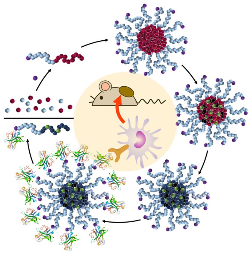
Figure 5. Concept
Conceptofofdelivering
deliveringsmall
smallmolecular
molecular drugs
drugssuch as as
such CatS inhibitors
CatS (depicted
inhibitors in green)
(depicted via
in green)
via covalent
covalent attachment
attachment to to reversibly
reversibly core-crosslinkedand
core-crosslinked andimmune
immunecell-targeting
cell-targetingnanogels
nanogels for
for cancer
immunotherapy.
immunotherapy. Reproduced
Reproduced with
with permission
permission [99].
[99]. Copyright 2020, Elsevier
Elsevier B.V.
B.V.
To that respect, the targeted delivery of cathepsin inhibitors has also been followed by Kos et
al. by adsorbing anti-cytokeratin antibodies onto their cystatin-loaded PLGA nanosphere systems
to enhance delivery to breast cancer cells in vitro [106,107]. However, even more attractive for
the novel CatS inhibitors would be targeting their delivery to more relevant immune cells of
the TME, i.e., immune-suppressive TAM and MDSC [108,109]. Targeting these cell populationsCells 2020, 9, 2021 10 of 17
could so far be demonstrated for surface-modified reactive-ester based nanogels, either by using
polymers with α-mannosyl end groups binding to the overexpressed TAM mannose (MMR, CD206)
receptor in vitro [110] or by targeting this receptor through nanogel-conjugated MMR/CD206-binding
nanobodies in vivo [105]. The latter is currently considered to be more effective in terms of
cheap recombinant production and precise chemical modification compared to full antibodies
or antibody–drug conjugates. More recently, also peptide-based targeting strategies [111,112] or
multivalent mannose derivatives [113] have been exploited to guide nanoparticle delivery toward
TAM. Interestingly, mannose targeting has also been directly applied to small molecule cathepsin
inhibitors by generating glycoconjugates of monomannose, trimannose, and heptamannose with
the pan-cathepsin inhibitor DCG-04 [114].
Therefore, targeted delivery strategies of cathepsin inhibitors to immune-suppressive myeloid
cells, especially for CatS, create a highly promising approach to a novel anti-cancer treatment.
The nanocarrier-mediated delivery of CatS inhibitors has so far not been reported. Consequently,
based on the novel, highly specific CatS inhibitors and substantial advances in the development of
suitable nanocarriers that show promising performance not only in vitro but also in vivo, we expect
that combining both strategies has become an exciting research area for developing novel therapeutics
to sustainably suppress cancer growth and progression.
5. Conclusion and Outlook
The inhibition of tumor-associated cathepsins, especially cathepsin S (CatS), has emerged as a novel
promising strategy in cancer immunotherapy. CatS is prominently expressed in tumor associated
M2-type macrophages (TAM), dendritic cells (DC), and myeloid-derived suppressor cells (MDSC)
of the TME. Its inhibition downregulates MHC class II expression on TAM and DC, and repolarizes
TAM, DC, and MDSC toward the M1 phenotype that promotes the proliferation and activity of CD8+
tumor-destroying cytotoxic T cells, and that abrogates CD4+ immune-suppressive regulatory T cells. A
secondary effect is the suppression of tumor angiogenesis. Therefore, further research on CatS selective
inhibitors and their targeting to M2-type TAM, DC, and MDSC is warranted. We propose that selective
CatS inhibition by specific small molecule inhibitors or siRNA can be targeted to TAM, DC, and
MDSC via precisely modified nanocarriers equipped for the directed delivery and functional release of
the inhibitors. Such an approach provides novel opportunities for effective adjunctive therapies to
promote anti-tumor immunity.
Author Contributions: T.S., L.N., and D.S. designed, wrote, and thoroughly edited this review. N.F. and M.M.
wrote parts of the review, prepared figures and tables, and edited the manuscript. All authors have read and agree
to the published version of the manuscript.
Funding: This research received no external funding.
Acknowledgments: All authors are grateful to the German Research Foundation (DFG) Collaborative Research
Center (CRC) SFB 1066 (projects B3+B4) for support. D.S. received additional project related support from CRC
1292 (project B8). L.N. acknowledges support by the Fonds der Chemischen Industrie (FCI) through their Liebig
program and the DFG through the Emmy-Noether program.
Conflicts of Interest: The authors declare no conflict of interest.
Abbreviations
APC antigen-presenting cell;
Cat cathepsin;
CatB cathepsin B;
CatK cathepsin K;
CatL cathepsin L;
CatS cathepsin S;
CCL2 CC-chemokine ligand-2;
CD206 cluster of differentiation 206;
COX2 cyclooxygenase 2;Cells 2020, 9, 2021 11 of 17
DC dendritic cell;
EC endothelial cell;
ECM extracellular matrix;
EPR enhanced permeability and retention;
FDA Food and Drug Administration;
HPMA poly(2-hydroxypropyl methacrylamide);
IL-6 interleukin 6;
JAM-B junctional adhesion molecule B;
LE ligand efficiency;
LHVS leucine homophenylalanine vinyl sulfone;
MC38 murine colon adenocarcinoma;
MCF7 Michigan Cancer Foundation–7;
MDSC myeloid-derived suppressor cell;
MHC major histocompatibility complex;
MMR mannose receptor;
MRI magnetic resonance imaging;
PDB protein database;
PEG polyethylene glycol;
PLGA PEG-block-poly-lactic-co-glycolic acid;
rhBMP-2 recombinant human bone morphogenetic protein-2;
SAR structure–activity relationship;
siRNA small interfering RNA;
TAM tumor-associated macrophages;
TME tumor microenvironment;
Treg regulatory T cell;
ZINC ZINC is not commercial;
References
1. Bararia, D.; Hildebrand, J.A.; Stolz, S.; Haebe, S.; Alig, S.; Trevisani, C.P.; Osorio-Barrios, F.; Bartoschek, M.D.;
Mentz, M.; Pastore, A.; et al. Cathepsin S Alterations Induce a Tumor-Promoting Immune Microenvironment
in Follicular Lymphoma. Cell Rep. 2020, 31, 107–522. [CrossRef] [PubMed]
2. Da Costa, A.C.; Santa-Cruz, F.; Mattos, L.A.R.; Aquino, M.A.R.; Martins, C.R.; Ferraz Álvaro, A.B.;
Figueiredo, J.L. Cathepsin S as a target in gastric cancer (Review). Mol. Clin. Oncol. 2019, 12, 99–103.
[CrossRef] [PubMed]
3. Dheilly, E.; Battistello, E.; Katanayeva, N.; Sungalee, S.; Michaux, J.; Duns, G.; Wehrle, S.; Sordet-Dessimoz, J.;
Mina, M.; Racle, J.; et al. Cathepsin S Regulates Antigen Processing and T Cell Activity in Non-Hodgkin
Lymphoma. Cancer Cell 2020, 37, 674–689.e12. [CrossRef] [PubMed]
4. Wilkinson, R.; Williams, R.; Scott, C.J.; Burden, R.E.; Williams, R. Cathepsin S: Therapeutic, diagnostic, and
prognostic potential. Biol. Chem. 2015, 396, 867–882. [CrossRef]
5. Jakoš, T.; Pišlar, A.; Jewett, A.; Kos, J. Cysteine Cathepsins in Tumor-Associated Immune Cells. Front.
Immunol. 2019, 10, 10. [CrossRef]
6. McDowell, S.H.; Gallaher, S.A.; Burden, R.E.; Scott, C.J. Leading the invasion: The role of Cathepsin S in
the tumour microenvironment. Biochim. Et Biophys. Acta (BBA) Bioenerg. 2020, 1867, 118–781. [CrossRef]
7. Farhood, B.; Najafi, M.; Mortezaee, K. CD8+ cytotoxic T lymphocytes in cancer immunotherapy: A review. J.
Cell. Physiol. 2018, 234, 8509–8521. [CrossRef]
8. Quaranta, V.; Schmid, M.C. Macrophage-Mediated Subversion of Anti-Tumour Immunity. Cells 2019, 8, 747.
[CrossRef]
9. Riese, R.J.; Wolf, P.R.; Brömme, D.; Natkin, L.R.; Villadangos, J.A.; Ploegh, H.L.; Chapman, H.A. Essential
Role for Cathepsin S in MHC Class II–Associated Invariant Chain Processing and Peptide Loading. Immunity
1996, 4, 357–366. [CrossRef]
10. Riese, R.J.; Mitchell, R.N.; Villadangos, J.A.; Shi, G.P.; Palmer, J.T.; Karp, E.R.; De Sanctis, G.T.; Ploegh, H.L.;
Chapman, H.A. Cathepsin S activity regulates antigen presentation and immunity. J. Clin. Investig. 1998, 101,
2351–2363. [CrossRef]Cells 2020, 9, 2021 12 of 17
11. Shi, G.-P.; Villadangos, J.A.; Dranoff, G.; Small, C.; Gu, L.; Haley, K.J.; Riese, R.; Ploegh, H.L.; Chapman, H.A.
Cathepsin S Required for Normal MHC Class II Peptide Loading and Germinal Center Development.
Immunity 1999, 10, 197–206. [CrossRef]
12. Hsing, L.C.; Rudensky, A.Y. The lysosomal cysteine proteases in MHC class II antigen presentation. Immunol.
Rev. 2005, 207, 229–241. [CrossRef] [PubMed]
13. Yan, X.; Wu, C.; Chen, T.; Santos, M.M.; Liu, C.-L.; Yang, C.; Zhang, L.; Ren, J.; Liao, S.; Guo, H.; et al.
Cathepsin S inhibition changes regulatory T-cell activity in regulating bladder cancer and immune cell
proliferation and apoptosis. Mol. Immunol. 2017, 82, 66–74. [CrossRef]
14. Prunk, M.; Kos, J. Nanoparticle Based Delivery of Protease Inhibitors to Cancer Cells. Curr. Med. Chem. 2018,
24, 4816–4837. [CrossRef] [PubMed]
15. Kos, J.; Vizin, T.; Fonović, U.P.; Pišlar, A. Intracellular signaling by cathepsin X: Molecular mechanisms and
diagnostic and therapeutic opportunities in cancer. Semin. Cancer Biol. 2015, 31, 76–83. [CrossRef] [PubMed]
16. Kos, J.; Lah, T.T. Cysteine proteinases and their endogenous inhibitors: Target proteins for prognosis,
diagnosis and therapy in cancer (review). Oncol. Rep. 1998, 5, 1349–1361. [CrossRef]
17. Mohamed, M.M.; Sloane, B.F. Cysteine cathepsins: Multifunctional enzymes in cancer. Nat. Rev. Cancer 2006,
6, 764–775. [CrossRef]
18. Sudhan, D.; Siemann, D.W. Cathepsin L targeting in cancer treatment. Pharmacol. Ther. 2015, 155, 105–116.
[CrossRef]
19. Gocheva, V.; Wang, H.-W.; Gadea, B.B.; Shree, T.; Hunter, K.E.; Garfall, A.L.; Berman, T.; Joyce, J.A. IL-4
induces cathepsin protease activity in tumor-associated macrophages to promote cancer growth and invasion.
Genes Dev. 2010, 24, 241–255. [CrossRef]
20. Kostoulas, G.; Lang, A.; Nagase, H.; Baici, A. Stimulation of angiogenesis through cathepsin B inactivation of
the tissue inhibitors of matrix metalloproteinases. FEBS Lett. 1999, 455, 286–290. [CrossRef]
21. Brömme, D.; Wilson, S. Role of Cysteine Cathepsins in Extracellular Proteolysis. Extracellular Matrix
Degradation 2011, 23–51. [CrossRef]
22. Wang, B.; Sun, J.; Kitamoto, S.; Yang, M.; Grubb, A.; Chapman, H.A.; Kalluri, R.; Shi, G.-P. Cathepsin S
Controls Angiogenesis and Tumor Growth via Matrix-derived Angiogenic Factors. J. Biol. Chem. 2005, 281,
6020–6029. [CrossRef] [PubMed]
23. Byrne, S.M.; Aucher, A.; Alyahya, S.; Elder, M.; Olson, S.T.; Davis, D.M.; Ashton-Rickardt, P.G. Cathepsin
B controls the persistence of memory CD8+ T lymphocytes. J. Immunol. 2012, 189, 1133–1143. [CrossRef]
[PubMed]
24. Gounaris, E.; Tung, C.-H.; Restaino, C.; Maehr, R.; Köhler, R.; Joyce, J.A.; Plough, H.L.; Barrett, T.A.;
Weissleder, R.; Khazaie, K. Live Imaging of Cysteine-Cathepsin Activity Reveals Dynamics of Focal
Inflammation, Angiogenesis, and Polyp Growth. PLoS ONE 2008, 3, e2916. [CrossRef]
25. Herroon, M.K.; Rajagurubandara, E.; Rudy, D.L.; Chalasani, A.; Hardaway, A.L.; Podgorski, I. Macrophage
cathepsin K promotes prostate tumor progression in bone. Oncogene 2012, 32, 1580–1593. [CrossRef]
26. Plüger, E.B.E.; Boes, M.; Alfonso, C.; Schröter, C.J.; Kalbacher, H.; Ploegh, H.L.; Driessen, C. Specific role for
cathepsin S in the generation of antigenic peptides in vivo. Eur. J. Immunol. 2002, 32, 467–476. [CrossRef]
27. Wilkinson, R.; Magorrian, S.M.; Williams, R.; Young, A.; Small, D.M.; Scott, C.J.; Burden, R.E. CCL2 is
transcriptionally controlled by the lysosomal protease cathepsin S in a CD74-dependent manner. Oncotarget
2015, 6, 29725–29739. [CrossRef]
28. Jevnikar, Z.; Obermajer, N.; Bogyo, M.; Kos, J. The role of cathepsin X in the migration and invasiveness of T
lymphocytes. J. Cell Sci. 2008, 121, 2652–2661. [CrossRef]
29. Somoza, J.R.; Zhan, H.; Bowman, K.K.; Yu, L.; Mortara, K.D.; Palmer, J.T.; Clark, J.M.; McGrath, M.E. Crystal
Structure of Human Cathepsin V. Biochemistry 2000, 39, 12543–12551. [CrossRef]
30. Kopitar, G.; Dolinar, M.; Strukelj, B.; Pungerčar, J.; Turk, V. Folding and Activation of Human Procathepsin S
from Inclusion Bodies Produced in Escherichia coli. JBIC J. Biol. Inorg. Chem. 1996, 236, 558–562. [CrossRef]
31. Shi, G.P.; Munger, J.S.; Meara, J.P.; Rich, D.H.; Chapman, H.A. Molecular cloning and expression of human
alveolar macrophage cathepsin S, an elastinolytic cysteine protease. J. Biol. Chem. 1992, 267, 7258–7262.
[PubMed]
32. Pauly, T.A.; Sulea, T.; Ammirati, M.; Sivaraman, J.; Danley, D.E.; Griffor, M.C.; Kamath, A.V.; Wang, I.-K.;
Laird, E.R.; Seddon, A.P.; et al. Specificity Determinants of Human Cathepsin S Revealed by Crystal
Structures of Complexes. Biochemistry 2003, 42, 3203–3213. [CrossRef] [PubMed]Cells 2020, 9, 2021 13 of 17
33. Kirschke, H.; Wiederanders, B.; Brömme, D.; Rinne, A. Cathepsin S from bovine spleen. Purification,
distribution, intracellular localization and action on proteins. Biochem. J. 1989, 264, 467–473. [CrossRef]
[PubMed]
34. Shi, G.P.; Webb, A.C.; Foster, K.E.; Knoll, J.H.; Lemere, C.A.; Munger, J.S.; Chapman, H.A. Human cathepsin
S: Chromosomal localization, gene structure, and tissue distribution. J. Biol. Chem. 1994, 269, 11530–11536.
35. Liu, W.-L.; Liu, D.; Cheng, K.; Liu, Y.-J.; Xing, S.; Chi, P.-D.; Liu, X.-H.; Xue, N.; Lai, Y.-Z.; Guo, L.; et al.
Evaluating the diagnostic and prognostic value of circulating cathepsin S in gastric cancer. Oncotarget 2016,
7, 28124–28138. [CrossRef]
36. Yixuan, Y.; Kiat, L.S.; Yee, C.L.; Huiyin, L.; Yunhao, C.; Kuan, C.P.; Hassan, A.; Ting, W.T.; Manuel, S.-T.;
Guan, Y.K.; et al. Cathepsin S Mediates Gastric Cancer Cell Migration and Invasion via a Putative Network
of Metastasis-Associated Proteins. J. Proteome Res. 2010, 9, 4767–4778. [CrossRef]
37. Sevenich, L.; Bowman, R.L.; Mason, S.D.; Quail, D.F.; Rapaport, F.; Elie, B.T.; Brogi, E.; Brastianos, P.K.;
Hahn, W.C.; Holsinger, L.J.; et al. Analysis of tumour- and stroma-supplied proteolytic networks reveals
a brain-metastasis-promoting role for cathepsin S. Nat. Cell Biol. 2014, 16, 876–888. [CrossRef]
38. Yang, M.; Liu, J.; Shao, J.; Qin, Y.; Ji, Q.; Zhang, X.; Du, J. Cathepsin S-mediated autophagic flux in
tumor-associated macrophages accelerate tumor development by promoting M2 polarization. Mol. Cancer
2014, 13, 43. [CrossRef]
39. Burden, R.E.; Gormley, J.A.; Kuehn, D.; Ward, C.; Kwok, H.F.; Gazdoiu, M.; McClurg, A.; Jaquin, T.J.;
Johnston, J.A.; Scott, C.J.; et al. Inhibition of Cathepsin S by Fsn0503 enhances the efficacy of chemotherapy
in colorectal carcinomas. Biochemistry 2012, 94, 487–493. [CrossRef]
40. Burden, R.E.; Gormley, J.A.; Jaquin, T.J.; Small, D.M.; Quinn, D.J.; Hegarty, S.M.; Ward, C.; Walker, B.;
Johnston, J.A.; Olwill, S.A.; et al. Antibody-Mediated Inhibition of Cathepsin S Blocks Colorectal Tumor
Invasion and Angiogenesis. Clin. Cancer Res. 2009, 15, 6042–6051. [CrossRef]
41. Fan, Q.; Wang, X.; Zhang, H.; Li, C.; Fan, J.; Xu, J. Silencing cathepsin S gene expression inhibits growth,
invasion and angiogenesis of human hepatocellular carcinoma in vitro. Biochem. Biophys. Res. Commun.
2012, 425, 703–710. [CrossRef] [PubMed]
42. Lang, Z.; Xu, J.; Li, N.; Ke, Z.; Liu, R.; Maubach, G. Cathepsin S is aberrantly overexpressed in human
hepatocellular carcinoma. Mol. Med. Rep. 2009, 2. [CrossRef]
43. Ryschich, E.; Lizdenis, P.; Ittrich, C.; Benner, A.; Stahl, S.H.; Hamann, A.; Schmidt, J.; Knolle, P.A.; Arnold, B.;
Hämmerling, G.J.; et al. Molecular Fingerprinting and Autocrine Growth Regulation of Endothelial Cells in
a Murine Model of Hepatocellular Carcinoma. Cancer Res. 2006, 66, 198–211. [CrossRef] [PubMed]
44. Otto, H.-H.; Schirmeister, T. Cysteine Proteases and Their Inhibitors. Chem. Rev. 1997, 97, 133–172. [CrossRef]
45. Lecaille, F.; Kaleta, J.; Brömme, D. Human and Parasitic Papain-Like Cysteine Proteases: Their Role in
Physiology and Pathology and Recent Developments in Inhibitor Design. Chem. Rev. 2002, 102, 4459–4488.
[CrossRef]
46. Kang, K.; Kim, W. Recent developments of cathepsin inhibitors and their selectivity. Expert Opin. Ther. Pat.
2002, 12, 419–432. [CrossRef]
47. Leung-Toung, R.; Li, W.; Tam, T.; Kaarimian, K. Thiol-Dependent Enzymes and Their Inhibitors: A Review.
Curr. Med. Chem. 2002, 9, 979–1002. [CrossRef]
48. Leroy, V. Cathepsin S inhibitors. Expert Opin. Ther. Pat. 2004, 14, 301–311. [CrossRef]
49. Gauthier, J.Y.; Black, W.C.; Courchesne, I.; Cromlish, W.; Desmarais, S.; Houle, R.; Lamontagne, S.; Li, C.S.;
Massé, F.; McKay, D.J.; et al. The identification of potent, selective, and bioavailable cathepsin S inhibitors.
Bioorganic Med. Chem. Lett. 2007, 17, 4929–4933. [CrossRef]
50. Lee-Dutra, A.; Wiener, D.K.; Sun, S. Cathepsin S inhibitors: 2004–2010. Expert Opin. Ther. Pat. 2011, 21,
311–337. [CrossRef]
51. Sterling, T.; Irwin, J.J. ZINC 15 – Ligand Discovery for Everyone. J. Chem. Inf. Model. 2015, 55, 2324–2337.
[CrossRef] [PubMed]
52. Wiener, J.J.M.; Sun, S.; Thurmond, R.L. Recent advances in the design of cathepsin S inhibitors. Curr. Top.
Med. Chem. 2010, 10, 717–732. [CrossRef]Cells 2020, 9, 2021 14 of 17
53. Ward, Y.D.; Thomson, D.S.; Frye, L.L.; Cywin, C.L.; Morwick, T.; Emmanuel, M.J.; Zindell, R.; McNeil, D.;
Bekkali, Y.; Hrapchak, M.; et al. Design and Synthesis of Dipeptide Nitriles as Reversible and Potent
Cathepsin S Inhibitors. J. Med. Chem. 2002, 45, 5471–5482. [CrossRef] [PubMed]
54. Tully, D.C.; Liu, H.; Alper, P.B.; Chatterjee, A.K.; Epple, R.; Roberts, M.J.; Williams, J.A.; Nguyen, K.T.;
Woodmansee, D.H.; Tumanut, C.; et al. Synthesis and evaluation of arylaminoethyl amides as noncovalent
inhibitors of cathepsin S. Part 3: Heterocyclic P3. Bioorganic Med. Chem. Lett. 2006, 16, 1975–1980. [CrossRef]
[PubMed]
55. Cai, J.; Fradera, X.; Van Zeeland, M.; Dempster, M.; Cameron, K.S.; Bennett, D.J.; Robinson, J.; Popplestone, L.;
Baugh, M.; Westwood, P.; et al. 4-(3-Trifluoromethylphenyl)-pyrimidine-2-carbonitrile as cathepsin S
inhibitors: N3, not N1 is critically important. Bioorganic Med. Chem. Lett. 2010, 20, 4507–4510. [CrossRef]
56. Jadhav, P.K.; Schiffler, M.A.; Gavardinas, K.; Kim, E.J.; Matthews, D.P.; Staszak, M.A.; Coffey, D.S.; Shaw, B.W.;
Cassidy, K.C.; Brier, R.A.; et al. Discovery of Cathepsin S Inhibitor LY3000328 for the Treatment of Abdominal
Aortic Aneurysm. ACS Med. Chem. Lett. 2014, 5, 1138–1142. [CrossRef]
57. Hilpert, H.; Mauser, H.; Humm, R.; Anselm, L.; Kuehne, H.; Hartmann, G.; Gruener, S.; Banner, D.W.; Benz, J.;
Gsell, B.; et al. Identification of Potent and Selective Cathepsin S Inhibitors Containing Different Central
Cyclic Scaffolds. J. Med. Chem. 2013, 56, 9789–9801. [CrossRef]
58. Ahmad, S.; Bhagwati, S.; Kumar, S.; Banerjee, D.; Siddiqi, M.I. Molecular modeling assisted identification
and biological evaluation of potent cathepsin S inhibitors. J. Mol. Graph. Model. 2020, 96, 107512. [CrossRef]
59. Kerns, J.K.; Nie, H.; Bondinell, W.; Widdowson, K.L.; Yamashita, D.S.; Rahman, A.; Podolin, P.L.;
Carpenter, D.C.; Jin, Q.; Riflade, B.; et al. Azepanone-based inhibitors of human cathepsin S: Optimization of
selectivity via the P2 substituent. Bioorganic Med. Chem. Lett. 2011, 21, 4409–4415. [CrossRef]
60. Kim, M.; Jeon, J.; Song, J.; Suh, K.H.; Kim, Y.H.; Min, K.-H.; Lee, K.-O. Synthesis of proline analogues as
potent and selective cathepsin S inhibitors. Bioorganic Med. Chem. Lett. 2013, 23, 3140–3144. [CrossRef]
61. Wilkinson, R.; Young, A.; Burden, R.E.; Williams, R.; Scott, C.J. A bioavailable cathepsin S nitrile inhibitor
abrogates tumor development. Mol. Cancer 2016, 15, 29. [CrossRef]
62. Tber, Z.; Wartenberg, M.; Jacques, J.-E.; Roy, V.; Lecaille, F.; Warszycki, D.; Bojarski, A.J.; Lalmanach, G.;
Agrofoglio, L.A. Selective inhibition of human cathepsin S by 2,4,6-trisubstituted 1,3,5-triazine analogs.
Bioorganic Med. Chem. 2018, 26, 4310–4319. [CrossRef] [PubMed]
63. Farokhzad, O.C.; Langer, R. Impact of Nanotechnology on Drug Delivery. Acs Nano 2009, 3, 16–20. [CrossRef]
[PubMed]
64. Peer, D.; Karp, J.M.; Hong, S.; Farokhzad, O.C.; Margalit, R.; Langer, R. Nanocarriers as an emerging platform
for cancer therapy. Nat. Nanotechnol. 2007, 2, 751–760. [CrossRef] [PubMed]
65. Wang, A.Z.; Langer, R.; Farokhzad, O.C. Nanoparticle Delivery of Cancer Drugs. Annu. Rev. Med. 2012, 63,
185–198. [CrossRef] [PubMed]
66. Matsumura, Y.; Maeda, H. A new concept for macromolecular therapeutics in cancer chemotherapy:
Mechanism of tumoritropic accumulation of proteins and the antitumor agent smancs. Cancer Res. 1986, 46,
6387–6392. [PubMed]
67. Maeda, H. Toward a full understanding of the EPR effect in primary and metastatic tumors as well as issues
related to its heterogeneity. Adv. Drug Deliv. Rev. 2015, 91, 3–6. [CrossRef] [PubMed]
68. Fang, J.; Islam, W.; Maeda, H. Exploiting the dynamics of the EPR effect and strategies to improve
the therapeutic effects of nanomedicines by using EPR effect enhancers. Adv. Drug Deliv. Rev. 2020.
[CrossRef]
69. Toth, I.; Skwarczynski, M. The immune system likes nanotechnology. Nanomedicine 2014, 9, 2607–2609.
[CrossRef]
70. Lepeltier, E.; Nuhn, L.; Lehr, C.; Zentel, R. Not just for tumor targeting: Unmet medical needs and
opportunities for nanomedicine. Nanomedicine 2015, 10, 3147–3166. [CrossRef]
71. Irvine, D.J.; Dane, E.L. Enhancing cancer immunotherapy with nanomedicine. Nat. Rev. Immunol. 2020, 20,
321–334. [CrossRef]
72. Martin, J.D.; Cabral, H.; Stylianopoulos, T.; Jain, R.K. Improving cancer immunotherapy using nanomedicines:
Progress, opportunities and challenges. Nat. Rev. Clin. Oncol. 2020, 17, 251–266. [CrossRef] [PubMed]You can also read


