Novel Two MRT Cell Lines Established from Multiple Sites of a Synchronous MRT Patient
←
→
Page content transcription
If your browser does not render page correctly, please read the page content below
ANTICANCER RESEARCH 40: 6159-6170 (2020)
doi:10.21873/anticanres.14636

                     Novel Two MRT Cell Lines Established from
                     Multiple Sites of a Synchronous MRT Patient
 YASUMICHI KUWAHARA1, TOMOKO IEHARA2, EISUKE ICHISE2, YOSHIKI KATSUMI2, KAZUTAKA OUCHI2,
        KUNIHIKO TSUCHIYA2, MITSURU MIYACHI2, EIICHI KONISHI3, HIROYASU SASAJIMA4,
        SATOAKI NAKAMURA5, SHIGEHISA FUMINO6, TATSURO TAJIRI6, PASCAL D. JOHANN7,
       MICHAEL C. FRÜHWALD8, TATSUSHI YOSHIDA1, TSUKASA OKUDA1 and HAJIME HOSOI2

                1Department of Biochemistry and Molecular Biology, Graduate School of Medical Science,
                                Kyoto Prefectural University of Medicine, Kyoto, Japan;
                            2Department of Pediatrics, Graduate School of Medical Science,

                                Kyoto Prefectural University of Medicine, Kyoto, Japan;
                            3Department of Pathology, Graduate School of Medical Science,

                                Kyoto Prefectural University of Medicine, Kyoto, Japan;
                          4Department of Neurosurgery, Graduate School of Medical Science,

                                Kyoto Prefectural University of Medicine, Kyoto, Japan;
                         5Department of Radiology, Kansai Medical University, Osaka, Japan;
                        6Department of Pediatric Surgery, Graduate School of Medical Science,

                                Kyoto Prefectural University of Medicine, Kyoto, Japan;
                           7Department of Pediatric Oncology, Hematology and Immunology,

                                 University Hospital Heidelberg, Heidelberg, Germany;
           8University Children’s Hospital Augsburg, Swabian Children’s Cancer Center, Augsburg, Germany

Abstract. Background/Aim: Malignant rhabdoid tumor                  KP-MRT-KSa, derived from different lesions and at a
(MRT) is a rare, aggressive neoplasm found in young                 different time from a synchronous multifocal 7-month-old
children, caused by inactivation of a single gene, SNF5             female MRT patient. Results: Both cells showed typical
(INI1, SMARCB1). MRT cases with multifocal tumors at                morphology of MRT, with a compound genomic mutation in
diagnosis are categorized as synchronous MRT, often with a          exons 2 and 5 of the SNF5 gene. The exon 2 mutation was
germline mutation of SNF5. The aim of this study was to             found in the germline. Conclusion: These cell lines could
establish new models useful in clarifying the biological basis      serve as powerful tools for unveiling the molecular
of synchronous MRT. Materials and Methods: We established           mechanism of refractory synchronous MRT.
two novel MRT cell lines, designated as KP-MRT-KS and
                                                                    Malignant rhabdoid tumor (MRT) is a rare and aggressive
                                                                    neoplasm encountered in neonates and young children. In
                                                                    1978, it was initially described as an unfavorable histological
This article is freely accessible online.                           type of renal tumor, a variant of Wilms tumor (1), and
                                                                    subsequently these kidney tumors with characteristic cells
Correspondence to: Tsukasa Okuda, Department of Biochemistry
and Molecular Biology, Graduate School of Medical Science, Kyoto    showing prominent large nuclei and eosinophilic cytoplasmic
Prefectural University of Medicine, 465 Kajii-cho, Hirokoji,        inclusion bodies, were re-categorized as malignant rhabdoid
Kamigyo-ku, Kyoto 602-8566, Japan. Tel: +81 752515316, Fax:         tumor of the kidney (2). Soon afterwards, MRT was reported
+81 752515315, e-mail: okuda@koto.kpu-m.ac.jp and Hajime            to be a tumor that can arise from any anatomical site (3-5)
Hosoi, Department of Pediatrics, Graduate School of Medical         including CNS that was also called as an atypical teratoid
Science, Kyoto Prefectural University of Medicine, 465 Kajii-cho,   rhabdoid tumor (AT/RT) (6). Although gradual improvement
Hirokoji, Kamigyo-ku, Kyoto 602-8566, Japan. Tel: +81
                                                                    of the clinical outcome has been achieved through extensive
752515571, Fax: +81 752511399, e-mail: hhosoi@koto.kpu-m.ac.jp
                                                                    clinical trials using various combinatorial chemo- and/or
Key Words: Synchronous rhabdoid tumor, cell line, germline          radio-therapies (7), the 5-year overall survival (OS) still
mutation, DNA methylation analysis.                                 remains as low as approximately 50% (4, 5, 8, 9).

                                                                                                                              6159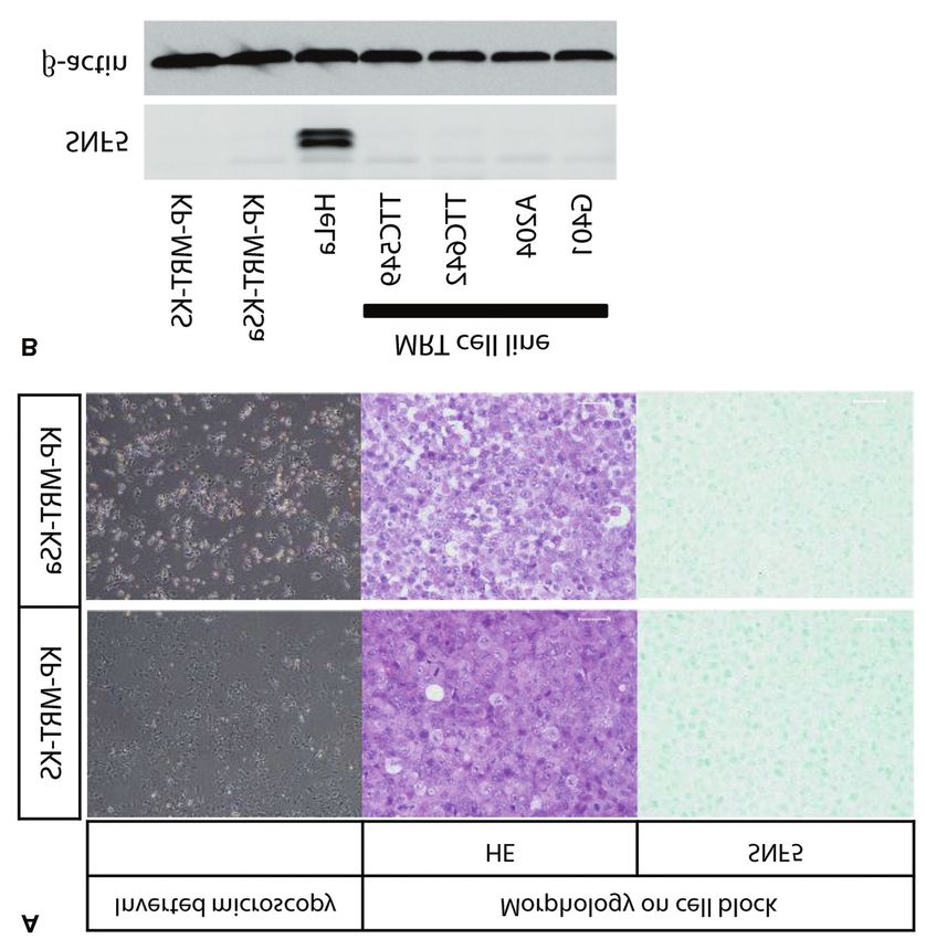
ANTICANCER RESEARCH 40: 6159-6170 (2020)
Bi-allelic inactivation of the SNF5 (SMARCB1, BAF47, Materials and Methods
and INI1) gene, that encodes a component of the SWI/SNF
chromatin remodeling complex on chromosome 22q11.2, has Patient. A 7-month-old female, who had no family history of
reportedly been found (10, 11) in nearly 100% of MRT and neoplasms, was referred to our hospital with the complaints of left
AT/RT cases. Importantly, SNF5 abnormality in MRT upper limb paralysis and Horner’s syndrome on the left side of her
face. Magnetic resonance imaging (MRI) and computed tomography
appears to be the single most important mutation, and
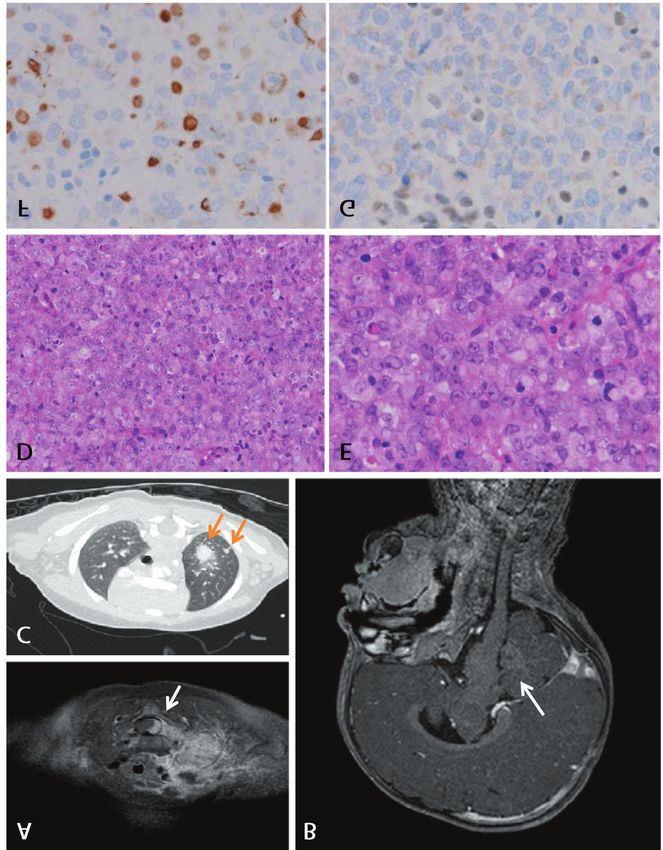
(CT) scanning showed multi-focal tumors: a tumor of the neck that
approximately 30% of children diagnosed with MRT and/or had invaded the spinal canal on C6/7 and C7/Th1 (Figure 1A), a
AT/RT exhibit heterogeneous germline inactivation of the cerebellar vermis tumor without meningeal metastasis (Figure 1B),
SNF5 gene (12). Recently, loss-of-function mutations of the and at least two tumors in the left apical pulmonary region (Figure
BRG-1 (SMARCA4) gene, which encodes another subunit of 1C). Biopsied specimens of the neck tumor were examined, and
the SWI/SNF complex, have been reported in ~5 % of histopathologic analysis established that the tumor cells were round
AT/RT and MRT cases without SNF5 mutation (13) as well with eccentrically placed nuclei with vesicular chromatin staining,
prominent nucleoli, and typical eosinophilic intracytoplasmic
as in familial rhabdoid patients (14, 15).
inclusion bodies (Figure 1D and E). Immunohistochemical
Bonnin et al. have reported the synchronous occurrence examination showed that the cells stained positive for vimentin,
of renal and CNS tumors in six MRT cases (16), followed epithelial membrane antigen, smooth muscle actin, cytokeratin 8
by occasional reports of similar multifocal MRT cases (8, (Figure 1F), and cytokeratin AE1/AE3, and negatively for desmin,
11, 17). Those who carry a germline mutation in either S100, GFAP and SNF5 (Figure 1G). On the basis of these findings,
SNF5 or BRG-1 tend to be at greater risks of developing the histological diagnosis of the specimen was malignant rhabdoid
single or multifocal rhabdoid tumors. This inherited tumor (MRT), and the patient was diagnosed as having synchronous
malignant rhabdoid tumor with both a CNS lesion (ATRT) and
condition is known as the rhabdoid tumor predisposition
extra-CNS lesions (eMRT).
syndrome (RTPS) (18-20). However, synchronous or The initial treatment consisted of three courses of combination
metachronous multifocal cases of rhabdoid tumors are so chemotherapy with intrathecal methotrexate. The patient also
scarce that details of the underlying mechanisms for the received radiation therapy to the CNS lesion followed by another
differential tumorigenesis among multiple primary sites course of radiation to the neck lesion between the 2nd and 3rd
remain unclear. chemotherapy course. These combination therapies had been
It was recently reported that ATRTs can be subdivided by effective for the neck tumor, CNS lesion and the lung masses,
however, the patient developed hydrocephalus without evidence of
unsupervised cluster analyses using methylation arrays into
CNS fluid metastasis. To control the hydrocephalus, she underwent
three epigenetically distinct subgroups, based on their ventriculoperitoneal (VP) shunt surgery, followed by a 4th course
genome-DNA methylation patterns: i) ATRT- tyrosinase of the chemotherapy, after which brain MRI showed new
(TYR), ii) -sonic hedgehog (SHH) and iii) - disseminated tumors in and around the cerebellum and midbrain.
Myelocytomatosis (MYC) subgroups (21-23). Pinto et al. Although we changed the chemotherapy regimen and continued the
have performed DNA-methylation profiling on their chemotherapy and intrathecal treatment, the CNS tumor rapidly
metachronous RT cases and reported that the CNS tumor regrew. Her tumor then progressed intraperitoneally accompanied
by ascites, and the patient died 10 months after diagnosis.
cells often show methylation profiles distinct from those in
extra-CNS tumor cells of the same patient (23). Data
Cell culture and cell lines. A tumor sample for cell culture was
obtained by means of DNA methylation profiling are obtained from the neck mass at initial biopsy and a sample of ascitic
expected to further contribute to new valuable insights for fluid was obtained after death. Two cell lines were successfully
a more profound understanding of molecular mechanisms established, one from the neck tumor and one from the ascites fluid,
of ATRTs and extra-CNS MRTs (9). and they were named KP-MRT-KS and KP-MRT-KSa, respectively.
Cell lines established from tumors generally serve as Cells were cultured in RPMI 1640 (Wako, Osaka, Japan) containing
powerful tools for further characterization of the biological penicillin (100 U/ml), streptomycin (100 μg/ml) (Gibco, Thermo
Fisher Scientific, Waltham, MA, USA), and 10% FBS (Gibco,
properties of a specific tumor type. As for MRT and AT/RT,
Thermo Fisher Scientific), at 37°C and 5% CO2 in a humidified
around 20 lines worldwide have so far been reported (24-28), atmosphere. Four MRT cell lines, TTC549, TTC642, A204 and
including the KP-MRT-NS (29), KP-MRT-HS( 30), KP- G401 were kindly provided by Dr Bernard E Weissman (33) form
MRT-YM (31), and the KP-MRT-RY (32) cell line the University of North Carolina at Chapel Hil, USAl. The HeLa
established by us. Although these cell lines are being used cell line was obtained from the Riken Cell Bank (Tsukuba, Japan).
by many researchers to explore their pathogenesis, no cell
lines have been established from multiple sites from one Western blot analysis. Cells were scraped into NP40 buffer [50
mmol/l Tris (pH 8.0) (Sigma Aldrich, St. Louis, MO, USA), 150
multifocal rhabdoid tumor patient.
mmol/l NaCl (Wako), 1% NP40 (Sigma Aldrich), 0.1% SDS
Here, we report a case of a 7-month-old female with (Sigma Aldrich), 0.5% sodium deoxycholate (Sigma Aldrich), 2
synchronous malignant rhabdoid tumor. We also describe and mmol/L EDTA (pH 8.0) (Sigma Aldrich), 1x protease inhibitor
characterize two MRT cell lines newly established from two cocktail (25955-11, Nakalai Tesque, Kyoto, Japan), 20 mmol/l
sites of this patient using genetic and epigenetic analyses. sodium fluoride (Sigma Aldrich), and 0.2 mmol/l sodium
6160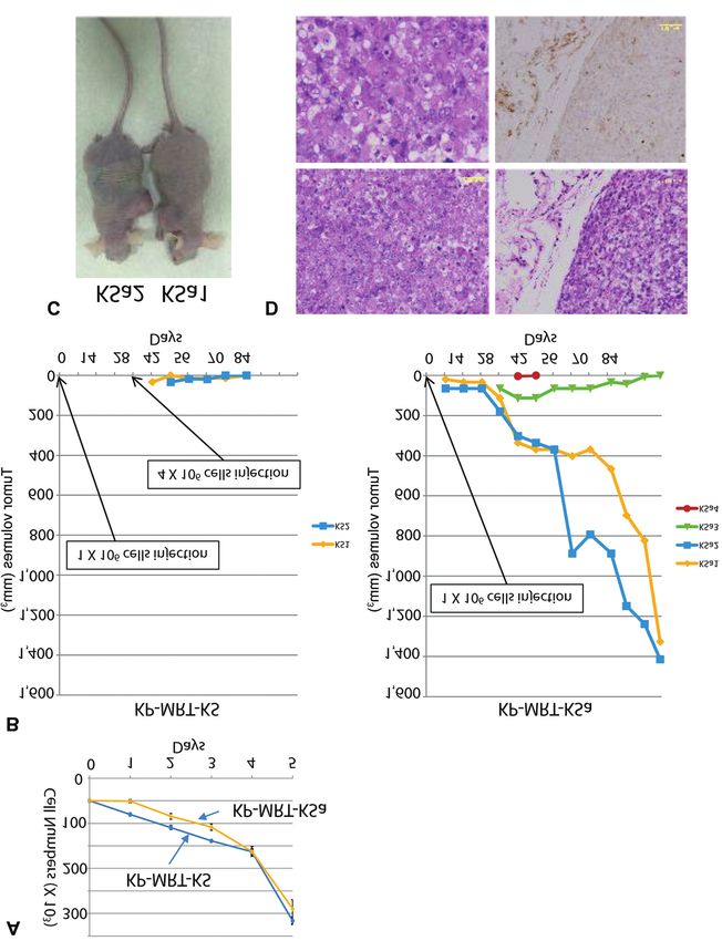
Kuwahara et al: A Pair of New MRT Cell Lines from Synchronous Case

Figure 1. Imaging examination and histopathological findings of the patient prior to treatment. A) Cervical magnetic resonance imaging (MRI)
[Gadolinium (Gd) enhanced T1), B) Cranial MRI (Gd enhanced T1), C) Lung computed tomography scan findings. Arrows indicate tumor lesions.
D) Hematoxylin and eosin (HE) staining of the biopsied clinical tissues (200×). E) Higher magnifications of the sections in D (400×). F)
Immunohistochemical staining of cytokeratin 8 and G) SNF5 (brown color; 200×).

                                                                                                                                      6161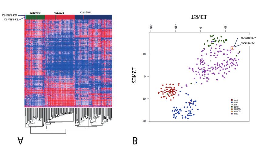
ANTICANCER RESEARCH 40: 6159-6170 (2020)

Figure 2. Structure and histopathological findings of KP-MRT-KS and KP-MRT-KSa cell lines. A) Images obtained with inverted microscopy showing
spindle-shaped cells with processes and flat cells. HE and immunohistochemical staining for SNF5 on constructed cell blocks. (B) Western blotting
analysis of SNF5 and β-actin expression in indicated cell lines.

orthovanadate (Sigma Aldrich)], and incubated for 30 minutes on           Cytogenetic analyses. Cytogenetic analyses were performed with a
ice. Thirty μg of protein was separated by electrophoresis on 10%         short-term culture method (SRL, Inc., Tokyo, Japan). Briefly,
SDS-polyacrylamide        gels   and    electro-transferred   onto        mechanically dispersed cells from the tumor or cell lines were
nitrocellulose membranes (GE Healthcare, Chicago, IL, USA).               cultured for 16 h with 0.06 μg/ml Colcemid (WAKO) to arrest cell
Proteins were analyzed by western blotting using anti SNF5                division at metaphase, followed by hypotonic treatment in 0.075 M
(1:1,000, 612110, BD Biosciences, San Jose, CA, USA), anti-β-             KCl for 25 min. The cells were then fixed with methanol-acetic acid
actin (1:2,000, PM053, MBL, Nagoya, Japan), and 1:2,000                   (3:1, v/v). The karyotype was analyzed by conventional G-banding.
horseradish peroxidase-conjugated anti-rabbit or anti-mouse IgG
(GE Healthcare). Individual proteins were detected using ECL              Histological examination of biopsied specimens. Four μm sections
chemiluminescence reagent (GE Healthcare) and ImageQuant LAS              made from formalin-fixed, paraffin-embedded tumor samples or cell
500 (GE Healthcare).                                                      blocks were stained with hematoxylin and eosin. For

6162
Kuwahara et al: A Pair of New MRT Cell Lines from Synchronous Case

                                                                          immunohistochemical analysis of clinical samples we used the
                                                                          automated Ventana Benchmark Ultra platform (Roche, Basel,
                                                                          Switzerland) as described previously (34). The sections were
                                                                          immuno-stained with i) vimentin (1:400, M7020, DAKO Denmark
                                                                          A/S, Glostrup, Denmark), ii) epithelial membrane antigen (1:200,
                                                                          M0613, DAKO), iii) smooth muscle actin (1:2,000, A2547, Sigma-
                                                                          Aldrich, St. Louis, MO, USA), iv) cytokeratin 8 (1:100, NCL-L-
                                                                          CK8-TS1, Leica Biosystems Inc., Lincolnshire, IL, USA), v)
                                                                          cytokeratin AE1/AE3 (1:100, NCL-L-AE1/AE3, Leica Biosystems),
                                                                          vi) desmin (1:100, M0760, DAKO), vii) S100 (1:2,000, Z0311,
                                                                          DAKO), viii) GFAP (1:2,000, N1506, DAKO), or ix) SNF5
                                                                          (1:2,000, ab58209, Abcam, Cambridge, UK). Written informed
                                                                          consent for use of clinical samples for research was obtained from
                                                                          the patient’s parents according to the protocol approved by the
                                                                          institutional review board of Kyoto Prefectural University of
                                                                          Medicine in accordance with the Declaration of Helsinki.

                                                                          DNA extraction and sequencing analysis; DNA methylation
                                                                          profiling by 850k array. Cells were suspended in an aliquot of
                                                                          Tris/EDTA/NaCl buffer (10 mM Tris pH8.0, 10 mM EDTA pH 8.0
                                                                          and 100 mM NaCl) with 0.5% SDS (Sigma Aldrich) and
                                                                          proteinase K (Sigma Aldrich), and incubated at 55˚C for 3 h, after
                                                                          which the genomic DNA was isolated using a phenol extraction
                                                                          protocol. Briefly, equal volume of Tris-EDTA buffered phenol
                                                                          (WAKO) was added and the mixture was spun down at 14000 rpm
                                                                          for 1 minute. The upper aqueous layer containing DNA was
                                                                          transferred to new tube and mixed with equal volume of
                                                                          chloroform. The mixture was centrifuged at 14000 rpm for 1
                                                                          minute. Then, DNA contained in the aqueous layer was isolated
                                                                          after ethanol precipitation. Sanger sequencing of the genomic
                                                                          DNA was performed with the aid of the BigDye Terminator v1.1
                                                                          Cycle Sequencing kit (Applied Biosystems, Foster City, CA, USA)
                                                                          and the ABI PRISM 3500 Sequence Detection System (Applied
                                                                          Biosystems). The primer sequences used for sequencing SNF5
                                                                          gene exons were kindly provided by Dr Jaclyn A. Biegel
                                                                          (Children’s Hospital Los Angeles) (11). DNA methylation patterns
                                                                          of cell lines were assessed using Illumina Human Methylation850
                                                                          Bead Chip arrays at the Genomics and Proteomics Core Facility
                                                                          of the German Cancer Research Center (DKFZ), according to the
                                                                          manufacturer’s instructions. Methylation analysis and
                                                                          unsupervised hierarchical subgrouping was carried out as
                                                                          described previously (21).

                                                                          Reverse transcription-PCR and single allele analysis. Total RNA was
                                                                          isolated from cultured cells and lymphocytes using the ISOGEN Kit
                                                                          (Nippon Gene, Tokyo, Japan). Single stranded cDNA synthesis from
                                                                          20 μg of template RNA with Oligo-dT (20) was performed using the
                                                                          SuperScript IV First-Strand Synthesis System according to the
                                                                          manufacturer’s instructions (18091050, Thermo Fisher Scientific). For
                                                                          RT-PCR amplification of cDNA, which corresponds to 1 μg RNA,
                                                                          AmpliTaq Gold 360 polymerase (Thermo Fisher Scientific) was used
                                                                          according to the manufacturer’s instructions. A pair of primers to
                                                                          amplify full-length SNF5/INI cDNA were designed according to the
                                                                          procedure described by Biegel et al. (11): INI1CD1 Forward: 5’-
Figure 3. Genetic findings of KP-MRT-KS and KP-MRT-KSa cell lines.        CTGAGCAAGACCTTCGGGCAG-3’; and INI1CD1 Reverse: 5’-
Representative karyotype of the two MRT cell lines, using G-banding. A)   GATGGCTGGCACAAACGTCAG-3’. The PCR products obtained
The karyotype of the tumor sample biopsied from the neck tumor: 46,XX,    with the INI1CD1 primer set were inserted into a T-vector (pRC 2.1
B) The karyotype of KP-MRT-KS: 93,XXXX,i(1)(q10),+2,-                     Vector, K2020, Thermo Fisher Scientific) according to the
4,+del(5)(q?),ins(5;?)(q13;?),-20,+mar1, C) The karyotype of KP-MRT-      manufacturer’s instructions. DH5α competent E. coli cells were
KSa: 46,XX,add(11)(p15),?t(12;17)(q24.1;q24),ins(17;?)(q11.2;?),+mar1.    subsequently transformed with the ligated vector that contains the

                                                                                                                                         6163
ANTICANCER RESEARCH 40: 6159-6170 (2020)
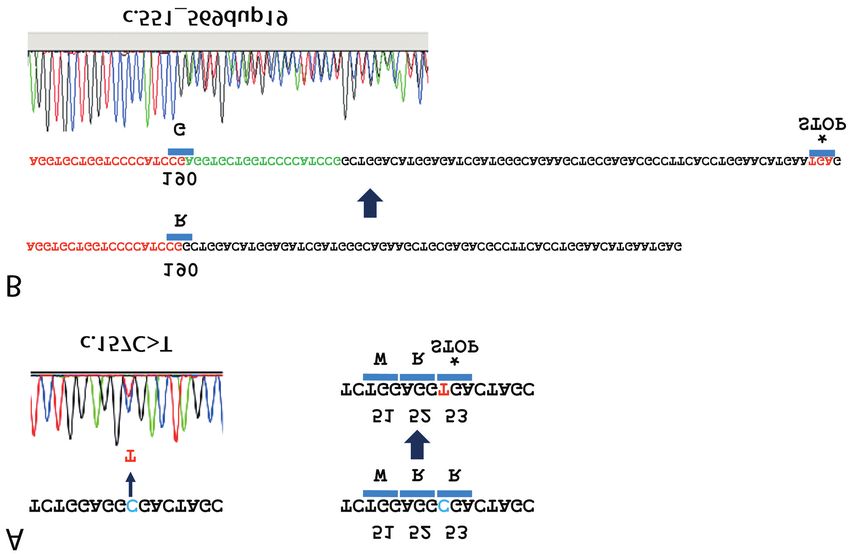
Figure 4. Chromatograms of DNA sequencing indicating alterations of the SNF5 gene for both KP-MRT-KS and KP-MRT-KSa cell lines. The
mutation in exon 2 (c.157 C>T) and the duplication in exon 5 (c.551_569dup19) were observed in both KP-MRT-KS and KP-MRT-KSa cell lines.
A) C→T transition resulted in a nonsense mutation at codon 157 (p.Arg53X). B) Duplication of 19 bp resulted in a frameshift mutation
(p.Arg190ProfsX27), causing the introduction of a premature stop codon.
PCR products. Plasmid DNAs were then extracted from each single Results
transformed colony and sequenced so that SNF5 coding sequences
per single allele could be differentially analyzed.
Establishment of the KP-MRT-KS and KP-MRT-KSa cell
Growth curves of the cell lines in vitro. The in vitro growth rates of lines. KP-MRT-KS and KP-MRT-KSa cells had a round or
the cell lines were assessed as previously described (35). Briefly, spindle-shaped appearance and grew in an adherent mono-
cells (5×10⁴) were plated in the RPMI1640 containing 10% FBS in layer, as previously described for several human MRT cell
triplicate and placed into 24-well cell dishes. Cells were harvested lines (Figure 2A) (27, 29, 33). The cell cultures were
by trypsinization every 24 h and counted using a hemocytometer. maintained for more than 50 passages over a 3-year period,
In Brief, cell suspension was placed between the hemocytometer suggesting that they have attained immortality as cell lines.
and cover glass, and the cell number was counted by use of an
We performed histological examinations using cell blocks
inverted microscope.
constructed from these two cell lines. The cells were small
In vivo growth assay. Six-week old female athymic mice (BALB/c, and round with vesicular nuclei, prominent nucleoli, and a
nu/nu) were injected subcutaneously at the dorsal area with aliquots typical eosinophilic cytoplasmic inclusion body observed in
of 200 μl (1×10⁶ cells/site) of the cell suspension. The mice were both cell lines (Figure 2A). Furthermore, both cell lines were
monitored daily for signs of distress and measured for tumor immunohistochemically positive for vimentin and cytokeratin
development. Tumor diameters were serially measured with calipers,
AE1/AE3 and negative for S100 or GFAP. These findings
and tumor volumes were calculated using the formula: π/6 × (larger
diameter) × (smaller diameter)2 (36). This experimental procedure was
show that the characteristics of both cell lines are consistent
approved by the Committee for Animal Research, Kyoto Prefectural with those of tumor cells from the primary cervical MRT
University of Medicine. After the tumors grew sufficiently, the mice tumor specimens (Figure 1E). We also found that SNF5 was
were euthanized and their xenografts were histologically examined. not expressed in cell block specimens from KP-MRT-KS and
6164Kuwahara et al: A Pair of New MRT Cell Lines from Synchronous Case
KP-MRT-KSa cell lines (Figure 2A). As expected, Western DNA methylation profiling of cell lines. To determine the
blot analysis showed the SNF5 protein was absent from these molecular subgroups of these cell lines, we used the Illumina
two cell lines as well as from the four comparative MRT cell Infinium Human 850K bead array for an unsupervised
lines, in comparison with the SNF5-positive HeLa cell line hierarchical clustering analysis of genome-wide DNA
(Figure 2B). These results indicate that the KP-MRT-KS and methylation. The resulting findings demonstrated that these
KP-MRT-KSa cell lines retained their characteristics as MRT cell lines could be classified as an MYC subgroup (Figure
cells manifested in the original tumor. 5A). This result was further substantiated by a t-distributed
stochastic neighbor embedding (tSNE)based analysis,
Loss of SNF5 expression and its gene alternations in cell lines. (Figure 5B), suggesting that these cell lines had originated
Next, we analyzed the mechanism of the loss of SNF5 from the same cell population.
expression. We first used cytogenetic G-banding analysis to
analyze the karyotypes of the KP-MRT-KS and KP-MRT-KSa In vitro and in vivo characterization of cell lines using a
cell lines as well as of the original tumor sample biopsied from xenograft model. We further examined whether these cell
the neck tumor. The original tumor cells from the neck lesion lines show different growth properties in culture. Figure 6A
showed a normal female karyotype, 46, XX (Figure 3A). In shows similar growth rates for their proliferation in culture
contrast, as shown in Figure 3B, KP-MRT-KS cell line showed with doubling time of approximately 48 h for both cell lines.
93, XXXX, i(1)(q10), +2, -4, +del(5)(q?), ins(5;?)(q13;?), -20, We next assessed the ability of each cell line to form
+mar1, indicating it was a hypo-tetraploid karyotype. Also, the tumors in immunocompromised mice after subcutaneous
KP-MRT-KSa cell line showed 46, XX, add(11)(p15), inoculation. As shown in the right panel of Figure 6B, on day
t(12;17)(q24;1;q24), ins(17;?)(q11.2;?), indicating it was a 7 after injection two of the four mice that had 1×10⁶ KP-
diploid karyotype with a few chromosomal abnormalities MRT-KSa cells injected showed a single subcutaneous mass.
(Figure 3C). Both cell lines as well as the primary tumor cells Four weeks after injection, these two xenograft masses had
showed, as determined by microscopic examination, that there grown exponentially, and 98 days after injection, the
were no alterations of chromosome 22 where the SNF5 gene xenografted mice were sacrificed, followed by resection of
localizes. the tumors (Figure 6C). On the other hand, none of the four
We then evaluated the SNF5 gene status by direct mice that had 1×10⁶ KP-MRT-KS cells injected had
sequencing of the genomic DNA of each of the developed tumors even by day 28 after injection (Figure 6B,
corresponding exons of SNF5. A mutation in exon 2 (c.157 left panel). We therefore injected an additional 4×10⁶ KP-
C>T) and a duplication in exon 5 (c.551_569dup19) were MRT-KS cells on the site opposite to the previous injection
observed in both the KP-MRT-KS and KP-MRT-KSa cell site. Although two of these four mice developed small
lines. The C→T transition had resulted in a nonsense palpable masses, these were transient and eventually
mutation at codon 157 (p.Arg53X) (Figure 4A). The disappeared. Since no tumors developed in any of the four
duplication of 19 bp had caused a frameshift mutation mice injected with KP-MRT-KS cells, these cells showed a
(p.Arg190ProfsX27), resulting in a premature stop codon difference with KP-MRT-KSa cells (Figure 6B, left panel).
(Figure 4B). Histological examination revealed that the tumors derived
Importantly, these mutations in SNF5 exons existed on from the KP-MRT-KSa cell line contained small round cells
different alleles. PCR products, amplified from cDNA with a typical eosinophilic intracytoplasmic inclusion body
reverse-transcribed from mRNA of either of the two cell with negative SNF5 stain, a finding which resembled that for
lines and with a primer set encompassing from exon 1 to the original tumor (Figure 6C).
exon 6 (INI1CD1), had either the mutation in exon 2 or the
duplication in exon 5 in a mutually exclusive fashion. Discussion
We confirmed that the primary neck tumor cells carried
the same compound mutation in exons 2 and 5 for both As a result of the study presented here, we could establish a
SNF5 alleles in their genomic DNA, while only the exon 2 pair of MRT cell lines, KP-MRT-KS and KP-MRT-KSa, that
mutation was present, and not the exon 5 duplication, in the were derived from two different sites of a synchronous MRT
genomic DNA samples extracted from the patient’s white patient suffering from RTPS, displaying similar histological
blood cells. Additional examination of family members was and genetic findings to the characteristic features of the
not possible because their consent could not be obtained. tumor cells from which the cell lines were derived.
Considering the fact that no other family members had The definition of RTPS specifies the presence of multiple
developed MRT or other pediatric tumors, we postulate that Rhabdoid tumors and molecular identification of a germline
the patient had been suffering from synchronous tumors in heterozygous pathogenic variant allele, either SNF5 or
multiple sites, as a case of RTPS with a de novo germline SMARCA4 (Brg-1) (13). Thus, it has been indicated that
mutation in exon 2. significant genes for tumor development exist not only in
6165ANTICANCER RESEARCH 40: 6159-6170 (2020) Figure 5. Identification by means of unsupervised cluster analyses of three distinct molecular subgroups of atypical teratoid rhabdoid tumor (ATRT). A) Unsupervised hierarchical clustering of ATRT methylation profiles. Both cell lines could be classified as an ATRT-MYC subgroup. B) A t- distributed stochastic neighbor embedding (tSNE)based analysis revealed that both cell lines were displayed at almost the same position. somatic mutatiosn but also in germline mutations (37). In our methylation signature of the genomic DNA differs between patient, a point-mutation in exon 2 of the SNF5 gene (c.157 CNS and extra-CNS lesions in most cases of synchronous C>T) was identified as a germline mutation. On the other or metachronous multifocal MRT tumors, even if these hand, a duplication in exon 5 of the SNF5 gene carry the same SNF5 alteration (23). In our case, the KP- (c.551_569dup19) had arisen in tumor cell as an additional MRT-KSa cells from ascites, containing relapsed CNS somatic mutation during the first step in tumor initiation. tumor cells and KP-MRT-KS cells from the neck tumor, Taken together, our case is compatible with RTPS with showed similar DNA methylation and gene expression synchronous tumors having a de novo germline mutation in patterns. From this point of view, the relapsed CNS lesions exon 2 in multiple sites. may have originated as a result of metastasis from the MRT The presence of the non-sense nucleotide substitution neck tumor after treatment, although the possibility remains c.157 C>T in exon 2 has been previously reported in two that the former lesion was derived from the initial cases of RTPS (19, 32) in another tumor cell line, KP-MRT- cerebellum lesion. Nevertheless, the important point is that RY, derived from the patient. Eaton et al. have also reported these two cell lines also had the same compound gene genomic duplication, c.548_566dup19 and c.553_565dup13 alteration, thus, providing further evidence that these two (19), which generate a premature stop codon that is similar cell populations originated from a single cell. These to the c.551_569dup19 of the present case. Although it is findings show that DNA methylation profiling analysis possible that there are hot spots for mutations in the SNF5 should serve as a useful tool not only for detecting MRT gene, which may feature a geographical distribution, the subgroups but also for investigating the pathogenesis of exact frequencies and variations of SNF5 gene alterations synchronous MRT patients. Elucidation of the mechanisms have not yet reported in any Japanese studies. Further through which epigenetic aberrations develop due to SNF5 examinations are, thus, needed to clarify those points with loss could lead us to a more profound understanding of regard to Japanese patients. MRT tumorigenesis. As for the question where the relapsed disseminating Although KP-MRT-KS and KP-MRT-KSa cells have a CNS lesions occurred in our patient, DNA methylation similar epigenetic background, as evidenced by the DNA profiling analysis and precise genotyping provided the methylation results, and share the same compound genetic answer. Pinto et al. have demonstrated that the DNA alteration at the SNF5 loci, their tumorigenic capability in 6166
Kuwahara et al: A Pair of New MRT Cell Lines from Synchronous Case

Figure 6. Cell growth characteristics and tumorigenicity of KP-MRT-KS and KP-MRT-KSa cell lines in vitro and in vivo. (A) Cell growth in vitro.
Both cell lines showed similar growth rates (proliferation) in culture. B) Xenograft model. Tumors developed in two of the four mice injected with
KP-MRT-KSa cells, but no tumor developed in any of the mice with KP-MRT-KS cells. (C) Two representative athymic mice with subcutaneous
tumor inoculated with KP-MRT-KSa cells. D) Histopatological findings of the KSa1 tumor. Left column shows H&E staining (top row: 200×, bottom
row: 400×) and right column shows images of the KSa1 tumor with SNF5 immunostaining (top row: H&E, bottom row: SNF5; brown colour).

                                                                                                                                            6167
ANTICANCER RESEARCH 40: 6159-6170 (2020)
vivo showed sharp differences. It is possible that KP-MRT- tumor. Results of the third National Wilms’ Tumor Study. Cancer
KSa cells, which were obtained at the end of the clinical 64(2): 349-360, 1989. PMID: 2544249. DOI: 10.1002/1097-
course, may have undergone additional mutations that may 0142(19890715)64:23.0.co;2-q
4 Reinhard H, Reinert J, Beier R, Furtwangler R, Alkasser M,
have contributed to their survival and/or growth potential to
Rutkowski S, Fruhwald M, Koscielniak E, Leuschner I, Kaatsch
resist anti-tumoral agents or irradiation. Further examination P and Graf N: Rhabdoid tumors in children: Prognostic factors
of the genomic differences between KP-MRT-KS and KP- in 70 patients diagnosed in germany. Oncol Rep 19(3): 819-823,
MRT-KSa cells can thus be expected to lead to a more 2008. PMID: 18288421. DOI: 10.3892/or.19.3.819
detailed understanding of the mechanisms of resistance to 5 Sultan I, Qaddoumi I, Rodriguez-Galindo C, Nassan AA,
chemotherapy and radiation therapy. Ghandour K and Al-Hussaini M: Age, stage, and radiotherapy,
In conclusion, this is the first report describing the but not primary tumor site, affects the outcome of patients with
malignant rhabdoid tumors. Pediatr Blood Cancer 54(1): 35-40,
establishment of a pair of cell lines from a synchronous MRT
2010. PMID: 19798737. DOI: 10.1002/pbc.22285
patient with RTPS. Now that MRT cell lines with their 6 Rorke LB, Packer R and Biegel J: Central nervous system
distinct epigenetic signature are becoming available, atypical teratoid/rhabdoid tumors of infancy and childhood. J
including the two cell lines described in this report, these Neurooncol 24(1): 21-28, 1995. PMID: 8523069. DOI:
will surely contribute to further elucidation of the 10.1007/BF01052653
tumorigenesis mechanism in relation to the epigenetic 7 Bartelheim K, Nemes K, Seeringer A, Kerl K, Buechner J, Boos
control of this aggressive and dismal pediatric tumor. J, Graf N, Durken M, Gerss J, Hasselblatt M, Kortmann RD,
Teichert von Luettichau I, Nagel I, Nygaard R, Oyen F, Quiroga
Conflicts of Interest
E, Schlegel PG, Schmid I, Schneppenheim R, Siebert R, Solano-
Paez P, Timmermann B, Warmuth-Metz M and Fruhwald MC:
Improved 6-year overall survival in AT/RT - results of the
The Authors declare no potential conflicts of interest. registry study rhabdoid 2007. Cancer Med 5(8): 1765-1775,
2016. PMID: 27228363. DOI: 10.1002/cam4.741
Authors’ Contributions 8 Tomlinson GE, Breslow NE, Dome J, Guthrie KA, Norkool P,
Li S, Thomas PR, Perlman E, Beckwith JB, D’Angio GJ and
YKu, YKa, KO, EK, TY, and JDP carried out the experiments, Green DM: Rhabdoid tumor of the kidney in the National
analyzed the data and prepared the manuscript. YKu, EI, MM, TI, Wilms’ Tumor Study: Age at diagnosis as a prognostic factor. J
HS, SN, SF, and TT were in charge of the treatment of the patient. Clin Oncol 23(30): 7641-7645, 2005. PMID: 16234525. DOI:
MCF, HH and TO edited and approved the manuscript. 10.1200/jco.2004.00.8110
9 Fruhwald MC, Hasselblatt M, Nemes K, Bens S, Steinbugl M,
Acknowledgements Johann PD, Kerl K, Hauser P, Quiroga E, Solano-Paez P,
Biassoni V, Gil-da-Costa MJ, Perek-Polnik M, van de Wetering
M, Sumerauer D, Pears J, Stabell N, Holm S, Hengartner H,
The Authors would like to thank Drs. Martin Hasselblatt, Kenta
Gerber NU, Grotzer M, Boos J, Ebinger M, Tippelt S, Paulus
Yamasaki, Kenjiro Tadagaki, and Noriko Kondo for their useful
W, Furtwangler R, Hernaiz-Driever P, Reinhard H, Rutkowski
comments regarding the experiments.
S, Schlegel PG, Schmid I, Kortmann RD, Timmermann B,
Warmuth-Metz M, Kordes U, Gerss J, Nysom K,
Grant Support Schneppenheim R, Siebert R, Kool M and Graf N: Age and
DNA-methylation subgroup as potential independent risk
This study was supported in part by grants from the JSPS Kakenhi factors for treatment stratification in children with atypical
Grants-in-Aid program (numbers JP15K09487, JP16K10038, teratoid/rhabdoid tumors. Neuro Oncol, 2019. PMID:
JP19K08257, and JP19K08846), and financial support for research 31883020. DOI: 10.1093/neuonc/noz244
projects related to childhood cancer by the Children’s Cancer 10 Versteege I, Sevenet N, Lange J, Rousseau-Merck MF, Ambros P,
Association of Japan. Handgretinger R, Aurias A and Delattre O: Truncating mutations
of hsnf5/ini1 in aggressive paediatric cancer. Nature 394(6689):
References 203-206, 1998. PMID: 9671307. DOI: 10.1038/28212
11 Biegel JA, Zhou JY, Rorke LB, Stenstrom C, Wainwright LM
1 Beckwith JB and Palmer NF: Histopathology and prognosis of and Fogelgren B: Germ-line and acquired mutations of INI1 in
wilms tumors: Results from the first national wilms’ tumor study. atypical teratoid and rhabdoid tumors. Cancer Res 59(1): 74-79,
Cancer 41(5): 1937-1948, 1978. PMID: 206343. DOI: 10.1002/ 1999. PMID: 9892189.
1097-0142(197805)41:53.0.co;2-u 12 Bourdeaut F, Lequin D, Brugieres L, Reynaud S, Dufour C, Doz
2 Haas JE, Palmer NF, Weinberg AG and Beckwith JB: F, Andre N, Stephan JL, Perel Y, Oberlin O, Orbach D, Bergeron
Ultrastructure of malignant rhabdoid tumor of the kidney. A C, Rialland X, Freneaux P, Ranchere D, Figarella-Branger D,
distinctive renal tumor of children. Hum Pathol 12(7): 646-657, Audry G, Puget S, Evans DG, Pinas JC, Capra V, Mosseri V,
1981. PMID: 7275104. DOI: 10.1016/s0046-8177(81)80050-0 Coupier I, Gauthier-Villars M, Pierron G and Delattre O:
3 D’Angio GJ, Breslow N, Beckwith JB, Evans A, Baum H, Frequent hSNF5/INI1 germline mutations in patients with
deLorimier A, Fernbach D, Hrabovsky E, Jones B, Kelalis P, rhabdoid tumor. Clin Cancer Res 17(1): 31-38, 2011. PMID:
Othersen HB, Teft M and Thomas PRM: Treatment of Wilms’ 21208904. DOI: 10.1158/1078-0432.ccr-10-1795
6168Kuwahara et al: A Pair of New MRT Cell Lines from Synchronous Case
13 Nemes K, Bens S, Bourdeaut F, Hasselblatt M, Kool M, Johann Zeller C, Barsyte-Lovejoy D, Lafay-Cousin L, Letourneau L,
P, Kordes U, Schneppenheim R, Siebert R and Fruhwald MC: Bourgey M, Yu M, Gendoo DM, Dzamba M, Barszczyk M,
Rhabdoid tumor predisposition syndrome. In: Genereviews®. Medina T, Riemenschneider AN, Morrissy AS, Ra YS,
Adam MP, Ardinger HH, Pagon RA, Wallace SE, Bean LJH, Ramaswamy V, Remke M, Dunham CP, Yip S, Ng HK, Lu JQ,
Stephens K and Amemiya A (eds.). University of Washington, Mehta V, Albrecht S, Pimentel J, Chan JA, Somers GR, Faria CC,
Seattle, pp. 1993-2020. PMID: 29215836. Roque L, Fouladi M, Hoffman LM, Moore AS, Wang Y, Choi SA,
14 Hasselblatt M, Gesk S, Oyen F, Rossi S, Viscardi E, Hansford JR, Catchpoole D, Birks DK, Foreman NK, Strother D,
Giangaspero F, Giannini C, Judkins AR, Fruhwald MC, Obser Klekner A, Bognar L, Garami M, Hauser P, Hortobagyi T, Wilson
T, Schneppenheim R, Siebert R and Paulus W: Nonsense B, Hukin J, Carret AS, Van Meter TE, Hwang EI, Gajjar A, Chiou
mutation and inactivation of smarca4 (BRG1) in an atypical SH, Nakamura H, Toledano H, Fried I, Fults D, Wataya T, Fryer
teratoid/rhabdoid tumor showing retained SMARCB1 (INI1) C, Eisenstat DD, Scheinemann K, Fleming AJ, Johnston DL,
expression. Am J Surg Pathol 35(6): 933-935, 2011. PMID: Michaud J, Zelcer S, Hammond R, Afzal S, Ramsay DA,
21566516. DOI: 10.1097/PAS.0b013e3182196a39 Sirachainan N, Hongeng S, Larbcharoensub N, Grundy RG, Lulla
15 Schneppenheim R, Fruhwald MC, Gesk S, Hasselblatt M, RR, Fangusaro JR, Druker H, Bartels U, Grant R, Malkin D,
Jeibmann A, Kordes U, Kreuz M, Leuschner I, Martin Subero McGlade CJ, Nicolaides T, Tihan T, Phillips J, Majewski J,
JI, Obser T, Oyen F, Vater I and Siebert R: Germline nonsense Montpetit A, Bourque G, Bader GD, Reddy AT, Gillespie GY,
mutation and somatic inactivation of SMARCA4/BRG1 in a Warmuth-Metz M, Rutkowski S, Tabori U, Lupien M, Brudno M,
family with rhabdoid tumor predisposition syndrome. Am J Hum Schuller U, Pietsch T, Judkins AR, Hawkins CE, Bouffet E, Kim
Genet 86(2): 279-284, 2010. PMID: 2820190. DOI: SK, Dirks PB, Taylor MD, Erdreich-Epstein A, Arrowsmith CH,
10.1016/j.ajhg.2010.01.013 De Carvalho DD, Rutka JT, Jabado N and Huang A: Integrated
16 Bonnin JM, Rubinstein LJ, Palmer NF and Beckwith JB: The (epi)-genomic analyses identify subgroup-specific therapeutic
association of embryonal tumors originating in the kidney and targets in cns rhabdoid tumors. Cancer Cell 30(6): 891-908, 2016.
in the brain. A report of seven cases. Cancer 54(10): 2137-2146, PMID: 27960086. DOI: 10.1016/j.ccell.2016.11.003
1984. PMID: 6091860. DOI: 10.1002/1097-0142(19841115) 23 Pinto EM, Hamideh D, Bahrami A, Orr BA, Lin T, Pounds S,
54:103.0.co;2-d Zambetti GP, Pappo AS, Gajjar A, Agnihotri S and Broniscer A:
17 Biegel JA, Tan L, Zhang F, Wainwright L, Russo P and Rorke Malignant rhabdoid tumors originating within and outside the
LB: Alterations of the hSNF5/INI1 gene in central nervous central nervous system are clinically and molecularly
system atypical teratoid/rhabdoid tumors and renal and heterogeneous. Acta Neuropathol 136(2): 315-326, 2018. PMID:
extrarenal rhabdoid tumors. Clin Cancer Res 8(11): 3461-3467, 6063764. DOI: 10.1007/s00401-018-1814-2
2002. PMID: 12429635. 24 Kuwahara Y, Hosoi H, Osone S, Kita M, Iehara T, Kuroda H and
18 Sredni ST and Tomita T: Rhabdoid tumor predisposition Sugimoto T: Antitumor activity of gefitinib in malignant
syndrome. Pediatr Dev Pathol 18(1): 49-58, 2015. PMID: rhabdoid tumor cells in vitro and in vivo. Clin Cancer Res
25494491. DOI: 10.2350/14-07-1531-misc.1 10(17): 5940-5948, 2004. PMID: 15355927. DOI: 10.1158/
19 Eaton KW, Tooke LS, Wainwright LM, Judkins AR and Biegel 1078-0432.ccr-04-0192
JA: Spectrum of SMARCB1/INI1 mutations in familial and 25 Kuwahara Y, Charboneau A, Knudsen ES and Weissman BE:
sporadic rhabdoid tumors. Pediatr Blood Cancer 56(1): 7-15, Reexpression of hSNF5 in malignant rhabdoid tumor cell lines
2011. PMID: 3086793. DOI: 10.1002/pbc.22831 causes cell cycle arrest through a p21cip1/waf1-dependent
20 Abu Arja MH, Patel P, Shah SH, Auletta JJ, Meyer EK, Conley mechanism. Cancer Res 70(5): 1854-1865, 2010. PMID:
SE, Aldrink JH, Pindrik JA and AbdelBaki MS: Synchronous 20179200. DOI: 10.1158/0008-5472.CAN-09-1922
central nervous system atypical teratoid/ rhabdoid tumor and 26 Suzuki A, Ohta S and Shimada M: Gene expression of malignant
malignant rhabdoid tumor of the kidney: Case report of a long- rhabdoid tumor cell lines by reverse transcriptase-polymerase
term survivor and review of the literature. World Neurosurg, chain reaction. Diagn Mol Pathol 6(6): 326-332, 1997. PMID:
2017. PMID: 29223518. DOI: 10.1016/j.wneu.2017.11.158 9559292. DOI: 10.1097/00019606-199712000-00004
21 Johann PD, Erkek S, Zapatka M, Kerl K, Buchhalter I, Hovestadt 27 Kuroda H, Moritake H, Sawada K, Kuwahara Y, Imoto I,
V, Jones DT, Sturm D, Hermann C, Segura Wang M, Korshunov Inazawa J and Sugimoto T: Establishment of a cell line from a
A, Rhyzova M, Grobner S, Brabetz S, Chavez L, Bens S, Groschel malignant rhabdoid tumor of the liver lacking the function of
S, Kratochwil F, Wittmann A, Sieber L, Georg C, Wolf S, Beck K, two tumor suppressor genes, hSNF5/INI1 and p16. Cancer Genet
Oyen F, Capper D, van Sluis P, Volckmann R, Koster J, Versteeg Cytogenet 158(2): 172-179, 2005. PMID: 15796965. DOI:
R, von Deimling A, Milde T, Witt O, Kulozik AE, Ebinger M, 10.1016/j.cancergencyto.2004.08.032
Shalaby T, Grotzer M, Sumerauer D, Zamecnik J, Mora J, Jabado 28 Kato H, Ohta S, Koshida S, Narita T, Taga T, Takeuchi Y and
N, Taylor MD, Huang A, Aronica E, Bertoni A, Radlwimmer B, Sugita K: Expression of pericyte, mesangium and muscle markers
Pietsch T, Schuller U, Schneppenheim R, Northcott PA, Korbel JO, in malignant rhabdoid tumor cell lines: Differentiation-induction
Siebert R, Fruhwald MC, Lichter P, Eils R, Gajjar A, Hasselblatt using 5-azacytidine. Cancer Sci 94(12): 1059-1065, 2003. PMID:
M, Pfister SM and Kool M: Atypical teratoid/rhabdoid tumors are 14662021. DOI: 10.1111/j.1349-7006.2003.tb01401.x
comprised of three epigenetic subgroups with distinct enhancer 29 Sugimoto T, Hosoi H, Horii Y, Ishida H, Mine H, Takahashi K,
landscapes. Cancer Cell 29(3): 379-393, 2016. PMID: 26923874. Abe T, Ohta S and Sawada T: Malignant rhabdoid-tumor cell
DOI: 10.1016/j.ccell.2016.02.001 line showing neural and smooth-muscle-cell phenotypes. Int J
22 Torchia J, Golbourn B, Feng S, Ho KC, Sin-Chan P, Vasiljevic A, Cancer 82(5): 678-686, 1999. PMID: 10417765. DOI:
Norman JD, Guilhamon P, Garzia L, Agamez NR, Lu M, Chan 10.1002/(sici)1097-0215(19990827)82:53.0.co;2-k
6169ANTICANCER RESEARCH 40: 6159-6170 (2020) 30 Mori T, Fukuda Y, Kuroda H, Matsumura T, Ota S, Sugimoto T, 35. Kuwahara Y, Mora-Blanco EL, Banine F, Rogers AB, Fletcher Nakamura Y and Inazawa J: Cloning and characterization of a C, Sherman LS, Roberts CW and Weissman BE: Establishment novel Rab-family gene, Rab36, within the region at 22q11.2 that and characterization of MRT cell lines from genetically is homozygously deleted in malignant rhabdoid tumors. engineered mouse models and the influence of genetic Biochem Biophys Res Commun 254(3): 594-600, 1999. PMID: background on their development. Int J Cancer 132(12): 2767- 9920784. DOI: 10.1006/bbrc.1998.9968 2777, 2013. PMID: 23197309. DOI: 10.1002/ijc.27976 31 Misawa A, Hosoi H, Imoto I, Iehara T, Sugimoto T and Inazawa 36 Ciardiello F, Bianco R, Damiano V, De Lorenzo S, Pepe S, De J: Translocation (1;22)(p36;q11.2) with concurrent del(22) Placido S, Fan Z, Mendelsohn J, Bianco AR and Tortora G: (q11.2) resulted in homozygous deletion of SNF5/INI1 in a Antitumor activity of sequential treatment with topotecan and newly established cell line derived from extrarenal rhabdoid anti-epidermal growth factor receptor monoclonal antibody tumor. J Hum Genet 49(10): 586-589, 2004. PMID: 15378398. c225. Clin Cancer Res 5(4): 909-916, 1999. PMID: 10213228. DOI: 10.1007/s10038-004-0191-y 37 Yadav S, Sarkar NDE, Kumari N, Krishnani N, Kumar A and 32 Katsumi Y, Kuwahara Y, Tamura S, Kikuchi K, Otabe O, Mittal B: Targeted gene sequencing of gallbladder carcinoma Tsuchiya K, Iehara T, Kuroda H, Hosoi H and Sugimoto T: identifies high-impact somatic and rare germline mutations. Trastuzumab activates allogeneic or autologous antibody- Cancer Genomics Proteomics 14(6): 495-506, 2017. PMID: dependent cellular cytotoxicity against malignant rhabdoid tumor 29109099. DOI: 10.21873/cgp.20059 cells and interleukin-2 augments the cytotoxicity. Clin Cancer Res 14(4): 1192-1199, 2008. PMID: 18281554. DOI: 10.1158/1078-0432.ccr-07-1661 33 Betz BL, Strobeck MW, Reisman DN, Knudsen ES and Weissman BE: Re-expression of hSNF5/INI1/BAF47 in pediatric tumor cells leads to G1 arrest associated with induction of p16ink4a and activation of RB. Oncogene 21(34): 5193-5203, 2002. PMID: 12149641. DOI: 10.1038/sj.onc.1205706 34 Ouchi K, Kuwahara Y, Iehara T, Miyachi M, Katsumi Y, Tsuchiya K, Konishi E, Yanagisawa A and Hosoi H: A NOXA/MCL-1 imbalance underlies chemoresistance of malignant rhabdoid Received August 21, 2020 tumor cells. J Cell Physiol 231(9): 1932-1940, 2016. PMID: Revised September 15, 2020 26680268. DOI: 10.1002/jcp.25293 Accepted September 16, 2020 6170
You can also read