Obesity Prevents S-Adenosylmethionine-Mediated Improvements in Age-Related Peripheral and Hippocampal Outcomes - MDPI
←
→
Page content transcription
If your browser does not render page correctly, please read the page content below
nutrients
Article
Obesity Prevents S-Adenosylmethionine-Mediated
Improvements in Age-Related Peripheral and
Hippocampal Outcomes
Jacob W. Vander Velden                    and Danielle M. Osborne *

                                           R.S. Dow Neurobiology Department, Legacy Research Institute, Portland, OR 97232, USA;
                                           jvandervelden1@student.gsu.edu
                                           * Correspondence: dosborne@downeurobiology.org

                                           Abstract: Background: Age predisposes individuals to a myriad of disorders involving inflamma-
                                           tion; this includes stress-related neuropsychiatric disorders such as depression and anxiety, and
                                           neurodegenerative diseases. Obesity can further exacerbate these effects in the brain. We investi-
                                           gated whether an inexpensive dietary supplement, s-adenosylmethionine (SAMe), could improve
                                           age- and/or obesity-related inflammatory and affective measures in the hippocampus. Methods:
                                           Mice were placed on their diets at six weeks of age and then aged to 14 months, receiving SAMe
                                           (0.1 g/kg of food) for the final six weeks of the experiment. Prior to tissue collection, mice were
                                           tested for anxiety-like behaviors in the open field test and for metabolic outcomes related to type
                                           2 diabetes. Results: SAMe treatment significantly improved outcomes in aged control mice, where
                                           fasting glucose decreased, liver glutathione levels increased, and hippocampal microglia morphology
                                           improved. SAMe increased transforming growth factor β-1 mRNA in both control mice, potentially

                                    accounting for improved microglial outcomes. Obese mice demonstrated increased anxiety-like
Citation: Vander Velden, J.W.;
                                           behavior, where SAMe improved some, but not all, open field measures. Conclusions: In summary,
Osborne, D.M. Obesity Prevents             SAMe boosted antioxidant levels, improved diabetic measures, and hippocampal inflammatory and
S-Adenosylmethionine-Mediated              behavioral outcomes in aged mice. The effects of SAMe in obese mice were more subdued, but it
Improvements in Age-Related                could still provide some positive outcomes for obese individuals dealing with anxiety and having
Peripheral and Hippocampal                 difficulty changing their behaviors to improve health outcomes.
Outcomes. Nutrients 2021, 13, 1201.
https://doi.org/10.3390/nu13041201         Keywords: aging; anxiety; s-adenosylmethionine; transforming growth factor b-1; TGFβ1; obesity;
                                           microglia; glutathione
Academic Editor: Roberto Iacone

Received: 27 February 2021
Accepted: 2 April 2021
                                           1. Introduction
Published: 6 April 2021
                                                Obesity is a chronic inflammatory disease; alone it perturbs metabolic function, but
Publisher’s Note: MDPI stays neutral
                                           also can exacerbate the negative effects of aging on the brain, and more specifically the
with regard to jurisdictional claims in
                                           hippocampus [1]. Age and obesity are both demarked by steady increases in several pro-
published maps and institutional affil-    inflammatory factors, effects largely induced by white adipose tissue driving activation
iations.                                   of pro-inflammatory kinases (i.e., JNK, NFκB, and IκB kinase β reviewed in [2,3]) that
                                           target insulin-sensitive organs. The brain is an insulin responsive tissue [4] that is sus-
                                           ceptible to inflammatory damage [5] by kinases known to be increased by aging [3], and
                                           made worse by obesity [6], and which activate the brain’s immune cells, microglia. Acti-
Copyright: © 2021 by the authors.
                                           vated ameboid-shaped microglia are triggered by metabolic dysfunction and implicated
Licensee MDPI, Basel, Switzerland.
                                           in hippocampal pathological conditions [7] and impact surrounding neurons by releasing
This article is an open access article
                                           cytokines (reviewed in [8]). Key to controlling microglia is transforming growth factor
distributed under the terms and            beta-1 (TGFβ1). TGFβ1 is necessary for maturating microglia, while TGFβ1 deficient mice
conditions of the Creative Commons         are completely void of brain microglia [9]. Neurodegenerative diseases suppress TGFβ1
Attribution (CC BY) license (https://      leading to perturbed microglial homeostasis [10], supporting that decreased TGFβ1 can
creativecommons.org/licenses/by/           indicate declining brain health and activated microglial states. Thus, therapeutics that
4.0/).                                     safely reduce activated microglia may help alleviate symptoms of neurological conditions.

Nutrients 2021, 13, 1201. https://doi.org/10.3390/nu13041201                                              https://www.mdpi.com/journal/nutrients
Nutrients 2021, 13, 1201 2 of 14
S-adenosylmethionine (SAMe) is an over-the-counter (OTC) dietary supplement and
is sought for its ability to increase levels of the antioxidant glutathione [11]. SAMe is
decreased in the livers of non-obese type 2 diabetic Goto-Kakizaki rats [12], db/db mice [13],
and mice fed a high-fat diet [14]. Inclusion of SAMe into a diet attenuated sucrose-facilitated
meal-induced insulin sensitization, an indicator of diabetic development, possibly due to
its ability to increase glutathione in the liver [15]. SAMe plasma levels are also decreased
in Type 2 Diabetic patients, while those with the lowest SAMe also demonstrated mild
cognitive impairment [16]. SAMe has other demonstrated abilities to affect the brain.
Cultured microglia treated with SAMe showed attenuated levels of LPS-induced increases
of IL-1β, TNF-1α, and IL-6 gene expression [17]. Several studies have demonstrated
cognitive and pathological improvements in Alzheimer’s disease transgenic mouse models
treated with SAMe [18–21]. Although SAMe has shown promise in clinical and pre-
clinical trials for ameliorating cognitive decline and depressive symptoms [22], it has not
been evaluated for anxiolytic/anxiogenic effects, another behavior with a strong link to
hippocampal function and a common co-morbidity among obese [23], aging [24], and/or
demented patients [25]. We hypothesized that feeding aged mice SAMe, with or without
diet-induced obesity (DIO), would improve peripheral measures of insulin resistance,
decrease inflammation in the hippocampus, and improve affective behaviors in both aged
and obese aged mice. Results supported that SAMe has the potential to improve blood
glucose regulation, increase hippocampal TGFβ1 levels, reduce reactive microglia, and
improve affect; however, the effects in obese mice were more limited.
2. Materials and Methods
All methods and procedures performed on mice were approved by the Institutional
Animal Use and Care Committee at Legacy Research Institute and conducted under pro-
tocol LRI-111-2017. Mice were maintained on a 12/12 h light/dark cycle, with lights on
at 0730.
2.1. Mice and Diets
C57BL/6 male mice were obtained from Jackson laboratories at five weeks of age
and housed 5/cage. Mice were randomly assigned to either control chow (Chow, Purina
5001) diet or diet-induced obesity (DIO) using an obesogenic diet, 60% high-fat food +10%
sucrose in the water, beginning at 6 weeks of age. Mice had ad libitum access to food
and water and were handled regularly for the duration of the experiment when they were
weighed/health checked weekly. SAMe administration was based on previously published
methods, which also found this dose increased brain levels of SAMe [19]. At 12 months of
age, half the mice were given a modified version of their existing diet containing 0.1 g of
SAMe/kg of food. Mice consumed approximately 400 µg of SAMe/day based on 4 g of
food consumption/mouse/day. SAMe was added to a standard 60% high-fat diet (D12492,
Research Diets) or a custom control diet formulated with the same macronutrient content as
Purina 5001. SAMe (derived from Denosyl 425 mg) was obtained from a commercial source.
Special diets were maintained until tissue was collection at 14 months of age. Mice were
euthanized by rapid cervical dislocation followed by decapitation. Blood was collected
and stored on ice until it was centrifuged and the supernatant removed and stored in a
separate collection tube. Samples were stored at −80 ◦ C. The dorsal hippocampus was
bilaterally dissected out, the rest of the brain was kept, and a small piece of liver was
removed. Samples were immediately frozen in liquid nitrogen and stored at −80 ◦ C.
2.2. Glucose Tolerance Testing (GTT)
Prior to SAMe treatment, as a means to determine the extent of metabolic dysfunction,
mice were tested for insulin resistance at 9 months of age to confirm phenotype. Mice were
fasted for six hours beginning at lights on at 0730, but maintained ad libitum access to
water (sucrose water was replaced with regular tap water in DIO mice). Mice were then
administered 1.5 g/kg i.p. of dextrose. Whole blood was tested using AgaMatrix PrestoNutrients 2021, 13, 1201 3 of 14
meter and testing strips at baseline and 15, 30, 60, 90, 120, and 180 min post-dextrose. At
completion of the final time point, mice were returned to their homecage with reinstated
ad libitum access to food and water.
2.3. Open Field
Mice were placed into the periphery of the open field box (40 cm × 40 cm × 40 cm) and
allowed to freely explore for 30 min. Speed and distance were analyzed using Ethovision
Software (Noldus, Wageningen, The Netherlands) in five minute bins, from a ceiling-
mounted camera. Anxiety-like behavior was determined based only on the first 10 min in
the box by measuring the latency to enter, and the duration of time, spent in the center of
the open field box.
2.4. Immunohistochemistry
Mice (n = 5–8/group) were put under anesthesia with isofluorane. Ice cold PBS was
perfused through each animal followed by ice cold 4% paraformaldehyde. Brains were
removed and placed in 4% paraformaldehyde for 24 h, then were transferred to 30% sucrose
until they sunk, and then stored at −80 ◦ C. Brains were sectioned at 40 microns. Sections
were permeabilized in 1% Triton and PBS for 1 h followed by 2N HCl for 30 min at 37 ◦ C
then neutralized with 0.1 M sodium tetraborate and incubated for a further 10 min at room
temperature. Slices were blocked with appropriate goat donkey blocking buffer for 1 h at
room temperature then primaries were added for overnight incubation at 4 ◦ C on rotator:
Iba1 (Abcam, 1:500). The next day slices were rinsed then incubated with appropriate
secondary antibody (AlexaFluor, Life Technologies) for 1 h at room temperature. Dapi was
applied in the coverslip mounting medium. Slides were imaged by confocal imaging Leica
LasX software system (Leica, Buffalo Grove, IL, USA).
Microglia size measurements were completed by Leica Application Suite analysis
software (Leica, Buffalo Grove, IL, USA) and Image J. Images were taken at 40x with a
resolution of 2048 × 2048 for maximum visibility centered on the CA1. All scoring was
completed by experimenters blind to conditions and based on previous methods [26]. For
soma measurements, Leica software was used to remove the background and a frame
(1200 × 1000 px; 153,938 µm2 , this same sized frame was used throughout for all morpho-
logical measurements) was placed within the CA1. Only microglia completely within the
frame were included in analyses (31–90 microglia in each image). Although the software
isolated microglia, every image was carefully scanned and compared with the original to
ensure whole visible microglia were included. For branch counting, ImageJ software was
used to create a skeletonized binary high-contrast image (Skeletonize3D plugin) to isolate
microglia (20–24 cells/image) and analyzed using the AnalyzeSkeleton plugin.
2.5. qPCR
All qPCR procedures were conducted by the Gene Profiling Shared Resource Center
at OHSU (Portland, OR) using largely automated methods. RNA was isolated from hip-
pocampus (n = 4–5/group) using Qiagen RNeasy mini kit with QIAcube automation. RNA
quality was assessed by Agilent 2100 Bioanalyzer with a Eukaryote total RNA Nano chip.
The core also performed reverse transcription using SuperScript VILO cDNA synthesis
kit (Invitrogen, #11754050) with ~16 ng RNA per well. Following cDNA synthesis, 2 µL
of cDNA was used in the PCR reaction using 10 µL of TaqMan Master Mix II (Invitrogen,
#4440040) and 1 µL of 20× gene specific TaqMan assay was loaded into QuantStudio
Real-time PCR System (Life Technologies) using a single mastermix per TaqMan probe
for Transforming Growth Factor beta 1 (TGFB1; Mm01178820_m1, ThermoFisher). Data
was collected using Applied Biosystems QuantStudio 12K Flex Software (v1.2.2). GAPDH
(Mm99999915_g1, ThermoFisher) was used as the control gene in calculating ∆CT. Ct levels
were all within the acceptable range, reference gene variance was stable across the samples,
and replicates showed inter-assay validity.Nutrients 2021, 13, 1201 4 of 14
2.6. Glutathione Assay
Measurement of reduced glutathione in brain and liver utilized a kinetic assay kit
(Sigma #CS0260) and followed manufacturer’s instructions (n = 7–8/group). In brief,
several hundred milligrams of tissue was required for this ELISA: liver and whole brains
(minus hippocampus used in qPCR) were dissected from mice following cervical disloca-
tion. Samples were immediately placed in liquid nitrogen and then transferred to −80 ◦ C
for long-term storage. For preparation, samples were pulverized, weighed (10–300 mg
required for assay) and then 3 volumes of 5-sulfosalicylic acid added and vortexed, and
another seven volumes of 5% SSA solution added. Samples were then homogenized with a
pestle in a glass tube until an even suspension was achieved. Samples were left on ice for
10 min prior to centrifuging at 10,000× g for 10 min. The volume of the supernatant was
measured. Samples were further diluted 1:20 with 5% SSA to obtain the working solution.
Blanks, standard curve and samples were plated (samples were counterbalanced across
conditions and organ type). Following manufacturer instructions, the plate was read in a
plate reader, measuring absorption at 412 nm at one-minute intervals for five minutes. All
samples were run in triplicate.
2.7. S-Adenosylmethionine ELISA
SAMe was measured in serum samples per manufacturer instructions (BioVision,
E4541). In brief, serum samples were diluted 1:1 with provided sample buffer and run
along with standards in duplicate. Plated samples were combined with biotin-detection
antibody and allowed to incubate for 45 min at 37 ◦ C. Solution was removed, and the plate
washed three times with 1× wash buffer. HRP-streptavidin conjugate was added to the
wells and incubated for a further 30 min at 37 ◦ C. Plate was emptied, and then washed
5 times with 1× wash buffer. TMB substrate was added to the wells and incubated for
15 min at 37 ◦ C, at which point STOP solution was added to terminate the reaction. Plate
was immediately read at 450 nm.
2.8. S-Adenosylhomocysteine Hydrolase ELISA
S-adenosylhomocysteine hydrolase (SAHH) was measured in serum samples per
manufacturer instructions (Aviva Systems Biology, Ahcy ELISA kit (Mouse), OKEH03618).
In brief, serum samples were diluted 1:3 with provided sample buffer and run along with
standards in duplicate. Samples were plated and incubated for 2 h at 37 ◦ C. Samples
were removed, and 1× biotinylated Ahcy detector antibody was added to the plate and
incubated for 1 h at 37 ◦ C. Fluid was removed and the plate was washed three times with
1× wash buffer. Avidin-HRP Conjugate was added to each well and the plate incubated
for 1 h at 37 ◦ C. Fluid was removed and the plate was washed five times with 1× wash
buffer. TMB solution was added to the wells and incubated for 22 min at 37 ◦ C, at which
point STOP solution was added to terminate the reaction. The plate was immediately read
at 450 nm.
2.9. Experimental Design and Statistical Analysis
Two-way ANOVAs were conducted for all comparisons (Diet x SAMe Treatment),
except for GTT open field distance which were mixed model repeated measures ANOVAs.
Statistical analysis and graphing was completed using GraphPad Prism 9.0 Software.
3. Results
3.1. SAMe Improves Fasting Glucose in Chow, but Not DIO, Mice
GTT was done prior to the administration of SAMe to confirm metabolic dysfunction
in DIO mice. DIO mice developed insulin resistance as indicated by significantly increased
and prolonged blood glucose levels following dextrose administration (significant interac-
tion, F6,564 = 4.3, p < 0.001; Figure 1A). With SAMe supplementation, Chow mice showed
a significant reduction in fasting glucose levels, an effect not observed in DIO mice (interac-
tion, F1,83 = 11.7, p < 0.005; Figure 1B). There was a main effect of diet, where DIO mice3.1. SAMe Improves Fasting Glucose in Chow, but Not DIO, Mice
GTT was done prior to the administration of SAMe to confirm metabolic dysfunction
in DIO mice. DIO mice developed insulin resistance as indicated by significantly increased
and prolonged blood glucose levels following dextrose administration (significant inter-
Nutrients 2021, 13, 1201 5 of 14
action, F6,564 = 4.3, p < 0.001; Figure 1A). With SAMe supplementation, Chow mice
showed a significant reduction in fasting glucose levels, an effect not observed in DIO
mice (interaction, F1,83 = 11.7, p < 0.005; Figure 1B). There was a main effect of diet, where
DIO micesignificantly
weighed weighed significantly more than
more than Chow mice, Chow mice,ofregardless
regardless of SAMe
SAMe treatment treatment
(F1,85 = 980.9,
(F1,85 = 980.9,
p < 0.0001; p < 0.0001;
Figure 1C). Figure 1C).
Figure 1.
Figure (A).Diet-induced
1. (A). Diet-inducedobesity
obesity(DIO)
(DIO)mice
micehad
hadsignificantly
significantlyimpaired
impairedglucose
glucoseregulation
regulationfol-
fol-
lowingglucose
lowing glucose tolerance
tolerance test
test (n
(n == 43–53/group).
43–53/group).(B).(B).Following
Followingsix
sixhours
hoursofoffasting,
fasting,Chow
Chowmicemice fed
fed
SAMe demonstrated
SAMe demonstrated improved glucose glucose homeostasis
homeostasiswith withreduced
reducedblood
bloodglucose
glucose levels relative
levels to all
relative to
all other
other condition
condition groups
groups (n =(n18–25/group).
= 18–25/group).(C).(C).
DIODIOmicemice weighed
weighed significantly
significantly moremore
than than
mice mice
fed a
fed
Chowa Chow
diet, diet, regardless
regardless of SAMeof SAMe treatment.
treatment. *: Significant
*: Significant difference.
difference.
3.2. Chow, but Not DIO, Mice Show Improved SAMe Utilization with SAMe Treatment
Chow + SAMe mice had significantly decreased serum levels of SAMe (F1,36 = 15.2,
p < 0.001; Figure 2A), while SAH levels were significantly increased (F1,36 = 6.5, p < 0.05;
Figure 2B). Chow mice treated with SAMe showed a significant increase in liver glutathione
levels, relative to other groups, while no change was observed in DIO mice (interaction,
F1,26 = 7.7, p < 0.05; Figure 2C). No changes in brain glutathione levels were detected in
either Chow or DIO mice (Figure 2D).3.2. Chow, but Not DIO, Mice Show Improved SAMe Utilization with SAMe Treatment
Chow + SAMe mice had significantly decreased serum levels of SAMe (F1,36 = 15.2,
p < 0.001; Figure 2A), while SAH levels were significantly increased (F1,36 = 6.5, p < 0.05;
Figure 2B). Chow mice treated with SAMe showed a significant increase in liver glutathi-
Nutrients 2021, 13, 1201 one levels, relative to other groups, while no change was observed in DIO mice (interac-6 of 14
tion, F1,26 = 7.7, p < 0.05; Figure 2C). No changes in brain glutathione levels were detected
in either Chow or DIO mice (Figure 2D).
Figure 2.
Figure SAMe (A)
2. SAMe (A) and
and (SAHH)
(SAHH) (B)
(B) were
were analyzed
analyzed in
in serum
serum samples
samples (n (n== 10/group). Liver (C)
10/group). Liver (C) and
and brain
brain (D)
(D) were
were collected
collected
for analysis
for analysis of
of total
total glutathione
glutathione levels
levels (n
(n == 7–8/group). InChow
7–8/group). In Chow mice
mice only,
only, SAMe
SAMe treatment
treatment lowered
lowered serum
serum SAMe,
SAMe, but
but
increased serum SAH and liver glutathione. Summary of key aspects of the transmethylation
glutathione. (E) Summary transmethylation and transsulferation
transsulferation
pathways that pertain to SAMe, SAHH, and glutathione levels. levels. *: Significant difference.
3.3. DIO
3.3. DIO and
and SAMe
SAMe Treatment
Treatment Affected
Affected Measures
Measures of
of Microglial
Microglial Activity
Activity in
in the
the CA1
CA1 Region
Region of
of
Dorsal Hippocampus
Dorsal Hippocampus
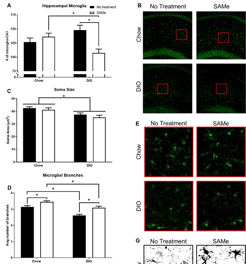
DIO negatively affected microglia soma area and branching, but not density or TGFB1
DIO negatively affected microglia soma area and branching, but not density or
transcripts; SAMe improved branching outcomes of both Chow and DIO mice, while
TGFB1 transcripts; SAMe improved branching outcomes of both Chow and DIO mice,
also affecting microglial density in DIO mice and TGFB1 transcripts in Chow mice only.
while also affecting microglial density in DIO mice and TGFB1 transcripts in Chow mice
SAMe treatment to DIO mice decreased the number of hippocampal microglia compared
only. SAMe treatment to DIO mice decreased the number of hippocampal microglia com-
to Chow + SAMe and DIO + No Treatment mice. There was a significant interaction
pared to Chow + SAMe and DIO + No Treatment mice. There was a significant interaction
(F1,19 = 11.8, p < 0.01; Figure 3A,B) and main effect of treatment (F1,19 = 4.5, p < 0.05) in
the number of microglia counted within the sub-neuronal area of CA1 region of the dorsal
hippocampus. Morphologically, DIO mice had significantly smaller microglial bodies,
commensurate with a heightened inflammatory state expected from obesity (significant
main effect, F1,22 = 13.3, p < 0.01; Figure 3C,D). There were significant main effects for diet
(F1,22 = 29.4, p < 0.0001) and treatment (F1,22 = 23.5, p < 0.0001); DIO mice had significantly
fewer microglial branches, while SAMe treatment increased branching in both Chow and
DIO mice (Figure 3E,F). Finally, there was a main effect of SAMe treatment (F1,15 = 6.1,(F1,19 = 11.8, p < 0.01; Figure 3A,B) and main effect of treatment (F1,19 = 4.5, p < 0.05) in
the number of microglia counted within the sub-neuronal area of CA1 region of the dorsal
hippocampus. Morphologically, DIO mice had significantly smaller microglial bodies,
commensurate with a heightened inflammatory state expected from obesity (significant
main effect, F1,22 = 13.3, p < 0.01; Figure 3C,D). There were significant main effects for diet
Nutrients 2021, 13, 1201 7 of 14
(F1,22 = 29.4, p < 0.0001) and treatment (F1,22 = 23.5, p < 0.0001); DIO mice had significantly
fewer microglial branches, while SAMe treatment increased branching in both Chow and
DIO mice (Figure 3E,F). Finally, there was a main effect of SAMe treatment (F1,15 = 6.1, p
< 0.05; Figurep3G) on TGFB1
< 0.05; transcripts.
Figure 3G) on TGFB1 Post hoc tests Post
transcripts. demonstrate
hoc tests that SAMe increased
demonstrate that SAMe increased
transcripts oftranscripts
TGFB1 in of theTGFB1
hippocampus of Chow mice
in the hippocampus ofonly,
Chownomice
differences
only, norelative to relative to
differences
untreated DIO mice were
untreated DIO observed.
mice were observed.
Figure 3. Hippocampal microglia were quantified within the sub-pyramidal area of CA1 for each condition (n = 5–7/group).
(A) SAMe treatment in DIO mice significantly reduced microglia density within the CA1, compared to Chow + SAMe and
untreated DIO counterparts. (B) Representative confocal images of hippocampal microglia. (C) Somal area of microglia
were measured. DIO significantly reduced the cell body size of microglia. (D) Representative confocal images of microglial
morphology. (E) Both DIO and SAMe affected the number of microglia branches. (F) Representative high contrast images.
(G) Transforming Growth Factor beta 1 (TGFb1) was measured by qPCR (n = 4–5/group) using hippocampal samples.
SAMe treatment significantly increased TGFb1 expression in Chow mice. *: Significant difference.tive confocal images of hippocampal microglia. (C) Somal area of microglia were measured. DIO
significantly reduced the cell body size of microglia. (D) Representative confocal images of micro-
glial morphology. (E) Both DIO and SAMe affected the number of microglia branches. (F) Repre-
sentative high contrast images. (G) Transforming Growth Factor beta 1 (TGFb1) was measured by
qPCR (n = 4–5/group) using hippocampal samples. SAMe treatment significantly increased TGFb1
Nutrients 2021, 13, 1201 8 of 14
expression in Chow mice. *: Significant difference.
3.4. SAMe Treatment Ameliorates some of the Increases in Anxiety-Like Behavior Caused by
DIO 3.4. SAMe Treatment Ameliorates Some of the Increases in Anxiety-Like Behavior Caused by DIO
Not surprisingly,Not DIO mice covered
surprisingly, less distance
DIO mice covered lessin the open field,
distance in theimportantly
open field, importantly
though, they moved
though, consistently
they moved throughout the entire
consistently testingthe
throughout period (main
entire effect
testing of diet,
period (main effect of
F3,50 = 21, pNutrients 2021, 13, 1201 9 of 14
from its glycogen stores [31]; in diabetics, the liver releases excessive glucose leading to
persistent hyperglycemia [30]. The improved glucose measure in Chow + SAMe mice
may reflect improved liver function, as is supported by the increase in SAHH and liver
glutathione that was observed exclusively in Chow + SAMe mice.
The elevated glutathione is likely a product of improved and increased SAMe metabolism
through the transmethylation and transsulferation pathways (Figure 2E). SAMe is metabolized
by NNMT into s-adenosylhomocysteine (SAH). As SAH is a potent inhibitor of SAMe-mediated
methylation reactions [32], and its accumulation often coincides with hyperhomocysteinemia,
SAHH is the only identified enzyme capable of SAH hydrolysis, making it vital to maintaining
cellular functions [33]. The decrease in serum SAMe in Chow + SAMe was unexpected, however,
with elevated SAHH, it supports that supplemental SAMe to Chow mice can enhance the
efficient metabolism of SAMe, and ultimately increase glutathione. Fuso et al. [19] administered
SAMe for three months and did observe a significant change in plasma SAMe levels. The
dietary delivery methods for this study were based on their design; however, their study
utilized three week old mice and a longer duration of SAMe delivery. The significant age
difference (3 months vs. 14 months) and the shorter administration period (12 weeks vs. 6
weeks) may account for the differing plasma results in control mice. DIO mice demonstrated
an inability to benefit from SAMe supplementation the same way as Chow mice. The insulin-
sensitizing drug Metformin has been shown to interfere with H19 long noncoding RNA-
mediated inhibition of SAHH [34], suggesting that DIO mice may be incapable of regulating
SAHH activity via this AMPK-dependent mechanism [34]. DIO diets were not deficient in
any dietary co-factors vital in SAMe metabolism, as such, the lack of changes to serum SAMe,
SAHH, or liver glutathione levels with SAMe supplementation reflects a general inability to
take up nutrients and perform basic metabolic functions within DIO mice. Obesity, and the
non-alcoholic fatty liver disease (NAFLD) and more severe nonalcoholic steatohepatitis, that
accompanies it, is associated with severe dysfunction of glutathione-related enzymes [35,36]
and inhibition of SAHH activity [37]. Conversely, genetic deletion of glutathione s-transferase
pi-isoform in mice results in significantly reduced glucose tolerance, increased JNK-mediated
inflammation, and increased hepatic gluconeogenesis; additionally, the high-fat diet mice were
even more susceptible to glutathione manipulations [36]. Although we do not know whether
insulin resistance was improved due to the lack of post-SAMe GTT results, the increase in
liver glutathione in Chow mice may have been sufficient to decrease age-related increases in
gluconeogenesis, thus lowering fasting glucose levels, while DIO mice experience a much more
severe level of liver dysfunction and probable NAFLD, such that sole use of a simple OTC
supplement is insufficient to overcome these liver deficiencies. With these results documenting
that SAMe can have positive effects on basic measures of glucose homeostasis, future studies
should pursue a more comprehensive evaluation of how SAMe interacts with multiple markers
of Type 2 Diabetes.
Microglia, as the immune cells of the brain, demonstrate profound dysfunction in
chronic pathological conditions, such as obesity. In particular, hippocampal microglia are
regionally distinct, such that they are more reactive, compared to cortical microglia, to
immune-activating events [38]. Type 2 Diabetes increases hippocampal pro-inflammatory
interferon-γ and Interleukin-1β levels along with activated microglia in Zucker fatty
rats [39]. The decrease in hippocampal microglia density in DIO + SAMe mice was unex-
pected. Microglia respond to a variety of both pro- (interleukin-1β [IL-1β], tumor necrosis
factor α [TNFα], etc.) and anti-inflammatory (interleukin-10 [IL-10]) cytokines. Obesity is
associated with increased hippocampal levels of these pro-inflammatory factors, leading
to activation and proliferation of hippocampal microglia [40]. Additionally, lipopolysac-
charide (LPS) is an endotoxin that is increased by obesity (metabolic endotoxemia) [41]
and may even drive diabetic phenotypes [42]. Twenty weeks of DIO increased the num-
ber of Iba1+ cells in the dentate gyrus, an effect driven by an increase in simple (i.e.,
fewer branched processes) microglia [43]. As people age, total CA1 microglia steadily
increase, with microglial morphology increasingly resembling microglia in disease states
such as Alzheimer’s, dementia, or encephalopathy [44]. In our results, all mice wereNutrients 2021, 13, 1201 10 of 14
older, while the number of microglia trended upward in DIO mice, but were amenable
to a dramatic decrease in response to SAMe. Information on how SAMe affects factors
related to microglial activity is limited, but macrophage studies provide some limited
insight. SAMe reduces plasma TNFα levels in LPS-treated mice [45]. SAMe treatment to
macrophages attenuates LPS-driven effects to increase NF-κB, IL-1β, IL-10, Nos2, and nitric
oxide production [46]. SAMe also aids in the LPS-induced increase in IL-10 production
in macrophage cell lines [47,48]. More established, is SAMe’s role as the primary methyl
donor. Prolonged ingestion of SAMe increases brain SAMe levels [19] potentially affecting
DNA methylation. Although increased methylation at promoters is known to decrease
gene expression, methylation at other intragenic sites (introns, and exons) can enhance
expression [49], as a result, SAMe can have both enhancing and diminishing effects on
gene expression. Altered DNA methylation affecting expression of cytokines cannot be
ruled out and may also be driving the effect of SAMe to increase TGFB1 transcripts in
Chow mice. Previous studies have linked TGFβ1 to SAMe in cultured fibroblasts [50], lung
macrophages [51], and hepatic stellate cells [52]. TGFβ1 is secreted by all cell types in the
brain, although predominately by neurons [53]. It is necessary in mediating microglial
quiescence, while suppression of TGFβ1 expression and signaling is a commonality across
Alzheimer’s Disease (human and mouse models), amyotrophic lateral sclerosis, multiple
sclerosis [10], and depression [54,55]. Restoration of TGFβ1-related signaling can attenuate
amyloid beta, tumor necrosis factor alpha, and IL-6 levels in the hippocampus of APP/PS1
mice [56], and was found to be essential for (S)-ketamine’s and fluoxetine’s antidepressant
effects in the hippocampus [54,55]. As such, therapeutics that increase TGFβ1 levels in the
brain, and specifically the hippocampus, may be advantageous in limiting tissue damage
(reviewed in [57]). The SAMe-induced increase in TGFβ1 corresponded to improved mi-
croglial morphology, albeit in the Chow mice only. This discrepancy suggests that TGFβ1
may control microglia through multiple pathways, some of which are more susceptible to
impairment with obesity, and also warrants continued investigation into SAMe’s ability
to modulate TGFβ1. Overall, these results support SAMe as a therapeutic for central
inflammatory activity, its mechanisms of action however, remain poorly understood and
warrant further investigation.
Considerable work has shown that SAMe can improve cognitive outcomes; however,
anxiety-like behaviors have been absent from the behavioral characterizations of its ef-
fects [22]. Obesity is associated with increased anxiety in clinical populations [58] and
animal models [59]. Relative to untreated Chow mice, DIO mice demonstrated significantly
decreased time spent in the center of the open field and longer latencies to enter the center.
Although DIO mice were not as fast moving as Chow mice, and therefore covered less
distance in the box, they were consistently mobile for the duration of the task, further
validating the affective behaviors of DIO mice. SAMe did not affect behavior in Chow
mice, presumably because they had no behavioral deficit related to anxiety. The increased
anxiety-like behavior of DIO mice was modestly improved with SAMe with a significantly
decreased latency to enter the center field in DIO mice. To our knowledge there is no
known etiological difference between these open field measures, although both time in,
and latency to enter, the center have been extensively used for decades [60]. Longer treat-
ment duration or increased SAMe dose may produce a stronger anxiolytic effect, but the
current dosing was not sufficient to ameliorate both measures of anxiety-like behavior in
DIO mice; especially as some aspects of affective disorders have been linked to the dorsal
hippocampus [61,62]. Chronic stress results in activation of microglia and recruitment of
mononuclear cells, resulting in increased activated microglia in the dorsal hippocampus [8]
and subsequent increased anxiety-like behavior in mice [63]. Furthermore, mice lacking
hippocampal PHD and ring finger domaine 1 (PHRF1), which is essential for TGFβ1 sig-
naling, displayed increased anxiety-like behavior [64]. SAMe’s effects to increase TGFβ1,
improve branching and decrease the quantity of microglia in the dorsal hippocampus may
have aided in reducing some anxiety-like behaviors in obese mice. Future work should alsoNutrients 2021, 13, 1201 11 of 14
examine SAMe effects on ventral hippocampal microglia, as this area of the hippocampus
also plays a prominent role in anxiety responses [62].
5. Conclusions
In summary, long-term supplementation with SAMe may provide some central and
peripheral benefits. Although aged non-obese mice reaped superior benefits from SAMe,
obese mice showed some improvements in hippocampal pathology and related anxiety
behaviors. Currently, this study cannot determine whether the beneficial effects of SAMe
were due to direct effects on the brain, or more indirectly by modulating peripheral sources
of inflammation. Future studies should address this gap. With a longer treatment regimen,
or as an adjuvant to exercise and diet changes, obese patients may benefit from taking this
OTC supplement that carries no known side effects.
Author Contributions: Conceptualization, D.M.O.; methodology, D.M.O.; software, J.W.V.V.; formal
analysis, D.M.O. and J.W.V.V.; writing—original draft preparation, D.M.O.; writing—review and
editing, J.W.V.V.; visualization, D.M.O.; supervision, D.M.O.; project administration, D.M.O.; funding
acquisition, D.M.O. All authors have read and agreed to the published version of the manuscript.
Funding: Funding for this work was provided by the Legacy Good Samaritan Foundation (Grant
number 750360599).
Institutional Review Board Statement: All methods and procedures performed on mice were
approved by the Institutional Animal Use and Care Committee at Legacy Research Institute and
conducted under protocol LRI-111-2017.
Informed Consent Statement: Not applicable.
Data Availability Statement: The data presented in this study are available on request from the
corresponding author.
Acknowledgments: The authors want to thank and acknowledge the assistance provided by the
GPSR Core at OHSU lead by Chris Harrington for their consultation and assistance in sample
preparations and troubleshooting, and Jiah Pearson-Leary for additional feedback and consultation.
Conflicts of Interest: The authors declare no conflict of interest.
References
1. Ronan, L.B.; Alexander-Bloch, A.F.; Wagstyl, K.; Farooqi, S.; Brayne, C.E.; Tyler, L.K.; Fletcher, P.C. Obesity associated with
increased brain age from midlife. Neurobiol. Aging 2016, 47, 63–70. [CrossRef] [PubMed]
2. Zatterale, F.; Longo, M.; Naderi, J.; Raciti, G.A.; Desiderio, A.; Miele, C.; Beguinot, F. Chronic Adipose Tissue Inflammation
Linking Obesity to Insulin Resistance and Type 2 Diabetes. Front. Physiol. 2020, 10, 1607. [CrossRef] [PubMed]
3. Deleidi, M.; Jaggle, M.; Rubino, G. Immune aging, dysmetabolism, and inflammation in neurological diseases. Front. Neurosci.
2015, 9, 172. [CrossRef] [PubMed]
4. Pearson-Leary, J.; McNay, E.C. Novel Roles for the Insulin-Regulated Glucose Transporter-4 in Hippocampally Dependent
Memory. J. Neurosci. 2016, 36, 11851–11864. [CrossRef]
5. Taga, M.; Minett, T.; Classey, J.; Matthews, F.E.; Brayne, C.; Ince, P.G.; Nicoll, J.A.; Hugon, J.; Boche, D. MRC CFAS Metaflam-
masome components in the human brain: A role in dementia with Alzheimer’s pathology? Brain. Pathol. 2017, 27, 266–275.
[CrossRef]
6. Taga, M.; Mouton-Liger, F.; Sadoune, M.; Gourmaud, S.; Norman, J.; Tible, M.; Thomasseau, S.; Paquet, C.; Nicoll, J.A.R.; Boche,
D.; et al. PKR modulates abnormal brain signaling in experimental obesity. PLoS ONE 2018, 13, e0196983. [CrossRef]
7. Chausse, B.; Kakimoto, P.A.; Kann, O. Microglia and lipids: How metabolism controls brain innate immunity. Semin. Cell Dev.
Biol. 2020, 112, 137–144. [CrossRef]
8. Pearson-Leary, J.; Osborne, D.M.; McNay, E.C. Role of Glia in Stress-Induced Enhancement and Impairment of Memory. Front.
Integr. Neurosci. 2016, 9, 63. [CrossRef]
9. Butovsky, O.; Jedrychowski, M.P.; Moore, C.S.; Cialic, R.; Lanser, A.J.; Gabriely, G.; Koeglsperger, T.; Dake, B.; Wu, P.M.; Doykan,
C.E.; et al. Identification of a unique TGF-beta-dependent molecular and functional signature in microglia. Nat. Neurosci. 2014,
17, 131–143. [CrossRef]
10. Krasemann, S.; Madore, C.; Cialic, R.; Baufeld, C.; Calcagno, N.; El Fatimy, R.; Beckers, L.; O’Loughlin, E.; Xu, Y.; Fanek, Z.; et al.
The TREM2-APOE Pathway Drives the Transcriptional Phenotype of Dysfunctional Microglia in Neurodegenerative Diseases.
Immunity 2017, 47, 566–581.e9. [CrossRef]Nutrients 2021, 13, 1201 12 of 14
11. Pajares, A.M.; Perez-Sala, D. Mammalian Sulfur Amino Acid Metabolism: A Nexus Between Redox Regulation, Nutrition,
Epigenetics, and Detoxification. Antioxid Redox Signal 2018, 29, 408–452. [CrossRef] [PubMed]
12. Jung, Y.S.; Yun, K.U.; Ryu, C.S.; Oh, J.M.; Kwak, H.C.; Lee, J.-Y.; Park, S.-K.; Kim, B.H.; Oh, S.J.; Kim, S.K. Alterations in hepatic
metabolism of sulfur amino acids in non-obese type-2 diabetic Goto-Kakizaki rats. Chem. Interact. 2013, 204, 80–87. [CrossRef]
13. Yun, K.U.; Ryu, C.S.; Lee, J.-Y.; Noh, J.-R.; Lee, C.-H.; Lee, H.-S.; Kang, J.S.; Park, S.K.; Kim, B.-H.; Kim, S.K. Hepatic metabolism
of sulfur amino acids in db/db mice. Food Chem. Toxicol. 2013, 53, 180–186.
14. Yun, K.U.; Ryu, C.S.; Oh, J.M.; Kim, C.H.; Lee, K.S.; Lee, C.-H.; Lee, H.-S.; Kim, B.-H.; Kim, S.K. Plasma homocysteine level and
hepatic sulfur amino acid metabolism in mice fed a high-fat diet. Eur. J. Nutr. 2012, 52, 127–134. [CrossRef] [PubMed]
15. Lautt, W.W.; Ming, Z.; Legare, D.J. Attenuation of age- and sucrose-induced insulin resistance and syndrome X by a synergistic
antioxidant cocktail: The AMIS syndrome and HISS hypothesis. Can. J. Physiol. Pharmacol. 2010, 88, 313–323. [CrossRef] [PubMed]
16. Zheng, M.; Zhang, M.; Yang, J.; Zhao, S.; Qin, S.; Chen, H.; Gao, Y.; Huang, G. Relationship between blood levels of methyl donor
and folate and mild cognitive impairment in Chinese patients with type 2 diabetes: A case-control study. J. Clin. Biochem. Nutr.
2014, 54, 122–128. [CrossRef] [PubMed]
17. Cruz-Carrillo, G.; Montalvo-Martínez, L.; Cárdenas-Tueme, M.; Bernal-Vega, S.; Maldonado-Ruiz, R.; Reséndez-Pérez, D.;
Rodríguez-Ríos, D.; Lund, G.; Garza-Ocañas, L.; Camacho-Morales, A. Fetal Programming by Methyl Donors Modulates Central
Inflammation and Prevents Food Addiction-Like Behavior in Rats. Front. Neurosci. 2020, 14, 452. [CrossRef]
18. Carmo, S.D.; Hanzel, C.E.; Jacobs, M.L.; Machnes, Z.; Iulita, M.F.; Yang, J.; Yu, L.; Ducatenzeiler, A.; Danik, M.; Breuillaud,
L.S.; et al. Rescue of Early bace-1 and Global DNA Demethylation by S-Adenosylmethionine Reduces Amyloid Pathology and
Improves Cognition in an Alzheimer’s Model. Sci. Rep. 2016, 6, 34051. [CrossRef]
19. Fuso, A.; Nicolia, V.; Ricceri, L.; Cavallaro, R.A.; Isopi, E.; Mangia, F.; Fiorenza, M.T.; Scarpa, S. S-adenosylmethionine reduces the
progress of the Alzheimer-like features induced by B-vitamin deficiency in mice. Neurobiol. Aging 2012, 33, e1–e16. [CrossRef]
20. Cuello, C.A.; Hall, H.; Do Carmo, S. Experimental Pharmacology in Transgenic Rodent Models of Alzheimer’s Disease. Front.
Pharmacol. 2019, 10, 189. [CrossRef]
21. Chan, A.; Rogers, E.; Shea, T.B. Dietary Deficiency in Folate and Vitamin E Under Conditions of Oxidative Stress Increases
Phospho-Tau Levels: Potentiation by ApoE4 and Alleviation by S-Adenosylmethionine. J. Alzheimer’s Dis. 2009, 17, 438–487.
[CrossRef] [PubMed]
22. Sharma, A.; Gerbarg, P.; Bottiglieri, T.; Massoumi, L.; Carpenter, L.L.; Lavretsky, H.; Muskin, P.R.; Brown, R.P.; Mischoulon, D.
S-Adenosylmethionine (SAMe) for Neuropsychiatric Disorders: A Clinician-Oriented Review of Research. J. Clin. Psychiatry 2017,
78, e656–e667. [CrossRef] [PubMed]
23. Jaremka, M.L.; Pacanowski, C.R. Social anxiety symptoms moderate the link between obesity and metabolic function. Psychoneu-
roendocrinology 2019, 110, 104425. [CrossRef] [PubMed]
24. Perna, G.; Iannone, G.; Alciati, A.; Caldirola, D. Are Anxiety Disorders Associated with Accelerated Aging? A Focus on
Neuroprogression. Neural. Plast. 2016, 2016, 8457612. [CrossRef] [PubMed]
25. Johansson, M.; Stomrud, E.; Lindberg, O.; Westman, E.; Johansson, P.M.; van Westen, D.; Mattsson, N.; Hansson, O. Apathy and
anxiety are early markers of Alzheimer’s disease. Neurobiol. Aging 2020, 85, 74–82. [CrossRef]
26. Pinto, B.; Morelli, G.; Rastogi, M.; Savardi, A.; Fumagalli, A.; Petretto, A.; Bartolucci, M.; Varea, E.; Catelani, T.; Contestabile, A.;
et al. Rescuing Over-activated Microglia Restores Cognitive Performance in Juvenile Animals of the Dp(16) Mouse Model of
Down Syndrome. Neuron 2020, 108, 887–904. [CrossRef]
27. Osborne, D.M.; Fitzgerald, D.P.; O0 Leary, K.E.; Anderson, B.M.; Lee, C.C.; Tessier, P.M.; McNay, E.C. Intrahippocampal adminis-
tration of a domain antibody that binds aggregated amyloid-beta reverses cognitive deficits produced by diet-induced obesity.
Biochim. Biophys. Acta 2016, 1860, 1291–1298. [CrossRef]
28. Fotuhi, M.; Do, D.; Jack, C.R. Modifiable factors that alter the size of the hippocampus with ageing. Nat. Rev. Neurol. 2012, 8,
189–202. [CrossRef]
29. Romanatto, T.; Fiamoncini, J.; Wang, B.; Curi, R.; Kang, J.X. Elevated tissue omega-3 fatty acid status prevents age-related glucose
intolerance in fat-1 transgenic mice. Biochim. Biophys. Acta (BBA) Mol. Basis Dis. 2014, 1842, 186–191. [CrossRef]
30. Rizza, R.A. Pathogenesis of Fasting and Postprandial Hyperglycemia in Type 2 Diabetes: Implications for Therapy. Diabetes 2010,
59, 2697–2707. [CrossRef]
31. Ter Horst, K.W.; Gilijamse, P.W.; Ackermans, M.T.; Soeters, M.R.; Nieuwdorp, M.; Romijn, J.A.; Serlie, M.J. Impaired insulin
action in the liver, but not in adipose tissue or muscle, is a distinct metabolic feature of impaired fasting glucose in obese humans.
Metabolism 2016, 65, 757–763. [CrossRef] [PubMed]
32. Turner, M.A.; Yang, X.; Yin, D.; Kuczera, K.; Borchardt, R.T.; Howell, P.L. Structure and function of S-adenosylhomocysteine
hydrolase. Cell Biochem. Biophys. 2000, 33, 101–125. [CrossRef]
33. Tehlivets, O.; Malanovic, N.; Visram, M.; Pavkov-Keller, T.; Keller, W. S-adenosyl-L-homocysteine hydrolase and methylation
disorders: Yeast as a model system. Biochim. Biophys. Acta (BBA) Mol. Basis Dis. 2013, 1832, 204–215. [CrossRef] [PubMed]
34. Zhong, T.; Men, Y.; Lu, L.; Geng, T.; Zhou, J.; Mitsuhashi, A.; Shozu, M.; Maihle, N.J.; Carmichael, G.G.; Taylor, H.S.; et al.
Metformin alters DNA methylation genome-wide via the H19/SAHH axis. Oncogene 2017, 36, 2345–2354. [CrossRef]
35. Kirpich, I.A.; Gobejishvili, L.N.; Homme, M.B.; Waigel, S.; Cave, M.; Arteel, G.; Barve, S.S.; McClain, C.J.; Deaciuc, I.V. Integrated
hepatic transcriptome and proteome analysis of mice with high-fat diet-induced nonalcoholic fatty liver disease. J. Nutr. Biochem.
2011, 22, 38–45. [CrossRef]Nutrients 2021, 13, 1201 13 of 14
36. Dastidar, S.G.; Jagatheesan, G.; Haberzettl, P.; Shah, J.; Hill, B.G.; Bhatnagar, A.; Conklin, D.J. Glutathione S-transferase P
deficiency induces glucose intolerance via JNK-dependent enhancement of hepatic gluconeogenesis. Am. J. Physiol. Metab. 2018,
315, E1005–E1018. [CrossRef]
37. Pogribny, I.P.; Dreval, K.; Kindrat, I.; Melnyk, S.; Jimenez, L.; De Conti, A.; Tryndyak, V.; Pogribna, M.; Ortega, J.F.; James, S.J.;
et al. Epigenetically mediated inhibition of S-adenosylhomocysteine hydrolase and the associated dysregulation of 1-carbon
metabolism in nonalcoholic steatohepatitis and hepatocellular carcinoma. FASEB J. 2018, 32, 1591–1601. [CrossRef]
38. Grabert, K.; Michoel, T.; Karavolos, M.H.; Clohisey, S.; Baillie, J.K.; Stevens, M.P.; Freeman, T.; Summers, K.M.; McColl, B.W.
Microglial brain region−dependent diversity and selective regional sensitivities to aging. Nat. Neurosci. 2016, 19, 504–516.
[CrossRef]
39. Hwang, I.K.; Choi, J.H.; Nam, S.M.; Park, O.K.; Yoo, D.Y.; Kim, W.; Yi, S.S.; Won, M.-H.; Seong, J.K.; Yoon, Y.S. Activation of
microglia and induction of pro-inflammatory cytokines in the hippocampus of type 2 diabetic rats. Neurol. Res. 2014, 36, 824–832.
[CrossRef]
40. Dey, A.; Hao, S.; Erion, J.R.; Wosiski-Kuhn, M.; Stranahan, A.M. Glucocorticoid sensitization of microglia in a genetic mouse
model of obesity and diabetes. J. Neuroimmunol. 2014, 269, 20–27. [CrossRef]
41. Boutagy, N.E.; McMillan, R.P.; Frisard, M.I.; Hulver, M.W. Metabolic endotoxemia with obesity: Is it real and is it relevant?
Biochimie 2016, 124, 11–20. [CrossRef]
42. Cani, P.D.; Amar, J.; Iglesias, M.; Poggi, M.; Knauf, M.; Bastelica, D.; Neyrinck, M.; Fava, F.; Tuohy, M.; Chabo, C.; et al. Metabolic
endotoxemia initiates obesity and insulin resistance. Diabetes 2007, 56, 1761–1772. [CrossRef] [PubMed]
43. Hao, S.; Dey, A.; Yu, X.; Stranahan, A.M. Dietary obesity reversibly induces synaptic stripping by microglia and impairs
hippocampal plasticity. Brain Behav. Immun. 2016, 51, 230–239. [CrossRef] [PubMed]
44. Shahidehpour, R.K.; Higdon, R.E.; Crawford, N.G.; Neltner, J.H.; Ighodaro, E.T.; Patel, E.; Price, D.; Nelson, P.T.; Bachstetter, A.D.
Dystrophic microglia are associated with neurodegenerative disease and not healthy aging in the human brain. Neurobiol. Aging
2021, 99, 19–27. [CrossRef] [PubMed]
45. Li, P.; Zhang, Z.; Gong, J.; Zhang, Y.; Zhu, X. S-Adenosylmethionine attenuates lipopolysaccharide-induced liver injury by
downregulating the Toll-like receptor 4 signal in Kupffer cells. Hepatol. Int. 2014, 8, 275–284. [CrossRef] [PubMed]
46. Jung, S.; Park, J.; Ko, K.S. Lipopolysaccharide-induced innate immune responses are exacerbated by Prohibitin 1 deficiency and
mitigated by S-adenosylmethionine in murine macrophages. PLoS ONE 2020, 15, e0241224. [CrossRef] [PubMed]
47. Song, Z.; Barve, S.; Chen, T.; Nelson, W.; Uriarte, S.; Hill, D.; McClain, C. S-adenosylmethionine (AdoMet) modulates endotoxin
stimulated interleukin-10 production in monocytes. Am. J. Physiol. Liver Physiol. 2003, 284, G949–G955. [CrossRef]
48. McClain, C.J.; Hill, D.B.; Song, Z.; Chawla, R.; Watson, W.H.; Chen, T.; Barve, S. S-Adenosylmethionine, cytokines, and alcoholic
liver disease. Alcohol 2002, 27, 185–192. [CrossRef]
49. Jones, P.A. Functions of DNA methylation: Islands, start sites, gene bodies and beyond. Nat. Rev. Genet. 2012, 13, 484–492.
[CrossRef]
50. Yoon, S.-Y.; Hong, G.H.; Kwon, H.-S.; Park, S.; Park, S.Y.; Shin, B.; Kim, T.-B.; Moon, H.-B.; Cho, Y.S. S-adenosylmethionine
reduces airway inflammation and fibrosis in a murine model of chronic severe asthma via suppression of oxidative stress. Exp.
Mol. Med. 2016, 48, e236. [CrossRef]
51. Gauthier, T.W.; Ping, X.-D.; Gabelaia, L.; Brown, L.A.S. Delayed neonatal lung macrophage differentiation in a mouse model of in
utero ethanol exposure. Am. J. Physiol. Cell. Mol. Physiol. 2010, 299, L8–L16. [CrossRef] [PubMed]
52. Wang, K.; Fang, S.; Liu, F.; Gao, J.; Wang, X.; Zhu, H.; Zhu, Z.; Ji, F.; Wu, J.; Ma, Y.; et al. TGF-beta1/p65/MAT2A pathway
regulates liver fibrogenesis via intracellular SAM. EBioMedicine 2019, 42, 458–469. [CrossRef]
53. Attaai, A.; Neidert, N.; von Ehr, A.; Potru, P.S.; Zöller, T.; Spittau, B. Postnatal maturation of microglia is associated with
alternative activation and activated TGFbeta signaling. Glia 2018, 66, 1695–1708. [CrossRef]
54. Zhang, K.; Yang, C.; Chang, L.; Sakamoto, A.; Suzuki, T.; Fujita, Y.; Qu, Y.; Wang, S.; Pu, Y.; Tan, Y.; et al. Essential role of
microglial transforming growth factor-beta1 in antidepressant actions of (R)-ketamine and the novel antidepressant TGF-beta1.
Transl. Psychiatry 2020, 10, 32. [CrossRef] [PubMed]
55. Torrisi, S.A.; Geraci, F.; Tropea, M.R.; Grasso, M.; Caruso, G.; Fidilio, A.; Musso, N.; Sanfilippo, G.; Tascedda, F.; Palmeri, A.; et al.
Fluoxetine and Vortioxetine Reverse Depressive-Like Phenotype and Memory Deficits Induced by Abeta1–42 Oligomers in Mice:
A Key Role of Transforming Growth Factor-beta1. Front. Pharmacol. 2019, 10, 693. [CrossRef] [PubMed]
56. Liu, C.; Liu, Q.; Song, L.; Gu, Y.; Jie, J.; Bai, X.; Yang, Y. Dab2 attenuates brain injury in APP/PS1 mice via targeting transforming
growth factor-beta/SMAD signaling. Neural Regen. Res. 2014, 9, 41–50. [CrossRef] [PubMed]
57. Spittau, B.; Dokalis, N.; Prinz, M. The Role of TGFbeta Signaling in Microglia Maturation and Activation. Trends Immunol. 2020,
41, 836–848. [CrossRef] [PubMed]
58. Whitworth, S.R.; Bruce, D.G.; Starkstein, S.E.; Davis, T.M.E.; Skinner, T.C.; Davis, W.A.; Bucks, R.S. Risk factors and outcomes
of anxiety symptom trajectories in type 2 diabetes: The Fremantle Diabetes Study Phase II. Diabet. Med. 2020, 37, 1688–1695.
[CrossRef]
59. Hunsche, C.; de Toda, I.M.; De la Fuente, M. Impacts of the late adulthood diet-induced obesity onset on behavior, immune
function, redox state and life span of male and female mice. Brain Behav. Immun. 2019, 78, 65–77. [CrossRef]
60. Walsh, N.R.; Cummins, R.A. The Open-Field Test: A critical review. Psychol. Bull. 1976, 83, 482–504. [CrossRef] [PubMed]Nutrients 2021, 13, 1201 14 of 14
61. Criado-Marrero, M.; Gebru, N.T.; Gould, L.A.; Smith, T.M.; Kim, S.; Blackburn, R.J.; Dickey, C.A.; Blair, L.J. Early Life Stress and
High FKBP5 Interact to Increase Anxiety-Like Symptoms through Altered AKT Signaling in the Dorsal Hippocampus. Int. J. Mol.
Sci. 2019, 20, 2738. [CrossRef] [PubMed]
62. Zhang, W.-N.; Bast, T.; Xu, Y.; Feldon, J. Temporary inhibition of dorsal or ventral hippocampus by muscimol: Distinct effects on
measures of innate anxiety on the elevated plus maze, but similar disruption of contextual fear conditioning. Behav. Brain Res.
2014, 262, 47–56. [CrossRef] [PubMed]
63. Ramirez, K.; Fornaguera-Trías, J.; Sheridan, J.F. Stress-Induced Microglia Activation and Monocyte Trafficking to the Brain Underlie the
Development of Anxiety and Depression; J.B. Metzler: Stuttgart, Germany, 2016; Volume 31.
64. Shih, T.W.; Lee, L.J.; Chang, H.C.; Lin, H.W.; Chang, M.S. An important role of PHRF1 in dendritic architecture and memory
formation by modulating TGF-beta signaling. Sci Rep. 2020, 10, 10857. [CrossRef] [PubMed]You can also read