OCULUS Keratograph 4 Topographer
←
→
Page content transcription
If your browser does not render page correctly, please read the page content below

OCULUS Keratograph 4
              Topographer

   We focus on progress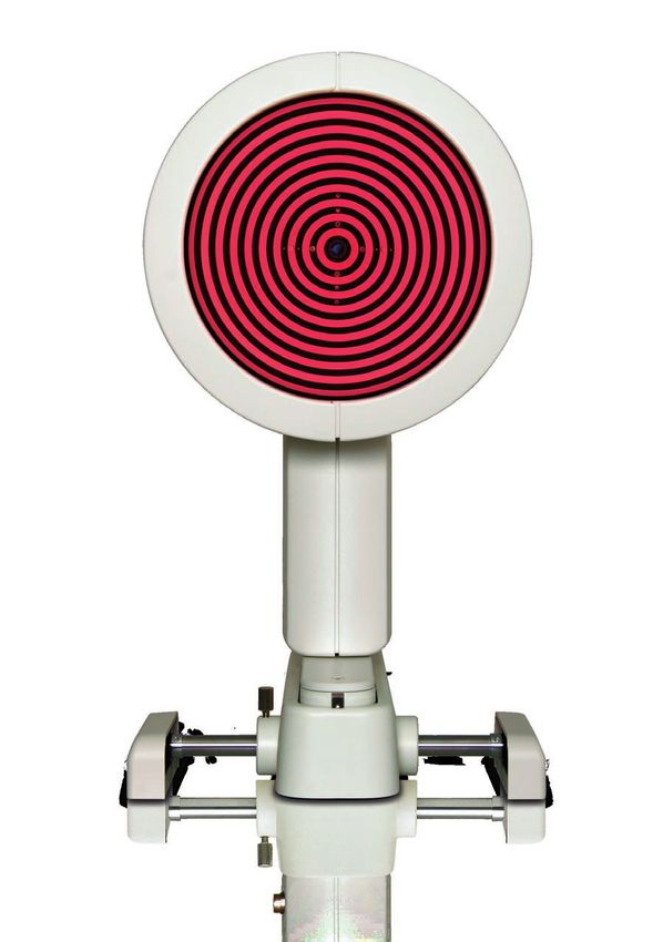
Ophthalmologist

Versatile and precise
For me the Keratograph 4 is an indispensable
device for diagnosis and surgical planning. Its
automatic measurement activation guarantees
fast, reproducible and accurate measurements.
This diagnostic device streamlines the workflow
in our clinic.
OCULUS Keratograph 4
From a measuring instrument to a consultation tool
Gold standard corneal topography – that’s what the Keratograph 4 is all about. It ensures reliability when it comes to
taking measurements, providing consultation and fitting contact lenses. The Keratograph accurate findings are something
you can count on. The integrated keratometer and automatic measurement activation guarantee perfect reproducibility.
In this way the Keratograph 4 also meets highest clinical standards for such procedures as tear film assessment and
qualitative cornea analysis. It stands out by virtue of its versatility.
Taking measurements with placido ring illumination
The cornea is represented across its entire surface and globally using thousands
of measuring points. Precise measurements form the basis for many modes of
analysis and representation, such as, automatic keratoconus detection and 3D
representation of the cornea.
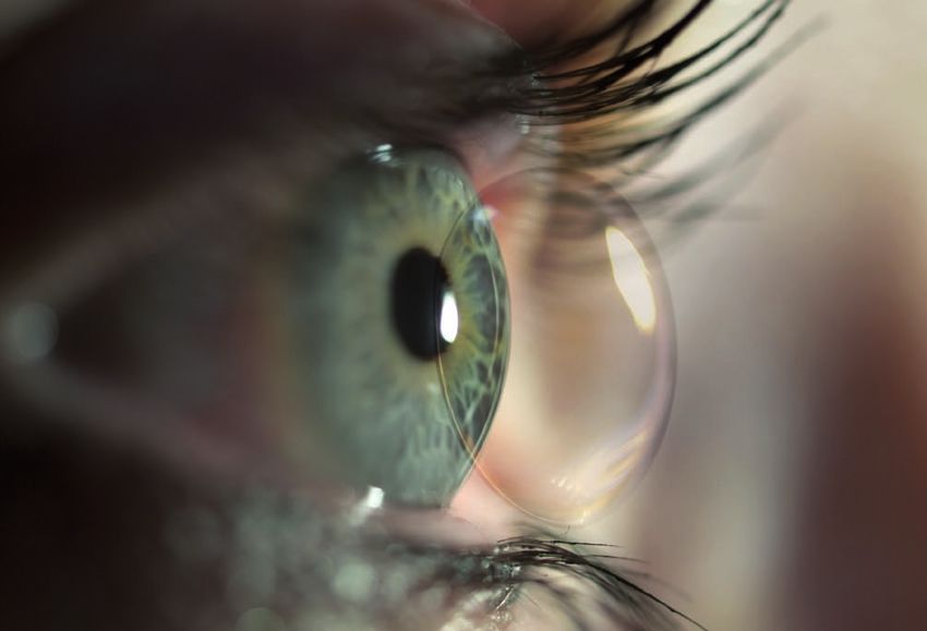
Taking measurements with the blue light emitting diode
Previously you may have taken static fluo images and videos using the slit
lamp – now you can also do so with the Keratograph 4 ! Use this
function to examine the fit and mobility of contact lenses.
Precise measurements, comprehensible presentation –
a picture says more than 1,000 words
Use the Keratograph 4 as a marketing tool and incorporate it actively into your consultations. With the Keratograph 4
software you can show images which your customers/patients have never seen before. Competent consultation (e.g. during
follow-up exams) builds trust and forms the basis for intensive customer/patient loyalty. The Keratograph 4 – not only a
measuring instrument but an ideal marketing tool.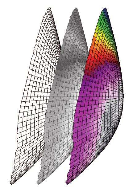
Diagnostics
Early detection is the key to preserve vision

> Non-contact, quick measurements
    Hygiene and time management are important for any successful
    ophthalmic practice.

               > Precise and reliable diagnosis and follow-up
                     With the Keratograph 4  accurate measurements, changes on the corneal
                     surface can often be detected in the early stage. Follow-up exams over longer
                     periods of time are equally important when you are dealing with changes in
                     the cornea.

> Corneal surgery
    The OCULUS Keratograph 4 provides reliable pre- and post-operative measurements.

> State-of-the-art Topography based
  keratoconus detection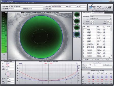
OCULUS Keratograph 4 –
high-performance functionality,
comprehensible representation.
Over 15 years of experience in assessing
topographic measurements.

                                                         Overview display
                                                         The integrated keratometer guarantees highest
                                                         measurement precision and reproducibility. After
                                                         measurements have been taken, the comprehensive
                                                         representation mode gives you a fast overview. Among
                                                         other parameters, the central radii, K values, corneal
                                                         astigmatism, eccentricity and corneal curvature are
                                                         displayed. The color topographic representation depicts
                                                         the curvature of the anterior segment of the cornea.
                                                         Irregularities can be seen and measured on the camera
                                                         image.

3D display
The 3D display depicts the curvature of the cornea.
Any corneal astigmatism and irregularities can be
demonstrated in an easy and comprehensible way. By
swivelling and rotating the 3D map, the cornea can be
viewed from various perspectives. Abnormalities can
be displayed easily, which helps with patient/customer
consultation.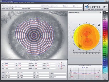
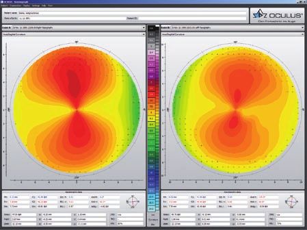
Fourier analysis
The Fourier analysis is an important tool for visualizing
the amount of corneal irregularities. Using the Fourier
analysis, the topography map is divided into individual
components. The first three are standard components
that represent lower order aberration and the fourth map
shows the amount of corneal irregularities or higher
order aberration.

                                                             Zernike analysis
                                                             Irregularities of the cornea can be depicted clearly with
                                                             Zernike analysis. If the given aberration coefficient is
                                                             increased, this is an indication for deterioration of the
                                                             eye’s optical imaging quality. The exact location of the
                                                             apex can also be identified easily using Zernike analysis.
                                                             The location of the apex is marked with a black cross.

Indices
Using the “Indices” display, topographic abnormalities can
be detected and diagnosed with ease. The measurements
are compared with normative data. If deviations are
found, the measurements are marked in yellow or
red. On the basis of the measurements, a topographic
classification is made. Abnormalities such as keratoconi
can be detected in the early stage. If keratoconus is
found, it is classified on the basis of the topographic
data. The Indices display helps compare follow-up exams
and shows whether an existing keratoconus remains in
the same stage or progresses.

      Careful monitoring ensures patient satisfaction which is the basis of patient loyalty.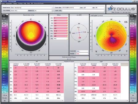
Show 2 exams                                                 Compare 3 exams
During the follow-up examinations, it is necessary to        With this feature, up to 3 examinations can be displayed
compare the results with the previous exams. With this       and compared side-by-side. This is very useful for the
feature you can compare the changes in the corneal           follow-up examinations and to assess the progression or
topography over time for the contact lens wearers or         changes in the color maps.
patients with progressive conditions, such as
Keratoconus.

Near portion height measurement                              Palpebral angle measurement
This software precisely simulates the near portion height    The measurement of the nasal lower palpebral angle
of rigid bifocal contact lenses and simplifies the complex   facilitates the identification of the expected inclination
fitting process.                                             or stabilization axis when fitting toric contact lenses.
                                                             Save time and money by giving this information to the
                                                             contact lens manufacturer when you place an order.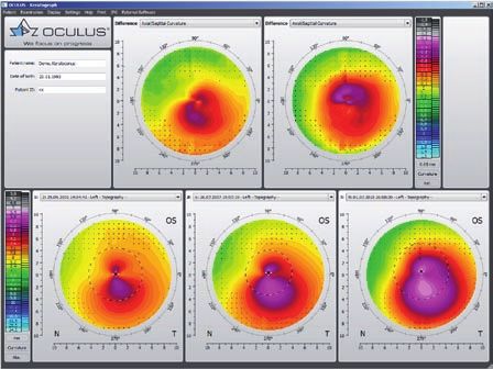
TF-Scan makes the
tear film visible
Patient consultation made easy. This software shows
the quality and quantity of the tear film

In cases of dry eye patients and contact lens wearers, the tear film should be examined carefully. Only an intact tear
film guarantees contact lens wearing comfort! The Keratograph 4 measures the tear film breakup time non-invasively
(quality assessment). You can show your patient the individual tear film quality using the color maps. In addition, you can
take another non-invasive measurement to determine the amount of tear film (tear film quantity).

Tear film quality (NIKBUT)
The Oculus Keratograph 4 determines the break-up time            Changes in the projected placido rings (displacement of
using the NIKBUT procedure (non-invasive-Keratograph-            margins of rings) give an indication of the break-up time
break-up time).                                                  of the tear film on the cornea.

> Projection of the placido rings

Tear film quantity (tear meniscus)

                                                                                         Consultation made easy –
                                                                                         color assessment of
                                                                                         break-up time
                                                                                           green = stable tear film
                                                                                           yellow = borderline tear film
                                                                                           red    = unstable tear film

                                                                 > Measure the tear meniscus height objectively with the
                                                                   OCULUS Keratograph 4.
OxiMap® – visualizing the
oxygen transmissibility
Professional patient consultation
The cornea needs oxygen and a good oxygen supply is fundamental for the comfort of a contact lens wearer. New materials
used for soft contact lenses offer excellent oxygen transmissibility. This can be shown with the new OCULUS OxiMap®
display. You can easily show these color maps to your patients and help them choose better contact lenses.

How much oxygen really reaches the cornea ?
Until now, only the oxygen transmissibility values for the center of a contact lens with –3.0 D were available. The OxiMap®
shows the oxygen transmissibility depending on the lens material and the lens thickness. The OxiMap® is available for
the most frequently sold spherical soft contact lenses. This impressive tool assists you in helping your patients select the
most suitable contact lens.

> Oxygen transmissibility for -3.00 D in comparison to the value for -10.00 D for identical type of contact lens

Plain and comprehensive visualization assures patient loyalty!
                                                                                                                                                        Dk/t > 125*
Contact lenses act as a potential
barrier to oxygen transport even                                                                            Dk/t > 87*                                  Continious Wear
when the eyes are open to the
                                                                                                            Extended Wear
atmosphere. Long hours of wearing                                  Dk/t > 35*
comfort can only be guaranteed
with a sufficient oxygen supply. The
                                                                   Daily Wear
color representation of the various
terms of oxygen transmissibility
is based on international
recommendations for daily,
                                                                                                                                                                    cm/sec
extended and continuous wear.                  93 x 10 9
                                                                                                                                                                mL 0²/mL x h Pa
                                               124 x 10 9         cm/sec
                                                                                                                                                                mL 0²/mL x mm Hg

                                             > The OxiMap color coding of the Dk/T-values and the recommended wearing time
                                                                   ®
Fitting contact lenses
Professional contact lens fitting is
an important expertise

> Fast, non contact measurements win customers over
       The recorded data and the informative displays form the basis for a
       long term relationship.

                     > Every eye is different
                             Whether someone opts for soft or rigid contact lenses depends on
                             many factors.

> Provide expert consultation
       The large number of options for taking measurements and automatic
       assessments offer best prerequisites for professional contact lens fitting.

> The high degree of wearing comfort afforded by professionally fitted contact lenses strengthens
  the customer/patient relationship significantly.
Contact lens fitting
Contact lenses are recommended on an individual basis
and displayed in a list. In order to avoid taking more
steps than necessary when fitting contact lenses, the fluo
image can be simulated beforehand. The contact lens can
be rotated and moved around. Fluo image simulation is
adjusted automatically. The integrated and expandable
database contains all customary types of contact lenses and
is updated on a regular basis. The user can determine the
order in which contact lens manufacturers appear.
Imaging
Using the “Imaging” option, real fluo images and videos
are recorded – similar to the way images can be seen with
a slitlamp using fluo drops. In this way you can check
and demonstrate the fit and mobility of contact lenses to
patients. All images and videos are saved automatically as
well as the suggested fit of contact lenses.
The Keratograph 4 provides all prerequisites
blue light emitting diodes in the illumination
beam path
a yellow filter in the observation beam path
Pupillometry
Using the “Pupillometry” option, the reaction of the pupil,
can be checked with and without blinding. This builds the
basis for selecting the proper treatment zone for laser
controlled surface ablation, multifocal contact lenses or
premium IOLs. The pupil reaction of the two eyes can be
compared.Technical data
Oculus Keratograph 4
Measurement range 3 – 38 mm, 9 – 99 D
Precision +/– 0.1 D
Reproducibility +/– 0.1 D
Number of rings 22
Working distance 80 mm
Number of measuring points 22,000
Camera digital CCD camera
Source of illumination placido illumination: red 650 nm
imaging illumination: blue 465 nm (UV-free)
pupillometer illumination: infrared 880 nm
Dimensions (H x W x D) 49 – 51.7 x 27.5 x 32 – 40 cm
Weight 15 lbs.
Power supply 100-240 VAC, 50-60 Hz
Minimum PC requirements Pentium IV 2.0 GHz, Windows XP, Windows Vista,
Windows 7, 1 GB RAM, graphic card 1.024 x 768 pixels,
USB connection
in accordance with Medical Device Directive 93/42/EEC
260 mm
Specifications and design are subject to change without notice and may vary depending on region.
490 - 517 mm
320 – 400 mm 275 mm
WWW.OCULUS.DE OCULUS Optikgeräte GmbH
Postfach • 35549 Wetzlar • GERMANY
Tel. +49-641-2005-0 • Fax +49-641-2005-295
E-Mail: export@oculus.de • www.oculus.de
06/0213/en/Ha
• OCULUS USA, sales@oculususa.com
P/70670/EN
• OCULUS Asia, info@oculus.hk
Oculus is certified by TÜV according to • OCULUS Czechia, oculus@oculus.cz
DIN EN ISO 13485/DIN EN ISO 9001 • OCULUS Iberia, info@oculus.esYou can also read