Oligonucleotides and microRNAs Targeting Telomerase Subunits in Cancer Therapy - MDPI
←
→
Page content transcription
If your browser does not render page correctly, please read the page content below
cancers
Review
Oligonucleotides and microRNAs Targeting
Telomerase Subunits in Cancer Therapy
Adam Eckburg , Joshua Dein , Joseph Berei, Zachary Schrank and Neelu Puri *
 Department of Biomedical Sciences, University of Illinois College of Medicine at Rockford,
 Rockford, IL 61107, USA; aeckbur2@uic.edu (A.E.); jdein2@uic.edu (J.D.); jberei2@uic.edu (J.B.);
 schrank.zachary@gmail.com (Z.S.)
 * Correspondence: neelupur@uic.edu; Tel.: +1-815-395-5678

 Received: 21 July 2020; Accepted: 17 August 2020; Published: 19 August 2020

 Abstract: Telomerase provides cancer cells with replicative immortality, and its overexpression serves
 as a near-universal marker of cancer. Anti-cancer therapeutics targeting telomerase have garnered
 interest as possible alternatives to chemotherapy and radiotherapy. Oligonucleotide-based therapies
 that inhibit telomerase through direct or indirect modulation of its subunits, human telomerase
 reverse transcriptase (hTERT) and human telomerase RNA gene (hTERC), are a unique and diverse
 subclass of telomerase inhibitors which hold clinical promise. MicroRNAs that play a role in the
 upregulation or downregulation of hTERT and respective progression or attenuation of cancer
 development have been effectively targeted to reduce telomerase activity in various cancer types.
 Tumor suppressor miRNAs, such as miRNA-512-5p, miRNA-138, and miRNA-128, and oncogenic
 miRNAs, such as miRNA-19b, miRNA-346, and miRNA-21, have displayed preclinical promise as
 potential hTERT-based therapeutic targets. Antisense oligonucleotides like GRN163L and T-oligos
 have also been shown to uniquely target the telomerase subunits and have become popular in the
 design of novel cancer therapies. Finally, studies suggest that G-quadruplex stabilizers, such as
 Telomestatin, preserve telomeric oligonucleotide architecture, thus inhibiting hTERC binding to the
 telomere. This review aims to provide an adept understanding of the conceptual foundation and
 current state of therapeutics utilizing oligonucleotides to target the telomerase subunits, including
 the advantages and drawbacks of each of these approaches.

 Keywords: anti-cancer therapy; oligonucleotides; microRNAs; antisense oligonucleotides; telomerase;
 hTERT; T-oligo; 6-thio-dG; G-quadruplex

1. Introduction
      The terminal portions of human chromosomes are capped by nucleoprotein structures called
telomeres [1–4]. Telomeres maintain genomic stability in normal somatic cells and protect against
chromosomal degradation [5,6]. Telomeres are progressively shortened by each round of cell
division, and, after approximately 50 divisions, they reach a critically short length that induces
DNA damage responses leading to cellular senescence or apoptosis [7–9]. Telomerase is an enzymatic
ribonucleoprotein complex that maintains telomere length by adding single-stranded TTAGGG
stretches to the 30 end of the chromosome [6,8,10–12]. Human telomerase is composed of two subunits,
a reverse transcriptase component called human telomerase reverse transcriptase (hTERT) and a
template RNA component called human telomerase RNA gene (hTERC) [11–13]. These two subunits
adopt a well-defined tertiary structure in the catalytic core of the telomerase complex and exhibit
limited protein-RNA interaction [12]. Telomerase activity is relatively quiescent in most normal tissue;
however, studies have found it to be significantly increased in up to 90% of malignancies [8,14–16].
The activation of telomerase allows cells to circumvent senescence and continue to divide while

Cancers 2020, 12, 2337; doi:10.3390/cancers12092337                                www.mdpi.com/journal/cancers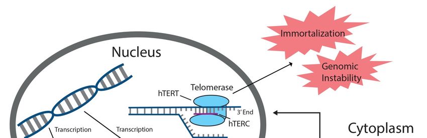
Cancers 2020, 12, 2337                                                                            2 of 16

accumulating oncogenic mutations that can ultimately lead to cancer [17,18]. Cancer therapeutics
specifically targeting telomerase-positive cells have garnered interest as a possible alternative to
traditional chemotherapy and radiotherapy methods, which are toxic to both cancerous and normal
cells [8,15]. The use of oligonucleotides and microRNAs (miRNAs) that inhibit the function of hTERC
and hTERT is a further specified subclass of telomerase-centered therapies that has shown promise in
treatment for a number of human malignancies [19–22]. This comprehensive review will elaborate on
the current state and underlying concepts of cancer therapeutics aimed at reducing telomerase activity
by targeting telomerase subunits with oligonucleotide-based approaches.
      miRNAs are endogenous oligonucleotides that play an important role in post-transcriptional
gene regulation. miRNAs often target genes involved in cancer-related processes, such as the
expression of hTERT, the catalytic subunit of telomerase [23]. Most tumor suppressor miRNAs
downregulate hTERT through direct interaction with the 30 -untranslated region (30 -UTR) of hTERT
mRNA, while oncogenic miRNAs indirectly upregulate hTERT activity through inhibition of genes
involved in the suppression of hTERT [22]. The therapeutic manipulation of miRNA-mediated hTERT
expression to treat human malignancies largely remains in preclinical stages; however, the array of
potential miRNA targets explored in this review demonstrates the clinical promise of this approach [15].
Antisense oligonucleotides (AS-ODNs) like GRN163L (Imetelstat) and T-oligos have also been utilized
to target telomerase activity through inhibitory interactions with hTERC and hTERT, respectively,
and GRN163L has progressed into clinical trials [15,19,21,24–28]. Furthermore, telomeric G-quadruplex
(G4) secondary structures formed by sequences rich in guanine serve as nucleotide sites intrinsic
to telomere architecture that can also be targeted to modulate the action and expression of the
telomerase subunits [25,29–31]. The therapeutic inhibition of telomerase through the activity of
the aforementioned oligonucleotide targets has been thoroughly explored in a number of cancer
types [28,32]. A comprehensive understanding of these anti-cancer approaches is crucial for the
development and improvement of new telomere-based cancer therapies that could serve as possible
alternatives to traditional chemotherapy-based treatment modalities.

2. MicroRNAs
     miRNAs comprise a non-coding class of RNA in eukaryotes that stretch 18–24 nucleotides
in length and function as endogenous post-transcriptional regulators of gene expression [8,33–35].
Primary miRNAs (pri-miRNAs) are transcribed and cleaved in the nucleus by RNAse III Drosha
to form hairpin-shaped pre-miRNAs (Figure 1) [36]. In the cytoplasm, these structures are cleaved
by RNAse III Dicer to form mature miRNAs, which then associate with the RNA-induced silencing
complex (RISC) (Figure 1) [34,37]. Once complexed to RISC, miRNA base pairs with a complementary
sequence on its target messenger-RNA site (mRNA), typically within the 30 -UTR, and induces its
degradation by RISC or prevents productive translation of the mRNA, thus effectively silencing the
associated gene (Figure 1) [8,33–35,37,38]. More than 50% of gene sequences encoding miRNAs exist
in regions associated with cancer and/or increased mutability [22]. As a result, miRNAs have been
the subject of recent clinical attention due to their involvement in driving cancer progression or in
silencing genes associated with cancer progression [35]. Oncogenic miRNAs facilitate cancer growth,
while tumor suppressor miRNAs inhibit proliferation [35,39]. Many tumor suppressor and oncogenic
miRNAs impact cancer development by directly or indirectly targeting hTERT, the reverse transcriptase
component of telomerase, causing it to be downregulated or upregulated, respectively. Increased
hTERT expression leads to increased telomerase activity and angiogenic characteristics, while hTERT
downregulation limits telomerase activity and causes antiproliferative effects. A vast amount of
therapeutic potential lies in the use of miRNA-induced gene modulation to regulate hTERT expression
and prevent the overexpression of telomerase, which is a common characteristic of human cancers [35].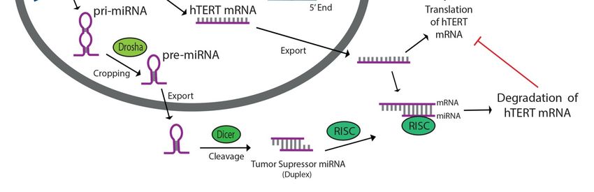
Cancers 2020, 12, 2337 3 of 16
Cancers 2020, 12, x 3 of 16
Figure 1. Tumor suppressor miRNAs targeting human telomerase reverse transcriptase (hTERT) inhibit
Figure 1. Tumor suppressor miRNAs targeting human telomerase reverse transcriptase (hTERT)
telomerase activity by binding to hTERT mRNA and preventing productive translation of the reverse
inhibit telomerase activity by binding to hTERT mRNA and preventing productive translation of the
transcriptase component of telomerase. miRNAs are transcribed in the nucleus, cleaved by Drosha and
reverse transcriptase component of telomerase. miRNAs are transcribed in the nucleus, cleaved by
Dicer, then associated with RNA-induced silencing complex (RISC) in the cytoplasm. Tumor suppressor
Drosha and Dicer, then associated with RNA-induced silencing complex (RISC) in the cytoplasm.
miRNAs prevent angiogenetic characteristics associated with increased telomerase activity in cancers.
Tumor suppressor miRNAs prevent angiogenetic characteristics associated with increased telomerase
activity
Some in cancers.
of the most well-studied hTERT-targeting tumor suppressor miRNAs include miRNA-1182,
miRNA-133a, miRNA-342, miRNA-491, miRNA-541, miRNA-512-5p, miRNA-138, and miRNA-128
Some of the most well-studied hTERT-targeting tumor suppressor miRNAs include miRNA-
(Table 1) [15,20]. miRNA-1182 has been shown to reduce gastric cancer cell proliferation and migration
1182, miRNA-133a, miRNA-342, miRNA-491, miRNA-541, miRNA-512-5p, miRNA-138, and
by targeting the open reading frame-1 (ORF-1) of hTERT [40]. Furthermore, an inverse correlation
miRNA-128 (Table 1) [15,20]. miRNA-1182 has been shown to reduce gastric cancer cell proliferation
between miRNA-1182 expression and hTERT protein levels in gastric cancer cells suggested that
and migration by targeting the open reading frame-1 (ORF-1) of hTERT [40]. Furthermore, an inverse
miRNA-1182 could be miRNA-1182
correlation between utilized as a biomarker
expression and and potential
hTERT protein treatment for in
levels thisgastric
form of cancer
cancer [40].
cells
Other studiesthat
suggested suggested
miRNA-1182that miRNA-1182
could be utilizedoverexpression
as a biomarker inhibited bladder
and potential cancer cell
treatment proliferation,
for this form of
colony formation, and invasion by binding to the 3 0 -UTR of hTERT transcripts [41]. miRNA-1182
cancer [40]. Other studies suggested that miRNA-1182 overexpression inhibited bladder cancer cell
has also been shown
proliferation, colonytoformation,
slow metastasis in ovarian
and invasion cancer to
by binding cells
thethrough
3′-UTR of hTERT
hTERT inhibition and[41].
transcripts thus
holds promise ashas
miRNA-1182 a potential
also been therapeutic
shown to target
slow for this form
metastasis inofovarian
cancer [42].
cancerIn cells
HeLathrough
adenocarcinoma
hTERT
cells, miRNA-133a was shown to bind directly to the 3 0 -UTR of hTERT mRNA, demonstrated through
inhibition and thus holds promise as a potential therapeutic target for this form of cancer [42]. In
inhibition of TERT 30 -UTR-driven
HeLa adenocarcinoma reporterwas
cells, miRNA-133a activity
shown [20]. In the
to bind same study,
directly miRNA-342,
to the 3′-UTR of hTERTmiRNA-491,
mRNA,
and miRNA-541through
demonstrated were each shownof
inhibition toTERT
bind the 30 -UTR of hTERT
3′-UTR-driven reporteras well [20].
activity [20]. Combining
In the same all three
study,
miRNAs
miRNA-342,yielded increasingand
miRNA-491, inhibitory
miRNA-541effects,weresuggesting
each shown eachtomiRNA
bind thebinds
3′-UTR to of
a unique
hTERT region
as wellof
30 -UTR
the[20]. Combining
on hTERT all mRNA
three miRNAs yielded increasing
[20]. miRNA-512-5p is anotherinhibitory
tumoreffects,
suppressorsuggesting
miRNAeach thatmiRNA
has been
bindsto
shown topost-transcriptionally
a unique region of theregulate
3′-UTR hTERT
on hTERT mRNA
through 30 -UTR binding
[20]. miRNA-512-5p
a unique is another
site ontumor
hTERT
suppressor
mRNA miRNA
[43]. In head andthatneck
has been showncell
squamous to post-transcriptionally
carcinoma (HNSCC) regulate hTERT through
cells, miRNA-512-5p a unique
inhibited cell
3′-UTR binding site on hTERT mRNA [43]. In head and neck squamous cell
growth by decreasing telomerase activity, shortening telomere length, and disrupting telomere-binding carcinoma (HNSCC) cells,
miRNA-512-5p
proteins [43]. Tumorinhibited cell growth
suppressor by decreasing
miRNA-138 has also telomerase
been associated activity,
withshortening telomere
antiproliferative length,
effects and
and disrupting telomere-binding proteins [43]. Tumor suppressor miRNA-138
inhibition of tumor growth as a result of direct targeting of hTERT [44,45]. In human anaplastic thyroid has also been
associatedcells,
carcinoma with miRNA-138
antiproliferative effects and inhibition
downregulation of tumor growth
was associated with hTERT as a result of direct targeting
overexpression [15,45].
of hTERT [44,45]. In human anaplastic thyroid carcinoma cells, miRNA-138
In cervical cancer cells, overexpression of miRNA-138 led to inhibition of cell proliferation, migration, downregulation was
andassociated
invasion, with hTERT
as well as overexpression [15,45]. Incaused
induction of apoptosis, cervicalby cancer
bindingcells,ofoverexpression
miRNA-138 to ofthe
miRNA-138
30 -UTR of
led to inhibition of cell proliferation, migration, and invasion, as well as induction of apoptosis,
hTERT mRNA [44]. Furthermore, upregulation of miRNA-138 proved to be more effective than hTERT
caused by binding of miRNA-138 to the 3′-UTR of hTERT mRNA [44]. Furthermore, upregulation of
knockdown in potentiating Apigenin-induced apoptosis, a process caused by the release and activation
miRNA-138 proved to be more effective than hTERT knockdown in potentiating Apigenin-induced
of caspases, in malignant neuroblastoma cells both in vivo and in vitro [46,47]. This suggests thatCancers 2020, 12, 2337 4 of 16
miRNA-138 upregulation may serve as a promising alternate pathway to control tumor growth [47].
miRNA-128 is another unique miRNA that can act as both an oncogenic miRNA and a tumor suppressor
miRNA depending on the tumor type [33,48,49]. miRNA-128 has been shown to interact with the
coding sequence of hTERT mRNA in HeLa and teratoma cells [48]. In HeLa cells, overexpression
of miRNA-128 led to reduced levels of hTERT mRNA and TERT protein, as well as reduced cell
proliferation [48]. On the other hand, downregulation of miRNA-128 has been associated with
metastasis and development of a number of malignancies, including glioma, prostate, head and neck,
lung, and colorectal cancers [33,48].
Table 1. A list of important hTERT-targeting tumor suppressor and oncogenic miRNAs, including
which cancers each has been studied in and the specific mechanism of action targeting hTERT.
miRNA Class Cancers Action (Targets)
miRNA-1182
Tumor Suppressor Bladder, Gastric Direct (ORF-1, 30 -UTR)
[40–42]
miRNA-19b Adenocarcinoma, Breast, Glioma, Lung,
Oncogenic Indirect (PITX1)
[50,51] Melanoma, Osteosarcoma
miRNA-128 Colorectal, Head and Neck, Lung,
Either Direct or Indirect
[33,48] Prostate, Teratoma,
miRNA-133a
Tumor Suppressor Adenocarcinoma Direct (30 -UTR)
[20]
miRNA-138
Tumor Suppressor Thyroid, Cervical, Neuroblastoma Direct (30 -UTR)
[15,44–47]
miRNA-21
Oncogenic Melanoma, Colorectal, Glioblastoma Indirect (PTEN, STAT3)
[15,52–54]
miRNA-342
Tumor Suppressor Adenocarcinoma Direct (30 -UTR)
[20]
miRNA-346
Oncogenic Cervical Direct (30 -UTR)
[55]
miRNA-491
Tumor Suppressor Adenocarcinoma Direct (30 -UTR)
[20]
miRNA-512-5p
Tumor Suppressor Head and Neck Direct (30 -UTR)
[43]
miRNA-541
Tumor Suppressor Adenocarcinoma Direct (30 -UTR)
[20]
Oncogenic miRNAs that directly or indirectly upregulate hTERT expression and drive cancer
metastasis and aggression through increased telomerase activity can also be targeted as a therapeutic
approach. Some of the most well-studied oncogenic miRNAs in this category include miRNA-19b,
miRNA-346, and miRNA-21 (Table 1) [8,51]. Studies suggest that miRNA-19b expression indirectly
upregulates hTERT expression through inhibition of a novel hTERT suppressor gene, paired-like
homeodomain1 (PITX1) [51]. PITX1 is a negative regulator in the RAS signaling pathway that binds
directly to the hTERT promoter region, repressing hTERT transcription and telomerase activity, as well
as preventing cellular proliferation [51]. miRNA-19b binds to a complementary sequence in the 30 -UTR
of PITX1 mRNA, repressing PITX1 translation and activating hTERT expression [51]. Overexpression
of miRNA-19b led to 1.5- to 1.7-fold increases in telomerase activity in melanoma cells and has
been associated with oncogenesis in lung cancer, melanoma, breast cancer, and osteosarcoma [50,51].
In cervical cancer cells, miRNA-346 has been shown to mediate hTERT upregulation through a
competitive process with miRNA-138, which binds to the same region of the 30 -UTR of hTERT
mRNA as miRNA-346 but promotes an opposite effect on hTERT expression [55]. The binding of
miRNA-346 to the 30 -UTR induces the binding of G-rich RNA sequence binding factor 1 (GRSF1)
to the miRNA-346 CCGCAU sequence [55]. This interaction forms a “bulge loop” that recruits
hTERT mRNA to ribosomes for translation [55]. By way of this translation process independent of
argonaute RISC catalytic component 2 (AGO2), miRNA-346 enhances proliferation in cervical cancer
cells [55]. Conversely, when miRNA-138 binds, it mediates the suppression of hTERT translationCancers 2020, 12, 2337 5 of 16
in an AGO2-dependent manner [55]. Studies suggest that miRNA-21 regulates hTERT expression
through several indirect pathways. miRNA-21 impacts telomerase activity through downregulation of
phosphate and tensin homolog deleted on chromosome 10 (PTEN), a tumor suppressor gene tied to
tumor progression through activation of the PI3K/AKT pathway [54]. In hypertrophic scar fibroblasts,
transfection with a miRNA-21 mimic led to increased hTERT mRNA and protein levels as well as
increased PI3K/AKT signaling [54]. Additionally, the introduction of PTEN cDNA inhibited the effects
of miRNA-21 overexpression [54]. In malignant melanoma cells, miRNA-21 was found to be associated
with hTERT upregulation, resulting in increased invasiveness, oxidative stress, genomic instability,
and cell proliferation, as well as evasion of apoptosis [52]. Furthermore, in colorectal cancer, a 3.4-fold
increase in miRNA-21 expression was correlated with a 2-fold increase in hTERT expression [53].
Studies have also shown that miRNA-21 enhances carcinogenesis through STAT3, a transcription factor
that plays a key role in Th17 helper T-cell differentiation [15]. When miRNA-21 was knocked down in
murine glioblastoma xenografts, reduced expression of both hTERT and STAT3 was observed, as well
as slowed tumor growth [15].
A number of different therapeutic approaches targeting miRNAs to reduce hTERT expression and
telomerase activity have been investigated. The administration of hTERT-targeting tumor suppressor
miRNAs to act as telomerase inhibitors in combination with chemotherapy drugs has been explored
in numerous clinical applications and shown to be more effective than use of chemotherapy drugs
alone [56]. The use of anti-miRNA oligonucleotides (AMOs), which are synthetic sequences antisense
to a specific miRNA, to reduce the activity of hTERT-targeting oncogenic miRNAs, has also garnered
significant interest [57]. Treatment with anti-miRNA-21 oligonucleotides successfully reduced STAT3
and hTERT levels in cancer cells, while administration of anti-miRNA-19b to melanoma cells reduced
hTERT mRNA expression and increased PITX1 mRNA expression [51,58]. Studies have shown that
unique subsets, or “families,” of miRNAs tend to be upregulated together in cancer [15]. miRNA sponges
are stretches of DNA with artificial miRNA binding sites incorporated into the 30 -UTR of a gene that
are utilized to simultaneously downregulate a group of hTERT-targeting miRNAs [59]. A recent study
in bladder cancer cells showed that synthetic miRNA sponges driven by a mutant hTERT promoter
were successful in downregulating four oncogenic miRNAs, thus inhibiting cell growth, decreasing
motility, and inducing apoptosis in cancer cells but not in normal cells [59]. Studies have investigated
the use of small RNA-specific ligands targeting the narrow groove of specific pre-miRNAs to inhibit
the formation of mature oncogenic miRNAs [60]. Finally, the use of CRISPR/Cas9 to knockdown genes
encoding oncogenic miRNAs, most notably miRNA-21, has also been explored extensively and was
shown to reduce the expression of oncogenic miRNAs by up to 96% [61,62].
A wide range of techniques that utilize tumor suppressor and oncogenic miRNAs to regulate
hTERT expression and limit telomerase activity have been explored; however, these therapies remain
largely in preclinical stages, likely due to inherent issues associated with telomerase inhibition. It often
takes several weeks for telomerase inhibition to cause critical telomere shortening and anti-cancer effects,
a long timeframe which compromises the efficacy of these therapies [63]. Furthermore, studies have
suggested that critical telomere shortening may lead to deleterious side effects such as cell crisis, genomic
instability, and cancer progression [64]. Previous research also suggested that telomerase may re-localize
under certain stress conditions and participate in responses that remain poorly characterized [64].
The long-term effects of telomerase inhibition are also not well-understood because the specific action
of telomerase after therapeutic targeting has not been fully characterized. Before miRNA therapies
targeting hTERT can be broadly implemented as a clinical approach, an added depth of understanding
and clarification regarding the exact mechanism of telomerase inhibition is required. Another issue
limiting the applicability and effectiveness of these therapies is the tissue-specific expression patterns
of different miRNAs [65]. Depending on the tissue type, some miRNAs can act as either a tumor
suppressor or an oncogenic miRNA [48,65,66]. Future studies should take these limitations into
account when designing miRNA-based therapies targeting telomerase for specific forms of cancer.
Some miRNA-based therapies have progressed to clinical trials; however, none mechanistically targetCancers 2020, 12, 2337 6 of 16
hTERT. Inhibition of miRNA-122 for treatment of hepatitis C has progressed to Phase II clinical
trials [15]. Additionally, MRG-106 oligonucleotides used as antisense inhibitors to miRNA-155 for
treatment of cutaneous T-cell lymphoma has moved into Phase I trials [15]. The clinical progression
of these therapies demonstrates the promise of miRNA-based treatment approaches. The design of
hTERT-targeting miRNAs that overcome the limitations of telomerase inhibition may hold significant
clinical promise. Furthermore, due to the tissue specificity of miRNAs, the use of miRNAs as a blood
and tumor biomarker in prostate, lung, and breast cancers has been thoroughly investigated [67–69].
Since hTERT upregulation also serves as a meaningful biomarker for several cancers, the diagnostic
and prognostic utilization of hTERT-targeting oncogenic or tumor suppressor miRNAs is an approach
with significant therapeutic potential [70].
3. Oligonucleotides
Oligonucleotides are molecules composed of short segments of DNA or RNA that have been used
for a variety of purposes in the fields of medicine and biomedical research. Because of their ability to
bind to complementary sequences of DNA or RNA, oligonucleotides have been employed as simple
nucleic acid probes using a number of basic science laboratory techniques, such as polymerase chain
reaction (PCR) and DNA sequencing, as well as in the specific targeting and regulation of the expression
and function of a wide range of proteins [71,72]. Due to their diverse functionality, oligonucleotides
have been at the center of many research studies in numerous disease models [73]. They have been
particularly useful in studying cancer biology, as their ability to bind and regulate specific nucleotide
sequences allows for the study of both the etiology and treatment of many different cancers [15,19,21].
Specific oligonucleotide molecules such as miRNAs and AS-ODNs, including T-oligos, have been
used to target distinct complementary DNA and RNA sequences in order to modulate both aberrant
protein levels and enzyme activities implicated in various cancers [8,15,74]. 6-Thio-20 -Deoxyguanosine
(6-Thio-dG) is an effective nucleoside analog and telomerase substrate which brings about several
anti-cancer effects through incorporation into DNA [8,15].
Much progress has been made in the use of oligonucleotides to target molecular events leading to
the upregulation of telomerase [23,75]. As previously mentioned, hTERT is the catalytic component of
telomerase that is essential for the replication of chromosomal telomeres, a process that confers cells
with perpetual replicative potential [23,75,76]. Oligonucleotides can be tailored to target hTERT at
specific steps in its synthesis and activity, including at the hTERT promoter, hTERT mRNA, or in its
protein form [15,77]. In order for hTERT to successfully perform its catalytic function, it must be able
to properly interact with telomere-related proteins in the shelterin complex and bind to the telomeric
nucleotide sequence using hTERC. Regulation of the hTERT protein itself has been accomplished
through oligonucleotide-based interference of hTERT at a number of different points [8,15].
AS-ODNs have shown promise in both preclinical and clinical trials as a potential treatment
for various cancers. A number of preclinical studies used AS-ODNs to target the hTERT initiator
sequence, pre-mRNA, or transcribed mRNA, which significantly decreased hTERT activity and
increased growth inhibition in human hepatoma, prostate adenocarcinoma, bladder cancer, and hepatic
lymphoma [24,78–80]. In particular, Folini et al. found that antisense oligonucleotide-mediated
inhibition of hTERT, but not hTERC, inhibited cell growth and induced apoptosis in human prostate
cancer cells, implying that telomerase can retain its role in telomere replication even in the absence of a
properly functioning RNA template. Another study found that the simultaneous in vitro targeting
of both hTERT and hTERC with AS-ODNS resulted in the synergistic inhibition of growth in human
colon cancer cells [81]. The results of these early preclinical studies showed that hTERT could be
successfully targeted at a number of regulatory points involved in its cellular production [81]. They also
demonstrated the wide applicability of specifically engineered oligonucleotides to target cells with
abnormal hTERT activity [81]. Additionally, various preclinical and clinical studies have revealed
the synergistic anti-cancer effects of AS-ODNs with other cancer drugs on the inhibition of abnormal
cancer cell growth [82–84].Cancers 2020, 12, 2337 7 of 16
Although AS-ODNs can be used to target the production of hTERT, one of the best-studied
AS-ODNs GRN163L (Imetelstat) acts as a competitive inhibitor of telomerase activity. GRN163L is
a 13-merthiophosphoramidate deoxyribo-oligonucleotide that is a complement and antagonist to
the hTERT-associated hTERC RNA template sequence. By binding to hTERC, Imetelstat prevents
hTERT-mediated telomere elongation, leading to progressive telomere attrition and eventual cell
death [8]. Imetelstat preferentially targets cancer cells due to their intrinsically high level of telomerase
activity and has been tested in a number of preclinical and clinical trials conducted to assess its effect
on different types of cancers, with varying levels of success [82,84,85].
In preclinical studies using both in vitro human cells and in vivo murine models, Imetelstat has
shown great promise in its ability to decrease the growth of many cancers, as well as downregulate
hTERT activity. Imetelstat alone significantly decreased telomere length through hTERT inhibition
and increased cell death of human myeloma and pancreatic cancer cells in an in vitro model [63,86].
Xenograft murine models showed similar results for both myeloma and malignant rhabdoid tumor cells,
demonstrating telomere shortening, decreased growth, and increased cell death [86,87]. Additionally,
Imetelstat has exhibited anti-metastatic properties in mouse models of lung cancer [88,89]. Imetelstat
has been tested in numerous preclinical trials involving other types of cancers, both by itself and in
combination with other anti-cancer therapeutics, with similar patterns of inhibition of telomerase and
decreasing cell viability being observed [15,28]. Despite the apparent effectiveness of Imetelstat in
preclinical studies, many preclinical trials using it have severe limitations which prevent its applicability
to human treatments. Some of these issues include significant differences observed between in vitro
and in vivo models for human cells as well as apparent variations between telomere structure and
maintenance in mice compared to humans [15].
Despite its success in preclinical trials, Imetelstat has not had as much success in clinical trials due
to its severe hematological side effects, including thrombocytopenia and myelosuppression, and as
a result has not been approved by the U.S. Food and Drug Administration (FDA) [1]. It has been
involved in numerous Phase I/Phase II clinical trials that have been completed as well as some Phase
II/Phase III trials that are still ongoing [15,28,90]. These studies have shown mixed results, with some
reporting no significant increase in long-term patient survival and others reporting no significant
reduction in tumor size despite a 95% reduction in telomerase activity [91,92].
Preclinical and clinical trials have tested the drug both alone and in combination with other
anti-cancer drugs in the treatment of various types of cancers [1]. The most promising results with
Imetelstat have been obtained when it is used alongside other chemotherapeutic agents, such as
3-aminobenzamide (3AB) and trastuzumab, or conventional radiation therapy, possibly through the
increased sensitization of cancer cells to these other treatments [15,93,94]. Future studies on Imetelstat
should investigate the synergistic targeting of hTERT by Imetelstat in combination with other drugs,
such as small molecule inhibitors, that target other sources of cancer aggression while simultaneously
minimizing unfavorable toxicity. Both preclinical and clinical trials are currently ongoing in these
areas [15,28].
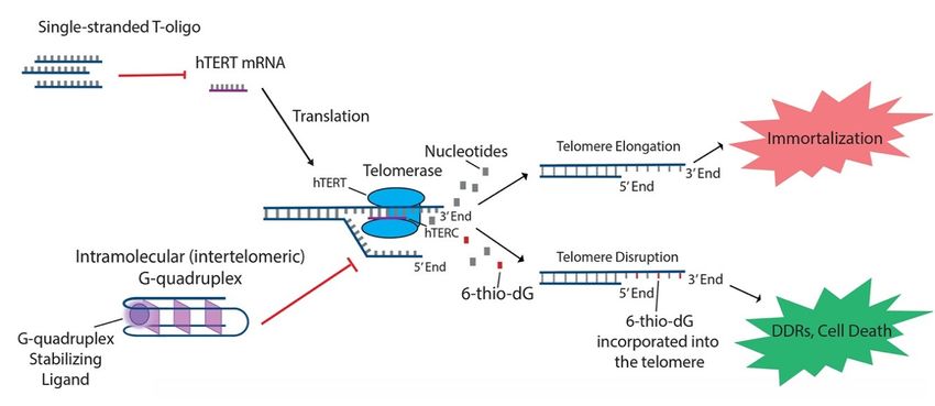
In addition to the aforementioned oligonucleotides, both T-oligos and 6-thio-dG have generated
interest due to their potential to regulate hTERT and possible use in treating various cancers. T-oligos
are oligonucleotides with a sequence homologous to the 30 telomere overhang. Their anti-cancer
effects are proposed to occur by activation of DNA damage responses (DDRs) and subsequent cell
death, either by mimicking damaged DNA or by triggering the dissociation of critical shelterin
proteins from telomeres [15,27]. T11 is a specific 11-base T-oligo that has been shown to demonstrate
anti-cancer properties in a number of different cancers [8,15,27,28]. T11 is proposed to function through
its resemblance to the telomeric overhang DNA sequence and displays promise because it has no
effect on normal cells (Figure 2) [27]. Administration of T11 also had no detectable toxic effects in
mice [28,95–97]. In preclinical studies, T-oligo was shown to inhibit mRNA expression of hTERT
by 50% in a melanoma cell model [98]. Additionally, concurrent administration of T-oligo with
vemurafenib in V600E-positive melanoma cells demonstrated an additive anti-cancer effect, implicatingCancers 2020, 12, 2337 8 of 16
Cancers 2020, 12, x 8 of 16
the potential benefit of T-oligo administration with chemotherapeutics in current use [98,99]. 6-thio-dG,
implicating the potential benefit of T-oligo administration with chemotherapeutics in current use
a nucleoside analog, has been shown to possess anti-cancer properties by being incorporated into
[98,99]. 6-thio-dG, a nucleoside analog, has been shown to possess anti-cancer properties by being
telomeric DNA, leading to the uncapping of telomeric DNA and dissociation of the shelterin complex
incorporated into telomeric DNA, leading to the uncapping of telomeric DNA and dissociation of the
(Figure 2). This results in telomere dysfunction-induced foci (TIFs) and subsequent cellular senescence
shelterin complex (Figure 2). This results in telomere dysfunction-induced foci (TIFs) and subsequent
and apoptosis [82]. Furthermore, this effect has been shown to be partly dependent on the activity
cellular senescence and apoptosis [82]. Furthermore, this effect has been shown to be partly
of hTERT, as TIFs were not seen in cells lacking hTERT activity [82]. Future studies should focus on
dependent on the activity of hTERT, as TIFs were not seen in cells lacking hTERT activity [82]. Future
the specific
studies molecular
should focus oninteractions of 6-thio-dG
the specific molecular and hTERT
interactions in order toand
of 6-thio-dG better establish
hTERT thetoeffect
in order betterof
6-thio-dGthe
establish oneffect
telomerase activity.
of 6-thio-dG on telomerase activity.
Figure 2. T-oligo, 6-thio-dG, and G-quadruplex stabilizing ligands like telomestatin function to
Figure
prevent2.telomere
T-oligo,elongation
6-thio-dG,through
and G-quadruplex stabilizing
unique processes related ligands like of
to inhibition telomestatin function
the telomerase to
subunits.
prevent telomere elongation through unique processes related to inhibition of the
T-oligo has been shown to inhibit expression of hTERT mRNA in melanoma cells with activated telomerase subunits.
T-oligo has been6-thio-dG
JNK signaling. shown to inhibit expression
incorporates of hTERT mRNA
into telomeric DNA and in melanoma
stimulatescells
TIFswith activated
in cells JNK
that exhibit
signaling. 6-thio-dG
hTERT activity. incorporates
G-quadruplex into telomeric
stabilizing DNA
ligands and hTERC
prevent stimulates
fromTIFs in cellstothat
binding the exhibit hTERT
single-stranded
activity. G-quadruplex
telomere overhang. stabilizing ligands prevent hTERC from binding to the single-stranded
telomere overhang.
4. G-Quadruplex
4. G-Quadruplex
The human genome contains DNA that is primarily in the duplex state, allowing for the formation
The human
of a double helix. genome
However,contains DNA increasingly
it is becoming that is primarilyapparentin the
thatduplex state, allowing
non-canonical structuresfor thein
arise
formation of a double helix. However, it is becoming increasingly apparent
DNA through non-Watson–Crick base pairing [100,101]. One such structure, the G-quadruplex (G4), that non-canonical
structures arise in
is formed when DNA through
a nucleic non-Watson–Crick
acid sequence with multiple base
runspairing [100,101].
of guanine basesOne
cansuch structure, the
spontaneously fold
G-quadruplex
into secondary(G4), is formed
structures. Thewhen a nucleic
G4 structure is acid
formedsequence
aroundwith multiple
a tetrad runs of guanine
of Hoogsteen bases can
hydrogen-bonded
spontaneously
guanine residuesfold thatinto secondary
are stabilized by structures.
monovalentThe ions G4 Na+isand
structure
including K+ [30].around
formed a tetrad
The tetrads stackofon
Hoogsteen
top of each hydrogen-bonded guanine residues
other, forming a multi-layered planarthat are stabilized
scaffold by monovalent
[26]. In vitro studies haveions including Na
demonstrated +
that
and K+ [30]. The
G4 structures aretetrads stack on top of eachstable
more thermodynamically other,than
forming a multi-layered
double-stranded DNA planar
and thescaffold
kinetics[26].
of In
G4
vitro studies have demonstrated that G4 structures are more thermodynamically
unfolding are considerably slower than that of double-stranded DNA or RNA hairpin structures [102]. stable than double-
stranded
Therefore, DNA
sinceandthethe kinetics of G4 unfolding
thermodynamic propertiesare of considerably
G4s are likelyslower than that
to interfere withofDNA
double-stranded
and RNA in
DNA or RNA hairpin structures [102]. Therefore, since the thermodynamic
transcription, translation, and genomic stability, these structures need some form of regulation properties of G4s[103].
are
likely
UnliketomiRNA
interfereandwith DNA and RNA
oligonucleotide cancer in therapies,
transcription,
which translation,
introduce and genomic
nucleotide stability,tothese
structures target
structures need some
different telomere andform of regulation
telomerase sites, G4[103].
servesUnlike
as anmiRNA and oligonucleotide
intramolecular DNA sequence cancer
thattherapies,
is inherent
which introduce
to telomere nucleotide
structure, structures
regulates to target
telomerase differentand
function, telomere
may be andtargeted
telomerase
by a sites, G4 serves
diverse rangeasof
an intramolecular
therapeutics DNA sequence that is inherent to telomere structure, regulates telomerase function,
[27,28].
and may
The be targeted by
sequencing of anumerous
diverse range
genomesof therapeutics
has revealed [27,28].
an abundance of DNA stretches that meet
the criteria for potential G4 formation [104]. Interestingly,abundance
The sequencing of numerous genomes has revealed an potential ofG4DNA stretches
structure that meet
sequences are
the criteria for potential
non-randomly distributedG4 formation [104]. Interestingly,
within genomes. Approximately potential
90%G4 of structure sequences
the origins are non-in
of replication
randomly
the humandistributed
genome containwithin genomes.
potential Approximately 90% of theAdditionally,
G4 structures [105,106]. origins of replication
potentialinG4 thestructure
human
genome contain potential G4 structures [105,106]. Additionally, potential
sequences have been found to be enriched near the ends of chromosomes, functioning in a regulatory G4 structure sequences
have been found to be enriched near the ends of chromosomes, functioning in a regulatory capacity
by providing a capping structure for telomeres [30]. Telomeric G4 structures were initially thoughtCancers 2020, 12, 2337 9 of 16
capacity by providing a capping structure for telomeres [30]. Telomeric G4 structures were initially
thought to impair telomerase function, but recent studies have suggested that they might also play a role
in telomerase recruitment [31,107]. Discovery of telomere repeats containing potential G4 sequences
has proposed a link to telomerase-mediated elongation of telomeres, leading researchers to inquire
about the use of G4-targeted therapies in cancer treatment through disrupting telomere maintenance.
The process of telomere maintenance has been linked to the function of G4 helicase enzymes [108].
During DNA replication, once the replication fork reaches the telomere sequence, G4 structures must
be unwound by helicase enzymes such as BLM, RTEL1, and WRN [26,109,110]. During the cell cycle
S phase, WRN has also been shown to localize to telomeres [111]. When WRN does not co-localize
to aid in unfolding G4 structures, the replication fork eventually stalls at lagging telomeres and
disrupts their replication [112]. The importance of properly unfolding G4 structures during DNA
replication highlights the therapeutic potential for G4-stabilizing molecules in the treatment of cancer
cells (Figure 2). A few molecules which have demonstrated an ability to disrupt telomere replication
and function as anti-cancer agents include BRACO-19, RHPS4, and telomestatin [113–115].
Telomestatin functions by stabilizing G4 structures through binding to guanine nucleotides at
the 30 and 50 ends of telomeres, thus forming a stable complex that inhibits the telomerase RNA
component, hTERC, from binding the single stranded overhang [116,117]. Stabilizing G4 structure
through telomestatin has resulted in telomerase inhibition in breast, cervical, neuroblastoma, and other
cancer cell lines during preclinical studies while also displaying minimal effect on non-cancerous
cells [28,32]. However, the drug’s low water solubility resulted in poor bioavailability and has impeded
research progress [118]. BRACO-19 and RHPS4 act through a similar mechanism as telomestatin in
stabilizing G4 structures, but they have demonstrated less tumor selectivity [119,120].
The repetitive nature of telomere sequences is conducive to many potential G4 structures,
providing a target for therapeutic-based G4-forming molecules. T-oligo has demonstrated the ability
to form stable intermolecular G4 structures in vitro and induce activation of the SAPK/JNK signaling
as well as inhibition of hTERT expression in melanoma cells [25]. Previous studies proposed that JNK
activation might result in decreased hTERT expression, thus the anti-cancer effects of T-oligo may
be in part due to JNK activation [25,121]. Further interactions with telomeres were highlighted after
T-oligo treatment resulted in the upregulation of shelterin complex proteins TRF2 and POT1, offering
an additional pathway to telomere dysfunction [25].
The targeting of G4 structures through hTERT promoter mutations has also recently garnered
interest. In melanoma, increases in hTERT expression were caused by the same mutations as in 70% of
other tumors [29]. Within these tumors, C-to-T sense strand mutations that correspond with G-to-A
antisense strand mutations in the promoter of hTERT activated transcription by altering the consensus
sequence [122]. Researchers found that these promoter mutations interfered with G4 structure folding
by disrupting a silencer element that reduced G4 transcriptional repression [123]. In an attempt
to counteract this mutation, a small pharmacological chaperone molecule that acts on the mutated
promoter has been proposed as a potential therapeutic [123]. This molecule, GTC365, functioned
by partially returning to wild type G4 folding and thus regaining some transcriptional repression
ability [123]. In melanoma cells, GTC365 treatment was able to induce cancer cell death through
apoptosis and senescence by inhibiting transcription of hTERT and decreasing telomerase activity,
ultimately leading to reduced telomere extension [123].
The study of DNA-based G4 structures during the past few decades has progressed from structural
analysis to cancer therapy studies in increasingly complex biological systems [124–127]. Researchers
have further uncovered the mechanisms of G4 structures that serve as key factors in a number of DNA
and RNA processes. While significant progress has been made in this area, additional studies are
needed to better understand G4 function. Some areas of future research should focus on the enrichment
of G4 structures near specific cancer genes and the exact mechanism through which these structures
affect transcription.Cancers 2020, 12, 2337 10 of 16
5. Conclusions
In this review, we have analyzed the current state of oligonucleotide-based anti-cancer therapeutics
that work by targeting telomerase subunits. The mechanistic diversity of this subclass of telomerase
inhibitors demonstrates the clinical promise of endogenously derived and exogenously synthesized
oligonucleotides for the treatment of various cancers. The administration of miRNAs and AS-ODNs
as well as the structural manipulation of telomeric G4s have proven to be effective means of
inhibiting hTERT or hTERC, reducing telomerase activity, and slowing tumor growth [24,57,78–80,121].
Novel therapies developed in light of these approaches hold the theoretical capacity to target cancer
cells expressing telomerase while leaving normal cells unharmed, making them possible alternatives
to traditional cancer methods. At the same time, several issues surrounding telomerase inhibition as a
potential cancer treatment must be navigated. The long lag time between administration and cancer
response, possible deleterious side effects associated with critical telomere shortening, as well as the
unknown long-term effects of telomerase inhibition comprise the common criticisms against telomerase
inhibition in the treatment of cancer [28,63]. Continued research in the field of oligonucleotide therapies,
including how different oligonucleotides interact with each other, telomerase, and telomere architecture,
is crucial for the sustained development of innovative cancer treatment regimens.
Funding: This research received no external funding.
Conflicts of Interest: The authors declare no conflict of interest.
References
1. Jafri, M.A.; Ansari, S.A.; Alqahtani, M.H.; Shay, J.W. Roles of telomeres and telomerase in cancer, and advances
in telomerase-targeted therapies. Genome Med. 2016, 8, 69. [CrossRef] [PubMed]
2. Blackburn, E.H.; Greider, C.W.; Szostak, J.W. Telomeres and telomerase: The path from maize, Tetrahymena
and yeast to human cancer and aging. Nat. Med. 2006, 12, 1133–1138. [CrossRef] [PubMed]
3. Harley, C.B. Telomerase and cancer therapeutics. Nat. Rev. Cancer 2008, 8, 167–179. [CrossRef] [PubMed]
4. Tian, X.; Chen, B.; Liu, X. Telomere and Telomerase as Targets for Cancer Therapy. Appl. Biochem. Biotechnol.
2009, 160, 1460–1472. [CrossRef] [PubMed]
5. Giardini, M.A.; Segatto, M.; Da Silva, M.S.; Nunes, V.S.; Cano, M.I.N. Telomere and Telomerase Biology.
Prog. Mol. Biol. Transl. Sci. 2014, 125, 1–40. [CrossRef] [PubMed]
6. Deng, Y.; Chang, S. Role of telomeres and telomerase in genomic instability, senescence and cancer.
Lab. Investig. 2007, 87, 1071–1076. [CrossRef]
7. Hayflick, L.; Moorhead, P. The serial cultivation of human diploid cell strains. Exp. Cell Res. 1961, 25, 585–621.
[CrossRef]
8. Berei, J.; Eckburg, A.; Miliavski, E.; Anderson, A.D.; Miller, R.J.; Dein, J.; Giuffre, A.M.; Tang, D.; Deb, S.;
Racherla, K.S.; et al. Potential Telomere-Related Pharmacological Targets. Curr. Top. Med. Chem.
2020, 20, 458–484. [CrossRef]
9. Shammas, M.A. Telomeres, lifestyle, cancer, and aging. Curr. Opin. Clin. Nutr. Metab. Care 2011, 14, 28–34.
[CrossRef]
10. Blackburn, E.H. Telomerases. Annu. Rev. Biochem. 1992, 61, 113–129. [CrossRef]
11. Jiang, J.; Chan, H.; Cash, D.D.; Miracco, E.J.; Loo, R.R.O.; Upton, H.E.; Cascio, D.; Johnson, R.O.; Collins, K.;
Loo, J.A.; et al. Structure of Tetrahymena telomerase reveals previously unknown subunits, functions, and
interactions. Science 2015, 350, aab4070. [CrossRef] [PubMed]
12. Nguyen, T.H.D.; Tam, J.; Wu, R.A.; Greber, B.J.; Toso, D.; Nogales, E.; Collins, K. Cryo-EM structure of
substrate-bound human telomerase holoenzyme. Nature 2018, 557, 190–195. [CrossRef] [PubMed]
13. Bajaj, S.; Kumar, M.S.; Peters, G.J.; Mayur, Y.C. Targeting telomerase for its advent in cancer therapeutics.
Med. Res. Rev. 2020, 40, 1871–1919. [CrossRef] [PubMed]
14. Kim, N.W.; A Piatyszek, M.; Prowse, K.R.; Harley, C.B.; West, M.D.; Ho, P.L.; Coviello, G.M.; Wright, W.E.;
Weinrich, S.L.; Shay, J.W. Specific association of human telomerase activity with immortal cells and cancer.
Science 1994, 266, 2011–2015. [CrossRef] [PubMed]Cancers 2020, 12, 2337 11 of 16
15. Schrank, Z.; Khan, N.; Osude, C.; Singh, S.; Miller, R.J.; Merrick, C.; Mabel, A.; Kuckovic, A.; Puri, N.
Oligonucleotides Targeting Telomeres and Telomerase in Cancer. Molecules 2018, 23, 2267. [CrossRef]
[PubMed]
16. Shay, J.; Bacchetti, S. A survey of telomerase activity in human cancer. Eur. J. Cancer 1997, 33, 787–791.
[CrossRef]
17. Shay, J.W. Role of Telomeres and Telomerase in Aging and Cancer. Cancer Discov. 2016, 6, 584–593. [CrossRef]
18. Wright, W.E.; Shay, J.W. Historical claims and current interpretations of replicative aging. Nat. Biotechnol.
2002, 20, 682–688. [CrossRef]
19. Dean, N.M.; Bennett, C.F. Antisense oligonucleotide-based therapeutics for cancer. Oncogene
2003, 22, 9087–9096. [CrossRef]
20. Hrdlicková, R.; Nehyba, J.; Bargmann, W.; Bose, J.H.R. Multiple Tumor Suppressor microRNAs Regulate
Telomerase and TCF7, an Important Transcriptional Regulator of the Wnt Pathway. PLoS ONE 2014, 9, e86990.
[CrossRef] [PubMed]
21. Stahel, R.A.; Zangemeister-Wittke, U. Antisense oligonucleotides for cancer therapy—An overview.
Lung Cancer 2003, 41, 81–88. [CrossRef]
22. Zhang, B.; Pan, X.; Cobb, G.P.; Anderson, T.A. microRNAs as oncogenes and tumor suppressors. Dev. Boil.
2007, 302, 1–12. [CrossRef]
23. Leao, R.; Apolónio, J.D.; Lee, D.; Figueiredo, A.; Tabori, U.; Castelo-Branco, P. Mechanisms of human
telomerase reverse transcriptase (hTERT) regulation: Clinical impacts in cancer. J. Biomed. Sci. 2018, 25, 22.
[CrossRef] [PubMed]
24. Folini, M.; Brambilla, C.; Villa, R.; Gandellini, P.; Vignati, S.; Paduano, F.; Daidone, M.G.; Zaffaroni, N.
Antisense oligonucleotide-mediated inhibition of hTERT, but not hTERC, induces rapid cell growth decline
and apoptosis in the absence of telomere shortening in human prostate cancer cells. Eur. J. Cancer
2005, 41, 624–634. [CrossRef] [PubMed]
25. Chhabra, G.; Wojdyla, L.; Frakes, M.; Schrank, Z.; Leviskas, B.; Ivancich, M.; Vinay, P.; Ganapathy, R.;
Ramirez, B.E.; Puri, N. Mechanism of Action of G-Quadruplex–Forming Oligonucleotide Homologous to the
Telomere Overhang in Melanoma. J. Investig. Dermatol. 2018, 138, 903–910. [CrossRef]
26. Crees, Z.; Girard, J.; Rios, Z.; Botting, G.M.; Harrington, K.; Shearrow, C.; Wojdyla, L.; Stone, A.L.; Uppada, S.B.;
DeVito, J.T.; et al. Oligonucleotides and G-quadruplex stabilizers: Targeting telomeres and telomerase in
cancer therapy. Curr. Pharm. Des. 2014, 20, 6422–6437. [CrossRef] [PubMed]
27. Ivancich, M.; Schrank, Z.; Wojdyla, L.; Leviskas, B.; Kuckovic, A.; Sanjali, A.; Puri, N. Treating Cancer by
Targeting Telomeres and Telomerase. Antioxidants 2017, 6, 15. [CrossRef] [PubMed]
28. Ruden, M.; Puri, N. Novel anticancer therapeutics targeting telomerase. Cancer Treat. Rev. 2013, 39, 444–456.
[CrossRef]
29. Horn, S.; Figl, A.; Rachakonda, P.S.; Fischer, C.; Sucker, A.; Gast, A.; Kadel, S.; Moll, I.; Nagore, E.;
Hemminki, K.; et al. TERT Promoter Mutations in Familial and Sporadic Melanoma. Science 2013, 339, 959–961.
[CrossRef]
30. Rhodes, D.; Lipps, H.J. G-quadruplexes and their regulatory roles in biology. Nucleic Acids Res.
2015, 43, 8627–8637. [CrossRef]
31. Zahler, A.M.; Williamson, J.R.; Cech, T.R.; Prescott, D.M. Inhibition of telomerase by G-quartet DMA
structures. Nature 1991, 350, 718–720. [CrossRef] [PubMed]
32. Tahara, H.; Shin-Ya, K.; Seimiya, H.; Yamada, H.; Tsuruo, T.; Ide, T. G-Quadruplex stabilization by telomestatin
induces TRF2 protein dissociation from telomeres and anaphase bridge formation accompanied by loss of
the 30 telomeric overhang in cancer cells. Oncogene 2005, 25, 1955–1966. [CrossRef] [PubMed]
33. Li, M.-L.; Fu, W.; Wo, L.; Shu, X.; Liu, F.; Li, C.-G. miR-128 and its target genes in tumorigenesis and metastasis.
Exp. Cell Res. 2013, 319, 3059–3064. [CrossRef] [PubMed]
34. Lund, E.; Guttinger, S.; Calado, A.; Dahlberg, J.E.; Kutay, U. Nuclear Export of MicroRNA Precursors. Science
2004, 303, 95–98. [CrossRef] [PubMed]
35. Mikhaı̆lova, R.I.; Bezrukov, V.M.; Komarova, Z.A.; Koroleva, N.B.; Burylina, O.M. [Use of collalysine++
phonophoresis in the combined treatment of hypertrophic and keloid scars of the face and neck]. Stomatologiia
(Mosk) 1986, 65, 48–50.
36. Han, J.; Lee, Y.; Yeom, K.-H.; Kim, Y.K.; Jin, H.; Kim, V.N. The Drosha-DGCR8 complex in primary microRNA
processing. Genes Dev. 2004, 18, 3016–3027. [CrossRef]Cancers 2020, 12, 2337 12 of 16
37. Sarin, K.Y.; Cheung, P.; Gilison, D.; Lee, E.; Tennen, R.I.; Wang, E.; Artandi, M.K.; Oro, A.E.; Artandi, S.E.
Conditional telomerase induction causes proliferation of hair follicle stem cells. Nature 2005, 436, 1048–1052.
[CrossRef]
38. Gleason, C.E.; Cholerton, B.; Carlsson, C.M.; Johnson, S.C.; Asthana, S. Alzheimer’s disease: The impact of
age-related changes in reproductive hormones. Cell. Mol. Life Sci. 2005, 62, 299–312. [CrossRef]
39. Esquela-Kerscher, A.; Slack, F.J. Oncomirs—Micrornas with a role in cancer. Nat. Rev. Cancer 2006, 6, 259–269.
[CrossRef]
40. Zhang, D.; Xiao, Y.F.; Zhang, J.W.; Xie, R.; Hu, C.J.; Tang, B.; Wang, S.M.; Wu, Y.Y.; Hao, N.B.; Yang, S.M.
miR-1182 attenuates gastric cancer proliferation and metastasis by targeting the open reading frame of
hTERT. Cancer Lett. 2015, 360, 151–159. [CrossRef]
41. Zhou, J.; Dai, W.; Song, J. miR-1182 inhibits growth and mediates the chemosensitivity of bladder cancer by
targeting hTERT. Biochem. Biophys. Res. Commun. 2016, 470, 445–452. [CrossRef] [PubMed]
42. Hou, X.S.; Han, C.Q.; Zhang, W. MiR-1182 inhibited metastasis and proliferation of ovarian cancer by
targeting hTERT. Eur. Rev. Med. Pharmacol. Sci. 2018, 22, 1622–1628. [CrossRef] [PubMed]
43. Li, J.; Lei, H.; Xu, Y.; Tao, Z.Z. miR-512-5p suppresses tumor growth by targeting hTERT in telomerase positive
head and neck squamous cell carcinoma in vitro and in vivo. PLoS ONE 2015, 10, e0135265. [CrossRef]
[PubMed]
44. Zhou, N.; Fei, D.; Zong, S.; Zhang, M.; Yue, Y. MicroRNA-138 inhibits proliferation, migration and invasion
through targeting hTERT in cervical cancer. Oncol. Lett. 2016, 12, 3633–3639. [CrossRef]
45. Mitomo, S.; Maesawa, C.; Ogasawara, S.; Iwaya, T.; Shibazaki, M.; Yashima-Abo, A.; Kotani, K.; Oikawa, H.;
Sakurai, E.; Izutsu, N.; et al. Downregulation of miR-138 is associated with overexpression of human
telomerase reverse transcriptase protein in human anaplastic thyroid carcinoma cell lines. Cancer Sci
2008, 99, 280–286. [CrossRef] [PubMed]
46. Shukla, S.; Fu, P.; Gupta, S. Apigenin induces apoptosis by targeting inhibitor of apoptosis proteins and
Ku70-Bax interaction in prostate cancer. Apoptosis 2014, 19, 883–894. [CrossRef]
47. Chakrabarti, M.; Banik, N.L.; Ray, S.K. miR-138 overexpression is more powerful than hTERT knockdown to
potentiate apigenin for apoptosis in neuroblastoma in vitro and in vivo. Exp. Cell Res. 2013, 319, 1575–1585.
[CrossRef] [PubMed]
48. Guzman, H.; Sanders, K.; Idica, A.; Bochnakian, A.; Jury, D.; Daugaard, I.; Zisoulis, D.G.; Pedersen, I.M.
miR-128 inhibits telomerase activity by targeting TERT mRNA. Oncotarget 2018, 9, 13244–13253. [CrossRef]
[PubMed]
49. Shen, L.; Chen, X.D.; Zhang, Y.H. MicroRNA-128 promotes proliferation in osteosarcoma cells by
downregulating PTEN. Tumour Biol. 2014, 35, 2069–2074. [CrossRef]
50. Liu, D.T.; Yao, H.R.; Li, Y.Y.; Song, Y.Y.; Su, M.Y. MicroRNA-19b promotes the migration and invasion
of ovarian cancer cells by inhibiting the PTEN/AKT signaling pathway. Oncol. Lett. 2018, 16, 559–565.
[CrossRef] [PubMed]
51. Ohira, T.; Naohiro, S.; Nakayama, Y.; Osaki, M.; Okada, F.; Oshimura, M.; Kugoh, H. miR-19b regulates
hTERT mRNA expression through targeting PITX1 mRNA in melanoma cells. Sci. Rep. 2015, 5, 8201.
[CrossRef] [PubMed]
52. Melnik, B.C. MiR-21: An environmental driver of malignant melanoma? J. Transl. Med. 2015, 13, 202.
[CrossRef] [PubMed]
53. Yang, Y.; Yang, J.J.; Tao, H.; Jin, W.S. MicroRNA-21 controls hTERT via PTEN in human colorectal cancer cell
proliferation. J. Physiol. Biochem. 2015, 71, 59–68. [CrossRef] [PubMed]
54. Zhu, H.Y.; Li, C.; Bai, W.D.; Su, L.L.; Liu, J.Q.; Li, Y.; Shi, J.H.; Cai, W.X.; Bai, X.Z.; Jia, Y.H.; et al. MicroRNA-21
regulates hTERT via PTEN in hypertrophic scar fibroblasts. PLoS ONE 2014, 9, e97114. [CrossRef]
55. Song, G.; Wang, R.; Guo, J.; Liu, X.; Wang, F.; Qi, Y.; Wan, H.; Liu, M.; Li, X.; Tang, H. miR-346 and
miR-138 competitively regulate hTERT in GRSF1- and AGO2-dependent manners, respectively. Sci. Rep.
2015, 5, 15793. [CrossRef]
56. Gandhi, N.S.; Tekade, R.K.; Chougule, M.B. Nanocarrier mediated delivery of siRNA/miRNA in combination
with chemotherapeutic agents for cancer therapy: Current progress and advances. J. Control Release
2014, 194, 238–256. [CrossRef]
57. Nguyen, D.D.; Chang, S. Development of Novel Therapeutic Agents by Inhibition of Oncogenic MicroRNAs.
Int. J. Mol. Sci. 2017, 19, 65. [CrossRef]Cancers 2020, 12, 2337 13 of 16
58. Wang, Y.Y.; Sun, G.; Luo, H.; Wang, X.F.; Lan, F.M.; Yue, X.; Fu, L.S.; Pu, P.Y.; Kang, C.S.; Liu, N.; et al. MiR-21
modulates hTERT through a STAT3-dependent manner on glioblastoma cell growth. CNS Neurosci. Ther.
2012, 18, 722–728. [CrossRef]
59. Zhuang, C.L.; Fu, X.; Liu, L.; Liu, Y.C.; Huang, W.R.; Cai, Z.M. Synthetic miRNA sponges driven by mutant
hTERT promoter selectively inhibit the progression of bladder cancer. Tumour Biol. 2015, 36, 5157–5163.
[CrossRef]
60. Thomas, J.R.; Hergenrother, P.J. Targeting RNA with Small Molecules. Chem. Rev. 2008, 108, 1171–1224.
[CrossRef]
61. Chang, H.; Yi, B.; Ma, R.; Zhang, X.; Zhao, H.; Xi, Y. CRISPR/cas9, a novel genomic tool to knock down
microRNA in vitro and in vivo. Sci. Rep. 2016, 6, 22312. [CrossRef]
62. Huo, W.; Zhao, G.; Yin, J.; Ouyang, X.; Wang, Y.; Yang, C.; Wang, B.; Dong, P.; Wang, Z.; Watari, H.;
et al. Lentiviral CRISPR/Cas9 vector mediated miR-21 gene editing inhibits the epithelial to mesenchymal
transition in ovarian cancer cells. J. Cancer 2017, 8, 57–64. [CrossRef] [PubMed]
63. Burchett, K.M.; Yan, Y.; Ouellette, M.M. Telomerase Inhibitor Imetelstat (GRN163L) Limits the Lifespan of
Human Pancreatic Cancer Cells. PLoS ONE 2014, 9, e85155. [CrossRef] [PubMed]
64. Jäger, K.; Walter, M. Therapeutic Targeting of Telomerase. Genes 2016, 7, 39. [CrossRef] [PubMed]
65. Guo, Z.; Maki, M.; Ding, R.; Yang, Y.; Zhang, B.; Xiong, L. Genome-wide survey of tissue-specific microRNA
and transcription factor regulatory networks in 12 tissues. Sci. Rep. 2014, 4, 5150. [CrossRef]
66. Frixa, T.; Donzelli, S.; Blandino, G. Oncogenic MicroRNAs: Key Players in Malignant Transformation. Cancers
2015, 7, 2466–2485. [CrossRef]
67. Bertorelle, R.; Briarava, M.; Rampazzo, E.; Biasini, L.; Agostini, M.; Maretto, I.; Lonardi, S.; Friso, M.L.;
Mescoli, C.; Zagonel, V.; et al. Telomerase is an independent prognostic marker of overall survival in patients
with colorectal cancer. Br. J. Cancer 2013, 108, 278–284. [CrossRef]
68. Bianchi, F.; Nicassio, F.; Marzi, M.; Belloni, E.; Dall’Olio, V.; Bernard, L.; Pelosi, G.; Maisonneuve, P.;
Veronesi, G.; Di Fiore, P.P. A serum circulating miRNA diagnostic test to identify asymptomatic high-risk
individuals with early stage lung cancer. EMBO Mol. Med. 2011, 3, 495–503. [CrossRef]
69. Moltzahn, F.; Olshen, A.B.; Baehner, L.; Peek, A.; Fong, L.; Stöppler, H.; Simko, J.; Hilton, J.F.; Carroll, P.;
Blelloch, R. Microfluidic-based multiplex qRT-PCR identifies diagnostic and prognostic microRNA signatures
in the sera of prostate cancer patients. Cancer Res. 2010, 71, 550–560. [CrossRef]
70. Deblakshmi, R.K.; Deka, M.; Saikia, A.K.; Sharma, B.K.; Singh, N.; Das, N.N.; Bose, S. Prognostic Relevance
of Human Telomerase Reverse Transcriptase (hTERT) Expression in Patients with Gall Bladder Disease and
Carcinoma. Asian Pac. J. Cancer Prev. 2015, 16, 2923–2928. [CrossRef]
71. Alm, E.W.; Oerther, D.B.; Larsen, N.; A Stahl, D.; Raskin, L. The oligonucleotide probe database.
Appl. Environ. Microbiol. 1996, 62, 3557–3559. [CrossRef] [PubMed]
72. Dalbadie-McFarland, G.; Cohen, L.W.; Riggs, A.D.; Morin, C.; Itakura, K.; Richards, J.H.
Oligonucleotide-directed mutagenesis as a general and powerful method for studies of protein function.
Proc. Natl. Acad. Sci. USA 1982, 79, 6409–6413. [CrossRef] [PubMed]
73. Corey, D.R. Telomerase inhibition, oligonucleotides, and clinical trials. Oncogene 2002, 21, 631–637. [CrossRef]
[PubMed]
74. Cimino-Reale, G.; Gandellini, P.; Santambrogio, F.; Recagni, M.; Zaffaroni, N.; Folini, M. miR-380-5p-mediated
repression of TEP1 and TSPYL5 interferes with telomerase activity and favours the emergence of an “ALT-like”
phenotype in diffuse malignant peritoneal mesothelioma cells. J. Hematol. Oncol. 2017, 10, 140. [CrossRef]
75. Hannen, R.; Bartsch, J.W. Essential roles of telomerase reverse transcriptase hTERT in cancer stemness and
metastasis. FEBS Lett. 2018, 592, 2023–2031. [CrossRef]
76. Lü, M.-H.; Liao, Z.-L.; Zhao, X.-Y.; Fan, Y.-H.; Lin, X.-L.; Fang, D.-C.; Guo, H.; Yang, S.-M. hTERT-based
therapy: A universal anticancer approach (Review). Oncol. Rep. 2012, 28, 1945–1952. [CrossRef]
77. Satyanarayana, A.; Manns, M.P.; Rudolph, K.L. Telomeres, telomerase and cancer: An endless search to
target the ends. Cell Cycle 2004, 3, 1136–1148. [CrossRef]
78. Yang, B.; Yu, R.-L.; Tuo, S.; Tuo, C.-W.; Liu, Q.-Z.; Zhang, N.; Lu, X.-C.; Chi, X.-H.; Lv, S.-B.; Cai, L.-L.
Antisense Oligonucleotide against hTERT (Cantide) Inhibits Tumor Growth in an Orthotopic Primary Hepatic
Lymphoma Mouse Model. PLoS ONE 2012, 7, e41467. [CrossRef]You can also read


