Original Article Therapeutic effects of recombinant human keratinocyte growth factor-2 on hyperoxia-induced bronchopulmonary dysplasia in neonatal ...
←
→
Page content transcription
If your browser does not render page correctly, please read the page content below
Int J Clin Exp Med 2019;12(2):1432-1442
www.ijcem.com /ISSN:1940-5901/IJCEM0086863

Original Article
Therapeutic effects of recombinant human
keratinocyte growth factor-2 on hyperoxia-induced
bronchopulmonary dysplasia in neonatal rats
Tao Han1,2, Chong Chen1,2, Yabo Mei2, Yan Wang2, Shaodong Hua2, Zhichun Feng1,2
1
 The Second Military Medical University of People’s Liberation Army, Shanghai, China; 2Affiliated BaYi Children’s
Hospital, The Seventh Medical Center of PLA General Hospital, Beijing, China
Received October 16, 2018; Accepted October 29, 2018; Epub February 15, 2019; Published February 28, 2019

Abstract: Background: Bronchopulmonary dysplasia (BPD) is one of the most devastating conditions in premature
babies; meanwhile, keratinocyte growth factor-2 (KGF-2) plays a key role in lung development. The aim of this study
was to examine the therapeutic effects of recombinant human KGF-2 (rhKGF-2) in a hyperoxia-induced BPD rat
model and explore the potential underlying mechanisms. Methods: A total of 75 newborn Sprague-Dawley (SD)
rats were assigned to 5 groups, including normoxia control, normoxia+rhKGF2, BPD (no treatment), BPD+saline
(NS) and BPD+rhKGF2 groups (n=15/group). For treatment, rats were intratracheally administered rhKGF2 (5 mg/
kg) or equal volume of saline, and sacrificed 2 weeks later. Weights and wet-to-dry weight ratio ((W/D), an indicator
of lung edema) values of lung specimens were measured. Then, IL-1β, IL-6, TNF-α, and macrophage inflammatory
protein-2 (MIP-2) levels were quantitated by enzyme-linked immunosorbent assay (ELISA) in bronchoalveolar fluid
(BALF) samples. Next, lung tissue specimens were assessed by H&E staining, immunohistochemistry (VEGFa and
NF-κB p65), quantitative real-time reverse transcription polymerase chain reaction (qRT-PCR; surfactant protein C or
SPC) and immunoblotting (VEGFa, NF-κB p65 and p-p65). Results: In the hyperoxia-induced BPD rat model, rhKGF-2
promoted lung growth and development, and reduced pulmonary edema. Treatment with rhKGF-2 resulted in re-
duced BALF levels of inflammatory cytokines, increased SPC mRNA levels and VEGF protein expression in the lung,
and slightly decreased lung NF-κB p65 nuclear expression and phosphorylation. Conclusion: rhKGF-2 alleviates
hyperoxia-induced BPD, likely through reduced inflammation and induction of SPC.

Keywords: Bronchopulmonary dysplasia, keratinocyte growth factor-2, neonatal rats, inflammation, hyperoxia

Introduction                                                   may cause oxygen toxicity and inflammation,
                                                               resulting in hyperoxic lung injury, alveolar sim-
Bronchopulmonary dysplasia (BPD) is one of                     plification and reduced-gas exchange, which is
the major causes of adverse health outcome in                  the hall mark of BPD [6]. Thus, it is imperative
premature infants [1]. As a rsult, long-standing               to develop novel therapeutic strategies that
consequences of BPD involve multiple organ                     can improve alveolar growth and development.
systems, inducing adverse effects on pulmo-                    Indeed, treatment should go beyond current
nary function [2]. Despite important advances                  clinical options, to address various stages of
in neonatology in the past two decades, BPD                    the disease process.
incidence remains above 30% in neonates with
a gestational age below 30 weeks [3].                          Keratinocyte growth factor-2 (KGF-2), also re-
                                                               ferred to as fibroblast growth factor-10 (FGF-
Currently, ventilation and oxygen therapy are                  10), is a member of the fibroblast growth factor
often employed for respiratory failure in pre-                 family that was first reported in 1996 [7]. KGF-2
term neonatesin order to improve survival [4].                 is a 20-kD heparin-binding protein, predomi-
However, the immature lung of neonates has                     nantly expressed by mesenchymal cells [8].
inadequate antioxidant and anti-inflammato-                    KGF-2 binds to a spliced variant of fibroblast
ry capacities [5]. Therefore, oxygen treatment                 growth factor receptor 2-IIIb (FGFR-2IIIb) with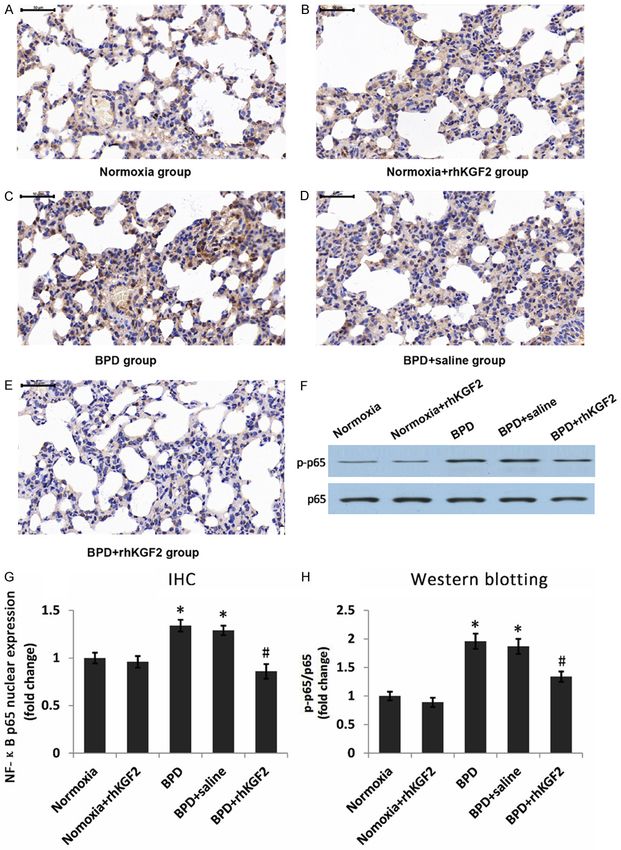
Therapeutic effects of rhKGF-2 on BPD

high affinity on epithelial cells, and reduced      Animal treatments
affinity for FGFR-1IIIb on epithelial/endothelial
cells [9, 10]. It mediates epithelial-mesenchy-     The animals were treated twice, at 1-week
mal interactions in a paracrine manner, and is      interval [18]. Briefly, after model establish-
essential for lung development [11]. The pro-       ment, rats were intraperitoneally anesthetized
tective effects of KGF-2 have been reported in      with chloralhydrate (300 mg/kg). Then, rhKGF2
various disease conditions, including bleomy-       (Newsummit Pharmaceutical Company, China)
cin-induced pulmonary fibrosis, high altitude       (diluted with saline to a concentration of 1 mg/
pulmonary edema, lipopolysaccharide (LPS)           ml; 5 mg/kg) was injected intratracheally in the
toxicity, mechanical ventilation and ischemia/      normoxia+rhKGF2 and BPD+rhKGF2 groups,
reperfusion-induced lung injury [12-16]. How-       with the same volume of saline administered to
ever, little is known about the therapeutic         the BPD+NS group, using an 18 G catheter
effects of KGF-2 on hyperoxia-induced lung          attached to a 1-ml syringe. Two weeks after the
injury in vivo.                                     last intratracheal injection, the animals were
                                                    sacrificed following anesthesia, and both lungs
Based on the above-mentioned properties of          in each animal were collected and weighed.
KGF-2, we hypothesized that it could alleviate      This was followed by lung lavage, for bronchoal-
BPD. Thus, this study aimed to assess the pro-      veolar lavage fluid (BALF) collection. Lung tis-
tective effects of rhKGF-2 on hyperoxia-induced     sues were subsequently stored at -80°C or
BPD in neonatal rats, exploring the potential       fixed with 10% paraformaldehyde for further
underlying mechanisms.                              use.

Materials and methods                               Lung wet-to-dry weight ratio (W/D)

Animals                                             The right main bronchus was ligated, and the
                                                    right lung was collected. After wet weight mea-
Sprague-Dawley (SD) rats were purchased from        surement, the middle lobe was placed in an
the Academy of Military Medical Sciences, and       oven at 60°C for 72 h, for determination of wet-
bred in a specific pathogen free (SPF) environ-     to-dry weight ratio.
ment, with rodent chow and water available ad
libitum. All experimental protocols were appr-      Bronchoalveolar lavage
oved by the Institutional Animal Care and Use
                                                    The left lung was lavaged for BALF collection. In
Committee of the PLA Army General Hospital.
                                                    brief, 2 ml of PBS (4°C) was slowly infused for
All procedures were performed according to the
                                                    1-2 min, and the fluid was slowly withdrawn, for
guidelines of the National Institutes of Health
                                                    3 cycles. The resultant fluid was collected and
concerning the care and use of laboratory
                                                    stored at -80°C for further analysis.
animals.
                                                    H&E staining and immunohistochemistry
Rat model of BPD and experimental groups
                                                    Lung tissue samples were fixed with 4% para-
The experimental BPD rat model was induced
                                                    formaldehyde, dehydrated in a series of graded
as described by O’Reilly M, et al [17]. Newborn     ethanol, paraffin embedded and sectioned (4
rats (n=75) were assigned to normoxia (21% O2,      μm). For H&E staining, the sections were treat-
n=30) and hyperoxia (85% O2, n=45) groups,          ed with xylene, a series of graded ethanol, and
and placed in plastic chambers with continu-        washed with distilled water. Hematoxylin and
ous O2 supply from postnatal day 1 to day 14.       eosin were used to stain the nucleus and the
The neonates were housed with their mothers,        cytoplasm, respectively. After dehydration and
and normoxia exposure in mother rats was per-       mounting, the sections were observed under a
formed with air break to prevent maternal O2        light microscope.
toxicity. The 75 neonates were assigned to 5
groups, including (1) normoxia control, (2)         Immunohistochemistry was used to detect
normoxia+rhKGF2, (3) BPD (no treatment), (4)        expression of VEGF and nuclear expression of
BPD+NS and (5) BPD+rhKGF2 groups (n=15/             NF-κB p65. For immunohistochemistry, endog-
group). The success of model establishment          enous peroxidase was blocked with 3% bovine
was verified by randomly selecting 2 rats for       serum albumin (BSA), and the sections were
pathological examination.                           incubated with primary antibodies, including

1433                                                     Int J Clin Exp Med 2019;12(2):1432-1442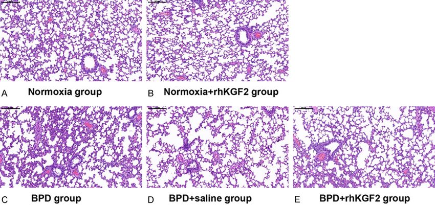
Therapeutic effects of rhKGF-2 on BPD

                                                                                 annealing at various tempera-
                                                                                 tures for 60 s and extension at
                                                                                 60°C for 5 min, followed by
                                                                                 the final single-peak melting
                                                                                 curve program. The compar-
                                                                                 ative threshold cycle method
                                                                                 (2-ΔΔCt) was used for data
                                                                                 analysis. The relative expres-
                                                                                 sion of SPC gene mRNA was
Figure 1. Pathological changes in lung tissues at 2 weeks post-partum (H&E
staining, 100×). A. Even alveolar septa, with no exudation in the normoxia
                                                                                 calculated and normalized
group; B. Irregular alveolar structure and enlarged alveolar space in the hy-    based on the GAPDH levels
peroxia group; some alveoli were merged, with reduced alveolus number,           using SDS software, version
and some alveoli showed inflammatory atelectasis (arrow). Scale bar: 200         1.4.
μm.
                                                                                 Western blotting

anti-VEGFa (Abcam, UK; 1:400) and anti-NF-κB                   Lung tissue samples were lysed with RIPA lysis
p65 (Cell signaling technology, USA; 1:400)                    buffer for 30min on ice. Total protein was quan-
antibodies, overnight at 4oC, followed by incu-                titated by the Bio-Rad protein assay. Equal
bation with secondary antibody for 50 min at                   amounts of total protein (20 μg) were separat-
room temperature. DAB kit (DAKO, Denmark)                      ed by SDS-PAGE and transferred onto PVDF
was used for visualization, according to the                   membranes. After blocking of nonspecific bind-
manufacturer’s instructions. The sections were                 ing sites with non-fat milk, the membranes
observed under a light microscope.                             were incubated with primary antibodies against
                                                               VEGFa (Abcam, UK; 1:1000), NF-κB p65 (Cell
Enzyme-linked immunosorbent assay (ELISA)                      signaling technology, USA; 1:1000) and NF-κB
                                                               p-p65 (Cell signaling technology, USA; 1:1000),
Two weeks after the last rhKGF-2 intratracheal                 respectively, at 4°C overnight. β-actin was used
administration, BALF samples were collected                    as an internal reference. Then, the membranes
and centrifuged at 2000-3000 rpm/min for 20                    were incubated with horseradish peroxidase
min at 4°C. The levels of cytokines, including                 (HRP)-conjugated goat anti-rabbit IgG for 1 h.
IL-1β, IL-6, TNF-α and macrophage inflammato-                  Immunoreactive bands were detected by the
ry protein-2 (MIP-2), were measured in the                     enhanced Chemiluminescence (ECL) method.
resulting supernatants by specific ELISA kits,
according to the manufacturer’s instructions.                  Statistical analysis

Quantitative real-time reverse transcription                   Statistical analyses were performed with SPSS,
polymerase chain reaction (qRT-PCR)                            version 22.0 (SPSS, USA). Data are expressed
                                                               as mean ± standard deviation (SD) and were
Lung tissues were homogenized in liquid nitro-                 assessed by an independent samples t-test
gen, and total RNA was extracted with TRIzol                   (group pairs) or one-way analysis variance
reagent (Thermo Fisher, USA), according to the                 (ANOVA; multiple groups) followed by a post hoc
manufacturer’s instructions. Total RNA was                     LSD t test. P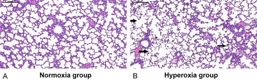
Therapeutic effects of rhKGF-2 on BPD

Figure 2. Pathological changes in lung tissues at 4 weeks after birth (H&E staining, 100×). A and B. In the normoxia
and normoxia+rhKGF2 groups, respectively, alveolar septa appeared even, with no exudation; C and D. In the BPD
and BPD+NS groups, respectively, lung tissues showed irregular alveolar structure, enlarged alveolar space and re-
duced number of alveoli. E. The alveolar structure was improved in the BPD+rhKGF2 group compared with the BPD
and BPD+NS groups, with increased number of alveoli, less hemorrhage and reduced infiltration of inflammatory.
Scale bar: 200 μm.

anxiety, shortness of breath, and cyanosis of                 weights in the BPD and BPD+NS groups were
the lips and toes immediately after disconti-                 129.15±8.92 g and 134.79±4.39 g, respec-
nuation of hyperoxia exposure. Meanwhile, the                 tively, which were significantly lower than those
normoxia group was active and had norm-                       of the normoxia group (t=-5.125, P
Therapeutic effects of rhKGF-2 on BPD
Figure 3. Effects of KGF-2 on inflammatory cytokine
expression in BALF (ELISA) and wet-to-dry weight ratios
of rat lungs. A. W/D ratios significantly increased in
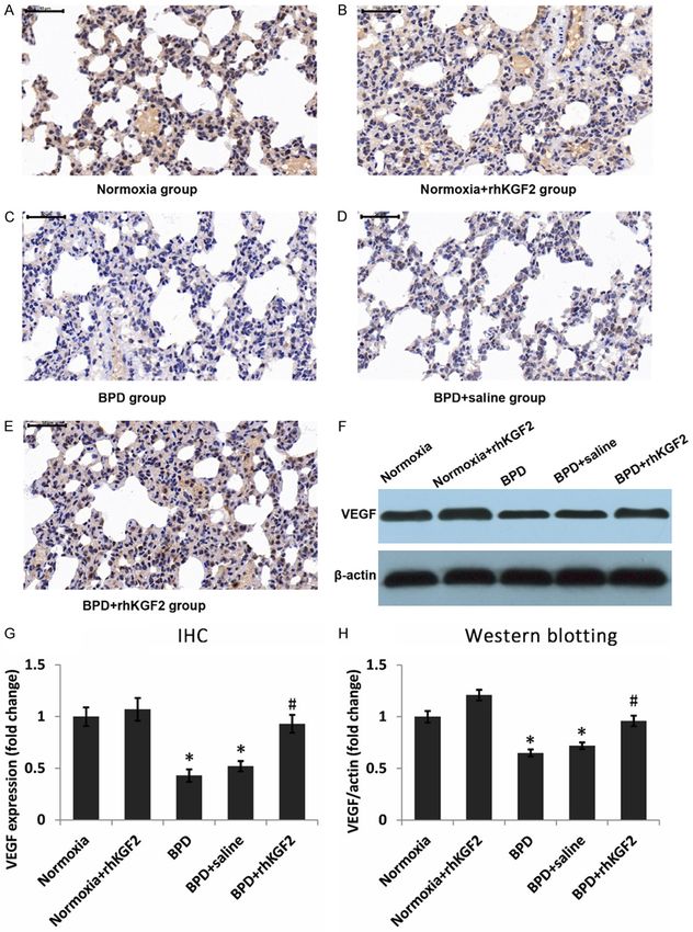
the BPD and BPD+NS groups (*#PTherapeutic effects of rhKGF-2 on BPD Figure 5. Effect of KGF-2 on VEGF protein expression in rat lung samples. High VEGF amounts were found in the normoxia (A) and normoxia+rhKGF-2 (B) groups, decreasing in the BPD (C) and BPD+NS (D) groups in comparison; the BPD+rhKGF-2 group (E) showed higher levels compared with the BPD group (DAB-based immunohistochemistry; 400×). Similar results were obtained by Western blotting (F). The expression of VEGF protein detected by immu- nohistochemistry was quantified as (G), and that detected by Western blotting was quantified as (H) (*P
Therapeutic effects of rhKGF-2 on BPD

Figure 6. Effect of KGF-2 on NF-κb p65 protein nuclear expression and phosphorylation in rat lung samples. Low
NF-κB p65 nuclear amounts were observed in the normoxia (A) and normoxia+rhKGF-2 (B) groups, increasing in
the BPD (C) and BPD+NS (D) groupsin comparison; the BPD+rhKGF-2 group (E) showed slightly reduced levels
compared with the BPD group (DAB-based immunohistochemistry; 400×). Similar results were obtained by Western
blotting (F). The nuclear expression of NF-κb p65 protein detected by immunohistochemistry was quantified as (G)

1438                                                              Int J Clin Exp Med 2019;12(2):1432-1442
Therapeutic effects of rhKGF-2 on BPD

(*P
Therapeutic effects of rhKGF-2 on BPD
we hypothesized that exogenous KGF-2 may group). This strongly indicated that KGF-2 pro-
protect against hyperoxia-induced BPD in neo- motes growth in type II alveolar epithelial cells.
natal rats. Indeed, we found that intratracheal The pathophysiological roles of VEGF have
administration of rhKGF2 promoted alveolar been intensively studied. For example, VEGF is
growth in BPD rats, improved clinical symptoms considered a potent endothelial cell-specific
and lung histology, suggesting that KGF-2 pre- mitogen that promotes vascular growth and
vents lung injury in hyperoxia-induced BPD. remodeling [30]. In addition, VEGF is involved in
the formation and maintenance of pulmonary
Previously, Abman and Matthay described and alveolar structures in infants and adults
KGF-2 as a potent alveolar type II cell mitogen [31], and immature lungs are sensible to VEGF
which plays an important role in preventing down-regulation [32]. Furthermore, reduced
alveolar epithelial cells from DNA damage and VEGF is associated with the pathology of BPD
apoptosis via the MAPK/ERK pathway [26]. In [33, 34]. In the current study, KGF-2 upregulat-
addition, pre-treatment with KGF-2 significantly ed VEGF expression in BPD animals, subse-
improves high-altitude pulmonary edema in quently preventing the pathological changes of
rats, most likely by reducing apoptosis and hyperoxia mediated BPD. It is reported that
inducing proliferation in type II alveolar cells KGF-2 is a major player in alveologenesis and
[13]. In this study, we wanted to explore the regeneration of the lung after injury, by upregu-
potential mechanisms underlying the therapeu- lating VEGF in the distal epithelium and direct-
tic effects of KGF-2 in hyperoxia mediated BPD. ing the differentiation of the bipotent progeni-
As shown above, intratracheal administration tor cells towards the AEC II lineage, but no
of KGF-2 significantly decreased lung W/D report has shown that KGF-2 has any effect on
ratios, indicating the therapeutic effect of normal lung tissue. The increased levels of SPC
KGF-2 on pulmonary edema. In addition, KGF-2 and VEGF were closely associated with the
markedly reduced the secretion levels of the therapeutic effects of KGF-2 in BPD rats, but
inflammatory cytokines IL-1β, IL-6, TNF-α and the levels of SPC and VEGF were not affected
MIP-2 in BALF of BPD rats, indicating that it pre- by KGF-2 in normal rats.
vents lung injury in these animals. These find-
ings suggested that KGF-2 may exert its pre- As a core transcription factor, NF-κB regulates
ventive effects by combining alleviation of pul- cellular inflammatory, participates in immune
monary edema, reduction of inflammatory cyto- responses, and regulates cell differentiation
kine secretion, and prevention of macrophage and apoptosis [35]. It is activated by a variety of
and neutrophil accumulation. These effects stimuli that include cytokines, growth factors
reduced lung damage and improved lung devel- and lymphokines. The various stimuli that acti-
opment in newborn rats. These findings cor- vate NF-κB cause phosphorylation of IκB, which
roborated previous studies showing that BPD is crucial to its role. Nuclear expression and
development is associated with increased phosphorylation of NF-κB p65 (a key compo-
amounts of inflammatory cells and pro-inflam- nent of NF-κB) are significantly increased in the
matory cytokines in the lung [27]. It is also lung tissues of BPD animals, whereas KGF-2
known that decreased alveologenesis with expression is markedly reduced [19]. Mean-
interstitial thickness is associated with incr- while, interactions between NF-κB p65 and
eased neutrophil cytokines in hyperoxia expo- SP3 inhibit KGF-2 expression [36]. In the cur-
sed rats [28]. rent study, KGF-2 reduced the secretion levels
of inflammatory cytokines. Thus, we hypothe-
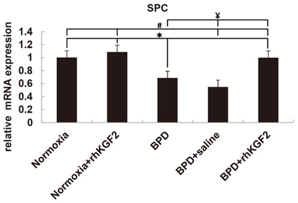
SPC is a key marker of type II alveolar epithelial sized that KGF-2 may downregulate NF-κB.
cells [29]. Therefore, we further evaluated SPC Indeed, as demonstrated above by immunohis-
expression in lung epithelial cells in hyperoxia- tochemistry and Western blotting, NF-κB p65
induced BPD rats. Because of the low expres- nuclear expression and phosphorylation were
sion of SPC in alveoli, it is difficult to detect it by slightly reduced after KGF-2 administration in
Western blotting. As the content of SPC in lung BPD rats. These findings suggested that KGF-2
lavage fluid is also low, we detect its mRNA by may also, by inhibiting NF-κB p65 nuclear
qRT-PCR. As expected, SPC mRNA levels were expression and phosphorylation, attenuate
reduced in the BPD group, and markedly inflammation, thereby promoting lung repair in
increased after KGF-2 treatment (BPD+KGF-2 neonatal rats after hyperoxia-induced BPD.
1440 Int J Clin Exp Med 2019;12(2):1432-1442Therapeutic effects of rhKGF-2 on BPD
Collectively, the current study demonstrated [3] Gortner L, Misselwitz B, Milligan D, Zeitlin J,
that rhKGF-2 prevents lung damage in hyperox- Kollee L, Boerch K, Agostino R, Van Reempts P,
ia-induced BPD rats via multiple routes; how- Chabernaud JL, Breart G, Papiernik E, Jarreau
ever, in terms of clinical application, further PH, Carrapato M, Gadzinowski J and Draper E.
investigation is required to comprehensively Rates of bronchopulmonary dysplasia in very
determine the therapeutic effects of KGF-2 and preterm neonates in Europe: results from the
MOSAIC cohort. Neonatology 2011; 99: 112-
the underlying mechanisms.
117.
This study had several limitations. First, we did [4] Iliodromiti Z, Zygouris D, Sifakis S, Pappa KI,
Tsikouras P, Salakos N, Daniilidis A, Siristatidis
not use a comprehensive approach to assess
C and Vrachnis N. Acute lung injury in preterm
various molecules and pathways involved in
fetuses and neonates: mechanisms and mo-
BPD pathology. In addition, we have only tenta-
lecular pathways. J Matern Fetal Neonatal Med
tively discussed the interactions of rhKGF2 and 2013; 26: 1696-1704.
NF-κB, but have not explored the mechanisms [5] Poggi C and Dani C. Antioxidant strategies and
of inhibiting inflammation in depth. Finally, respiratory disease of the preterm newborn:
whether results generated in SD rats would an update. Oxid Med Cell Longev 2014; 2014:
reflect the clinical situation remains unknown. 721043.
Therefore, further investigation is required to [6] Laube M, Stolzing A, Thome UH and Fabian C.
confirm our findings. Therapeutic potential of mesenchymal stem
cells for pulmonary complications associated
Conclusion with preterm birth. Int J Biochem Cell Biol
2016; 74: 18-32.
In summary, KGF-2 promotes lung development [7] Yamasaki M, Miyake A, Tagashira S and Itoh N.
in hyperoxia-mediated BPD in neonatal rats. Structure and expression of the rat mRNA en-
These protective effects may combine allevia- coding a novel member of the fibroblast growth
tion of pulmonary edema, reduction of inflam- factor family. J Biol Chem 1996; 271: 15918-
matory cytokine secretion, and prevention of 15921.
macrophage and neutrophil accumulation. [8] Huang Z, Zhu G, Sun C, Zhang J, Zhang Y,
These findings provide potential options for Zhang Y, Ye C, Wang X, Ilghari D and Li X. A
future clinical studies aiming at treating BPD novel solid-phase site-specific PEGylation en-
patients to prevent long-term sequelae. hances the in vitro and in vivo biostabilty of
recombinant human keratinocyte growth fac-
Acknowledgements tor 1. PLoS One 2012; 7: e36423.
[9] Emoto H, Tagashira S, Mattei MG, Yamasaki M,
This work was supported by the National Na- Hashimoto G, Katsumata T, Negoro T, Naka-
tural Science Foundation of China (81401248). tsuka M, Birnbaum D, Coulier F and Itoh N.
Structure and expression of human fibroblast
Disclosure of conflict of interest growth factor-10. J Biol Chem 1997; 272:
23191-23194.
None. [10] Ware LB and Matthay MA. Keratinocyte and
hepatocyte growth factors in the lung: roles in
Address correspondence to: Dr. Zhichun Feng, The lung development, inflammation, and repair.
Second Military Medical University of People’s Am J Physiol Lung Cell Mol Physiol 2002; 282:
Liberation Army, Yangpu District, Shanghai 200433, L924-940.
China. Tel: 010-66721786; E-mail: bzfengzc@yahoo. [11] Benjamin JT, Smith RJ, Halloran BA, Day TJ,
com Kelly DR and Prince LS. FGF-10 is decreased in
bronchopulmonary dysplasia and suppressed
References by Toll-like receptor activation. Am J Physiol
Lung Cell Mol Physiol 2007; 292: L550-558.
[1] Davidson LM and Berkelhamer SK. Broncho- [12] Gupte VV, Ramasamy SK, Reddy R, Lee J,
pulmonary dysplasia: chronic Lung disease of Weinreb PH, Violette SM, Guenther A, War-
infancy and long-term pulmonary outcomes. J burton D, Driscoll B, Minoo P and Bellusci S.
Clin Med 2017; 6: 6. Overexpression of fibroblast growth factor-10
[2] Hwang JS and Rehan VK. Recent advances in during both inflammatory and fibrotic phases
bronchopulmonary dysplasia: pathophysiolo- attenuates bleomycin-induced pulmonary fi-
gy, prevention, and treatment. Lung 2018; brosis in mice. Am J Respir Crit Care Med
196: 129-138. 2009; 180: 424-436.
1441 Int J Clin Exp Med 2019;12(2):1432-1442Therapeutic effects of rhKGF-2 on BPD
[13] She J, Goolaerts A, Shen J, Bi J, Tong L, Gao L, [24] Wheeler G. Repifermin. Human Genome Sci-
Song Y and Bai C. KGF-2 targets alveolar epi- ences/GlaxoSmithKline. IDrugs 2001; 4: 813-
thelia and capillary endothelia to reduce high 819.
altitude pulmonary oedema in rats. J Cell Mol [25] Upadhyay D, Correa-Meyer E, Sznajder JI and
Med 2012; 16: 3074-3084. Kamp DW. FGF-10 prevents mechanical
[14] Tong L, Bi J, Zhu X, Wang G, Liu J, Rong L, Wang stretch-induced alveolar epithelial cell DNA
Q, Xu N, Zhong M, Zhu D, Song Y and Bai C. damage via MAPK activation. Am J Physiol
Keratinocyte growth factor-2 is protective in li- Lung Cell Mol Physiol 2003; 284: L350-359.
popolysaccharide-induced acute lung injury in [26] Abman SH and Matthay MA. Mesenchymal
rats. Respir Physiol Neurobiol 2014; 201: stem cells for the prevention of bronchopulmo-
7-14. nary dysplasia: delivering the secretome. Am J
[15] Bi J, Tong L, Zhu X, Yang D, Bai C, Song Y and Respir Crit Care Med 2009; 180: 1039-1041.
[27] Niver D. Bronchopulmonary dysplasia: struc-
She J. Keratinocyte growth factor-2 intratra-
tural challenges and stem cell treatment po-
cheal instillation significantly attenuates venti-
tential. Adv Neonatal Care 2014; 14: e1-e11.
lator-induced lung injury in rats. J Cell Mol Med
[28] Thebaud B and Abman SH. Bronchopulmonary
2014; 18: 1226-1235.
dysplasia: where have all the vessels gone?
[16] Fang X, Wang L, Shi L, Chen C, Wang Q, Bai C Roles of angiogenic growth factors in chronic
and Wang X. Protective effects of keratinocyte lung disease. Am J Respir Crit Care Med 2007;
growth factor-2 on ischemia-reperfusion-in- 175: 978-985.
duced lung injury in rats. Am J Respir Cell Mol [29] Wang D, Haviland DL, Burns AR, Zsigmond E
Biol 2014; 50: 1156-1165. and Wetsel RA. A pure population of lung alve-
[17] Buch S, Han RN, Cabacungan J, Wang J, Yuan olar epithelial type II cells derived from human
S, Belcastro R, Deimling J, Jankov R, Luo X, Lye embryonic stem cells. Proc Natl Acad Sci U S A
SJ, Post M and Tanswell AK. Changes in ex- 2007; 104: 4449-4454.
pression of platelet-derived growth factor and [30] Johnson KE and Wilgus TA. Vascular endothe-
its receptors in the lungs of newborn rats ex- lial growth factor and angiogenesis in the re-
posed to air or 60% O(2). Pediatr Res 2000; gulation of cutaneous wound repair. Adv
48: 423-433. Wound Care (New Rochelle) 2014; 3: 647-661.
[18] Tong L, Zhou J, Rong L, Seeley EJ, Pan J, Zhu X, [31] Balasubramaniam V, Mervis CF, Maxey AM,
Liu J, Wang Q, Tang X, Qu J, Bai C and Song Y. Markham NE and Abman SH. Hyperoxia reduc-
Fibroblast growth factor-10 (FGF-10) mobilizes es bone marrow, circulating, and lung endothe-
lung-resident mesenchymal stem cells and lial progenitor cells in the developing lung: im-
protects against acute lung injury. Sci Rep plications for the pathogenesis of bronchopul-
2016; 6: 21642. monary dysplasia. Am J Physiol Lung Cell Mol
[19] Madurga A, Mizikova I, Ruiz-Camp J and Morty Physiol 2007; 292: L1073-1084.
RE. Recent advances in late lung development [32] Scott CL, Walker DJ, Cwiklinski E, Tait C, Tee AR
and the pathogenesis of bronchopulmonary and Land SC. Control of HIF-1{alpha} and vas-
dysplasia. Am J Physiol Lung Cell Mol Physiol cular signaling in fetal lung involves cross talk
2013; 305: L893-905. between mTORC1 and the FGF-10/FGFR2b/
[20] Olaloko O, Mohammed R and Ojha U. Spry2 airway branching periodicity clock. Am J
Physiol Lung Cell Mol Physiol 2010; 299: L455-
Evaluating the use of corticosteroids in pre-
471.
venting and treating bronchopulmonary dys-
[33] Hiscott J, Kwon H and Genin P. Hostile take-
plasia in preterm neonates. Int J Gen Med
overs: viral appropriation of the NF-kappaB
2018; 11: 265-274.
pathway. J Clin Invest 2001; 107: 143-151.
[21] Gough A, Spence D, Linden M, Halliday HL and
[34] Northway WH Jr, Rosan RC and Porter DY.
McGarvey LPA. General and respiratory health Pulmonary disease following respirator thera-
outcomes in adult survivors of bronchopulmo- py of hyaline-membrane disease. Bronchopu-
nary dysplasia: a systematic review. Chest lmonary dysplasia. N Engl J Med 1967; 276:
2012; 141: 1554-1567. 357-368.
[22] Short EJ, Klein NK, Lewis BA, Fulton S, [35] Benjamin JT, Carver BJ, Plosa EJ, Yamamoto Y,
Eisengart S, Kercsmar C, Baley J and Singer LT. Miller JD, Liu JH, van der Meer R, Blackwell TS
Cognitive and academic consequences of and Prince LS. NF-kappaB activation limits air-
bronchopulmonary dysplasia and very low way branching through inhibition of Sp1-
birth weight: 8-year-old outcomes. Pediatrics mediated fibroblast growth factor-10 expres-
2003; 112: e359. sion. J Immunol 2010; 185: 4896-4903.
[23] Bellusci S, Grindley J, Emoto H, Itoh N and [36] Carver BJ, Plosa EJ, Stinnett AM, Blackwell TS
Hogan BL. Fibroblast growth factor 10 (FGF10) and Prince LS. Interactions between NF-
and branching morphogenesis in the embry- kappaB and SP3 connect inflammatory signal-
onic mouse lung. Development 1997; 124: ing with reduced FGF-10 expression. J Biol
4867-4878. Chem 2013; 288: 15318-15325.
1442 Int J Clin Exp Med 2019;12(2):1432-1442You can also read