Osteochondral Regeneration Using Adipose Tissue-Derived Mesenchymal Stem Cells - MDPI
←
→
Page content transcription
If your browser does not render page correctly, please read the page content below
International Journal of
            Molecular Sciences

Review
Osteochondral Regeneration Using Adipose
Tissue-Derived Mesenchymal Stem Cells
Daiki Murata 1, *, Ryota Fujimoto 1,2 and Koichi Nakayama 1
 1    Center for Regenerative Medicine Research, Faculty of Medicine, Saga University, Honjo-machi,
      Saga 840-8502, Japan; R-FUJIMOTO@nakayama-labs.co.jp (R.F.); nakayama@me.saga-u.ac.jp (K.N.)
 2    Department of Oral and Maxillofacial Surgery, Faculty of Medicine, Saga University, Nabeshima 5-1-1,
      Saga 849-8501, Japan
 *    Correspondence: daiki_net_official@yahoo.co.jp; Tel.: +81-952-28-8480

 Received: 15 March 2020; Accepted: 15 May 2020; Published: 19 May 2020

 Abstract: Osteoarthritis (OA) is a major joint disease that promotes locomotor deficiency during
 the middle- to old-age, with the associated disability potentially decreasing quality of life. Recently,
 surgical strategies to reconstruct both articular cartilage and subchondral bone for OA have been
 diligently investigated for restoring joint structure and function. Adipose tissue-derived mesenchymal
 stem cells (AT-MSCs), which maintain pluripotency and self-proliferation ability, have recently
 received attention as a useful tool to regenerate osteocartilage for OA. In this review, several studies
 were described related to AT-MSC spheroids, with scaffold and scaffold-free three-dimensional
 (3D) constructs produced using “mold” or “Kenzan” methods for osteochondral regeneration.
 First, several examples of articular cartilage regeneration using AT-MSCs were introduced. Second,
 studies of osteochondral regeneration (not only cartilage but also subchondral bone) using AT-MSCs
 were described. Third, examples were presented wherein spheroids were produced using AT-MSCs
 for cartilage regeneration. Fourth, osteochondral regeneration following autologous implantation of
 AT-MSC scaffold-free 3D constructs, fabricated using the “mold” or “Kenzan” method, was considered.
 Finally, prospects of osteochondral regeneration by scaffold-free 3D constructs using AT-MSC
 spheroids were discussed.

 Keywords: adipose tissue-derived mesenchymal stem cells; cell construct; Kenzan method;
 mold method; osteoarthritis; osteochondral regeneration; scaffold-free; spheroid

1. Introduction
     Articular cartilage constitutes hyaline cartilage, which is generally encompassed by perichondrium
consisting of collagen fibers and early mesenchymal progenitor cells that can differentiate into
chondrocytes [1]. However, articular cartilage is instead covered by horizontally arranged
collagen-containing proteoglycans, such as lubricin [2]. Therefore, the articular surface exhibits lubricity
but does not possess self-healing ability, owing to the absence of vascularity and perichondrium [3].
Cartilage defects are frequently observed in young and active patients, and the repaired tissue is fibrous
due to the poor self-healing capacity of articular cartilage [4,5]. Notably, this fibrous tissue does not have
the functional properties of natural hyaline cartilage. Thus, the defect leads to cartilage degradation
and osteochondral lesions, defined as subchondral bone sclerosis/deformation, which usually develops
into osteoarthritis (OA) [6]. OA constitutes a major joint disease that causes movement disorders in
the middle- and old-age [7–9], and the associated disabilities can reduce the quality of life. Therefore,
surgical strategies to rebuild both bone and cartilage to restore joint structure and function have been
eagerly studied [7].

Int. J. Mol. Sci. 2020, 21, 3589; doi:10.3390/ijms21103589                           www.mdpi.com/journal/ijms
Int. J. Mol. Sci. 2020, 21, 3589                                                                      2 of 23

      A particular focus in recent studies has been the complete regeneration of hyaline cartilage
covering the subchondral bone in OA. Several treatments for damaged bone and cartilage, including
mosaicplasty [10], microfracture [11], and autologous chondrocyte implantation [12], have been used
in patients to relieve pain and improve joint function. However, there are several associated problems,
such as limitations of available donor sites based on the required size and shape of the osteochondral
autograft [9], dedifferentiation of chondrocytes during passaging in culture [13]. To solve these problems,
mesenchymal stem cells (MSCs) have recently received increasing attention as promising options for
osteochondral regeneration [14,15]. In numerous previous studies, chondrogenic differentiation ability
was evaluated in vitro, especially using MSCs derived from bone marrow (BM), adipose tissue (AT),
and other sources [15–17]. Among these stem cells, AT-derived MSCs (AT-MSCs) can be isolated most
abundantly, and their cellular proliferation rate may be higher in mature animals [15,18]. Although it
has been reported that AT-MSCs rarely differentiate into chondrocytes [19,20], AT-MSCs have been
specifically shown to differentiate into cartilage in vitro [16]. Additionally, liposuction, a surgical
method used to aspirate AT, is commonly utilized in the cosmetic field and is a globally accepted
method for obtaining AT [21,22]. Therefore, the advantages of using AT-MSCs for cartilage regeneration
are not only that 1) the stem cells can differentiate into chondrocytes but also that 2) they can be isolated
more abundantly than other mesenchymal stem cells. Furthermore, 3) the cell proliferation rate is very
high, and 4) they can be collected by liposuction accepted worldwide. However, conventional methods
for transplanting cell suspensions have been unsuccessful in reconstructing osteochondral defects in
large animals because MSCs administered to the defect do not settle and survive [20]. Therefore, it is
considered indispensable to develop methods for providing MSCs in a three-dimensional (3D) format
to ensure that cells are placed in the defect.
      In recent years, advances in tissue engineering of functional articular cartilage have been facilitated
by several methods using synthetic or biological scaffolds to achieve sufficient thickness and mechanical
function, along with supporting cell attachment, migration, proliferation, and differentiation [23,24].
Studies involving surgical procedures using a combination of artificial bone and autologous
chondrocytes seeded into a collagen scaffold have also demonstrated favorable bone and cartilage
restoration compared to outcomes using chondrocyte suspensions [25,26]. Previous studies indicated
that scaffolds composed of materials, such as collagen and hyaluronic acid, could be useful for promoting
cell adhesion, proliferation, and chondrogenic differentiation [27,28] and might also facilitate stem cell
seeding for implantation into osteochondral defects [29,30]. Bone regeneration using AT-MSCs seeded
into hydroxyapatite has also been investigated [31]. However, the bone itself is notably self-restorative,
while articular cartilage is less so [3,32]. While scaffold or scaffold-free systems have recently been
investigated for the purpose of cartilage regeneration [23–30,33–35], it has been difficult to create
sufficient thickness to fill in the osteochondral defects with or without using a scaffold. For example,
a minimum thickness of 5 mm is necessary to be filled in a full-thickness articular cartilage defect of the
knee. With or without a scaffold, it is even more challenging with the current methods to regenerate
both articular cartilage and subchondral bone in the context of an osteochondral defect.
      Alternatively, adherent cells grown in suspension can form aggregates (spheroids) in nature,
avoiding death through cell-to-cell attachment [36]. The majority of the approaches using these
spheroids incorporate a specific cylindrical mold to produce constructs of desired shapes [37,38].
Certain cell types, such as AT-MSCs, possess the capacity to synthesize and release components of
the extracellular matrix (ECM), such as collagen, in vitro. Notably, this capacity is accelerated under
confluence or 3D culture conditions because the cell cycle stops, and the cells produce ECM components.
As this phenomenon has been confirmed in studies using AT-MSCs, a novel method for fusing the
spheroids by using a specific cylindrical “mold” was developed to fabricate scaffold-free 3D columnar
constructs consisting of AT-MSCs in vitro [39–41]. Two studies have reported the regeneration of
articular cartilage and subchondral bone using these constructs consisting of autologous AT-MSC
spheroids in minipigs [39,40]. Other studies have evaluated the outcome of using autologous AT-MSCs
for osteochondral regeneration, as well [42,43]. In contrast, the use of allogeneic AT-MSCs of large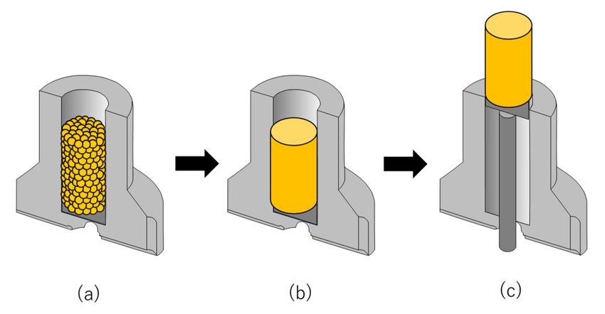
Int. J. Mol. Sci. 2020, 21, 3589                                                                    3 of 23

animals has not yet been reported, although previous studies demonstrated the safety of the immune
reaction and oncogenesis risk when using allogeneic MSCs. Besides, these cells offer some advantages,
including avoidance of donor site morbidity and reducing overall cost [44,45]. Despite this, data related
to whether osteochondral defects could be healed histologically by implanting 3D cell constructs made
of allogeneic AT-MSCs engrafted into osteochondral defects are only available in rabbit models [41].
Moreover, to create more sophisticated constructs, the use of automatic spheroid deposition using
bio-3D printing with a specific needle-array, termed the “Kenzan” method, is recommended [46].
Bio-3D printing constitutes a technology to create tissues and organs with an internal structure
by 3D-stacking of spheroids at appropriate positions in the array, according to the original design.
The “Kenzan,” which plays the role of a temporary artificial scaffold to fix the position of the spheroids
placed by bio-3D printing, is removed prior to implantation after the spheroids fuse using the increased
ECM. With this technique, spheroids are uniformly dispensed in contact with one another at a regular
distance. Using these advantages, a newer method for fusing the spheroids using “Kenzan” has
been developed to fabricate scaffold-free 3D tubular constructs consisting of autologous AT-MSCs.
These constructs have been used for the regeneration of articular cartilage and subchondral bone in
minipigs [47].
      In this review, recent studies, demonstrating cartilage and osteochondral regeneration following
implantation of AT-MSCs, were first introduced [48–56]. Second, the most recent examples of
assembling AT-MSCs in three dimensions with or without scaffold for cartilage regeneration were
discussed [57–60]. Third, osteochondral regeneration through implantation of scaffold-free constructs,
consisting of autologous swine AT-MSCs, fabricated using the “mold”, into osteochondral defects
in minipigs was discussed [39,40]. Fourth, a study describing a construct comprised of allogeneic
AT-MSCs engrafted into osteochondral defects in rabbit models was presented [41]. This was in addition
to examples of histological examination of MSC constructs prepared by using the “mold” method [40].
Finally, a representative study was presented that evaluated the histology of AT-MSC constructs
prepared by bio-3D printing using the Kenzan method and analyzed osteochondral regeneration
following the autologous implantation of two swine AT-MSC constructs [47].

2. AT-MSCs for Cartilage Regeneration
      Several studies addressing articular cartilage regeneration using AT-MSCs were reviewed here
and summarized in Table 1. First, stem cell-chondrocyte interactions for cartilage regeneration
modulated by a combinatorial extracellular matrix containing hydrogel were described [48]. Next,
a transforming growth factor (TGF)-β3 encapsulated polylactide-co-caprolactone (PLCL) scaffold by
a supercritical carbon dioxide (CO2 )-1,1,1,3,3,3-hexafluoro-2-propanol (HFIP) co-solvent system for
cartilage tissue engineering was mentioned [49]. We also introduced an approach to promote cartilage
formation of AT-MSCs seeded in polylactic-co-glycolic acid (PLGA) by dynamic compression combined
with the exogenous sex-determining region Y-box (SOX)-9 [50]. Subsequently, an example was
described in which cartilage in an osteoarthritis model was regenerated using AT-MSC differentiated
chondrocytes [51]. In addition, chondrogenesis of AT-MSCs enhanced in hyaluronic acid-modified
thermoresponsive poly N-isopropyl acrylamide (HA-PNIPAAm-CL) hydrogel for cartilage regeneration
was introduced [52]. We also showed an example, illustrating enrichment of CD146+ AT-MSCs
combined with articular cartilage extracellular matrix (ACECM) scaffold that promotes cartilage
regeneration [53]. Finally, we discussed cartilage regeneration in human knee osteoarthritis using
autologous AT-MSCs and autologous extracellular matrix [54].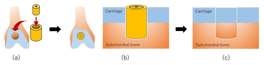
Int. J. Mol. Sci. 2020, 21, 3589 4 of 23
Table 1. Cartilage and osteocartilage regeneration using AT-MSCs.
Target Tissue
Number of Materials of a Shape of Approximate Size of Other
(Study Design) AT-MSCs Pros and Cons
AT-MSCs Scaffold a Scaffold a Scaffold Materials
[Reference]
Cartilage P; CS-MA scaffold maximize synergistic cartilage
(in vitro) N/A 7.5 × 10⁵ cells CS-MA Gel 50 µL NChons formationC; CS-MA decrease the mechanical
[48] properties of the scaffold
Cartilage P; Multifunctional scaffold
D: 9.0 mm
(in vitro) N/A 1.0 × 10⁶ cells PLCL Discoid TGF-β3,Fibrin gel C; The possibility of immune-foreign body reaction
T: 3.0 mm
[49] to the capsules
P; Dynamic compression combined with SOX-9 and
Cartilage
Sox-9 gene PLGA scaffold.
(in vitro) N/A 1.25 × 10⁷ cells PLGA Columnar N.D.
transfection C; N.D. of the detailed mechanism and the proper
[50]
pore density
Cartilage P; High regenerative potential of DCs
Autologous
(in vivo) 1.0 × 10⁵ cells N/A N/A N/A N/A from AT-MSCs
DCs
[51] C; N.D. of the outcomes in vivo using large animals
Cartilage P; Suitable microenvironment for chondrogenesis
(in vivo) Allogenic 2.0 × 10⁵ cells HA-PNIPAAm-CL Gel 200 µL N/A in vitro and in vivo
[52] C; N.D. on long-term regeneration
Cartilage
Xenogenic D: 3.5 mm P; Good biocompatibility with the scaffold
(in vivo) 5.0 × 10⁵ cells ACECM Columnar N/A
CD146+ H: 2.0 mm C; Small amount of CD146+ AT-MSCs
[53]
Cartilage P; Good clinical outcomes of three OA patients
Autologous Other cells, PRP,
(Clinical) Autologous N.D. Liquid ~ 14 mL C; Small number of cases and no histopathological
ECM and HA
[54] outcomes
L: 10.0 mm
Cancellous
1.0 × 10⁷ cells Rectangular W: 8.0 mm P; Bi-layered scaffold for the fabrication of
bone osteochondral tissue
Osteocartilage H: 5.0 mm
(in vitro) N/A N/A C; N.D. of transplanting effect using animals
L: 10.0 mm
[55]
1.0 × 10⁷ cells Hydrogel Rectangular W: 8.0 mm
H: 3.0 mm
Osteocartilage L: 2.0 mm NH2 and COOH P; NH2 and COOH functionalization of scaffolds
(in vivo) Xenogenic N.D. POSS Tabular W: 2.0 mm functionalization, C; N.D. of chondro- and osteogenesis pathway of
[56] H: N.D. CAM the chemicals
AT-MSCs, adipose tissue-derived mesenchymal stem cells; DCs, differentiated chondrocytes from AT-MSCs; CS-MA, chondroitin sulfate methacrylate; PLCL, polylactide-co-caprolactone;
PLGA, polylactic-co-glycolic acid; HA-PNIPAAm-C, hyaluronic acid-modified thermoresponsive poly N-isopropyl acrylamide; ACECM, articular cartilage extracellular matrix; ECM,
extracellular matrix; POSS, polyhedral oligomeric silsesquioxane; D, diameter; H, height; T, thickness; L, length; W, width; NChons, neonatal chondrocytes; TGF-β3, transforming growth
factor-beta3; SOX-9, sex-determining region Y-box-9; PRP, platelet-rich plasma; HA, hyaluronan; CAM, chorioallantoic membrane; OA, osteoarthritis; N/A, not applicable; N.D., no data.Int. J. Mol. Sci. 2020, 21, 3589 5 of 23
Biochemical cues provided by methacrylated ECM molecules in hydrogels, as well as the
mechanical properties of hydrogels, impact chondrogenic gene expression of AT-MSCs in 3D culture [61].
Wang et al. [48] recently reported that AT-MSCs could catalyze cartilage formation by neonatal
chondrocytes (NChons) when co-cultured in biomimetic hydrogels using a 3D co-culture model.
AT-MSCs and NChons were co-encapsulated in 39 combinatorial hydrogel compositions with decoupled
biochemical and mechanical properties to modulate stem cell-chondrocyte interactions for cartilage
repair. Both chondroitin sulfate (CS) and hyaluronic acid (HA) led to robust articular cartilage matrix
deposition, as shown by the intense staining of aggrecan and type II collagen. In soft hydrogels (15 kPa),
CS led to the highest amount of sulfated glycosaminoglycan deposition and increased compressive
moduli. Findings from this study might guide optimal scaffold design to maximize synergistic
cartilage formation using mixed cell populations. However, the hydrogels supported robust new
cartilage matrix deposition by mixed AT-MSC–NChons co-culture in CS and HA containing hydrogels,
and the compressive modulus in cell-containing hydrogels remained substantially lower than that
of native cartilage. This showed a decrease in mechanical properties of hydrogels containing mixed
AT-MSCs and NChons over time, likely due to the active degradation of hydrogels by chondrocytes
to make space for a new matrix. Future studies with long-term culture using leading hydrogel
compositions would help validate the increase in mechanical properties of tissue-engineered cartilage
over time. Using enzymatically degradable PEG hydrogels may accelerate the speed of neocartilage
deposition and improve the interconnectedness of the cell deposited matrix so that the hydrogel
construct will become mechanically stronger over the culture period. Therefore, future work will
include the incorporation of specific ECM molecules into hydrogels in distinct spatial zones to direct
cells to secrete a cartilage-specific matrix that mimics the zones of native cartilage. On the other
hand, animal experiments in which the mixed AT-MSC–NChons co-cultured in CS and HA containing
hydrogels are transplanted subcutaneously in mice have been performed [62]. In the future, it is
expected that additional in vivo studies in the knee joint using larger experimental animals shall
provide future insight.
Mechanical cues and sustained biological cues are important factors, particularly in load-bearing
tissues, such as articular cartilage [63–65]. Carriers, including hydrogels and nanoparticles, have been
investigated for the sustained release of protein drugs [66–69]. However, it is difficult to apply such
carriers alone as scaffolds for cartilage regeneration because of their weak mechanical properties;
they must be combined with other biomaterials that have adequate mechanical strength. Therefore,
Kim et al. [49] developed a multifunctional scaffold that had similar mechanical properties to those
of native cartilage and encapsulated TGF-β3 for chondrogenesis. Tissue regeneration efficiency of
the TGF-β3 encapsulated PLCL scaffold was investigated using human AT-MSCs in vitro and in vivo.
Based on TGF-β3 release studies, it was confirmed that TGF-β3 molecules were released by 8 weeks
and remained in the PLCL matrix. Explants of TGF-β3 encapsulated scaffolds by a co-solvent system
exhibited distinct improvement in the compressive E-modulus and deposition of the extracellular
matrix. Long-term delivery of TGF-β3 resulted in the formation of a hyaline cartilage-specific lacunae
structure and prevented hypertrophy of differentiated chondrocytes. However, there was the possibility
of immune-foreign body reaction to the capsules, including TGF-β3. Therefore, it is necessary to
monitor the inflammation or degradation of them in vivo for the long-term.
Dynamic compression, as a physical stimulus, is also an important factor in regulating the
proliferation and differentiation of AT-MSCs [70,71]. Zhang et al. [50] demonstrated that dynamic
compression combined with exogenous SOX-9 promoted chondrogenesis of AT-MSCs in a 3D porous
PLGA scaffold. Although this might benefit articular cartilage regeneration, the detailed mechanism
of how the combination of exogenous SOX-9 with the gradual PLGA composite scaffold affected the
metabolism and distribution of AT-MSCs has been unknown. It was assumed that the increased
hypoxia-inducible factor (HIF)-1α promoted the chondrogenic differentiation of AT-MSCs because
HIF-1α might play a crucial role in regulating chondrogenic differentiation and proliferation of AT-MSCs.Int. J. Mol. Sci. 2020, 21, 3589 6 of 23
However, the lack of studies about the proper pore density to maintain HIF-1 α expression within the
physiological range and in vivo animal experiments was the limitation to be further investigated.
Recently, differentiated chondrocytes (DCs) derived from AT-MSCs have been proposed as an
alternative to AT-MSCs [72,73]. Latief et al. [51] investigated the regenerative potential of AT-MSCs
and DCs in the repair of damaged cartilage in osteoarthritic rats. The DCs showed better survival and
regeneration potential as compared with AT-MSCs in rats and, thus, might offer a better option for
regeneration of osteoarthritic cartilage. However, further studies are needed to compare DCs with
AT-MSCs at the molecular level to elucidate the mechanisms involved in the repair of cartilage tissue.
Outcomes after implantation of DCs and AT-MSCs in vivo using large animals, such as rabbits and
pigs, should be investigated.
HA, as a microenvironmental factor, can both initiate and enhance cell aggregation and the
chondrogenesis of AT-MSCs and, subsequently, facilitate hyaline cartilaginous matrix synthesis [74].
Therefore, Wang et al. [52] investigated the chondrogenic inductive potential of HA-PNIPAAm-CL,
which is a newly developed and modified HA hydrogel, on enhancing rabbit AT-MSC chondrogenesis
in vitro. AT-MSCs/HA-PNIPAAm-CL hydrogel constructs injected in vivo showed hyaline cartilage
formation in the synovial cavity of rabbits. These results suggested that the HA-PNIPAAm-CL hydrogels
provided a suitable microenvironment to enhance AT-MSC chondrogenesis for articular cartilage tissue
engineering applications. Furthermore, the cell tracking images of the AT-MSC/HA-PNIPAAm-CL
hydrogel demonstrated the presence of injected AT-MSCs after 3 weeks of implantation, which suggested
that HA-PNIPAAm-CL hydrogel might provide a biocompatible microenvironment to facilitate the
survival of transplanted/injected AT-MSCs in vivo. However, further in vivo studies must determine
whether this HA-PNIPAAm-CL hydrogel combined with AT-MSCs is beneficial for long-term articular
cartilage regeneration in a focal defect.
CD146+ MSCs are the natural ancestors of MSCs, and the expression of CD146+ indicates greater
pluripotency and self-renewal potential [75–78]. Furthermore, CD146+ chondroprogenitors express
higher levels of an MSC-specific marker and have better chondrogenic differentiation capacity [79].
Hence, Li et al. [53] sorted a CD146+ subpopulation from AT-MSCs for cartilage regeneration
and combined CD146+ AT-MSCs with the ACECM scaffold. CD146+ AT-MSCs exhibited good
biocompatibility with the ACECM scaffold, and this combination promoted cartilage regeneration in 6
months. However, the detail of the regulation in chondrogenic differentiation of CD146+ and CD146−
cells remains to be further investigated. In addition, sufficient numbers of CD146+ subpopulation is
needed to be obtained without requiring serial sub-culturing in vitro and should be more feasible for
future clinical applications. Furthermore, the variability of CD146+ cells between donors should be
explored to enable the selection of the proper subpopulation for therapy.
For AT-MSC injections, platelet-rich plasma (PRP) has been used as a source of growth factors
and as a differentiating agent. PRP contains various growth factors, which have been shown to
have positive effects on the growth and differentiation of stem cells to chondrocyte formation [80,81].
HA has been used as scaffolding and to enhance stem cell penetration of the cartilage matrix [16,82].
Pak et al. [54] demonstrated that percutaneous injections of autologous AT-MSCs and autologous ECM
in the form of an adipose stromal vascular fraction, along with HA and PRP activated by calcium
chloride, could regenerate cartilage-like tissue in human knee OA patients. The resulting mixture was
injected weekly into the knees of three patients with OA for 3 weeks. Although this study demonstrated
that their method was a safe and potentially effective minimally-invasive therapy for OA of human
knees, their paper showed that a clinical study was only performed on three patients, and the study was
evaluated using magnetic resonance (MR) imaging without histological analysis. Moreover, the basic
data in the animal experiments that led them to conduct clinical research were not described in the
paper and references. Therefore, it is necessary to carry out some additional studies for understanding
the outcomes from their method scientifically.Int. J. Mol. Sci. 2020, 21, 3589 7 of 23
3. AT-MSCs for Osteochondral Regeneration
First, we showed the fabrication and development of artificial osteochondral constructs based on
the cancellous bone/hydrogel hybrid scaffold [55]. Second, we described that chemical group-dependent
plasma polymerization preferentially directs AT-MSC differentiation toward osteogenic or chondrogenic
lineages [56].
It is difficult to produce osteochondral composite tissue with an optimal interface between bone
and cartilage tissue [83]. Therefore, the selection and compatibilities of biomaterials and cells need to
be carefully investigated, screening cell attachment and differentiation on the surface chemistries [84].
Song et al. [55] fabricated an artificial osteochondral construct to treat large osteochondral defects
using tissue engineering techniques. Porcine cancellous bones and chitosan/gelatin hydrogel scaffolds
were used as substitutes to mimic bone and cartilage, respectively. The cancellous bone and hydrogel
composite scaffold is a promising biomaterial, which shows essential physical performance and strength
attributes with excellent osteochondral tissue interaction in situ. The bi-layered scaffold significantly
enhanced AT-MSC proliferation compared to the cells seeded on either single scaffold. Therefore,
a bi-layered composite scaffold is an appropriate candidate for the fabrication of osteochondral
tissue. However, the effect of transplanting this composite scaffold into an osteochondral defect
must be carefully verified in experiments using animals. Although artificial osteochondral constructs
have shown promising results in terms of biochemical characteristics and morphology, its potential
application in humans still requires comprehensive studies.
Nanocomposite scaffold, which encompasses polyhedral oligomeric silsesquioxane (POSS)
nanoparticles within a polyurethane backbone, can support the adhesion and growth of AT-MSCs
in vitro [85]. POSS with NH2 and COOH functionalization can be modified using plasma
polymerization [86]. Moreover, allylamine modification may increase the osteogenic differentiation
of AT-MSCs [87]. Therefore, Griffin et al. [56] tested the hypothesis that different modifications of
chemical groups on the surface of a nanocomposite polymer could increase the adhesion of human
AT-MSCs and selectively enhance their osteogenic and chondrogenic differentiation. They showed
that COOH modification significantly promoted initial cell adhesion and proliferation over 14 d
compared to NH2 surfaces. In addition, chondrogenic differentiation was enhanced, as indicated
by the up-regulation of aggrecan and collagen II transcripts. In contrast, NH2 group functionalized
scaffolds promoted osteogenic differentiation with significantly enhanced expression of collagen I,
alkaline phosphatase, and osteocalcin, at both the gene and protein levels. Finally, chorioallantoic
membrane grafting demonstrated that both NH2 and COOH functionalized scaffolds seeded with
AT-MSCs were biocompatible and supported vessel ingrowth apparently to a degree greater than
unmodified scaffolds. These studies showed the ability to direct AT-MSC chondrogenic and osteogenic
differentiation by deposition of different chemical groups through plasma surface polymerization.
Future work will be aimed at understanding how NH2 and COOH affect AT-MSC chondrogenesis and
osteogenesis pathway. Moreover, the repair of critical size defects in long-term in vivo studies will also
need to be performed to fully understand the ability of modified scaffolds to maintain the differentiated
phenotype of the AT-MSCs. Besides, using plasma polymerization has the potential to functionalize
other biomaterial surfaces, including metals and ceramics. However, a detailed investigation to
determine the stability of chemical groups on different surfaces and optimize the plasma modification
process for each biomaterial is required.
4. Spheroid Formation for Cartilage Regeneration Using AT-MSCs
The use of spheroid formation for cartilage and osteochondral regeneration using AT-MSCs
has been reviewed in this section and summarized in Table 2. First, we introduced the preparation
and characterization of directed, one-day-self-assembled millimeter-sized spheroids of AT-MSCs [57].
Second, enhanced cartilage formation via 3D cell engineering of AT-MSCs was introduced [58]. Third,
microwell-mediated microcartilage-like tissue formation of AT-MSCs was presented [59]. Finally,Int. J. Mol. Sci. 2020, 21, 3589 8 of 23
the substrate-dependent regeneration capacity of AT-MSC spheroids derived on various biomaterial
surfaces was described [60].
Spheroid preparation technology has enabled preparation of size-controlled (>1 mm diameter) cell
spheroids on a large scale by merely seeding large numbers of cells on polyion complexes-coated culture
dishes [88]. Three-dimensional cell spheroids prepared using no artificial scaffold materials are desirable
for cell-based transplants. Iwai et al. [57] prepared one-day-self-assembled millimeter-sized spheroids
of AT-MSCs by controlling the spheroid size (diameter range: 0.4–2.5 mm). Most spheroid-derived
AT-MSCs were viable and produced adhesion molecules and growth factors, which play an important
role in tissue regeneration. Spheroid-derived AT-MSCs could infiltrate and recellularize collagenous
tissue membranes in vitro. Therefore, it was concluded that the AT-MSC spheroids developed in this
study could be directly used for cell-based tissue regeneration therapy. Furthermore, the rapid scale-up
process and non-cytotoxic generation of spheroids would also support other applications, such as
their use as screening models for drug discovery [57]. However, the authors did not discuss what
kind of organ/tissue and how the spheroids could be applied as regenerative medicine tools at all.
Furthermore, what kind of drug and disease the spheroids are applied as a drug screening model
should be investigated and discussed.
Pellet culture is inappropriate for large-scale culture to obtain an adequate number of cells for
clinical applications; pellet culture produces only one pellet in each culture tube [89]. In contrast,
cultivation using 3D bioreactors (e.g., spinner flasks) can produce a large number of cell spheroids
or pellets and facilitate culture on a large-scale [90]. Yoon et al. [58] developed an effective method
for large-scale in vitro chondrogenic differentiation, which is the procedure required for clinical
applications, and subsequent in vivo cartilage formation of human AT-MSCs. In vitro chondrogenic
differentiation of AT-MSCs was enhanced by spheroid culture compared with monolayer culture,
and the enhanced chondrogenesis was probably attributable to hypoxia-related cascades and enhanced
cell–cell interactions in AT-MSC spheroids. When AT-MSCs were loaded into fibrin gel and transplanted
into the subcutaneous space of athymic mice for 4 weeks, in vivo cartilage formation was enhanced
compared with that of monolayer cultured AT-MSCs. These results indicated that spheroid culture
might be an effective method for large-scale in vitro chondrogenic differentiation of AT-MSCs and
subsequent in vivo cartilage formation. However, this method is not suitable for constructing a
three-dimensional tissue body because the size of the spheroid is non-uniform and small. Furthermore,
the outcomes after spheroids implantation into cartilage defects using experimental animals should be
further addressed.
Recently, polyethylene glycol (PEG) hydrogel microwells have been developed to manipulate
3D cell aggregates, and stem cell differentiation can be modulated by controlling the size of the cell
aggregate [91]. This provides the sizes and shapes of homogeneous 3D cell aggregate in a controlled
manner. Kim et al. [59] developed scaffold-free 3D micro-cartilage-like tissue via microwell-mediated
cell spheroid formation and 3D dynamic chondrogenic culture in a bioreactor. Homogenous micro-cell
spheroids were generated by the self-condensation of AT-MSCs in microfabricated PEG hydrogel
microwells. Subsequently, chondrogenic differentiation of the micro AT-MSC spheroids was induced in
the presence of TGF-β3 under dynamic 3D culture conditions using a high aspect ratio vessel bioreactor.
The 3D dynamic chondrogenic culture of AT-MSCs in the bioreactor facilitated the chondrogenic
mRNA expression of proteins, such as sox-9, runt-related transcription factor 2, osterix, type II collagen,
and aggrecan, and the good deposition of glycosaminoglycan and type II collagen, which finally
generated micro-cartilage-like tissue [59]. Therefore, the hydrogel microwell arrays could be useful for
efficiently deriving initial cell condensation-mediated chondrogenic differentiation and for developing
3D cell-based micro-cartilage-like tissue with stem cells in a controlled manner. However, there is not
such a small defect as large as the microtissue clinically, so it is necessary to prepare a large cartilage
tissue fusing them. Nevertheless, there can be trouble in the fusion of the micro-cartilage-like tissues
because the tissue may be wrapped with type I collagen, such as cartilage particles.Int. J. Mol. Sci. 2020, 21, 3589 9 of 23
Table 2. Spheroid formation for cartilage regeneration using AT-MSCs.
Target Tissue AT-MSCs Number of Materials for Approximate Size of Other
Pros and Cons
[Ref.] (Implantation) AT-MSCs a Spheroid Formation a Spheroid (Days) Materials
P; One-day-self-assembled
Cartilage Rat 0.5 × 10⁵ 0.4–2.5 mm millimeter-size spheroids
PCCCD N/A
[57] (N/A) −2.5 × 10⁶ cells (1 day) C; N.D. for regenerative medicine tools
and drug screening models
P; Large-scale spheroid for in vitro and
Cartilage Human N.D.
1.0 × 10⁵ cells Silicon TGF-β3 in vivo chondrogenesis
[58] (N/A) (3 days)
C; N.D. of cartilage repair in vivo
P; 3D micro-cartilage-like tissue and 3D
Cartilage Human 290 µm Chondrogenic dynamic chondrogenic culture
1.0 × 10⁶ cells Silicon
[59] (N/A) (5 days) medium 1 C; N.D. of fabrication methods for large
size of cartilage defect
P; ChS-HA-derived spheroids
Cartilage Rabbit
5.0 × 10⁴ cells ChS-HA N.D. N/A C; N.D. of long-term monitoring
[60] (Allogenic)
after implantation
AT-MSCs, adipose tissue-derived mesenchymal stem cells; PCCCD, polyion complexes-coated culture dishes; ChS-HA, chitosan-hyaluronan; TGF-β3, transforming growth factor-beta 3;
N/A, not applicable; N.D., no data. 1 Chondrogenic medium comprised high-glucose Dulbecco’s modified Eagles medium, 10% (v/v) FBS, 100 units/mL penicillin, 100 µg/mL streptomycin,
100 ng/mL dexamethasone, 50 µg/mL ascorbic acid, 40 µg/mL L-proline, 50 mg/mL insulin-transfferin-selenium, and transforming growth factor-beta3.Int. J. Mol. Sci. 2020, 21, 3589 10 of 23
Different in vitro properties have been demonstrated between self-assembled spheroids and the
spheroids made from the hanging drop or non-adherent Petri dish [92], but it remains unclear
if the self-assembled MSC spheroids are superior to other MSC spheroids in vivo. Therefore,
Huang et al. [60] cultured AT-MSCs on a non-adherent Petri dish with polyvinyl alcohol, chitosan (ChS),
or chitosan-hyaluronan (ChS-HA) to form 3D spheroids. These results demonstrated that AT-MSC
spheroids derived on the ChS or ChS-HA surface had greater expression of N-cadherin and better
migration ability. Moreover, animal studies also revealed significantly better cartilage repair in defects
loaded with ChS-HA-derived spheroids. This suggested that AT-MSC spheroids derived on different
surfaces might have distinct in vitro and in vivo properties, which appeared to be associated with the
surface-bound calcium and the calcium-dependent N-cadherin and C-X-C chemokine receptor type 4
signaling. However, the animal experiment presented here only serves as a pilot study to support the
different chondrogenic capacities of various biomaterial-derived spheroids. One month is too short for
complete cartilage repair, and also the possibility of hypertrophic differentiation at a later stage could
happen. Furthermore, outcomes after implantation of the spheroids using large animals, such as pigs
and dogs, require further investigation.
5. Osteochondral Regeneration Using Scaffold-Free 3D Constructs Produced by the
“Mold” Method
Osteochondral regeneration through the implantation of scaffold-free constructs, consisting of
autologous swine AT-MSCs, fabricated using the “mold”, into osteochondral defects in minipigs was
reviewed here [39,40]. More specifically, a study of a construct comprised of allogeneic AT-MSCs
engrafted into osteochondral defects in rabbit models was presented [41], and examples of histological
examination for MSC constructs prepared by using the “mold” method were presented [40]. Details of
the methods to produce scaffold-free columnar construct and associated references are summarized
in Table 3.
5.1. Scaffold-Free 3D Constructs Produced Using “Mold”
Skeletally mature micromini-pigs (MMPigs) and minipigs were used in studies of osteochondral
regeneration following implantation of constructs of autologous swine AT-MSC spheroids fabricated
using the “mold”, as described in Table 3 [39,40]. This “mold” is made of Teflon, and the price is
cheaper than 100 dollars. Liposection was performed to obtain cervical subcutaneous ATs of MMPigs
with a surgical knife and a scissors [39], and Liposuction was to gain gluteal subcutaneous ATs of
minipigs with a cannula and a syringe [40]. These AT samples were digested with a proteolytic enzyme,
and the collected cell suspensions were cultured until construct creation. At least 4 × 10⁷ AT-MSCs
were used to produce each autologous construct for both MMPigs and minipigs (Table 3). The cells
were inoculated into eight low attachment 96-well plates with 5 × 10⁴ cells/well. Following incubation
for 48 h, the cells formed spheroids with a diameter of approximately 700 or 500 µm in the bottom of
the wells. About 760 spheroids were placed into a cylindrical “mold” (4 and 5 mm in diameter for
MMPigs and minipigs, respectively) (Figure 1a; Table 3) [39,40]. The spheroids were then incubated in
the “molds” in a culture medium until implantation (Figure 1b; Table 3) [39,40]. When the “mold” was
carefully removed, a columnar construct for MMPigs was revealed of 4 mm in diameter and 6 mm in
height (Figure 1c; Table 3) [39]. In comparison, for minipigs, the construct was 5 mm in diameter and
5 mm in height (Figure 1c; Table 3) [40]. The two types of constructs were then used for subsequent
autologous implantation [39,40].Int. J. Mol. Sci. 2020, 21, 3589 11 of 23
Table 3. Fabrication conditions for the bio-3D constructs.
Approximate Size
Cell Number of
Animal [Ref.] AT-MSCs of a Spheroid Mold or Kenzan (Shape) Construct Shape Construct Size Medium Pros and Cons
a Spheroid
(2 Days)
MMPig Mold D; 4.0 mm P; Mold price is reasonable
Autologous 5 × 10⁴ cells 700 µm Columnar DMEM + 10% FBS
[39] (Cylindrical) H; 6.0 mm C; Construct is fragile
Minipig Mold D; 5.0 mm P; Mold price is reasonable
Autologous 5 × 10⁴ cells 500 µm Columnar DMEM + 10% FBS
[40] (Cylindrical) H; 5.0 mm C; Construct is fragile
Rabbit Mold D; 4.6 mm P; Mold price is reasonable
Allogenic 5 × 10⁴ cells 700 µm Columnar DMEM + 10% FBS
[41] (Cylindrical) H; 3.0 mm C; Construct is fragile
T; 1.5 mm
Kenzan
Tubular iD; 2.0 mm P; Construct is elastic
(13 × 13 Circular)
Minipig H; 4.0 mm C; Prices of Kenzan and Bio-3D
Autologous 1.0 × 10⁴ cells 550 µm Combined medium 1
[47] printer are much more
T; 1.5 mm
Kenzan expensive than “mold”
Tubular iD; 0.5 mm
(9 × 9 Circular)
H; 4.0 mm
MMPig, microminipig; AT-MSCs, adipose tissue-derived mesenchymal stem cells; D, diameter; H, height; T, thickness; iD, inner diameter; DMEM, Dulbecco’s modified Eagle’s medium;
FBS, fetal bovine serum; P, pros; C, cons. 1 Combined culture medium: Xeno-free MSC culture medium and serum-free MSC culture medium at a ratio of 1:1.Int. J. Mol. Sci. 2020, 21, 3589 12 of 23
Figure 1. The fabrication of scaffold-free adipose tissue-derived mesenchymal stem cell (AT-MSC)
construct using the “mold”. (a) Spheroids are piled up into a “mold” and employed to fabricate
scaffold-free columnar cell constructs. (b) Spheroids are cultured and matured in the “mold” to fuse.
(c) The scaffold-free cell construct is retrieved from the “mold” and used for implantation and analysis.
In this figure, half of the mold is illustrated for the convenience of explanation.
On the other hand, skeletally mature rabbits were used in a study of osteochondral regeneration
following implantation of constructs of allogenic rabbit AT-MSC spheroids fabricated using the “mold”,
as shown in Table 3 [41]. The constructs were made in the same manner as the swine AT-MSC constructs
and used for allogenic implantation [41].
Microscopic observations of the columnar constructs revealed that the spheroids agglutinated
partially with each other within the construct [39–41]. Numerous cell nuclei were confirmed both inside
and around the spheroids without fragmentation or chromatin condensation, suggesting that the cells
in the constructs were viable and did not undergo apoptosis [40]. However, it was necessary to carefully
grip the constructs using surgery tweezers because ECM, such as type I collagen, was present in only
small amounts [39–41]. The constructs showed low expression of proteoglycan, which suggested that
the cells in the constructs had not differentiated into chondrocytes before implantation [40]. Based on
these results, it was confirmed that AT-MSCs secreted type I collagen to form a self-generated scaffold,
which migrated to fill in the interval spaces between each spheroid and then formed a columnar
construct before implantation [39–41].
5.2. Swine Osteochondral Regeneration with Autologous AT-MSCs
Using a surgical trephine with an outer diameter of 4 mm, articular cartilage and subchondral bone
were drilled to a depth of 6 mm at the center of the groove in MMPigs [39]. After removing a column
of cartilage and bone, a cylindrical osteochondral defect was created in each groove (Figure 2a) [39].
The columnar construct was autografted into the osteochondral defect in the right hind limb (Figure 2b),
whereas no graft was implanted into the defect in the left limb as control defects [39]. Alternatively,
the surgery was performed under general anesthesia to create a cylindrical osteochondral defect (5 mm
in depth) at the center of the groove using a bone chisel with an outer diameter of 5.2 mm in minipigs
(Figure 2a) [40]. The columnar construct was carefully autografted into the osteochondral defect in
one of the two defects as an implanted defect (Figure 2b), and nothing was implanted into the second
defect as a control [40]. Pigs were scanned by computed tomography (CT) every 3 months following
implantation and then euthanized at 24 or 48 weeks to confirm the osteochondral regeneration process
by macroscopic and histological evaluation [39].Int. J. Mol. Sci. 2020, 21, 3589 13 of 23
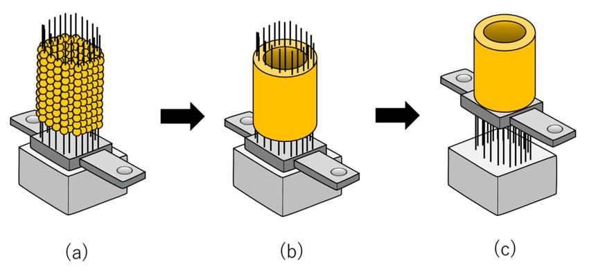
Figure 2. The production of scaffold-free AT-MSC constructs using the “mold” for osteochondral
regeneration in vivo: (a) Scaffold-free cell construct and osteochondral defects are prepared for
implantation. (b) A scaffold-free cell construct is implanted into an osteochondral defect. (c) AT-MSCs
in the construct are differentiated to chondrocytes and osteocytes, followed by regeneration of articular
cartilage in the surface layer and formation of subchondral bone in the deep layer at the implanted site.
CT images of MMPigs revealed a reduction in the subchondral radiolucent area of the implanted
site that became more dramatic at 2 or 3 months post-surgery compared with that at the control site [39].
A radiopaque area emerged from the boundary between the bone and the implant, and the area
increased more steadily upward and inward for the implanted defect as time passed until 6 months
post-surgery, compared with the control site. Thereafter, the radiopaque area of the implant gradually
progressed and then filled the entire osteochondral defect at 12 months post-surgery. In contrast, in the
control site, a radiopaque area emerged in the shallow layer, but the subchondral bone formation was
not completed to any degree in the deep layer. Macroscopic examination at 6 months post-implantation
revealed that the surface of the implanted defect was covered with abundant cartilaginous white
tissues, whereas cartilaginous tissue was scarce, and the surface was depressed in the control site.
Histopathological sections showed that thickened fibrocartilage had developed over the
subchondral bone regenerating in the implanted site [39,40]. The surface of the cartilage was
smooth, and the boundary with the surrounding normal cartilage was obscure in the implanted
site. In comparison, the surface was collapsed and irregular in the control site. At 12 months
post-implantation, the surface was uniformly covered with abundant cartilaginous white tissues,
and the boundary of the surrounding normal cartilage was unclear in the implanted site upon
macroscopic examination [39,40]. Histological examination indicated that the surface of the cartilage
was smooth, and the boundary with the surrounding normal cartilage was obscure, although small
areas of endochondral ossification persisted at the center of the implanted site (Figure 2c) [39,40].
Additionally, the subchondral bone was symmetrically reconstructed in the implanted defect and was
covered by a mixed matrix of hyaline cartilage and fibrocartilage (Figure 2c) [39,40]. In the control
site, although fibrocartilage had immediately covered the defect, the subchondral ossification was
poor [39,40].
5.3. Rabbit Osteochondral Regeneration with Allogenic AT-MSCs
A study of an allogeneic AT-MSC construct engrafted into osteochondral defects was carried
out using skeletally mature female Japanese white rabbits as the cell donor and defect recipients
(Table 3) [41]. AT-MSCs were extracted from the AT of the interscapular fat pad of the donor female [41].
The harvested AT was washed, cut into small pieces, and digested in 0.12% type I collagenase,
and the resulting solution was filtered and centrifuged [41]. The pellet was resuspended in culture
medium containing DMEM with 10% FBS and then plated onto culture dishes and cultured for one
week [41]. To generate AT-MSC spheroids, the cells were seeded into low attachment 96-well plates at
5 × 10⁴ cells/well (Table 3) [41]. Two days following incubation, the cells aggregated into a spheroid
formation approximately 700 mm in diameter (Table 3) [41]. About 800 spheroids were added into
a “mold” of 4.6 mm in diameter in culture medium (Figure 1a; Table 3) [41]. The loaded spheroids
adhered to one another and formed an AT-MSC construct after further culturing for several days
(Figure 1b) [41]. The implant surgery was performed using aseptic techniques, and an osteochondralInt. J. Mol. Sci. 2020, 21, 3589 14 of 23
lesion (4.8 mm diameter and 3 mm depth) was created at the center of the trochlear groove using a drill
(Figure 2a) [41]. For the implanted group, the allogenic AT-MSC constructs were gently implanted into
the defect (Figure 2b) [41]. In the control group, the osteochondral defect was left empty [41]. Rabbits
were euthanized 4, 8, and 12 weeks following implantation for macroscopic and histological evaluation
of healed tissues [41].
The microscopic findings of the osteochondral defects revealed maturation of articular cartilage at
four weeks, which increased gradually over time in the experimental group (Figure 2c) [41]. In contrast,
the surface of the created defect was covered with fibrous tissue, and healing tissue with large fissures
was observed at 4 weeks post-operation in the control group [41]. Therefore, the experimental group
showed greater evidence of integration through cartilage-like tissue. In addition, inflammatory cells
were not detected in the implanted defects. In comparison, fissures partially filled with fibrous tissue
were observed in the control group.
6. Osteochondral Regeneration Using Scaffold-Free 3D Constructs Produced by the
“Kenzan” Method
AT-MSC constructs prepared using bio-3D printing and “Kenzan” were analyzed for osteochondral
regeneration following autologous implantation of two swine AT-MSC constructs as reviewed
below [47]. Details of the methods to produce scaffold-free tubular construct and associated references
are summarized in Table 3.
6.1. Scaffold-Free 3D Constructs Produced Using “Kenzan”
Autologous AT was aseptically excised from skeletally mature male minipigs under general
anesthesia (Table 3) [47]. The AT was immediately minced and digested in collagenase, and the
collected cells were resuspended in a special culture medium in which a xeno-free and serum-free MSC
culture medium and a reduced serum (2%) MSC culture medium were mixed (Table 3) [47]. The cells
were seeded into a culture flask and cultured until construct creation, with at least 3.0 × 10⁷ autologous
AT-MSCs being used to fabricate each construct. For this purpose, the AT-MSCs were resuspended in
the special culture medium, and 1.0 × 10⁴ cells/well were dispensed into low attachment 96-well plates.
Following incubation for 24 h, the cells gathered in the center and formed spheroids by cell adhesion
with a diameter of approximately 550 mm. After the AT-MSC spheroids were prepared, a Bio-3D
printer (Regenova® ; Cyfuse Biomedical K.K., Tokyo, Japan) was used to assemble the AT-MSCs into
scaffold-free tubular tissue constructs according to a 3D model predesigned on a computer system using
a bio-3D designer (B3D; Cy-fuse Biomedical K.K., Tokyo, Japan) [47]. The bio-3D printer automatically
skewered the AT-MSCs spheroids onto two types of “Kenzan”, comprising needle grids of 9 × 9
(small) and 13 × 13 (large) with a needle diameter of 0.17 mm and an interneedle interval of 0.4 mm
(Table 3) [47]. The usable size of the small needle array was 3.4 mm2 × 10 mm high, and that of the large
needle array was 5.0 mm2 × 10 mm high (Table 3) [47]. The needle of “Kenzan” is made of tungsten,
and the price is about 1000 dollars. In this system, the MSC spheroids were picked up separately from
the 96-well plate by a robotically controlled fine suction 26-gauge nozzle. Sequentially, the nozzle was
inserted into the needle array to skewer the spheroid onto the “Kenzan” (Figure 3a). To create two
constructs according to the predesigned tubular configuration, a total of 3000 spheroids were prepared.
After skewering the spheroids onto the “Kenzan”, they were placed into the perfusion chamber and
cultured in the special culture medium for 1 week to fuse the spheroids (Figure 3b). The flow rate of
the medium was 4–4.5 mL/min. After the spheroids were fused, constructs were extracted from the
“Kenzan”, revealing a small tubular construct of 3.5 mm diameter, 4 mm height, and 1.5 mm thickness
and a large construct of 5 mm, 4 mm, and 1.5 mm, respectively (Figure 3c; Table 3).Int. J. Mol. Sci. 2020, 21, 3589 15 of 23
Figure 3. The fabrication of scaffold-free AT-MSC constructs using the “Kenzan”. (a) Spheroids
are skewered onto the “Kenzan” automatically using a bio-3D printer and employed to fabricate
scaffold-free tubular cell constructs. (b) Spheroids are cultured on the microneedles of the “Kenzan” to
fuse with each other. (c) The scaffold-free cell construct is retrieved from the “Kenzan” and additionally
cultured on tubular support for further maturation.
Histology of the small tubular constructs showed that the spheroids were fused within the
construct, and sufficient type I collagen was produced in the constructs and almost filled in the spaces
between the spheroids [47]. The constructs exhibited moderate hardness so that they could be gripped
easily with surgery tweezers. The constructs also exhibited low expression of proteoglycan, whereas
the majority of the cells in the construct were negative for TdT-mediated dUTP nick end labeling
(TUNEL) staining with very few positive cells [47]. Based on these results, it was confirmed that
AT-MSCs secreted abundant type I collagen to form a self-generated scaffold that formed a rigid
columnar construct.
6.2. Swine Osteochondral Regeneration with Autologous AT-MSCs
The implant surgery was performed in pigs under general anesthesia, and articular cartilage and
subchondral bone were holed to a depth of 4 mm at the center of the groove in both hind limbs using a
bone chisel with an outer diameter of 5.2 mm (Figure 4a) [47]. A large tubular construct was inserted
into the defect site in the right hind limb, with a small construct inserted inside the large construct to
complete the graft (Figure 4b). The left hind limb was left untreated as a control. Pigs were scanned by
CT and MR imaging every three months following implantation and then euthanized at 24 weeks to
confirm the osteochondral regeneration process by macroscopic and histological evaluation.
Figure 4. The production of scaffold-free AT-MSC constructs using “Kenzan” for osteochondral
regeneration in vivo: (a) Scaffold-free cell construct is prepared for implantation. (b) A scaffold-free cell
construct is implanted into an osteochondral defect. (c) Mesenchymal stem cells (MSCs) in the construct
are differentiated to chondrocytes and osteocytes, followed by regeneration of articular cartilage in the
surface layer and formation of subchondral bone in the deep layer at the implanted site.
A radiopaque area was found at the boundary between the bone and the graft, which increased
steadily upward and inward in the defect sites implanted with constructs as compared with the controlInt. J. Mol. Sci. 2020, 21, 3589 16 of 23
sites [47]. In both implanted and control defect sites, the area was reduced at three and six months
post-surgery as compared with the area immediately after surgery. Besides, the area at three months
was lower in the implanted defects than that in the control defects. MR images of the implanted
defect sites showed restoration of the articular cartilage, with signal patterns similar to that of the
surrounding normal cartilage. Nevertheless, new bone formation under the cartilaginous tissue was
incomplete. The distinction between the superficial and deep cartilage layers, as shown by the high
and the low signal intensity layers, respectively, was nearly restored, and AT predominantly occupied
the subchondral area in the implanted sites. Macroscopic examination revealed that the surfaces of
the osteochondral defects were better restored in the implanted sites than those of the control sites.
Histopathology showed smooth hyaline cartilage, and subchondral bone formation was also noted
in the implanted sites (Figure 4c). In contrast, a deeply recessed surface and a lower rate of bone
formation were observed in the control defects.
7. Conclusions
In this review, recent studies, demonstrating cartilage and osteochondral regeneration following
implantation of AT-MSCs [48–56], were described. This included the latest examples of AT-MSCs
assembling in three dimensions with or without scaffold [57–60]. Osteochondral regeneration through
implantation of scaffold-free constructs of autologous swine AT-MSCs fabricated using the “mold”
was also detailed [39,40]. A construct comprised of allogeneic AT-MSCs engrafted into osteochondral
defects in rabbit models was presented [41]. In addition, examples of histological examination for MSC
constructs prepared by using the “mold” method were described [40]. Finally, a representative study
was indicated that evaluated the histology of AT-MSC constructs prepared using bio-3D printing using
the Kenzan method and analyzed osteochondral regeneration following the autologous implantation
of two swine AT-MSC constructs [47].
By using the scaffold-free MSC constructs produced by the “mold”, successful achievement of the
simultaneous regeneration of articular cartilage and subchondral bone for up to one year has been
confirmed in small and large animals [39,40]. Additionally, the scaffold-free allogeneic MSC constructs
implanted into the osteochondral defects have survived, adhered to the defect, and regenerated
articular cartilage and subchondral bone in vivo [41]. Although allogeneic cells have the limitation
that they will be foreign to the immune system of the recipient, it has been suggested that AT-MSCs
and their secretions afford the potential to induce immunologic tolerance [93] and that allogeneic
MSC transplantation does not enhance the hyper-response of T cells against donor antigens [45].
Furthermore, implantation of an artificial scaffold-free autologous MSC construct fabricated using a
bio-3D printer with the “Kenzan” method into an osteochondral defect can aid in the regeneration of
articular cartilage and subchondral bone in a large animal [47].
Artificial materials may, however, induce xenobiotic reactions through the tissue immune
response [94], and they may not support the completion of osteochondral regeneration [36,95].
The collagen scaffold remaining in the implanted sites for long periods can prevent the regeneration
of hyaline cartilage besides promoting its re-placement of fibrous cartilage [36]. Although numerous
studies have reported the successful use of various biomaterials for scaffold construction, no ideal
biomaterial has yet been identified. Considering the potential influence of scaffolds on the
surrounding microenvironment [96], various concerns remain that need to be solved, including
immunogenicity [97,98], the long-term safety of scaffold degradation products [99], and risk of
infection or transmission of disease [100]. Based on these concerns, artificial scaffolds may, in principle,
be unfavorable for regenerating articular cartilage; therefore, further investigation into scaffold-free
cell constructs to promote cartilage regeneration is necessary.
Nevertheless, the studies described here suggest that it will be possible to produce cell constructs
exhibiting optimal functions as implants for osteochondral reconstruction by optimizing the cell
types and medium for construct fabrication. Moreover, it is also expected that studies using larger
osteochondral defect models will be performed, and the mechanism of articular cartilage regenerationYou can also read