Outcomes of Infected versus Symptomatic Sterile Walled-Off Pancreatic Necrosis Treated with a Minimally Invasive Therapy
←
→
Page content transcription
If your browser does not render page correctly, please read the page content below
Gut and Liver, Vol. 13, No. 2, March 2019, pp. 215-222

ORiginal Article

Outcomes of Infected versus Symptomatic Sterile Walled-Off Pancreatic
Necrosis Treated with a Minimally Invasive Therapy

Jong Jin Hyun1,2, Nadav Sahar1, Anand Singla3, Andrew S. Ross1, Shayan S. Irani1, S. Ian Gan1, Michael C. Larsen1,
Richard A. Kozarek1, and Michael Gluck1
1
 Digestive Disease Institute, Virginia Mason Medical Center, Seattle, WA, USA, 2Division of Gastroenterology and Hepatology, Korea University
College of Medicine, Seoul, Korea, and 3Division of Gastroenterology, Northwestern University, Chicago, IL, USA

                                                                                        mally invasive therapy; Spontaneous pancreatic fistulae;
 See editorial on page 135.
                                                                                        Walled-off necrosis

Background/Aims: Acute pancreatitis complicated by                                      INTRODUCTION
walled-off necrosis (WON) is associated with high morbidity
and mortality, and if infected, typically necessitates interven-                           Necrotizing pancreatitis occurs in approximately 15% of
tion. Clinical outcomes of infected WON have been described                             patients admitted to the hospital with acute pancreatitis, and
as poorer than those of symptomatic sterile WON. With                                   walled-off necrosis (WON) develops as a sequela of acute ne-
the evolution of minimally invasive therapy, we sought to                               crotic collections.1-3 Complications of WON can be serious, with
compare outcomes of infected to symptomatic sterile WON.                                patients developing persistent organ failure that can lead to
Methods: We performed a retrospective cohort study exam-                                prolonged hospital stays, require treatment with multiple pro-
ining patients who were undergoing dual-modality drainage                               cedures, and even result in death.4,5 In a retrospective study of
as minimally invasive therapy for WON at a high-volume                                  99 patients with necrotizing pancreatitis, Perez et al .6 showed
tertiary pancreatic center. The main outcome measures in-                               that 37% of affected patients had infected WON and worse out-
cluded mortality with a drain in place, length of hospital stay,                        comes than were found in patients with sterile WON. In a 15-
admission to intensive care unit, and development of pan-                               year study of 167 patients with necrotizing pancreatitis who
creatic fistulae. Results: Of the 211 patients in our analysis,                         underwent surgery, Rodriguez et al .7 demonstrated that proven
98 had infected WON. The overall mortality rate was 2.4%.                               or assumed infection was the indication in 51% of the cases.
Patients with infected WON trended toward higher mortality                              Moreover, among the subset of patient who underwent surgery
although not statistically significant (4.1% vs 0.9%, p=0.19).                          for “persistent unwellness,” infection was documented in 42%,
Patients with infected WON had longer length of hospitaliza-                            further indicating that outcomes and prognosis are worse in
tion (29.8 days vs 17.3 days, p
216 Gut and Liver, Vol. 13, No. 2, March 2019
vasive techniques are currently preferred.8-14 While many studies Review Board of Virginia Mason Medical Center (IRB08120). In-
have reported outcomes of various interventions for WON, few formed consent was waived. The indications for managing WON
studies have specifically examined differences in clinical out- are unrelenting pain, continued clinical deterioration, gastric/
comes between infected versus sterile WON as treatments have duodenal/biliary obstruction, and recurrent fevers or persistent
gravitated toward more minimally invasive therapies. The aim leukocytosis without other causes (Fig. 1). Among all patients
of this study was to compare the clinical outcomes associated who develop WON, our database was built to capture only those
with symptomatic sterile versus infected WON in patients un- who undergo minimally invasive therapy, since approximately
dergoing minimally invasive therapy at Virginia Mason Medical 70% of these patients are transferred to our hospital at some
Center. point in their disease and the disease process prior to referral
is not always clear. In addition, patients who died during the
MATERIALS AND METHODS initial stage of acute necrotizing pancreatitis and thus could not
undergo minimally invasive therapy are also not included. From
1. Patients
our database, we retrospectively reviewed the data obtained
The data related to all patients undergoing minimally invasive in patients who underwent combined percutaneous and endo-
therapy for symptomatic or infected WON are maintained in a scopic drainage (dual modality drainage, DMD) as a minimally
prospectively collected database approved by the Institutional invasive therapy between November 2007 and February 2017.
Symptomatic WON (n=211)
- Unrelenting pain
- Failure to thrive
- Gastric outlet obstruction
- Biliary obstruction
- Recurrent fevers without other causes
- Persistent leukocytosis without other causes
Dual modality drainage
Culture of fluid aspirates
Culture ( ) Culture (+)
Sterile WON (n=113) Infected WON (n=98)
Culture directed antibiotics
Drain tube change or additional tube for
WON enlargement/loculation outside of
tube or signs of worsening infection
Complete resolution of WON on follow-up CT scan?
Yes No
Drain clamping Drain maintenance
Follow-up CT scan 2 weeks later
Complete resolution (+) Complete resolution ( ) Fig. 1. Therapeutic flow diagram of
patients who underwent minimally
invasive therapy for walled-off pan-
Drain removal Reopen drain
creatic necrosis (WON).Hyun JJ, et al: Outcomes of Dual Modality Therapy for Infected WON 217
The database captures patient characteristics, including age at ing a 19-gauge needle or EUS-directed transenteric drainage
presentation, sex, and body mass index. Clinical information system (Navix; Boston Scientific, Natick, MA). After a guidewire
and factors related to necrotizing pancreatitis, including the is placed within the cavity, the tract is dilated, and two 7-Fr
most likely etiology of pancreatitis and the American Society double pigtail stents or, more recently, lumen-apposing metal
of Anesthesiologists (ASA) classification, were obtained. When stents (LAMS) (AXIOS; Boston Scientific, Natick, MA, USA) are
available, the size of fluid collection was determined using the placed across the gastric or duodenal wall. After the procedure,
largest reported dimension of the largest collection on cross- percutaneous drains are flushed with 10 to 20 mL of saline 3
sectional imaging along with the modified computed tomogra- times a day. Although every attempt is made to postpone the
phy severity index (CTSI). Symptomatic sterile collections were drainage procedure until after 4 weeks from initiation of acute
defined as those with gastric, duodenal, or biliary obstruction necrotizing pancreatitis, drainage is carried out within 4 weeks
and those obtained in patients who did not progress clinically if the patient has unrelenting pain, develops recurrent fevers
due to an inability to eat or failure to thrive. The following strict without other cause, or shows continued clinical deterioration
definition of infected necrosis was used: positive culture results despite intensive conservative management. Repeat percutane-
from fluid aspirates obtained during initial placement of a per- ous drain changes are performed for analogous reasons, that is,
cutaneous or endoscopic drain. if the size of the WON increases, fever develops with no other
cause, or the patient shows clinical deterioration (Fig. 1). Apart
2. Drainage techniques
from DMD, endoscopic necrosectomy was not performed in
Over the past two decades, our institutional approach to the any of the patients. When complete resolution of WON was
treatment of symptomatic and infected WON involving the use documented on follow-up CT scan, the drains are clamped. If
of minimally invasive therapy has evolved from percutaneous a follow-up CT scan taken 2 weeks later demonstrates an ab-
drainage alone to DMD, and this change has resulted in good sence of residual fluid, the percutaneous drains are removed. A
clinical outcomes, as described previously.15-18 Briefly, in DMD single dose of intravenous prophylactic antibiotics (cefazolin or
(Fig. 2), a percutaneous drainage catheter is placed into the levofloxacin) is administered prior to drainage. Afterwards, the
WON under computed tomography (CT) guidance after taking choice of type and the duration of antibiotic use, if indicated, is
into consideration multiple factors, such as the location of fluid tailored based on the culture results and after consultation with
collection, the trajectory of the catheter, the ease of drainage, infectious disease specialists (Fig. 1).
and accessibility for wound care. If the WON is considered to be
3. Outcomes
accessible from the gastrointestinal tract (i.e., is located within 2
cm of the gastric or duodenal wall), the patient is then immedi- The main outcome measures used to compare infected and
ately transferred to an endoscopy suite for additional endoscop- symptomatic sterile WON were mortality with a drain in place
ic drainage. The WON is entered either endoscopically with a (unresolved WON), the length of hospital stay, the use of critical
needle-knife sphincterotome (Cook Endoscopy, Winston-Salem, care services (i.e., admission to an intensive care unit), and the
NC, USA) or under endoscopic ultrasound (EUS) guidance us- development of spontaneous pancreatic fistulae. Development
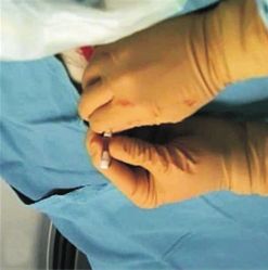
A B C
Fig. 2. Dual-modality drainage te
chnique. (A) Walled-off necrosis
(WON) is noted around the pan-
creas. (B) The area of WON was first
accessed percutaneously for drain-
age under computed tomography
(CT) guidance. (C) The percutaneous
drainage tube is seen on a fluoro-
scopic image. (D) Drainage between
the gastrointestinal tract and the
D E F area of WON was then performed
endoscopically, and two double pig-
tail stents were inserted. (E) Follow-
up CT scan taken 7 days after dual-
modality drainage shows intact
drainage tubes within the cyst, with
a decrease in cyst size. (F) CT scan
taken 4 months after the procedure
demonstrates complete remission of
WON.218 Gut and Liver, Vol. 13, No. 2, March 2019
of spontaneous pancreatic fistulae was defined as the spontane- mean±standard deviation or number (%) values. Comparative
ous formation of fistulae from the necrotic collection to an ex- statistical analyses were performed using unpaired Student t-
trapancreatic location, that is, the biliary tract, gastrointestinal tests for continuous variables and chi-square tests for categori-
tract, pleural space, and peritoneum. For patients undergoing cal variables. p-values 1 percutaneous drain-
age, the interval between drain placement and removal, the RESULTS
number of total tube checks, the presence of disconnected duct
1. Baseline characteristics
syndrome, the occurrence of other complications (e.g., bleeding,
gastric outlet obstruction, colonic obstruction, biliary stricture, A total of 211 patients were included in this analysis. Of
and acute renal failure), and the number of total CT scans. The these, 98 (46.4%) were classified as infected WON, and 113
presence of disconnected duct syndrome was diagnosed by en- (53.6%) were classified as symptomatic sterile WON (Table 1).
doscopic retrograde cholangiopancreatography performed at the Infected WON patients were older at presentation than symp-
time of drainage or magnetic resonance cholangiopancreatogra- tomatic sterile WON patients (mean age: 56.7 years vs 51.4
phy performed later in the course of treatment.16,17 years, p=0.01). Approximately two-thirds of the patients were
male, and there was no difference in the sex ratio between
4. Statistical analysis
the two groups. There was also no difference between the two
Data were analyzed using IBM SPSS Statistics version 23.0 groups with regards to etiologies of pancreatitis. The most com-
(IBM Corp., Armonk, NY, USA). Data are expressed as the mon etiologies were gallstones (52.1%), followed by alcohol
Table 1. Baseline Characteristics of Patients with Infected and Symptomatic Sterile WON
Characteristic Infected WON (n=98) Symptomatic sterile WON (n=113) p-value
Age, yr 56.71±15.46 51.35±15.76 0.01
Female sex 33 (33.7) 34 (30.1) 0.66
BMI, kg/m2 31.20±6.34 28.92±6.95 0.01
Pancreatitis etiology 0.58
Gallstones 52 (53.1) 58 (51.3)
Alcohol 17 (17.3) 26 (23.0)
Triglycerides 7 (7.1) 3 (2.7)
Medications 2 (2.0) 0
Post-ERCP 2 (2.0) 2 (1.8)
Surgical/trauma 3 (3.1) 4 (3.5)
Ampullary adenoma 1 (1.0) 2 (1.8)
Pancreas divisum 2 (2.0) 5 (4.4)
Idiopathic 12 (12.2) 13 (11.5)
ASA classificationHyun JJ, et al: Outcomes of Dual Modality Therapy for Infected WON 219 (20.4%). With regard for physical status, more patients had procedures (31.6% vs 14.2%, p
220   Gut and Liver, Vol. 13, No. 2, March 2019

examined clinical outcomes in treated WON, specifically with            endoscopically, even in those with a median Sequential Organ
respect for the presence of infection. Several studies have dem-        Failure Assessment score of 0.26 As most of the patients in this
onstrated that mortality is higher in those with infected necro-        series were transferred from outside institutions, obtaining true
sis. However, previous studies have all had small sample sizes,         mortality figures would require researchers to determine the
and many did not directly compare infected WON with sterile             number of patients admitted with acute pancreatitis and the
WON.9,19-22 To our knowledge, our study is the largest study to         concomitant mortality rates across multiple centers with vari-
compare clinical outcomes between infected and sterile WON              able expertise and resources for treating such patients.
in cases treated with minimally invasive therapy. The results of           In addition to requiring longer hospital stays and more criti-
our study confirm that compared to sterile WON, infected WON            cal care services and showing a trend toward higher mortality,
predicts worse outcomes and showed a non-significant trend              patients with infected WON also more frequently developed
toward higher mortality (4.1% vs 0.9%, p=0.19). Surprisingly,           spontaneous pancreatic fistulae. Even after adjusting for patient
overall mortality was very low (2.4%) even when only infected           characteristics, the etiology of pancreatitis and the presence of
WON patients were considered (4.1%). This finding demon-                multiple percutaneous drains, infected WON patients had 3-fold
strates that WON, which was once considered a highly morbid             higher odds of spontaneous pancreatic fistulae formation. The
and significant cause of death, can be treated with good out-           reason for this association remains unclear. Spontaneous pan-
comes using minimally invasive therapy regardless of whether            creatic fistulae can result from severe necrotizing pancreatitis as
it is infected or sterile. The authors fully recognize that mortality   well as interventional pancreatic procedures. Since all patients
is not the same between treated WON and severe acute pan-               in our study underwent placement of a percutaneous drain and
creatitis (SAP) since a percentage of patients do not survive the       endoscopic drainage, it is not clear whether procedural manipu-
inflammatory response and organ failure that occur during the           lation of pancreatic fluid collection contributed to the occur-
initial stages of SAP. WON develops at a later stage, often when        rence of spontaneous pancreatic fistulae in structures other than
a patient may be stabilizing.                                           the skin and the stomach or duodenum. Alternatively, infection
    Currently, DEN and VARD are the most common procedures              of the necrotic collection could lead to increased inflammation
used to drain symptomatic WON.13 However, a variable subset             of contiguous structures, resulting in damage to the surround-
of these patients require the addition of a percutaneous drain          ing structures and spontaneous fistulae formation. It is also
because of nonresolution, infection, or the extension of WON            possible that infection of the fluid collection results from spon-
into areas not amenable to conventional drainage or endoscopic          taneous pancreatic fistulae developing in a nonsterile structure,
debridement. Although DMD is not a modality that is currently           such as the colon, leading to bacterial colonization of the fluid
utilized as the standard of practice for treating WON at other          collection. In a prior study, Ho and Frey27 found that 25 out of
pancreatic centers, the fact that the use of DMD led to single          136 (18%) patients with SAP developed spontaneous pancreatic
digit mortality implies that our minimally invasive therapy is as       fistulae. However, the rate was much higher in patients who
effective as and representative of minimally invasive therapy for       underwent surgical treatment for necrotizing pancreatitis.28
WON. At our institution, either two 7-Fr double pigtail stents or       For patients undergoing pancreatic resection for any reason,
a LAMS are placed across the gastric or duodenal wall during            the incidence of pancreatic fistulae ranged between 9.9% and
DMD. Although LAMS have recently been inserted at a higher              28.5%.29
rate, when we compared the two endoscopic transgastric stent               Infected WON, in our patient population, was an indication
techniques, we found no differences between the treatment               for intervention. It should be noted that there is some evidence
outcomes for WON.23 Since the implementation of DMD for the             suggesting that prompt intervention may not always be neces-
treatment of WON at our institution, no patient has required            sary in infected WON. Some authors have advocated that con-
open surgical necrosectomy, undergone VARD or needed a                  servative management with antibiotics and careful monitoring
distal pancreatectomy for a persistent percutaneous fistula,16,18       may be adequate.30,31 However, this could increase the incidence
a situation common in previous series in patients treated with          of multidrug-resistant organisms and Clostridium difficile infec-
percutaneous drainage alone.24,25 Nevertheless, our study fails         tion, and care should be taken to avoid the overuse of antibiot-
to define the total mortality in SAP since only a select group          ics.32,33 The subset of patients who seem most appropriate for
of patients survived the initial severe disease, presented with         noninvasive management are clinically stable patients, a group
WON, and then underwent DMD were included in the analysis.              not found in our series as the vast majority were transferred to
Patients who presented with organ failure and died early during         our hospital due to clinical instability. Recent guidelines suggest
the course of illness were not included. This could be one of the       that patients with sterile WON can appear to be as clinically ill
reasons for the low mortality of 2.4% observed in the current           as those with infected WON; thus, symptomatic sterile WON is
study, which was lower than that reported in a recently pub-            also drained.
lished multicenter randomized trial that found a mortality rate            This study is not without its limitations. Our data represent
of 18% in patients with infected collections that were treated          the experience of a single high-volume, tertiary referral center
Hyun JJ, et al: Outcomes of Dual Modality Therapy for Infected WON 221
for patients with complex pancreaticobiliary disorders. Given tis. Pancreatology 2013;13:e1-e15.
the availability of advanced endoscopists and experienced in- 6. Perez A, Whang EE, Brooks DC, et al. Is severity of necrotizing
terventional radiologists, our results may not apply to lower pancreatitis increased in extended necrosis and infected necrosis?
volume centers that do not have the same dedicated resources Pancreas 2002;25:229-233.
or expertise in treating pancreatic diseases. Analyses of large, 7. Rodriguez JR, Razo AO, Targarona J, et al. Debridement and
administrative datasets have shown that a higher hospital vol- closed packing for sterile or infected necrotizing pancreatitis: in-
ume of acute pancreatitis is associated with better clinical out- sights into indications and outcomes in 167 patients. Ann Surg
comes.34 In addition, the patients in the current study had fluid 2008;247:294-299.
collections that could be reached endoscopically, and these data 8. Clancy TE, Ashley SW. Current management of necrotizing pan-
therefore cannot be applied to WON that occurs more than 2 cm creatitis. Adv Surg 2002;36:103-121.
from the gastric or duodenal wall on CT scan. Our cohort does 9. Besselink MG, Verwer TJ, Schoenmaeckers EJ, et al. Timing
not reflect the potential differences in outcomes that may occur of surgical intervention in necrotizing pancreatitis. Arch Surg
between patients with infected and sterile WON located away 2007;142:1194-1201.
from the lumen of the gastrointestinal tract; however, these are 10. Besselink MG, van Santvoort HC, Nieuwenhuijs VB, et al. Mini-
infrequently found in clinical practice. Therefore, our conclu- mally invasive ‘step-up approach’ versus maximal necrosectomy
sions appear valid for the subset of patients with WON in whom in patients with acute necrotizing pancreatitis (PANTER trial):
combined percutaneous and endoscopic drainage is feasible. design and rationale of a randomised controlled multicenter trial
In conclusion, infected WON patients have longer length of [ISRCTN13975868]. BMC Surg 2006;6:6.
stay, a higher use of critical care services, and a higher inci- 11. van Santvoort HC, Bakker OJ, Bollen TL, et al. A conservative and
dence of spontaneous fistula than were found in symptomatic minimally invasive approach to necrotizing pancreatitis improves
sterile WON. Although a longer, more complicated hospital outcome. Gastroenterology 2011;141:1254-1263.
course can be anticipated in patients with infected WON, pro- 12. Gardner TB, Chahal P, Papachristou GI, et al. A comparison of di-
viders can nevertheless expect good outcomes in this era of rect endoscopic necrosectomy with transmural endoscopic drain-
minimally invasive therapy. age for the treatment of walled-off pancreatic necrosis. Gastroin-
test Endosc 2009;69:1085-1094.
CONFLICTS OF INTEREST 13. Boumitri C, Brown E, Kahaleh M. Necrotizing pancreatitis: current
management and therapies. Clin Endosc 2017;50:357-365.
No potential conflict of interest relevant to this article was 14. Kawakami H, Itoi T, Sakamoto N. Endoscopic ultrasound-guided
reported. transluminal drainage for peripancreatic fluid collections: where
are we now? Gut Liver 2014;8:341-355.
ACKNOWLEDGEMENTS 15. Gluck M, Ross A, Irani S, et al. Endoscopic and percutaneous
drainage of symptomatic walled-off pancreatic necrosis reduces
The authors thank Terri Davis Smith for administrative, tech- hospital stay and radiographic resources. Clin Gastroenterol Hepa-
nical, and material support. tol 2010;8:1083-1088.
16. Ross A, Gluck M, Irani S, et al. Combined endoscopic and percu-
REFERENCES taneous drainage of organized pancreatic necrosis. Gastrointest
Endosc 2010;71:79-84.
1. Banks PA, Freeman ML; Practice Parameters Committee of the 17. Gluck M, Ross A, Irani S, et al. Dual modality drainage for symp-
American College of Gastroenterology: practice guidelines in acute tomatic walled-off pancreatic necrosis reduces length of hospi-
pancreatitis. Am J Gastroenterol 2006;101:2379-2400. talization, radiological procedures, and number of endoscopies
2. Banks PA, Bollen TL, Dervenis C, et al. Classification of acute pan- compared to standard percutaneous drainage. J Gastrointest Surg
creatitis-2012: revision of the Atlanta classification and definitions 2012;16:248-256.
by international consensus. Gut 2013;62:102-111. 18. Ross AS, Irani S, Gan SI, et al. Dual-modality drainage of infected
3. Zhao K, Adam SZ, Keswani RN, Horowitz JM, Miller FH. Acute and symptomatic walled-off pancreatic necrosis: long-term clini-
pancreatitis: revised Atlanta classification and the role of cross- cal outcomes. Gastrointest Endosc 2014;79:929-935.
sectional imaging. AJR AM J Roentgenol 2015;205:W32-W41. 19. Beger HG, Rau B, Isenmann R. Natural history of necrotizing pan-
4. Tenner S, Baillie J, DeWitt J, Vege SS; American College of creatitis. Pancreatology 2003;3:93-101.
Gastroenterology. American College of Gastroenterology guide- 20. Beger HG, Bittner R, Block S, Büchler M. Bacterial contamination
line: management of acute pancreatitis. Am J Gastroenterol of pancreatic necrosis. A prospective clinical study. Gastroenterol-
2013;108:1400-1415. ogy 1986;91:433-438.
5. Working Group IAP/APA Acute Pancreatitis Guidelines. IAP/APA 21. Petrov MS, Kukosh MV, Emelyanov NV. A randomized controlled
evidence-based guidelines for the management of acute pancreati- trial of enteral versus parenteral feeding in patients with predicted222 Gut and Liver, Vol. 13, No. 2, March 2019 severe acute pancreatitis shows a significant reduction in mortality necrotizing pancreatitis. Arch Surg 1995;130:48-52. and in infected pancreatic complications with total enteral nutri- 29. Bassi C, Butturini G, Molinari E, et al. Pancreatic fistula rate af- tion. Dig Surg 2006;23:336-344. ter pancreatic resection: the importance of definitions. Dig Surg 22. Bhansali SK, Shah SC, Desai SB, Sunawala JD. Infected necrosis 2004;21:54-59. complicating acute pancreatitis: experience with 131 cases. Indian 30. Garg PK, Sharma M, Madan K, Sahni P, Banerjee D, Goyal R. J Gastroenterol 2003;22:7-10. Primary conservative treatment results in mortality comparable to 23. Sahar N, Kozarek R, Kanji ZS, et al. Do lumen-apposing metal surgery in patients with infected pancreatic necrosis. Clin Gastro- stents (LAMS) improve treatment outcomes of walled-off pan- enterol Hepatol 2010;8:1089-1094. creatic necrosis over plastic stents using dual-modality drainage? 31. Mouli VP, Sreenivas V, Garg PK. Efficacy of conservative treat- Endosc Int Open 2017;5:E1052-E1059. ment, without necrosectomy, for infected pancreatic necro- 24. Freeny PC, Hauptmann E, Althaus SJ, Traverso LW, Sinanan M. sis: a systematic review and meta-analysis. Gastroenterology Percutaneous CT-guided catheter drainage of infected acute necro- 2013;144:333-340. tizing pancreatitis: techniques and results. AJR Am J Roentgenol 32. Barnes SL, Rock C, Harris AD, Cosgrove SE, Morgan DJ, Thom 1998;170:969-975. KA. The impact of reducing antibiotics on the transmission of 25. Traverso LW, Kozarek RA. Interventional management of peripan- multidrug-resistant organisms. Infect Control Hosp Epidemiol creatic fluid collections. Surg Clin North Am 1999;79:745-757. 2017;38:663-669. 26. van Brunschot S, van Grinsven J, van Santvoort HC, et al. Endo- 33. Trikudanathan G, Munigala S. Impact of Clostridium difficile in- scopic or surgical step-up approach for infected necrotising pan- fection in patients hospitalized with acute pancreatitis: a popula- creatitis: a multicenter randomised trial. Lancet 2018;391:51-58. tion based cohort study. Pancreatology 2017;17:201-202. 27. Ho HS, Frey CF. Gastrointestinal and pancreatic complications as- 34. Singla A, Simons J, Li Y, et al. Admission volume determines sociated with severe pancreatitis. Arch Surg 1995;130:817-822. outcome for patients with acute pancreatitis. Gastroenterology 28. Tsiotos GG, Smith CD, Sarr MG. Incidence and management of 2009;137:1995-2001. pancreatic and enteric fistulas after surgical management of severe
You can also read