Pain in hypermobile Ehlers-Danlos syndrome: New insights using new criteria - SED in France
←
→
Page content transcription
If your browser does not render page correctly, please read the page content below

Received: 12 December 2018               Revised: 22 February 2019             Accepted: 7 April 2019
DOI: 10.1002/ajmg.a.61175

ORIGINAL ARTICLE

Pain in hypermobile Ehlers-Danlos syndrome: New insights
using new criteria

Karelle Bénistan1                           | Valeria Martinez2,3,4

1
 Centre de référence des syndromes d'Ehlers-
Danlos non vasculaires, Hôpital Raymond                        Abstract
Poincaré, Garches, Assistance Publique                         Features of the pain in hypermobile Ehlers-Danlos syndrome (hEDS) are complex and
Hôpitaux de Paris, Garches, France
2                                                              insufficiently known by clinicians. We enrolled 37 hEDS patients. Disease status was
  Département d'anesthésie et consultation
douleur, Hôpital Raymond Poincaré, Garches,                    ascertained using revised 2017 International Classification criteria, in the EDS French
Assistance Publique Hôpitaux de Paris,
                                                               National Reference Center. Patients were evaluated with a clinical examination,
Garches, France
3
  Centre d'Evaluation et de Traitement de la                   quantitative sensory testing, and validated questionnaires. Thirty-seven patients
Douleur, F-92100 France, INSERM, U-987,                        were evaluated. Pain had appeared at 10 ± 5 years old and became chronic at 20
Hôpital Ambroise Paré, Boulogne-Billancourt,
France                                                         ± 9 years old. hEDS was diagnosed at only 24 ± 10 years old. Ninety-seven percent
4
  Université Versailles Saint-Quentin,                         of them had severe chronic pain, which gradually increased over time in 75% of them.
Montigny-Le-Bretonneux, France
                                                               The main location of pain was in joints and predominated in lower limbs. Patients
Correspondence                                                 with a generalized presentation of pain had older chronic pain and a higher impact on
Karelle Benistan, Centre de référence des
                                                               the affective component. Neuropathic pain was frequent in the most painful joint
syndromes d'Ehlers-Danlos non vasculaires,
Hôpital Raymond Poincaré, 104 bd Poincaré,                     and associated with heat hypoesthesia. An asymmetric proprioception was found in
Assistance Publique Hôpitaux de Paris,
                                                               one third of the patients. A very high rate of attempted suicide was observed. To
F-92380, 92380 Garches, France.
Email: karelle.benistan@aphp.fr                                conclude, pain in hEDS is severe, chronic, and disabling. Sensorial and proprioceptive
                                                               sensibilities are also affected. Peripheral neuropathic pain is frequent and central sen-
                                                               sitization appears to be a key step in the evolution of disease.

                                                               KEYWORDS
                                                               hyperalgesia, hypermobile Ehlers-Danlos syndrome, neuropathic pain, pain, recurrent
                                                               dislocation

1 | I N T RO D UC T I O N                                                                       Ehlers-Danlos syndrome (hEDS), which reduces the number of hEDS
                                                                                                diagnoses across the world in particular among patients with chronic
Ehlers-Danlos syndromes (EDS) are a heterogeneous group of heredi-                              pain and limits confusion around hEDS, joint hypermobility and
tary connective tissue disorders characterized by skin laxity, joint                            related musculoskeletal manifestations (Challal, Minichiello, Funalot, &
hypermobility, and tissue fragility. They are due to mutations in genes                         Boissier, 2015; Malfait et al., 2017). On the other end, this classifica-

encoding structural proteins of connective tissue, for collagen modifiers                       tion introduces the term “hypermobility spectrum disorders” to give
                                                                                                some diagnostic label to other individuals suffering from chronic pain
or enzymes involved in their metabolism. The prevalence for all types
                                                                                                and presenting additional musculoskeletal features probably related to
of EDS collectively is estimated at 1/5,000 (Tinkle et al., 2017), if one
                                                                                                a pre-existent joint hypermobility, but who do not respect the criteria
does not take into account other hypermobility spectrum disorders.
                                                                                                for any EDS. hEDS is the most frequent type of EDS; it affects over
     The 2017 International Classification of the EDS now recognizes
                                                                                                80% of EDS patients and is now well characterized (Tinkle et al.,
13 subtypes and includes a set of stricter criteria for hypermobile
                                                                                                2017). Unlike the other subtypes, its diagnosis remains only clinical
                                                                                                because genetic etiology has not been found yet (haploinsufficiency
The corresponding author certifies that all authors approved the entirety of the submitted
material and contributed actively to the study.                                                 of TNXB could explain 5% of hEDS (Zweers et al., 2003); possible

Am J Med Genet. 2019;1–9.                                           wileyonlinelibrary.com/journal/ajmga                            © 2019 Wiley Periodicals, Inc.     1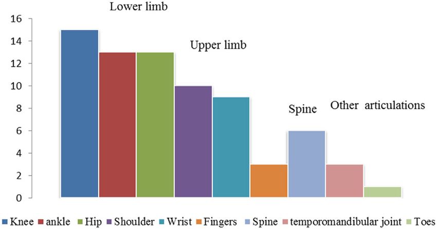
2                                                                                                                         BÉNISTAN AND MARTINEZ

candidate region on chromosome 8p22-8p21.1 (Syx et al., 2015). It          subjective pain experience, describing the sensory and the affective
has an autosomal dominant mode of inheritance. Symptoms begin              dimensions. Fatigue intensity was evaluated by a visual analog scale
during infancy. The diagnostic criteria of hEDS are summarized in          (VAS). In the most painful joint, we searched for neuropathic charac-
Appendix. So far, there has been no specific etiological treatment         teristics with the validated DN4 questionnaire (Bouhassira et al.,
available, but only symptomatic treatments and preventive measures         2005). Patients with NP had to fill in the neuropathic pain symptom
designed to avoid complications. Although patients with hEDS suffer        inventory (NPSI; Bouhassira et al., 2004) which includes 10 symptoms
from pain, its underlying mechanisms are still unresolved and remain       rated on 0–10 numerical scales (burning, squeezing, pressure, electric
complex (Castori et al., 2017). Pain is the symptom by which patients      shocks, stabbing, pain evoked by brush, pressure, cold, tingling, pins,
most often enter the disease. The characteristics of pain in hEDS are      and needles). The total score is based on a 100-point scale.
now better described. However, literature has each time been inter-           We also assessed psychological vulnerability with the hospital
ested in only certain aspects of these pains. In this study, we wanted     anxiety and depression (HAD) scale which includes an anxiety sub-
to present an overview of the pain with all its components, and fur-       scale (HAD-A) and a depression subscale (HAD-D), both of them con-
ther explore neurophysiological aspects of pain in hEDS.                   taining seven intermingled items, providing a total score for each
                                                                           subscale ranging from 0 to 21 (Zigmond & Snaith, 1983). Digital palpa-

2 | METHOD                                                                 tion of 18 specified locations of tenderness described in 1990 by the
                                                                           American College of Rheumatology for the diagnosis of fibromyalgia.
Patients were consecutively recruited during 1 year in 2017 and dis-       We also performed a manual palpation of quadriceps to test for a
ease status has been systematically ascertained using validated diag-      muscular pain.
nostic criteria in the French EDS National Reference Center
(Raymond Poincaré Hospital, Garches). Inclusion criterion was a hEDS
                                                                           2.2 | Quantitative sensory testing
diagnosis according to the new 2017 EDS criteria (Malfait et al., 2017;
Appendix). We collected demographic data, medical history, pain his-       Quantitative sensory testing was performed on the area of the joint
tory, physical examination, echocardiography, and completed ques-          described by the patient as the most painful. Contralateral joint were
tionnaires. Evaluations were always performed by the two same              used as control. Brush-induced allodynia was assessed using a paint-
senior investigators, on the one hand, the EDS specialist for the physi-   brush (Somedic AB, Stockholm, Sweden); intensity was assessed on a
cal examination, and on the other hand, the pain specialist for the pain   100 mm VAS. Thermal sensations and pains were assessed with a
evaluation. In this observational study, the data collection was           Somedic thermotest (Somedic AB, Stockholm, Sweden), using the
approved by National Commission for Informatics and Liberties              Marstock method according to the method of limits (Fruhstorfer, Lin-
(CNIL). All data were anonymized.                                          dblom, & Schmidt, 1976). The baseline temperature of the thermode
                                                                           was set at 32 C. The maximum and minimum temperatures were set
2.1 | Assessment of pain and its impact                                    at 50 C for heat, 10 C for cold detection, and 4 C for cold pain. A
                                                                           thermal rate of change of 1 C/s was used. All thresholds were calcu-
All the patients were asked to describe their pain and its impacts. We
                                                                           lated as the average of three successive determinations. Hypoesthesia
asked them at what age the first symptoms occurred, their age at
                                                                           to mechanical, warm, or cold stimuli was considered in case of
hEDS diagnosis, their age at chronic pain onset, and the duration of
                                                                           increased warm detection thresholds or decreased cold detection
their pain. We asked patients to enumerate currently painful joints,
                                                                           thresholds of at least 2 SD in comparison with contralateral sides.
and to rank the three most painful. They were also asked about the
frequency of subluxations, luxations, or sprains during the last month
and the number of emergency department visits in the last 12 months.       2.3 | Vibratory perception sense
Patients were asked to record school or absenteeism at work mea-
                                                                           Patients underwent testing for vibratory perception sense by assess-
sured in days off work/school in the last 12 months. We also
questioned them about diagnostic wandering, suicide attempts, anal-        ment of the vibratory perception threshold (VPT). Therefore, a bio-

gesic treatment history, and previous orthopedic surgery. The Brief        thesiometer (Somedic AB, Stockholm, Sweden) was used according to

Pain Inventory short form was used to assess the interference of pain      previously published methods (Frenette, Mergler, & Ferraris, 1990).

in the daily life (Cleeland & Ryan, 1994). Average and worst pain          The tractor of the device was applied with uniform pressure on three
intensity over the last 24 hr were assessed using numerical rating         points bilaterally: trochanter at the hip, femoral condyle at the knee,
scale (NRS; 0: no pain; 10: worst possible pain). The areas of pain        and fibula condyle at the ankle. Patients were asked to inform the
including maximal pain were reported on the body map from the Brief        examiner of the first sensation of vibration as the amplitude of vibra-
Pain Inventory (BPI) (Cleeland & Ryan, 1994). The QDSA scale               tion was slowly increased by one vibration per second. The average
(Questionnaire Douleur de Saint-Antoine, the validated French ver-         (V) of the three VPT measurements at each site was calculated. An
sion of the McGill Pain Questionnaire; Boureau, Luu, & Doubrere,           asymmetry of proprioception was considered if a difference of two
1992) was used to evaluate the different components of the                 standard deviations was observed between the two sides.
BÉNISTAN AND MARTINEZ                                                                                                                                     3

2.4 | Temporal summation tests with Von Frey                                    TABLE 1          Clinical characteristics of patients
filaments                                                                         Demography

This test evaluates the pain triggered by the application of a 180 g                Sexe (F/M)                                           36/37 (95%)

Von Frey filament (Bioseb, in vivo Research Instruments, http://www.                Age (years)                                         26 ± 10 (10–53)

bioseb.com) to the inner surface of the right upper arm until the fila-             Body mass index (kg/m2)                              23 ± 4 (16–31)
ment curves. The pain induced was evaluated on an 11-point NRS                    Rheumatology
after the first stimulation, and then again after 10 consecutive applica-           Beighton score ≥5/9                                  37/37 (100%)
tions to an area of 1 cm2, at a frequency of 1 Hz, as described by                  Number of painful joints                              9 ± 3 (2–20)
Weissman-Fogel et al. (2009). The difference between the NRS scores                 Number of dislocation during last month              11 ± 6 (0–300)
obtained after 1 and 10 stimulations was calculated (ΔNRS10–1).                     Age of first sprain/dislocation (years)              10 ± 5 (2–27)
                                                                                    Scoliosis ≥20                                        1/37 (2.7%)

2.5 | Statistical analysis                                                          Congenital hip dislocation                            8/37 (22%)
                                                                                    Clumsiness                                           36/37 (97%)
Quantitative data were described as mean and standard deviation
                                                                                  Dermatology
(min–max) and qualitative data as frequencies (percentages). Chi-squared
                                                                                    Unusual soft or velvety skin                         12/37 (32%)
tests (or Fisher's exact tests if necessary) and Wilcoxon tests were
                                                                                    Unexplained and pathologic striae                     5/37 (13%)
performed for univariate analyses. All statistical tests were bilateral and a
p-value 1                                 15/37 (40%)
                                                                                  Echocardiography
3 | RESULTS
                                                                                    Mitral prolapse                                       5/24 (20%)
                                                                                    Aortic root dilatation                                0/24 (0%)
3.1 | Patient characteristics, clinical data, and
                                                                                  Other
psychometric testing
                                                                                    Unexplained pelvic floor prolapse                     1/37 (2.5%)
Thirty-seven consecutive patients fulfilling the diagnostic criteria of
                                                                                    Abdominal hernias                                     6/37 (16%)
hEDS from 34 different families were included. Baseline characteris-
                                                                                    Urinary dysfunction                                   9/19 (47%)
tics are summarized in Table 1. Hypermobile EDS was diagnosed at
                                                                                    Ogival palate                                        19/37 (51%)
24 ± 10 years old. Twenty-nine (78%) patients had a family history of
hEDS. All patients suffered from chronic pain. The pain duration has            Note. Data are reported as mean ± SD (minimum–maximum) or as
                                                                                number (%).
been 7 ± 2.1 years. Seventy-five percent described overall pain as
gradually increasing, pain appeared at 10 ± 5 years old, became
                                                                                recurrent absenteeism (54%), and about one third had a long sick
chronic at 20 ± 8 years old. Abdominal pain is present in 55% of cases,
headaches in 63%, and insensitivity to local anesthetic was reported            leave or permanent disability status. Eight patients reported having

in 48% of cases.                                                                attempted suicide at a very young age (10, 13, 24, 15, 15, 16, 23, and

    Previous orthopedic surgery was performed in 35% (13/37) of                 25 years old).

patients in following joints: ankle (5/13), knee (4/13), hip (3/13), and
shoulder (1/13). Three patients had two or three surgical procedures            3.2 | Pain characteristics: Experimental findings
on the same joint and three patients had surgical procedures on differ-
ent joints. All surgeries were performed before the hEDS diagnosis.             Eleven (27%) patients had severe pain (pain intensity ≥7/10) and six-

Patients stated that they consult the emergency department 3 (0–7.5)            teen (40%) had moderate pain (pain intensity between 3 and 7). Aver-

times per year. Diagnostic wandering was reported for 60% of patients.          age worst pain was 8.5 ± 1.2/10 and lowest pain was 4.7 ± 2.3/10. The

The most frequent diagnoses previously evoked were fibromyalgia                 total QDSA score was 20 ± 9 over a total score of 60. Pain was
(26%), psychological diseases (21%), and inflammatory/degenerative              described as sharp (70%), stabbing (70%), tender (65%), or exhausting
joint diseases (16%).                                                           (90%). Three different types of body map representation of pain have
    The impact of pain on quality of life was important with a median           been reported (Figure 2). Patients with widespread pain had a more
BPI score of 61 ± 23 and interfered with all aspects of life (Figure 1).        length duration of pain and a higher impact on the affective component
Six patients (15%) were suspected of depression and three patients              of pain (Table 2). The worst pain was most often located in the lower
(7.5%) of anxiety defined by HAD questionnaire. About 36/37 (97%)               limb 15/37 (40%), followed by the upper limb 10/37 (27%) and spine
of patients complained of fatigue with a mean NRS of 7 ± 1 (6.5–8).             5/ 37 (13% ; Figure 3). Pain had neuropathic characteristics in the most
The fatigue was severe for 28/37 (75%) of them. Only three (8%)                 painful joint in 67% of the patients (25/37) and was severe with a total
patients had normal working or studying activities. More than half had          score of NPSI of 43 ± 12. Average intensity of neuropathic symptoms
4 BÉNISTAN AND MARTINEZ
F I G U R E 1 Brief pain inventory,
interference of pain with daily life.
Bar graphs showing the mean and
standard deviation of impact of pain
in different component of quality of
life measured with the BPI
questionnaire
was 4.3 ± 3 for burning pain, 5 ± 3.5 for squeezing pain, 5.3 ± 2.8 for 35 examined had reported a significant increase of pain (more than
pressure pain, 5.2 ± 3.9 for electric shocks, 4.4 ± 3.1 for stabbing pain, 3 points).
4.5 ± 3.9 for pain evoked by brushing, 2.7 ± 2 for pain evoked by pres- Hypoesthesia to thermal stimulation was observed in 9/35
sure, 1.3 ± 3.2 for pain evoked by cold, 5.0 ± 3.5 for tingling, and 6.7 patients (26%) when comparing the most painful joint and contralat-
± 3.5 for pins and needles. eral joint. Heat hypoesthesia was the most frequent, one patient had
About a third of patients (11/37) were treated with strong opioid both cold and heat hypoesthesia in the most painful joint. Thermal
medication, one third (13/37) with antidepressants, and another third hyperalgesia was not observed (Table 3).
11/37 with gabapentinoids. The combination of two or more of these An asymmetry in the vibratory perception threshold was reported
different classes were reported for 13/37 (35%) patients. Some in nine patients among the 28 examined. One patient had an asymme-
patients (4/37) reported using cannabis regularly to alleviate pain. try of the vibratory perception threshold on the three joints examined
Nineteen patients complained of spontaneous muscular pain. The (hip, knee, and ankle), a second patient had an asymmetry both on the
quadriceps palpation had evoked pain in 73% of them. However, less hip and knee. The other patients had an asymmetry of perception in
than a third of the 37 patients examined reported tenderness in the only one joint (four on the knee and three in the hip).
11 or more of 18 possible “tender points of fibromyalgia.” Twenty
patients complained of cutaneous hyperesthesia during touching or 4 | DISCUSSION
stroking. Among them, a repetitive caress on the forearm led to pain
in 16 of them. In mechanical temporal summation tests with a Von Our cohort study highlights the special features of pain in hEDS
Frey filament, the first stimulation had not induced pain in any of the patients, meeting the new criteria for hEDS according to the 2017
patients. After 10 consecutive applications, 22 patients among the International Classification (Malfait et al., 2017). Previous studies had
F I G U R E 2 Pain drawing assessment. (a) Typical presentation: The most frequent drawing corresponding to 29 patients (78%) was as follows:
Pain located in several small and large joints, both in lower and upper limbs, bilaterally. (b) Widespread presentation: Six patients (16%) reported a
widespread pain located in joints and also in muscles. (c) Other presentations: Only two patients (6%) had another representation suggesting
lomboradiculalgiaBÉNISTAN AND MARTINEZ 5
TABLE 2 Pain characteristics depending on pain profile (Figure 1) lower incidence (90% in various EDS; Voermans, Knoop, Bleijenberg, &
van Engelen, 2010). Our study underlines a diagnosis delay in hEDS
Typical Widespread
presentation presentation p despite an old painful joint instability, evolving since childhood. The
Number of patients 29 6 pain, initially localized, gradually spreads to all joints and becomes per-
manent. Three quarters of the patients described a very progressive
Total duration of chronic pain 6±5 11 ± 7 .05
(years) entry into chronic pain in frequency, intensity, and duration. When
Worst pain (NRS/10) 8.4 ± 1.2 8.8 ± 0.7 .4 they visit the pain center for the first time, their chronic pain has been
Least pain (NRS/10) 4.5 ± 2.3 4.8 ± 1.6 .7 evolving for years with frequent visits to the emergency department.
BPI total (/100) 58.7 ± 21 72.2 ± 21 .1 As reported in literature, besides musculoskeletal pain, patients often
suffered from headaches, gastro-intestinal, genito-urinary, and pelvic
QDSA total (/60) 19.35 ± 8.2 27.75 ± 7.8 .07
pain (Castori et al., 2012, 2013; Syx, De Wandele, Rombaut, & Malfait,
QDSA sensorial (/40) 16.7 ± 6.6 20 ± 5.6 .35
2017; Tinkle et al., 2017).
QDSA affective/ (/20) 4.5 ± 2.4 8 ± 2.6 .01
Patients frequently experienced misdiagnosis and medical wan-
Notes. In this table, pain characteristics of the two most frequent “pain dering. They shared some symptoms with fibromyalgia (10), and this
drawing presentation” (i.e., Figure 1) were compared. The total duration of
diagnosis was frequently wrongly made because of widespread pain
chronic pain and the affective impact were higher in the widespread
presentation. Data are reported as mean ± SD (minimum–maximum). and major asthenia. But hEDS patients have joint hypermobility and
p < .05 was considered as significant. recurrent joint dislocations since childhood, which help physicians to
distinguish those diagnoses. Other doctors do not believe patients
about their invisible pain (Syx et al., 2017) and misdiagnose them as
having a psychological disease.
The pain history is also marked by the frequency of surgeries per-
formed on one-third of the patients because of joint instability, before
the diagnosis of hEDS has been made. This figure is worrisome
because the anesthetic and surgical management of these patients
should ideally be adapted to their pathology to avoid complications
(risk of failure of the surgery, bleeding, poor wound healing, infection;
Fogel, 2013).
Nociceptive pain is directly due to joint instability leading to repeti-
tive joint dislocations and sprains. Muscle cramps, periarticular inflam-
F I G U R E 3 Most painful joints reported by patients. On the Y axis:
Number of time where joints are cited in the top three most painful mation, entesopathies can also increase nociceptive pain (Rombaut, De
[Color figure can be viewed at wileyonlinelibrary.com] Paepe, Malfait, Cools, & Calders, 2010). In our study, the pain was pre-
sent in small and large joints, as already shown (6), predominating in the
included joint hypermobility syndrome and hEDS patients, considering “supporting” joints of the lower limbs, whereas other studies reported
these as overlapping clinical phenomena. comparable pain in lower and superior limbs (Voermans, Knoop,
The first episode of pain in hEDS is most often consecutive to a Bleijenberg, et al., 2010). Common additional complaints included burn-
sprain or dislocation. Then joint events multiply during adolescence ing sensations, generalized hyperalgesia, allodynia, and hypersensitivity
and chronic pain becomes one of the predominant symptoms, often to a various stimuli, as previously reported (Syx et al., 2017).
described as diffuse (Castori et al., 2013; Sacheti et al., 1997; Among relevant data we found, a significant increase of pain after
Voermans, Knoop, van de Kamp, et al., 2010), leading to disabilities 10 consecutive applications of Von Frey filament, thermal hypo-
and reduced activity. Sacheti found a 100% incidence of pain in hEDS esthesia, heat hypoesthesia, and asymmetry in vibratory perception
patients (Sacheti et al., 1997) whereas other authors found a slightly threshold. One limit of our cohort is the absence of a control group.
TABLE 3 Thermal quantitative sensory testing
Most painful Contralateral Difference of threshold more
Thermal quantitative sensory testing joint side than 2 SD between the two joints
Heat detection threshold (degree) 33 ± 2.8 35 ± 1.7* 9/35 (25%)
Cold detection threshold (degree) 28 ± 1.75 30 ± 2 1/35 (3%)
Hot pain threshold a (degree) 42 ± 3.2 41.6 ± 2.9 1/35 (3%)
Cold pain threshold (degree) 13 ± 8.3 15 ± 9.4 0/35 (0%)
Notes. Comparison of detection and pain thermal threshold between most painful joint and contralateral side. Data are reported as mean ± SD (minimum–
maximum) or as number (%).
*
p < .05.6 BÉNISTAN AND MARTINEZ
However, the abnormalities in the quantitative sensory testing seem Our study, as others, shows how pain interferes with socialization
sufficiently obvious to enlighten the heterogeneity of the clinical and activities of daily life (Voermans, Knoop, van de Kamp, et al.,
symptoms of hEDS. Indeed, the presence of neuropathic pain has 2010). We found a link between diffuse hyperalgesia and decrease of
been reported several times among hEDS patients (Camerota, Celletti, the quality of life, as already reported (Cazzato et al., 2016). Severe
Castori, Grammatico, & Padua, 2011; Cazzato et al., 2016; Rombaut fatigue was a predominant symptom in almost all patients, confirming
et al., 2015), although it was not confirmed in another study that was previous findings (Castori et al., 2013; Celletti et al., 2011). Severe
rather in favor of hyperalgesia (Di Stefano et al., 2016). Neuropathic kinesiophobia was spontaneously mentioned in three patients, as
pain was present in 60% of the patients in a study (Camerota et al., reported in the literature with a correlation to pain and fatigue (Hall
2011). Our data confirm neuropathic pain using the validated DN4 et al., 1995). The percentage of depressed patients remained low. This
questionnaire in the most painful joint in 75% of the cases and sus- result contrasts with the high rate of suicide attempts (22%) at a very
pect lesions of small nerve fibers with warm thermal hypoesthesia in young age. This alarming rate, not reported in literature, is probably a
one of four patients. Previous studies already found a decreased intra- reflection of the extreme distress that patients face before the diag-
epidermal nerve fiber density in EDS skin biopsies, providing evidence nosis is made. Pain in hEDS is considered to be a multifactorial per-
for small fiber neuropathy (Cazzato et al., 2016; Pascarella et al., ception dependent on biological, psychological, and environmental
2016). Patients with hEDS have defects in different components of substrates (Rombaut et al., 2015). Currently, no specific treatment is
the extracellular matrix, which could impact the central and peripheral available for hEDS. The treatment is only symptomatic, relying essen-
nervous system (Syx et al., 2017) and contribute to increase the vul- tially on analgesics, orthotics, physical medicine, and rehabilitation,
nerability of peripheral nerves to stretching or pressure (Voermans & with multidisciplinary and personalized care (Syx et al., 2017). Our
Knoop, 2011). Besides these nociceptive and neuropathic compo- experience shows that it is essential to raise awareness among pain
nents, central sensitization has been described in patients with hEDS specialists of the necessity of early detection of hEDS, in collaboration
(Di Stefano et al., 2016; Rombaut et al., 2015; Scheper et al., 2017; with EDS reference centers, using the 2017 new diagnostic criteria, to
Syx et al., 2017). A study (Scheper et al., 2017) found that generalized distinguish hEDS from other dysfunctional pains. Pain killers have to
hyperalgesia is already present in childhood and suggested an involve- be adapted to the type of pain. Strong opioid could be used against
ment of the central nervous system in the development of chronic acute pain, but with caution and for a limited time to avoid misuse
pain. Some authors (Rombaut et al., 2015) provided evidence for the
and addiction. The use of specific treatments for neuropathic pain
presence of hyperalgesia even in asymptomatic areas and for a ner-
should be offered if necessary (Finnerup et al., 2015). Clinicians
vous system sensitization phase, which is responsible for the onset of
should use a validated questionnaire to detect neuropathic pain, and
chronicity. Our results suggest the presence of central sensitization
search for loss of sensitivity and diffuse hyperalgesia with an inter-
with the presence of an increased wind-up ratio, in 37% of patients.
view and clinical exam. A careful assessment of the impact of pain on
This mechanical sensitization, previously described as a central sensiti-
the quality of life and on the suicide risk should be mandatory.
zation (Di Stefano et al., 2016), could share similar mechanisms with
those underlying dysfunctional pain syndrome like fibromyalgia.
Widespread pain is more common in patients with a long history of 5 | C O N CL U S I O N
pain, 11 years versus 6 years. This could be explained by long-term
changes in the nervous system (development of synaptic plasticity Patients with hEDS suffer from intense and recurrent pain, evolving
in the peripheral and central nervous system neurons), observed toward chronic pain. Pains in hEDS are nociceptive, neuropathic, or
in cases of intense and repeated pain (Woolf, 2007) but could be a more frequently mixed. Diffuse hyperalgesia is not rare. Unfortu-
consequence of the continuous stimulation of peripheral nociceptors nately, hEDS diagnosis is often delayed due to the small number of
by mediators released from the aberrant Extracellular Matrix (ECM) doctors evoking this diagnosis and the lack of genetic testing. The use
(Kawasaki et al., 2008; Syx et al., 2017; Tajerian & Clark, 2015). of the revised 2017 International Classification criteria should clarify
Diffuse hyperalgesia is therefore clearly one more step in the some diagnoses, allowing to adapt medical care. Due to the phenotyp-
evolution of hEDS. Proprioception helps to protect the joints from ical variability, an individual pain management seems necessary.
hyperextending and damaging the ligaments, reducing joint instability
and the risk of injury (Celletti et al., 2011; Clayton, Cressman, &
ACKNOWLEDG MENTS
Henriques, 2013; Hall, Ferrell, Sturrock, Hamblen, & Baxendale,
1995). The excessive joint mobility in hEDS may damage joint's pro- The authors thank Michèle Gautron for her help with the quantitative
prioceptive receptors and generate chronic pain. Proprioceptive acuity sensory testing. The authors also thank Caroline de Percin and Anne-
could have effects on the trajectory of pain (Felson et al., 2009). In Laure de Percin for copyediting the manuscript; and EDS Society for
this study, we have found an asymmetry of proprioception, which allowing us to use Appendix.
reinforces the idea that joint complications could be correlated with
the lack of proprioception. Physical exercises to enhance propriocep-
CONFLIC T OF INT ER E ST
tion could reduce pain (Ferrell et al., 2004). Wearing somatosensory
compressive garments could also help (Dupuy et al., 2017). None.BÉNISTAN AND MARTINEZ 7
ORCID enhancement of proprioception in patients with joint hypermobility
syndrome. Arthritis and Rheumatism, 50(10), 3323–3328.
Karelle Bénistan https://orcid.org/0000-0002-2193-457X Finnerup, N. B., Attal, N., Haroutounian, S., McNicol, E., Baron, R.,
Dworkin, R. H., … Wallace, M. (2015). Pharmacotherapy for neuro-
pathic pain in adults: A systematic review and meta-analysis. Lancet
RE FE R ENC E S Neurology, 14(2), 162–173.
Fogel, S. (2013). Surgical failures: Is it the surgeon or the patient? The all
Bouhassira, D., Attal, N., Alchaar, H., Boureau, F., Brochet, B., Bruxelle, J., too often missed diagnosis of Ehlers-Danlos syndrome. The American
… Vicaut, E. (2005). Comparison of pain syndromes associated with Surgeon, 79(6), 608–613.
nervous or somatic lesions and development of a new neuropathic Frenette, B., Mergler, D., & Ferraris, J. (1990). Measurement precision of a
pain diagnostic questionnaire (DN4). Pain, 114(1–2), 29–36. portable instrument to assess vibrotactile perception threshold.
Bouhassira, D., Attal, N., Fermanian, J., Alchaar, H., Gautron, M., European Journal of Applied Physiology and Occupational Physiology, 61
Masquelier, E., … Boureau, F. (2004). Development and validation of (5–6), 386–391.
the neuropathic pain symptom inventory. Pain, 108(3), 248–257. Fruhstorfer, H., Lindblom, U., & Schmidt, W. C. (1976). Method for quanti-
Boureau, F., Luu, M., & Doubrere, J. F. (1992). Comparative study of the tative estimation of thermal thresholds in patients. Journal of Neurol-
validity of four French McGill Pain Questionnaire (MPQ) versions. ogy, Neurosurgery, and Psychiatry, 39(11), 1071–1075.
Pain, 50(1), 59–65. Hall, M. G., Ferrell, W. R., Sturrock, R. D., Hamblen, D. L., &
Camerota, F., Celletti, C., Castori, M., Grammatico, P., & Padua, L. (2011). Baxendale, R. H. (1995). The effect of the hypermobility syndrome on
Neuropathic pain is a common feature in Ehlers-Danlos syndrome. knee joint proprioception. British Journal of Rheumatology, 34(2),
Journal of Pain and Symptom Management, 41(1), e2–e4. 121–125.
Castori, M., Morlino, S., Celletti, C., Celli, M., Morrone, A., Colombi, M., … Kawasaki, Y., Xu, Z. Z., Wang, X., Park, J. Y., Zhuang, Z. Y., Tan, P. H., …
Grammatico, P. (2012). Management of pain and fatigue in the joint Ji, R. R. (2008). Distinct roles of matrix metalloproteases in the early-
hypermobility syndrome (a.k.a. Ehlers-Danlos syndrome, hypermobility and late-phase development of neuropathic pain. Nature Medicine, 14
type): Principles and proposal for a multidisciplinary approach. Ameri- (3), 331–336.
can Journal of Medical Genetics. Part A, 158A(8), 2055–2070. Malfait, F., Francomano, C., Byers, P., Belmont, J., Berglund, B., Black, J., …
Castori, M., Morlino, S., Celletti, C., Ghibellini, G., Bruschini, M., Tinkle, B. (2017). The 2017 international classification of the Ehlers-
Grammatico, P., … Camerota, F. (2013). Re-writing the natural history Danlos syndromes. American Journal of Medical Genetics. Part C, Semi-
of pain and related symptoms in the joint hypermobility syndrome/- nars in Medical Genetics, 175(1), 8–26.
Ehlers-Danlos syndrome, hypermobility type. American Journal of Med- Pascarella, A., Provitera, V., Lullo, F., Stancanelli, A., Saltalamacchia, A. M.,
ical Genetics. Part A, 161A(12), 2989–3004. Caporaso, G., & Nolano, M. (2016). Evidence of small fiber neuropathy
Castori, M., Tinkle, B., Levy, H., Grahame, R., Malfait, F., & Hakim, A. in a patient with Ehlers-Danlos syndrome, hypermobility-type. Clinical
(2017). A framework for the classification of joint hypermobility and Neurophysiology, 127(3), 1914–1916.
related conditions. American Journal of Medical Genetics. Part C, Semi- Rombaut, L., De Paepe, A., Malfait, F., Cools, A., & Calders, P. (2010). Joint
nars in Medical Genetics, 175(1), 148–157. position sense and vibratory perception sense in patients with Ehlers-
Cazzato, D., Castori, M., Lombardi, R., Caravello, F., Bella, E. D., Danlos syndrome type III (hypermobility type). Clinical Rheumatology,
Petrucci, A., … Lauria, G. (2016). Small fiber neuropathy is a common 29(3), 289–295.
feature of Ehlers-Danlos syndromes. Neurology, 87(2), 155–159. Rombaut, L., Scheper, M., De Wandele, I., De Vries, J., Meeus, M.,
Celletti, C., Castori, M., La Torre, G., Grammatico, P., Morico, G., & Malfait, F., … Calders, P. (2015). Chronic pain in patients with the
Camerota, F. (2011). Reassessment of oral frenula in Ehlers-Danlos hypermobility type of Ehlers-Danlos syndrome: Evidence for general-
syndrome: A study of 32 patients with the hypermobility type. Ameri- ized hyperalgesia. Clinical Rheumatology, 34(6), 1121–1129.
can Journal of Medical Genetics. Part A, 155A(12), 3157–3159. Sacheti, A., Szemere, J., Bernstein, B., Tafas, T., Schechter, N., &
Challal, S., Minichiello, E., Funalot, B., & Boissier, M. C. (2015). Ehlers- Tsipouras, P. (1997). Chronic pain is a manifestation of the Ehlers-
Danlos syndrome in rheumatology: Diagnostic and therapeutic chal- Danlos syndrome. Journal of Pain and Symptom Management, 14(2),
lenges. Joint, Bone, Spine, 82(5), 305–307. 88–93.
Clayton, H. A., Cressman, E. K., & Henriques, D. Y. (2013). Proprioceptive Scheper, M. C., Pacey, V., Rombaut, L., Adams, R. D., Tofts, L., Calders, P.,
sensitivity in Ehlers-Danlos syndrome patients. Experimental Brain … Engelbert, R. H. (2017). Generalized hyperalgesia in children and
Research, 230(3), 311–321. adults diagnosed with hypermobility syndrome and Ehlers-Danlos syn-
Cleeland, C. S., & Ryan, K. M. (1994). Pain assessment: Global use of the drome hypermobility type: A discriminative analysis. Arthritis Care &
brief pain inventory. Annals of the Academy of Medicine, Singapore, 23 Research, 69(3), 421–429.
(2), 129–138. Syx, D., De Wandele, I., Rombaut, L., & Malfait, F. (2017). Hypermobility,
Di Stefano, G., Celletti, C., Baron, R., Castori, M., Di Franco, M., La Cesa, S., the Ehlers-Danlos syndromes and chronic pain. Clinical and Experimen-
… Camerota, F. (2016). Central sensitization as the mechanism under- tal Rheumatology, 107(5), 116–122.
lying pain in joint hypermobility syndrome/Ehlers-Danlos syndrome, Syx, D., Symoens, S., Steyaert, W., De Paepe, A., Coucke, P. J., &
hypermobility type. European Journal of Pain, 20(8), 1319–1325. Malfait, F. (2015). Ehlers-Danlos syndrome, hypermobility type, is
Dupuy, E. G., Leconte, P., Vlamynck, E., Sultan, A., Chesneau, C., linked to chromosome 8p22-8p21.1 in an extended Belgian family.
Denise, P., … Decker, L. M. (2017). Ehlers-Danlos syndrome, hyper- Disease Markers, 2015, 828970.
mobility type: Impact of somatosensory orthoses on postural control Tajerian, M., & Clark, J. D. (2015). The role of the extracellular matrix in
(a pilot study). Frontiers in Human Neuroscience, 11, 283. chronic pain following injury. Pain, 156(3), 366–370.
Felson, D. T., Gross, K. D., Nevitt, M. C., Yang, M., Lane, N. E., Torner, J. C., Tinkle, B., Castori, M., Berglund, B., Cohen, H., Grahame, R.,
… Hurley, M. V. (2009). The effects of impaired joint position sense on Kazkaz, H., & Levy, H. (2017). Hypermobile Ehlers-Danlos syndrome
the development and progression of pain and structural damage in (a.k.a. Ehlers-Danlos syndrome Type III and Ehlers-Danlos syndrome
knee osteoarthritis. Arthritis and Rheumatism, 61(8), 1070–1076. hypermobility type): Clinical description and natural history. Ameri-
Ferrell, W. R., Tennant, N., Sturrock, R. D., Ashton, L., Creed, G., can Journal of Medical Genetics. Part C, Seminars in Medical Genetics,
Brydson, G., & Rafferty, D. (2004). Amelioration of symptoms by 175(1), 48–69.8 BÉNISTAN AND MARTINEZ
Voermans, N. C., & Knoop, H. (2011). Both pain and fatigue are important Woolf, C. J. (2007). Central sensitization: Uncovering the relation between
possible determinants of disability in patients with the Ehlers-Danlos pain and plasticity. Anesthesiology, 106(4), 864–867.
syndrome hypermobility type. Disability and Rehabilitation, 33(8), Zigmond, A. S., & Snaith, R. P. (1983). The hospital anxiety and depression
706–707. scale. Acta Psychiatrica Scandinavica, 67(6), 361–370.
Voermans, N. C., Knoop, H., Bleijenberg, G., & van Engelen, B. G. (2010). Zweers, M. C., Bristow, J., Steijlen, P. M., Dean, W. B., Hamel, B. C.,
Pain in ehlers-danlos syndrome is common, severe, and associated Otero, M., … Schalkwijk, J. (2003). Haploinsufficiency of TNXB is asso-
with functional impairment. Journal of Pain and Symptom Management, ciated with hypermobility type of Ehlers-Danlos syndrome. American
40(3), 370–378. Journal of Human Genetics, 73(1), 214–217.
Voermans, N. C., Knoop, H., van de Kamp, N., Hamel, B. C.,
Bleijenberg, G., & van Engelen, B. G. (2010). Fatigue is a frequent and
clinically relevant problem in Ehlers-Danlos syndrome. Seminars in
How to cite this article: Bénistan K, Martinez V. Pain in
Arthritis and Rheumatism, 40(3), 267–274.
hypermobile Ehlers-Danlos syndrome: New insights using new
Weissman-Fogel, I., Granovsky, Y., Crispel, Y., Ben-Nun, A., Best, L. A.,
Yarnitsky, D., & Granot, M. (2009). Enhanced presurgical pain temporal criteria. Am J Med Genet Part A. 2019;1–9. https://doi.org/10.
summation response predicts post-thoracotomy pain intensity during 1002/ajmg.a.61175
the acute postoperative phase. The Journal of Pain, 10(6), 628–636.BÉNISTAN AND MARTINEZ 9 APPENDIX
You can also read