PEG-OligoRNA Hybridization of mRNA for Developing Sterically Stable Lipid Nanoparticles toward In Vivo Administration - MDPI
←
→
Page content transcription
If your browser does not render page correctly, please read the page content below
molecules
Article
PEG-OligoRNA Hybridization of mRNA for
Developing Sterically Stable Lipid Nanoparticles
toward In Vivo Administration
Shota Kurimoto 1,2 , Naoto Yoshinaga 1,2 , Kazunori Igarashi 3 , Yu Matsumoto 3 ,
Horacio Cabral 1,2, * and Satoshi Uchida 1,2, *
 1   Department of Bioengineering, Graduate School of Engineering, The University of Tokyo, 7-3-1 Hongo,
     Bunkyo-ku, Tokyo 113-8655, Japan; kurimoto@bmw.t.u-tokyo.ac.jp (S.K.);
     yoshinaga@bmw.t.u-tokyo.ac.jp (N.Y.)
 2   Innovation Center of NanoMedicine (iCONM), Kawasaki Institute of Industrial Promotion,
     3-25-14 Tonomachi, Kawasaki-ku, Kawasaki 210-0821, Japan
 3   Department of Otorhinolaryngology and Head and Neck Surgery, Graduate School of Medicine and Faculty
     of Medicine, The University of Tokyo, 7-3-1 Hongo, Bunkyo-ku, Tokyo 113-0033, Japan;
     tempesta.50.1@gmail.com (K.I.); yumatsumoto@mac.com (Y.M.)
 *   Correspondence: horacio@bmw.t.u-tokyo.ac.jp (H.C.); suchida@bmw.t.u-tokyo.ac.jp (S.U.);
     Tel.: +81-3-5841-7138 (H.C. & S.U.)

 Academic Editor: Roger Strömberg

 Received: 27 February 2019; Accepted: 2 April 2019; Published: 3 April 2019

 Abstract: Lipid nanoparticles (LNPs) exhibit high potential as carriers of messenger RNA
 (mRNA). However, the arduous preparation process of mRNA-loaded LNPs remains a huge
 obstacle for their widespread clinical application. Herein, we tackled this issue by mRNA
 PEGylation through hybridization with polyethylene glycol (PEG)-conjugated RNA oligonucleotides
 (PEG-OligoRNAs). Importantly, mRNA translational activity was preserved even after hybridization
 of 20 PEG-OligoRNAs per mRNA. The straightforward mixing of the PEGylated mRNA with
 lipofectamine LTX, a commercial lipid-based carrier, just by pipetting in aqueous solution, allowed
 the successful preparation of mRNA-loaded LNPs with a diameter below 100 nm, whereas the use of
 non-PEGylated mRNA provided large aggregates above 100- and 1000-nm. In vivo, LNPs prepared
 from PEG-OligoRNA-hybridized mRNA exhibited high structural stability in biological milieu,
 without forming detectable aggregates in mouse blood after intravenous injection. In contrast, LNPs
 from non-PEGylated mRNA formed several micrometer-sized aggregates in blood, leading to rapid
 clearance from blood circulation and deposition of the aggregates in lung capillaries. Our strategy of
 mRNA PEGylation was also versatile to prevent aggregation of another type of mRNA-loaded LNP,
 DOTAP/Chol liposomes. Together, our approach provides a simple and robust preparation method
 to LNPs for in vivo application.

 Keywords: mRNA delivery; mRNA-engineering; lipid nanoparticles; polyethylene glycol;
 hybridization; RNA oligonucleotide; steric stabilization

1. Introduction
      Messenger RNA (mRNA) encoding therapeutic proteins has recently demonstrated its potential
to treat a variety of diseases in preclinical and clinical studies [1,2]. For in vivo mRNA delivery, lipid
nanoparticles (LNPs) are widely used to prevent mRNA degradation before translating proteins in
target cells, and exhibit promising outcome especially in mRNA delivery to the liver and the spleen,
owing to their intrinsic tropism to these organs [3–5]. On the other hand, a critical issue for the
widespread clinical application of LNPs is the complicated procedures for their formulation. The main

Molecules 2019, 24, 1303; doi:10.3390/molecules24071303                        www.mdpi.com/journal/molecules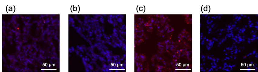
Molecules 2019, 24, 1303                                                                         2 of 16

reason for such intricate processes is the uncontrollable aggregation of cationic lipids with large and
highly charged anionic molecules, such mRNA, resulting after directly mixing these components,
as extensively demonstrated for LNPs loading plasmid DNA (pDNA) [6]. Thus, the preparation of
uniform nanometer-sized mRNA-loaded LNPs often requires special devices or techniques, such as
microfluidics, for mixing the lipid components and mRNA, which allows controlling the size of
LNPs [7,8] and replacing organic solvents with buffers for biological usage [9,10]. On the other
hand, the development of a simple and scalable technique capable of preventing such aggregate
formation in buffer would be highly valuable for the straightforward formulation of clinically relevant
mRNA-loaded LNPs.
      Herein, we proposed a simple yet robust and versatile strategy of LNP preparation by installing
polyethylene glycol (PEG) onto the mRNA molecules. The steric repulsive forces between PEG
chains are expected to prevent LNP aggregation after mixing parent LNPs with mRNA. Besides
the undemanding assembly, this strategy would allow easy preparation of mRNA-loaded LNPs in
bedside just by mixing two aqueous solutions of LNPs and mRNA, without the need of special devices
and buffer replacement, as well as eliminating long-term storage of mRNA-loaded LNPs in aqueous
solution, which is often challenging. Moreover, the LNPs resulting from the proposed method are
PEGylated, which contributes to LNP steric stabilization under biological milieu [11,12]. In addition,
the PEG density on LNPs can be easily tuned by changing the number of PEG chains installed onto
mRNA molecules. Therefore, once the parent LNPs are established, this strategy would allow easy
optimization of PEG density on LNPs to obtain desirable biodistribution after in vivo administration.
      For pursuing the proposed approach, the PEGylation of mRNA without affecting its translation
activity is a critical issue. While chemically modified bases, such as 5-methylcytosine, and
N1 -methyl-pseudouracil, are widely used for manipulating mRNA chemical structures [13–15],
even subtle base modification could lead to drastic decrease in translation efficiency, depending
on the formulations of modification [16,17]. We have recently developed an mRNA-engineering
strategy based on the installation of functional moieties to mRNA through hybridization of the
mRNA with short RNA oligonucleotides [18,19]. While double stranded RNA (dsRNA) structures
prepared in this strategy can also result in the inhibition of mRNA translation, as well as the
induction of inflammatory responses [20,21], these undesirable outcomes were effectively avoided
by shortening hybridization lengths to 17 nt. Thus, based on these studies, we hybridized mRNA
with PEGylated 17 nt RNA oligonucleotides (PEG-OligoRNAs) for mRNA PEGylation in this study.
Using a commercially-available reagent, lipofectamine LTX, and a widely-used liposomal formulation,
1,2-bis(dioleoyloxy)-3-(trimethylamonio)propane (DOTAP)/cholesterol (Chol), as model LNPs to load
mRNA hybridized with PEG-OligoRNAs (PEG-OligoRNAs/mRNA), we studied the feasibility of our
strategy through detailed physicochemical characterization and functional analyses using cultured
cells and mice.

2. Results

2.1. Hybridization of mRNA with PEG-OligoRNAs
      RNA oligonucleotides were designed to have a 12 kDa PEG chain on the 50 end of a 17 nt
hybridization sequence. We prepared mRNA hybridized with 5, 10 and 20 different PEG-OligoRNAs.
The PEG-OligoRNAs were designed to target the positions throughout 50 to 30 ends of 783 nt Gaussia
luciferase (GLuc) mRNA (Figure 1). In the selection of target sequences in GLuc mRNA, dsRNA
region was avoided (Supplementary Table S1), by using a software that predicts RNA secondary
structure [22], unless no unstructured regions were found nearby, because endogenous base-paring
in mRNA might hamper the hybridization of PEG-OligoRNAs to the mRNA. The mRNA was
hybridized with PEG-OligoRNAs with molar ratio of each sequence of PEG-OligoRNA to mRNA of
1:1. Then, gel permeation chromatography (GPC) was performed to measure hybridization efficiency.
Molecules 2019, 24, 1303 3 of 16
Molecules 2019, 24 FOR PEER REVIEW 3
The peak intensity derived from unhybridized free PEG-OligoRNAs was very weak, revealing high
PEG-OligoRNAs
(a) 2019, 24 FOR
Molecules
hybridization PEER REVIEW
efficiency in every tested formulation (Table 1, Supplementary Figure S1). 3
of 87–95%
5´ cap GLuc mRNA
PEG-OligoRNAs Poly A
(a) Start codon Stop codon
(b) GLuc mRNA
5´ cap Poly A
Start codon Stop codon
(b)
(c)
(c)
100 nt
Figure 1. Positions in GLuc mRNA to which each PEG-OligoRNA hybridizes. (a) 5 PEG-
OligoRNAs/mRNA. (b) 10 PEG-OligoRNAs/mRNA. (c) 20 PEG-OligoRNAs/mRNA. A scale 100 bar
nt
shows the length of 100 nt.
Figure 1. Positions
Figure 1. Positionsin in GLuc
GLuc mRNAmRNA to which
to which each PEG-OligoRNA
each PEG-OligoRNA hybridizes.
hybridizes. (a) 5 PEG-
(a) 5 PEG-OligoRNAs
OligoRNAs/mRNA. (b)Table
10 PEG-OligoRNAs/mRNA.
/mRNA. (b) 10 PEG-OligoRNAs/mRNA. (c) 20 (c) 20 PEG-OligoRNAs/mRNA.
PEG-OligoRNAs/mRNA. A scale
1. Hybridization efficiency determined by HPLC. bar A
shows scale bar
the length
shows
of 100 the
nt. length of 100 nt.
Number of PEG-OligoRNAs 5 10 20
Table 1. Hybridization efficiency determined by HPLC.
Table 1.Hybridization
Hybridizationefficiency
efficiency(%) 87 95by 94
determined HPLC.
Number of PEG-OligoRNAs 5 10 20
Number of PEG-OligoRNAs 5 10 20
2.2. Translation Activity of mRNA after Hybridization with PEG-OligoRNAs
Hybridization efficiency (%)
Hybridization efficiency87(%) 87 95 94
95 94
Translation activity of PEG-OligoRNAs/mRNA was measured in cell free translational system
composed
2.2.
2.2. Translationof Activity
Translation rabbit reticulocyte
Activity of mRNA
of mRNA after lysate.
after Luciferase
Hybridization
Hybridization withprotein
with expression levels decreased with the
PEG-OligoRNAs
PEG-OligoRNAs
increase in PEG-OligoRNA numbers hybridizing to mRNA (Figure 2). However, more than 60% of
Translation activity
Translation activity of
of PEG-OligoRNAs/mRNA
PEG-OligoRNAs/mRNAwas wasmeasured
measuredin incell
cellfree
free translational
translational system
system
translational activity was maintained even after hybridizing 20 PEG-OligoRNAs, compared to
composed of rabbit reticulocyte lysate. Luciferase protein expression levels decreased
composed of rabbit reticulocyte lysate. Luciferase protein expression levels decreased with the with the increase
unhybridized mRNA. This result is consistent with our previous report, showing that mRNA
in PEG-OligoRNA
increase numbers numbers
in PEG-OligoRNA hybridizing to mRNA to
hybridizing (Figure
mRNA 2). (Figure
However,2).more than 60%
However, moreof than
translational
60% of
hybridized with a large number of cholesterol-installed RNA oligonucleotides preserved its
activity was maintained
translational activity was even after hybridizing
maintained 20 PEG-OligoRNAs,
even after hybridizing 20compared to unhybridized
PEG-OligoRNAs, comparedmRNA.to
translational activity [19].
This result is consistent with our previous report, showing that mRNA hybridized
unhybridized mRNA. This result is consistent with our previous report, showing that mRNA with a large number
of cholesterol-installed
hybridized with a large RNA oligonucleotides
number preserved its translational
of cholesterol-installed activity [19]. preserved its
RNA oligonucleotides
translational activity [19].
20000
Relative luminescence unit
15000
20000
Relative luminescence unit
10000
15000
5000
10000
5000 0
0 5 10 20
Number of PEG-OligoRNA
0
Figure Translation efficiency
Figure2.2. Translation efficiency0from
frommRNA
mRNAin 5incell
cell free
free system.
system.
10 GLuc GLuc
mRNA mRNA
20 hybridized
hybridized with with
PEG-
PEG-OligoRNAs
OligoRNAs was was incubated
incubated in rabbit Number
in rabbit of PEG-OligoRNA
reticulocyte
reticulocyte lysate.After
lysate. After4 4hh of
of incubation, GLuc
GLuc protein
protein
expression
expressionlevels
levelswere measured.nn==3.3.Data
weremeasured. Dataare
arepresented
presentedas mean±± standard error of the
asmean the mean.
mean.
Figure 2. Translation efficiency from mRNA in cell free system. GLuc mRNA hybridized with PEG-
2.3. Characterization
was of Lipofectamine LTX-based LNPs lysate.
LoadingAfter
PEG-OligoRNAs/mRNA
2.3.OligoRNAs
Characterization incubated in rabbit
of Lipofectamine reticulocyte
LTX-based LNPs Loading 4 h of incubation, GLuc protein
PEG-OligoRNAs/mRNA
expression levels were measured.
PEG-OligoRNAs/mRNA wasn then
= 3. Data are presented
mixed as mean ± standard
with lipofectamine LTX, aerror of the mean.available
commercially
PEG-OligoRNAs/mRNA was then mixed with lipofectamine LTX, a commercially available
lipid-based transfection reagent, just by pipetting in aqueous solution. Two LNP formulations
lipid-based
2.3. transfection
Characterization reagent, just
of Lipofectamine by pipetting
LTX-based LNPs in aqueous
Loading solution. Two LNP formulations were
PEG-OligoRNAs/mRNA
prepared by mixing mRNA with minimal or maximal amount of lipofectamine LTX solution that the
PEG-OligoRNAs/mRNA was then mixed with lipofectamine LTX, a commercially available
lipid-based transfection reagent, just by pipetting in aqueous solution. Two LNP formulations were
prepared by mixing mRNA with minimal or maximal amount of lipofectamine LTX solution that theMolecules 2019, 24, 1303 4 of 16
Molecules 2019, 24 FOR PEER REVIEW 4
manufacturer
were preparedsuggests
by mixing tomRNA
use. The withresulting
minimalLNPs are designated
or maximal amount ofaslipofectamine
Lipo-Min and LTXLipo-Max,
solution
respectively. Gel electrophoresis
that the manufacturer suggests of to the
use.LNPsThewas performed
resulting LNPs to are
check successfulasloading
designated of PEG-
Lipo-Min and
OligoRNAs/mRNA to the LNPs. Bands corresponding to free PEG-OligoRNAs/mRNA
Lipo-Max, respectively. Gel electrophoresis of the LNPs was performed to check successful loading of were
undetectable in every tested
PEG-OligoRNAs/mRNA toformulation,
the LNPs. Bands demonstrating
correspondingthat almost
to freeall PEG-OligoRNAs/mRNAwere
PEG-OligoRNAs/mRNA was
associated
undetectable with lipofectamine
in every LTX reagent
tested formulation, both in Lipo-Min
demonstrating and Lipo-Max
that almost (Supplementary Figure
all PEG-OligoRNAs/mRNA was
S2).
associated with lipofectamine LTX reagent both in Lipo-Min and Lipo-Max (Supplementary Figure S2).
The
The sizes
sizes ofof the
the LNPs
LNPs loaded
loaded with
with mRNA
mRNA werewere measured
measured using
using dynamic
dynamic light
lightscattering
scattering(DLS).
(DLS).
The addition of unhybridized mRNA to the parent Lipofectamine LTX shifted
The addition of unhybridized mRNA to the parent Lipofectamine LTX shifted the size from around the size from around
50
50 nm
nm upup to 5000 nm
to 5000 nm when
when we we used
used Lipo-Min,
Lipo-Min, and to several
and to several hundred
hundred nanometers
nanometers when when wewe used
used
Lipo-Max
Lipo-Max (Figure
(Figure 3a,b, Table 2).
3a,b, Table 2). Hybridization
Hybridization of of PEG-OligoRNAs
PEG-OligoRNAs was was effective
effective in preventing
in preventing
aggregate formation in
aggregate formation inaamanner
mannerdependent
dependentonon thethe number
number of PEG-OligoRNAs.
of PEG-OligoRNAs. Eventually,
Eventually, Lipo-
Lipo-Min
Min and Lipo-Max loading 20 PEG-OligoRNA/mRNA exhibited sizes
and Lipo-Max loading 20 PEG-OligoRNA/mRNA exhibited sizes of 78 nm and 57 nm with narrow of 78 nm and 57 nm with
narrow size distribution,
size distribution, respectively.respectively.
Notably, Notably,
LNP size LNP
shouldsize
beshould
ideallybe ideally
below 200below
nm to 200
obtainnmprolonged
to obtain
prolonged blood circulation with escaping the uptake by reticuloendothelial
blood circulation with escaping the uptake by reticuloendothelial system (RES) [23,24]. Moreover, system (RES) [23,24].
size
Moreover, size control in the sub-100 nm range is important for achieving
control in the sub-100 nm range is important for achieving deep and broad distribution in some tissues, deep and broad
distribution in some
such as fibrotic cancer tissues,
tissuessuch[25].as fibrotic cancer tissues [25].
(a) (b)
30 30
0 0
5 Number of 5 Number of
25 25 PEG-OligoRNAs
10 PEG-OligoRNAs 10
20 20
20 mRNA (-) 20 mRNA (-)
Volume (%)
Volume (%)
15 15
10 10
5 5
0 0
1 10 100 1000 10000 1 10 100 1000 10000
Size (d, nm) Size (d, nm)
(c) (d)
30 30
0 0
5 Molar amount of Molar amount of
25 25 5
PEG-OligoA relative PEG-OligoA relative
10 10
to mRNA (eq.) to mRNA (eq.)
20
20 20 20
Volume (%)
Volume (%)
15 15
10 10
5 5
0 0
1 10 100 1000 10000 1 10 100 1000 10000
Size (d, nm) Size (d, nm)
Figure 3. Dynamic
Dynamic light scattering measurement of Lipofectamine
Lipofectamine LTX/mRNALTX/mRNA LNPs. Two Two LNP
formulations were
formulations were prepared
prepared by by mixing
mixing mRNA
mRNAwithwith(a,
(a,c) minimal or
c) minimal or (b,
(b,d)
d) maximal
maximal volume of
lipofectamine LTX
LTX solution
solutionthat
thatthe
themanufactures
manufacturessuggest
suggesttotouseuse((a,c)
((a,Lipo-Min
c) Lipo-Minandand
(b,d)(b,
Lipo-Max).
d) Lipo-
(a,b) The size of the LNP without mRNA addition (mRNA ( − )), and that loading
Max). (a, b) The size of the LNP without mRNA addition (mRNA (−)), and that loading mRNA mRNA hybridized
with 0, 5, 10,
hybridized withor0,205, PEG-OligoRNAs was measured.
10, or 20 PEG-OligoRNAs (c,d) PEG-OligoA
was measured. was used
(c, d) PEG-OligoA as used
was a control
as a
PEG-OligoRNA
control that does
PEG-OligoRNA notdoes
that hybridize to mRNA.
not hybridize to 5mRNA.
eq., 10 eq., and
5 eq., 1020eq.,
eq.and
of PEG-OligoA relative to
20 eq. of PEG-OligoA
mRNA was
relative addedwas
to mRNA to mRNA
added for preparation
to mRNA of Lipo-Min
for preparation ofand Lipo-Max.
Lipo-Min and Lipo-Max.Molecules 2019, 24, 1303 5 of 16
Molecules 2019, 24 FOR PEER REVIEW 5
Table 2. Dynamic light scattering measurement of Lipofectamine LTX/mRNA lipoplexes.
Table 2. Dynamic light scattering measurement of Lipofectamine LTX/mRNA lipoplexes.
Lipo-Min Lipo-Max
Lipo-Min Lipo-Max
Number
Number ofof PEG-OligoRNAs
PEG-OligoRNAs Number
Numberofof
PEG-OligoRNAs
PEG-OligoRNAs
mRNA (−) mRNA (−)
mRNA (−) mRNA (−)
0 0 55 10
10 20
20 00 55 10 10 20 20
Size (nm) a
Size (nm) a 43 43 4071
4071 234234 90
90 78 25
25 254
254 132
132 68 68 57 57
PDI PDI
b b 0.420.42 0.260.26 0.19
0.19 0.13
0.13 0.21
0.21 0.45
0.45 0.20
0.20 0.19
0.19 0.12 0.180.18
0.12
a Volume
a Volumeaveraged
averaged size.
b Polydispersity index.
size. b Polydispersity index.
To see whether the hybridization process is required to prevent aggregation using PEG-
To see we
OligoRNAs, whether the hybridization
used PEGylated process(PEG-OligoA),
17 nt oligoadenine is required as to a prevent aggregation using
control PEG-OligoRNA that
PEG-OligoRNAs, we used PEGylated 17 nt oligoadenine (PEG-OligoA),
does not hybridize to mRNA. Lipo-Min and Lipo-Max were prepared from the mixture of mRNA as a control PEG-OligoRNA
that PEG-OligoA
and does not hybridize
for DLS to mRNA. Lipo-Min
measurement. Theand Lipo-Max
addition were prepared
of PEG-OligoA fromtothe
failed mixture
inhibit LNPof
mRNA and in
aggregation PEG-OligoA
both Lipo-Min for DLS
and measurement.
Lipo-Max, evenThe addition
at the of PEG-OligoA
same molar amount offailed to inhibit
PEG-OligoA as LNP
that
aggregation in both Lipo-Min and Lipo-Max, even at the same molar
of PEG-OligoRNAs used to prepare 20 PEG-OligoRNAs/mRNA (Figure 3c,d). This result amount of PEG-OligoA as that of
PEG-OligoRNAs used to prepare 20 PEG-OligoRNAs/mRNA (Figure
demonstrates that PEG-OligoRNAs should be hybridized to mRNA to prevent LNP aggregation.3c,d). This result demonstrates
that Interestingly,
PEG-OligoRNAs should be hybridized
in Lipo-Max, the additionto mRNA
of mRNA to prevent LNP aggregation.
and PEG-OligoA to Lipofectamine LTX
Interestingly, in Lipo-Max, the addition of mRNA and PEG-OligoA to Lipofectamine
solution resulted in the preparation of large particles with the size of 1,000 nm or even larger LTX solution
(Figure
3d), while the size of Lipo-Max loaded with unhybridized mRNA was originally several3d),
resulted in the preparation of large particles with the size of 1,000 nm or even larger (Figure while
hundred
the size of Lipo-Max
nanometers loaded
(Figure 3b). Thus, with
the unhybridized mRNA wasprompted
presence of PEG-OligoA originallythe
several hundredofnanometers
aggregation Lipo-Max.
(Figure 3b). Thus, the presence of PEG-OligoA prompted the
However, addition of PEG-OligoA to the parent Lipofectamine LTX solution without aggregation of Lipo-Max. However,
the presence of
additionfailed
mRNA of PEG-OligoA to theaggregation
to induce such parent Lipofectamine
(SupplementaryLTX solution
Figurewithout the on
S3). Based presence of mRNA failed
these observations, it
to induce such aggregation (Supplementary Figure S3). Based on these observations,
is reasonable to assume that the binding of PEG-OligoA to Lipofectamine LTX causes the change in it is reasonable to
assume that
binding the binding
behavior of PEG-OligoA
of non-PEGylated to Lipofectamine
mRNA LTX causes
to Lipofectamine LTX,thewhich
change in binding
then results behavior
in LNP
of non-PEGylated mRNA to Lipofectamine LTX, which then results in LNP aggregation.
aggregation.
Then, LNP
Then, LNP ζ-potential
ζ-potential waswas measured
measured to to examine
examine the the degree
degree of
of LNP
LNP PEGylation,
PEGylation, which which may
may
influence the
influence the ζ-potential. Lipo-Min and
ζ-potential. Lipo-Min and Lipo-Max
Lipo-Max loaded
loaded with
with unhybridized
unhybridized mRNAmRNA exhibited
exhibited high
high
ζ-potential of +12 mV and +31 mV, respectively (Figure 4). The ζ-potential
ζ-potential of +12 mV and +31 mV, respectively (Figure 4). The ζ-potential decreased after decreased after hybridization
of mRNA with
hybridization of PEG-OligoRNAs,
mRNA with PEG-OligoRNAs, and the dropand correlated
the dropwith the number
correlated with theof PEG-OligoRNAs
number of PEG-
hybridizing hybridizing
OligoRNAs to mRNA. Lipo-Min
to mRNA. andLipo-Min
Lipo-Maxand loading 20 PEG-OligoRNA
Lipo-Max showed almostshowed
loading 20 PEG-OligoRNA neutral
ζ-potential of +0.2 mV and +2.3 mV, respectively (Figure 4). Thus, the cationic
almost neutral ζ-potential of +0.2 mV and +2.3 mV, respectively (Figure 4). Thus, the cationic surface surface charge of
Lipofectamine LTX/mRNA LNPs was masked after hybridization of
charge of Lipofectamine LTX/mRNA LNPs was masked after hybridization of mRNA with PEG- mRNA with PEG-OligoRNAs,
indicating the
OligoRNAs, successful
indicating PEGylation
the successfulof the LNPs. of the LNPs.
PEGylation
(a) 15 (b) 35
30
10 25
ζ-potential
ζ-potential
20
5
15
10
0
5
0 5 10 20
Number of PEG-OligoRNAs 0
-5 0 5 10 20
Number of PEG-OligoRNAs
Figure 4.
Figure 4. ζ-potential measurement of
ζ-potentialmeasurement of Lipofectamine
Lipofectamine LTX/mRNA
LTX/mRNALNPs.LNPs.TwoTwoLNP LNPformulations
formulations were
were
prepared by mixing mRNA with (a) minimal or (b) maximal volume of lipofectamine
prepared by mixing mRNA with (a) minimal or (b) maximal volume of lipofectamine LTX solution LTX solution
that the
that the manufactures
manufactures suggest
suggest toto use
use ((a)
((a) Lipo-Min
Lipo-Min and
and (b)
(b) Lipo-Max).
Lipo-Max). ζ-potential
ζ-potential of ofthe
theLNPs
LNPswithout
without
mRNA addition (mRNA ( − )), and those loading mRNA hybridized with 0,
mRNA addition (mRNA (−)), and those loading mRNA hybridized with 0, 5, 10, or 20 PEG-5, 10, or 20 PEG-OligoRNAs
were measured.
OligoRNAs werenmeasured.
= 6. Data are
n = presented mean ± standard
6. Data areaspresented as mean ±error of theerror
standard mean.of the mean.
2.4. Transmission Electron Microscopic Observation of Lipofectamine LTX-based LNPsMolecules 2019, 24, 1303 6 of 16
Molecules 2019, 24 FOR PEER REVIEW 6
2.4. Transmission Electron Microscopic Observation of Lipofectamine LTX-based LNPs
Direct
Directobservation
observationofofLipo-Min
Lipo-Minandand Lipo-Max
Lipo-Maxwas was performed
performedusing using transmission
transmissionelectron
electron
microscopy
microscopy(TEM).
(TEM).Representative
Representativeimages
imagesof ofLipo-Min
Lipo-Min loaded
loaded with
with 5–20
5–20 PEG-OligoRNAs/mRNA
PEG-OligoRNAs/mRNA
and
andLipo-Max
Lipo-Maxloaded
loadedwith
with0–200–20PEG-OligoRNAs/mRNA
PEG-OligoRNAs/mRNAare areshown
shownininFigure
Figure5,5,while
whileaggregation
aggregation
hampered TEM observation of Lipo-Min loaded with unhybridized mRNA
hampered TEM observation of Lipo-Min loaded with unhybridized mRNA (see Figure 3a). (see Figure 3a).Images
Imagesof
ofLipo-Min
Lipo-Minloaded
loadedwith
with55PEG-OligoRNAs/mRNA
PEG-OligoRNAs/mRNA and and Lipo-Max
Lipo-Maxloaded
loadedwith
withunhybridized
unhybridizedmRNA mRNA
revealed the formation of large LNPs with the size over 100 nm, which were
revealed the formation of large LNPs with the size over 100 nm, which were prepared through prepared through
association
associationofofseveral
severalsmaller
smallerparticles
particles(see
(seedotted
dottedcircle
circlein
in Figure
Figure 5a,d).
5a,d). InInaccordance
accordancewith withthis
this
observation,
observation,a aprevious
previousresearch
researchreported
reportedthatthataddition
additionofofpDNA
pDNAtotocationic
cationicLNP LNPinduced
inducedthe the
aggregation of the LNP by crosslinking of several LNPs through pDNA strands [6].
aggregation of the LNP by crosslinking of several LNPs through pDNA strands [6]. Thus, aggregationThus, aggregation
ofof LNPs loadingnon-PEGylated
LNPs loading non-PEGylated mRNA
mRNA or mRNA
or mRNA hybridized
hybridized with awith
small anumber
small of number of PEG-
PEG-OligoRNAs
OligoRNAs may be explained
may be explained by crosslinking
by crosslinking of severalofLNPs
several LNPs through
through mRNA strands.
mRNA strands. In contrast,
In contrast, mRNA
mRNA
hybridization with a large number of PEG-OligoRNAs leads to LNP coating with high densitydensity
hybridization with a large number of PEG-OligoRNAs leads to LNP coating with high of PEG
ofchains,
PEG chains, as indicated
as indicated by ζ-potential
by ζ-potential neutralization
neutralization of LNP4),(Figure
of LNP (Figure which 4),
may which may
inhibit theinhibit the
association
association between LNPs, thereby preventing the LNP aggregation
between LNPs, thereby preventing the LNP aggregation (Figure 5b,c,f,g). (Figure 5b,c,f,g).
(a) (b) (c)
(d) (e) (f) (g)
Figure
Figure5.5.Transmission
Transmissionelectron
electronmicroscopic
microscopicimaging
imagingofofLipofectamine
LipofectamineLTX/mRNA
LTX/mRNALNPs. LNPs.TwoTwoLNPLNP
formulations were prepared by mixing mRNA with (a–c) minimal or
formulations were prepared by mixing mRNA with (a–c) minimal or (d–g) maximal volume(d–g) maximal volume of
lipofectamine
of lipofectamineLTX LTX
solution that the
solution thatmanufactures suggest
the manufactures to useto
suggest ((a–c)
use Lipo-Min and (d–g)
((a–c) Lipo-Min andLipo-
(d–g)
Max). (a–c) Lipo-Min
Lipo-Max). loadedloaded
(a–c) Lipo-Min with (a) 5 PEG-OligoRNAs/mRNA,
with (a) 5 PEG-OligoRNAs/mRNA, (b) 10 (b)
PEG-OligoRNAs/mRNA,
10 PEG-OligoRNAs/mRNA, and
(c)
and20 (c)
PEG-OligoRNAs/mRNA.
20 PEG-OligoRNAs/mRNA. (d–g) Lipo-Max loaded loaded
(d–g) Lipo-Max with (d) unhybridized
with mRNA,mRNA,
(d) unhybridized (e) 5 PEG-
(e) 5
OligoRNAs/mRNA,
PEG-OligoRNAs/mRNA, (f) 10 PEG-OligoRNAs/mRNA,
(f) 10 PEG-OligoRNAs/mRNA, and (g)and
20 PEG-OligoRNAs/mRNA.
(g) 20 PEG-OligoRNAs/mRNA. Dotted Dotted
circle
showed large particles
circle showed prepared
large particles through through
prepared association of severalofsmaller
association severalparticles. Scale bars:Scale
smaller particles. 100 nm.
bars:
100 nm.
2.5. Loading of PEG-OligoRNA/mRNA to DOTAP/Chol liposome
2.5. Loading of PEG-OligoRNA/mRNA to DOTAP/Chol liposome
To investigate the versatility of our approach, PEG-OligoRNAs/mRNA was then merged with
To investigate
DOTAP/Chol the versatility
liposome. Two aqueousof our solutions
approach, of PEG-OligoRNAs/mRNA
PEG-OligoRNAs/mRNA was then
and merged with
DOTAP/Chol
DOTAP/Chol liposome. Two aqueous solutions of PEG-OligoRNAs/mRNA
liposome were mixed just by pipetting, at amine groups in DOTAP to phosphate groups in PEG- and DOTAP/Chol
liposome were mixed
OligoRNAs/mRNA (N/P)just
ratioby
of pipetting, at amine groups
3. Gel electrophoresis experimentin DOTAP
confirmed to successful
phosphateassociation
groups in
PEG-OligoRNAs/mRNA (N/P) ratio of 3. Gel electrophoresis experiment
of almost all PEG-OligoRNAs/mRNA to the liposomes in all tested formulations (Figure 6a). confirmed successful
In DLS
association of almost all PEG-OligoRNAs/mRNA to the liposomes in all tested formulations
measurement, addition of unhybridized mRNA led to the formation of two types of aggregates with (Figure 6a).
In DLS
the size measurement,
around 1000 nm addition of unhybridized
and 5000 mRNA(Figure
nm, respectively led to the
6b,formation
Table 3).ofHybridization
two types of aggregates
of PEG-
OligoRNAs was effective in preventing aggregate formation, and the ability3).to Hybridization
with the size around 1000 nm and 5000 nm, respectively (Figure 6b, Table prevent the
of PEG-OligoRNAs
aggregation correlatedwaswitheffective in preventing
the number aggregateEventually,
of PEG-OligoRNAs. formation, LNPsand the ability
loaded withto20prevent
PEG-
the aggregation correlated with the number of PEG-OligoRNAs.
OligoRNA/mRNA exhibited the average size of 132 nm with narrow size distribution. Eventually, LNPs loaded
In thewith
ζ-
potential measurement, LNPs loaded with unhybridized mRNA had high cationic charge of +24 mV.
The cationic charge was successfully masked after hybridization of mRNA with 5-20 PEG-
OligoRNAs, indicating the successful PEGylation of the LNPs (Figure 6c). Together, our approach ofMolecules 2019, 24, 1303 7 of 16
20 PEG-OligoRNA/mRNA exhibited the average size of 132 nm with narrow size distribution. In the
ζ-potential measurement, LNPs loaded with unhybridized mRNA had high cationic charge of +24 mV.
The cationic charge was successfully masked after hybridization of mRNA with 5-20 PEG-OligoRNAs,
Molecules 2019, 24 FOR PEER REVIEW 7
indicating the successful PEGylation of the LNPs (Figure 6c). Together, our approach of PEG-OligoRNA
hybridization allowed
PEG-OligoRNA easy preparation
hybridization allowed of PEGylated
easy DOTAP/Chol-based
preparation LNPs loaded with mRNA
of PEGylated DOTAP/Chol-based LNPs
with relatively small sizes and almost neutral surface charges, as is the case of Lipofectamine
loaded with mRNA with relatively small sizes and almost neutral surface charges, as is the case of LTX-based
LNPs (Figures LTX-based
Lipofectamine 3–5). LNPs (Figures 3–5).
(a) Naked DOTAP/Chol
Number of
PEG-OligoRNAs
0 5 10 20 0 5 10 20
(b) 0
(c)
30
5 Number of
30
10 PEG-OligoRNAs
25
20
25
mRNA (-)
20
20
Volume (%)
ζ-potential
15 15
10 10
5
5
0
0 0 5 10 20
1 10 100 1000 10000 -5
Size (d, nm) Number of PEG-OligoRNAs
Characterization of DOTAP/Chol
Figure 6. Characterization DOTAP/Chol LNPsLNPs loaded with PEG-OligoRNAs/mRNA.
PEG-OligoRNAs/mRNA. (a) (a) Gel
electrophoresis of unhybridized
unhybridized mRNA
mRNAandandmRNA
mRNAhybridized
hybridizedwith with5,5,10,
10,oror2020PEG-OligoRNAs,
PEG-OligoRNAs,inina
anaked
naked form
formoror
after complexation
after with
complexation DOTAP/Chol
with DOTAP/Chol liposomes.
liposomes.(b)(b)
TheThe
sizes andand
sizes (c) ζ-potential
(c) ζ-potential of the
of
DOTAP/Chol LNPs without mRNA addition (mRNA ( − )), and that loading
the DOTAP/Chol LNPs without mRNA addition (mRNA (−)), and that loading mRNA hybridizedmRNA hybridized with 0,
5, 10, or 20 PEG-OligoRNAs were measured. n = 6 for (c). Data are presented as mean
with 0, 5, 10, or 20 PEG-OligoRNAs were measured. n = 6 for (c). Data are presented as mean ± ± standard error
of the mean.
standard error of the mean.
Table 3. Dynamic light scattering measurement of DOTAP/Chol LNPs.
Table 3. Dynamic light scattering measurement of DOTAP/Chol LNPs.
Number of PEG-OligoRNAs
mRNA (−) Number of PEG-OligoRNAs
mRNA (−) 0 5 10 20
0 5 10 20
Size (nm) a
(nm) a 84 84 1698 1698 853 853 152152 132132
Size
PDI bb
PDI 0.150.15 0.31 0.31 0.47 0.47 0.100.10 0.08
0.08
a aVolumeaveraged
Volume size.bb Polydispersity
averaged size. Polydispersity index.
index.
2.6. Introduction
Introduction of Lipofectamine LTX-based
LTX-Based LNPs to Cultured Cells
Biological function of Lipofectamine LTX-based
LTX-based LNPs
LNPs loading
loading 20
20 PEG-OligoRNAs/mRNA
PEG-OligoRNAs/mRNA was
then investigated
investigatedininvitro
vitrousing
using HuH-7
HuH-7 cells.
cells. First,
First, mRNA
mRNA introduction
introduction efficiency
efficiency was evaluated
was evaluated using
using GLuc mRNA as a reporter. LNP loaded with non-PEGylated mRNA exhibited 2–4 orders higher
efficiency of GLuc protein expression compared to that loaded with 20 PEG-OligoRNAs/mRNA
(Figure 7a). To study the mechanism, efficiency of mRNA cellular uptake was evaluated by
measuring GLuc mRNA amount in the cells through quantitative real-time PCR (qRT-PCR). The
efficiency of mRNA uptake decreased by 3–4 orders after hybridization of 20 PEG-OligoRNAsMolecules 2019, 24, 1303 8 of 16
GLuc mRNA as a reporter. LNP loaded with non-PEGylated mRNA exhibited 2–4 orders higher
efficiency of GLuc protein expression compared to that loaded with 20 PEG-OligoRNAs/mRNA
(Figure 7a). To study the mechanism, efficiency of mRNA cellular uptake was evaluated by measuring
GLuc mRNA amount in the cells through quantitative real-time PCR (qRT-PCR). The efficiency of
mRNA uptake decreased by 3–4 orders after hybridization of 20 PEG-OligoRNAs compared to LNP
Molecules 2019, 24 FOR PEER REVIEW 8
loaded with unhybridized mRNA (Figure 7b), showing the profile consistent to the measurement of
GLuc
the expression. Thus,
measurement the translational
of GLuc expression. efficiency
Thus, thestandardized
translationalwith the amount
efficiency of mRNA with
standardized takenthe
up
by cells was
amount comparable
of mRNA takenregardless of was
up by cells the hybridization of 20 PEG-OligoRNAs.
comparable regardless This result
of the hybridization of suggests
20 PEG-
that the decrease in GLuc protein expression efficiency after LNP PEGylation is largely
OligoRNAs. This result suggests that the decrease in GLuc protein expression efficiency after LNP attributed
to limited cellular
PEGylation uptake
is largely of the LNPs,
attributed rather than
to limited reduced
cellular uptaketranslational activity
of the LNPs, of mRNA.
rather Indeed,
than reduced
in cell free translational
translational activity of system,
mRNA.hybridization of PEG-OligoRNAs
Indeed, in cell free translationalinduced
system,minimal effect onofmRNA
hybridization PEG-
translational activity (Figure 2).
OligoRNAs induced minimal effect on mRNA translational activity (Figure 2).
(a) 100000000 unPEGylated 20 PEG-OligoRNAs
(b) 100
unPEGylated 20 PEG-OligoRNAs
10000000
10
mRNA uptake (% dose)
1000000
Relative GLuc expression
1
100000
10000 0.1
1000
0.01
100
0.001
10
1 0.0001
Lipo-Min Lipo-Max Lipo-Min Lipo-Max
Introduction of Lipofectamine LTX/mRNA
Figure 7. Introduction
Figure LTX/mRNALNPs LNPsto to cultured
cultured cells.
cells. Two
Two LNP
LNP formulations
formulations
were prepared
preparedby bymixing
mixing GLuc mRNA
GLuc mRNA withwith
minimal or maximal
minimal volume
or maximal of lipofectamine
volume LTX solution
of lipofectamine LTX
that the manufactures
solution suggest tosuggest
that the manufactures use (Lipo-Min and Lipo-Max).
to use (Lipo-Min LNP loaded
and Lipo-Max). LNP with non-PEGylated
loaded with non-
mRNA andmRNA
PEGylated that loaded with
and that 20 PEG-OligoRNAs/mRNA
loaded with 20 PEG-OligoRNAs/mRNAwere transfected to HuH-7to
were transfected cells.
HuH-7(a) GLuc
cells.
protein concentration in the culture medium 4 h after the transfection. n = 6. (b) GLuc mRNA
(a) GLuc protein concentration in the culture medium 4 h after the transfection. n = 6. (b) GLuc mRNA uptake
to HuH-7
uptake cells measured
to HuH-7 using using
cells measured qRT-PCR 4 h after
qRT-PCR 4 h the
aftertransfection. n = 4.
the transfection. n =Data areare
4. Data presented
presentedas
mean
as mean± ±standard
standard error
errorofof
the mean.
the mean.
2.7. In Vivo Behavior of Lipofectamine LTX-Based LNPs after Systemic Delivery
2.7. In Vivo Behavior of Lipofectamine LTX-based LNPs after Systemic Delivery
Lastly, the in vivo behavior of the Lipofectamine LTX-based LNPs was evaluated after their
Lastly, the in vivo behavior of the Lipofectamine LTX-based LNPs was evaluated after their
intravenous injection into mice. Real-time behavior of LNPs loaded with Cy5-labeled mRNA in the
intravenous injection into mice. Real-time behavior of LNPs loaded with Cy5-labeled mRNA in the
blood circulation was observed using intravital confocal microscopy. In both cases of Lipo-Min and
blood circulation was observed using intravital confocal microscopy. In both cases of Lipo-Min and
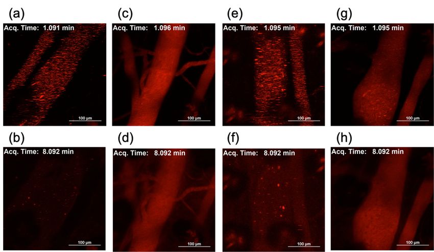
Lipo-Max loaded with non-PEGylated mRNA, several micrometer-sized aggregates were observed
Lipo-Max loaded with non-PEGylated mRNA, several micrometer-sized aggregates were observed
1 min after the injection (Figure 8a,e). Note that minimal detectable size of the aggregate is theoretically
1 min after the injection (Figure 8a,e). Note that minimal detectable size of the aggregate is
estimated to be above 700 nm, based on the pixel size of 620 nm × 620 nm, and on the Cy5
theoretically estimated to be above 700 nm, based on the pixel size of 620 nm × 620 nm, and on the
detection wavelength of 700 nm. The fluorescence signal in the blood was rapidly decreased,
Cy5 detection wavelength of 700 nm. The fluorescence signal in the blood was rapidly decreased, and
and almost no signal was detected as early as 8 min after the injection (Figure 8b,f, Videos S1
almost no signal was detected as early as 8 min after the injection (Figure 8b,f, Video S1, S3), revealing
and S3), revealing rapid clearance of non-PEGylated Lipo-Min and Lipo-Max. In contrast, when
rapid clearance of non-PEGylated Lipo-Min and Lipo-Max. In contrast, when 20 PEG-
20 PEG-OligoRNAs/mRNA was loaded to Lipo-Min and Lipo-Max, the fluorescence signal in the
OligoRNAs/mRNA was loaded to Lipo-Min and Lipo-Max, the fluorescence signal in the blood was
blood was uniform without detectable aggregation throughout the observation periods, and high
uniform without detectable aggregation throughout the observation periods, and high fluorescence
fluorescence intensity was observed in the blood even 8 min or later after the injection (Figure 8c,d,g,h,
intensity was observed in the blood even 8 min or later after the injection (Figure 8c,d,g,h, Video S2,
Videos S2 and S4). This observation indicates effective steric stabilization of Lipo-Min and Lipo-Max
S4). This observation indicates effective steric stabilization of Lipo-Min and Lipo-Max by mRNA
by mRNA PEGylation also in the biological milieu, which resulted in prolonged blood circulations.
PEGylation also in the biological milieu, which resulted in prolonged blood circulations.Molecules 2019, 24, 1303 9 of 16
Molecules 2019, 24 FOR PEER REVIEW 9
Figure
Figure 8. 8.Intravital
Intravitalreal-time
real-time confocal
confocal observation
observationofofLipofectamine
LipofectamineLTX-based
LTX-based LNPs
LNPs in inthethe
blood.
blood.
Using GLuc mRNA labeled with Cy5, two LNP formulations were prepared
Using GLuc mRNA labeled with Cy5, two LNP formulations were prepared by mixing the mRNA withby mixing the mRNA
with minimal
minimal or maximal
or maximal volume volume of lipofectamine
of lipofectamine LTX solution
LTX solution thatmanufacture
that the the manufacture suggests
suggests to use
to use ((a–d)
((a–d) Lipo-Min
Lipo-Min and (e–h)and (e–h) Lipo-Max).
Lipo-Max). Then, theThen,
LNPsthe
wasLNPs was intravenously
intravenously injected
injected to to for
the mice the observation
mice for
of observation of thein
the blood vessel blood vessel inusing
the earlobes the earlobes using
intravital intravital
confocal confocal microscopy.
microscopy. (a, c, 1e,min
(a,c,e,g) Images g) Images
after the
1 min after the injection. (b, d, f, h) Images 8 min after the injection. (a, b) Lipo-Min loaded with non-
injection. (b,d,f,h) Images 8 min after the injection. (a,b) Lipo-Min loaded with non-PEGylated mRNA.
PEGylated mRNA. (c, d) Lipo-Min loaded with 20 PEG-OligoRNAs/mRNA. (e, f) Lipo-Max loaded
(c,d) Lipo-Min loaded with 20 PEG-OligoRNAs/mRNA. (e,f) Lipo-Max loaded with non-PEGylated
with non-PEGylated mRNA. (g, h) Lipo-Max loaded with 20 PEG-OligoRNAs/mRNA.
mRNA. (g,h) Lipo-Max loaded with 20 PEG-OligoRNAs/mRNA.
According to a previous report, the aggregation of nucleic acid complexes in the blood induced
According to a previous report, the aggregation of nucleic acid complexes in the blood induced
lung embolism, even causing the death of experimental animals [26]. Then, we observed the
lung embolism, even causing the death of experimental animals [26]. Then, we observed the
distribution of Lipo-Min and Lipo-Max in the tissue section of the lung after injection of Lipo-Min
distribution of Lipo-Min and Lipo-Max in the tissue section of the lung after injection of Lipo-Min and
and Lipo-Max loaded with Cy5-labeled mRNA. For Lipo-Min and Lipo-Max loaded with non-
Lipo-Max loaded with Cy5-labeled mRNA. For Lipo-Min and Lipo-Max loaded with non-PEGylated
PEGylated mRNA, several micrometer-sized red dots were observed throughout the lung tissue 5
mRNA, several micrometer-sized red dots were observed throughout the lung tissue 5 min after
min after injection, which presumably showed deposition of aggregates in the lung capillary (Figure
injection,
9a,c). Onwhich presumably
the other showed
hand, such deposition
deposition of aggregates
of LNP aggregatesininthethelung
lungcapillary (Figure prevented
was effectively 9a,c). On the
other hand, such deposition of LNP aggregates in the lung was effectively prevented
after hybridization of 20 PEG-OligoRNAs to mRNA (Figure 9b,d). Similarly, several-micrometer- after hybridization
of sized
20 PEG-OligoRNAs to mRNA (Figure
aggregates from unPEGylated Lipo-Max 9b,d).
were Similarly, several-micrometer-sized
deposited also aggregates
in the spleen and the liver, while
from
Lipo-Max from 20 PEG-OligoRNAs/mRNA showed reduced accumulation to these Lipo-Max
unPEGylated Lipo-Max were deposited also in the spleen and the liver, while organs
from 20 PEG-OligoRNAs/mRNA
(Supplementary Figure S4). Theseshowed reduced
observations accumulation
are consistent withto these
the organs
real-time LNP(Supplementary
imaging in
Figure
bloodS4). These observations
circulation, demonstrating are
theconsistent with
effectiveness of the real-time LNP
PEG-OligoRNA imaging infor
hybridization blood circulation,
inhibiting the
demonstrating
LNP aggregation the effectiveness of PEG-OligoRNA
in blood (Figure 8). With avoidance hybridization for inhibiting
of tissue deposition the LNP
and clearance aggregation
mechanism,
in the
bloodPEGylated
(Figure 8).LNP showed
With prolonged
avoidance of tissueblood circulation
deposition comparedmechanism,
and clearance to non-PEGylated LNPs as
the PEGylated LNP
shownprolonged
showed in Figure 8.blood circulation compared to non-PEGylated LNPs as shown in Figure 8.Molecules 2019, 24, 1303 10 of 16
Molecules 2019, 24 FOR PEER REVIEW 10
Figure9.
Figure 9. Distribution
Distribution of
of Lipofectamine
Lipofectamine LTX-based
LTX-based LNPs
LNPs inin the
thelung.
lung.Using
UsingGLuc
GLucmRNA
mRNAlabeled
labeledwithwith
Cy5,two
Cy5, twoLNP
LNPformulations
formulationswere
wereprepared
preparedbybymixing
mixingthethemRNA
mRNAwith withminimal
minimalorormaximal
maximalvolumevolumeof
of lipofectamine
lipofectamine LTXLTX solution
solution thatthat
thethe manufacture
manufacture suggests
suggests to use
to use ((a,Lipo-Min
((a,b) b) Lipo-Min
andand
(c,d)(c,Lipo-Max).
d) Lipo-
Max). Then, the LNPs were intravenously injected to the mice. Five min after the
Then, the LNPs were intravenously injected to the mice. Five min after the injection, the lunginjection, the lung
was
was excised for preparation of frozen tissue sections. Blue: nucleus (DAPI). (a) Lipo-Min
excised for preparation of frozen tissue sections. Blue: nucleus (DAPI). (a) Lipo-Min loaded with loaded with
non-PEGylated mRNA.
non-PEGylated mRNA. (b)(b) Lipo-Min
Lipo-Min loaded
loaded with
with 20
20 PEG-OligoRNAs/mRNA.
PEG-OligoRNAs/mRNA. (c) (c)Lipo-Max
Lipo-Maxloadedloaded
withnon-PEGylated
with non-PEGylated mRNA.
mRNA. (d)(d) Lipo-Max
Lipo-Max loaded
loaded with
with 20
20 PEG-OligoRNAs/mRNA.
PEG-OligoRNAs/mRNA.
3.3.Discussion
Discussion
LNP
LNPloading
loading of of mRNA requires complicated
complicated procedures,
procedures,which
whichhamper
hamperthe thewidespread
widespreadclinical
clinical
application
applicationof of the
the mRNA LNPs. In In this
this study,
study, wewe developed
developedaasimple,
simple,robust
robustandandversatile
versatileprocedure
procedure
for
formRNA
mRNA LNP LNP preparation
preparation by mRNA PEGylation.PEGylation. UsingUsingthis
thisstrategy,
strategy,mRNA-loaded
mRNA-loadedLNPs LNPswith with
regulated sizes and close-to-neutral surface charges were prepared just
regulated sizes and close-to-neutral surface charges were prepared just by mixing two aqueous by mixing two aqueous
solutions of
solutions of parent
parent LNPLNP andand mRNA withoutwithout thethe need
need of of buffer
buffer exchange
exchangefor forsubsequent
subsequentbiological
biological
usage(Figures
usage (Figures3–6). 3–6).
SuchSuch
easyeasy procedure
procedure allowsallows LNP preparation
LNP preparation even atwhich
even at bedside, bedside, which
additionally
additionally provides a solution to the issue of mRNA storage. Although
provides a solution to the issue of mRNA storage. Although lyophilization is an attractive approach lyophilization is an
attractive
for long-term approach
storage for long-term
of mRNA withoutstorage of mRNA
degradation without
[27], degradation
lyophilization [27], lyophilization
of mRNA-loaded LNPs could of
mRNA-loaded
influence LNPs could influence
their physicochemical properties.their
On thephysicochemical properties. ofOn
other hand, lyophilization mRNAthe molecule
other hand, is an
lyophilization
established of mRNA
technique, molecule
allowing is an established
long-term mRNA storage technique,
even atallowing long-term[28].
room temperature mRNA Ourstorage
strategy
even at
would room
allow temperature
preparation [28]. Our strategy
of mRNA-loaded LNPs would allow after
immediately preparation of mRNA-loaded
rehydration LNPs
of lyophilized mRNA,
immediately after rehydration of lyophilized
minimizing time of mRNA storage in aqueous solution. mRNA, minimizing time of mRNA storage in aqueous
solution.
For mRNA PEGylation, 17 nt PEG-OligoRNAs were hybridized to mRNA. Importantly,
For of
addition mRNA PEGylation,
PEG-OligoA, 17 nt PEG-OligoRNAs
an unhybridizing control, failed were hybridized
to prevent LNP to mRNA. Importantly,
aggregation (Figure 3c,d),
addition of PEG-OligoA, an unhybridizing control, failed to prevent
demonstrating the importance of mRNA PEGylation through hybridization of PEG-OligoRNAs. LNP aggregation (Figure 3c,d),
demonstrating the importance of mRNA PEGylation through hybridization
Moreover, hybridization of 20 PEG-OligoRNAs per mRNA induced minimal influence on translational of PEG-OligoRNAs.
Moreover,
activity of mRNA hybridization
(Figure 2).ofThis
20 result
PEG-OligoRNAs per mRNA
is consistent with induced
our previous minimal
report influence on
of mRNA-engineering,
translational
showing thatactivity
short RNAof mRNA (Figure 2). Thispossessing
oligonucleotides result is consistent withmoieties
functional our previous
can report of mRNA-to
be hybridized
engineering, showing that short RNA oligonucleotides possessing
mRNA with preserved translational activities [19]. Cellular mechanisms of unwinding dsRNA functional moieties caninbean
hybridized to mRNA with preserved translational activities [19]. Cellular
ATP-consuming manner presumably contributed to efficient translation from mRNA hybridized with mechanisms of unwinding
dsRNA in an ATP-consuming manner presumably contributed to efficient translation from mRNA
short RNA oligonucleotides [29].
hybridized with short RNA oligonucleotides [29].
LNP PEGylation showed large influence on in vivo blood circulation profile after systemic
LNP PEGylation showed large influence on in vivo blood circulation profile after systemic
injection (Figure 8). LNPs loaded with non-PEGylated mRNA formed aggregates immediately after the
injection (Figure 8). LNPs loaded with non-PEGylated mRNA formed aggregates immediately after
injection, leading to rapid clearance of the LNPs from the blood circulation. Notably, the non-PEGylated
the injection, leading to rapid clearance of the LNPs from the blood circulation. Notably, the non-
LNPs were deposited in the lung tissues, indicating the occlusion of lung capillary by the aggregates.
PEGylated LNPs were deposited in the lung tissues, indicating the occlusion of lung capillary by the
Importantly, such lung embolism can cause the death of mice [26]. On the other hand, LNP PEGylation
aggregates. Importantly, such lung embolism can cause the death of mice [26]. On the other hand,
through PEG-OligoRNA hybridization to mRNA is effective in preventing LNP aggregation in the
LNP PEGylation through PEG-OligoRNA hybridization to mRNA is effective in preventing LNP
blood, and LNP deposition in the lung, demonstrating high structural stability of the LNPs in biological
aggregation in the blood, and LNP deposition in the lung, demonstrating high structural stability of
milieu and their improved safety for in vivo usage. Such structural stabilization after PEGylation
the LNPs in biological milieu and their improved safety for in vivo usage. Such structural
may be explained by PEG steric repulsive force preventing the association of LNP with surrounding
stabilization after PEGylation may be explained by PEG steric repulsive force preventing the
proteins, blood cells or LNPs in the blood [30].
association of LNP with surrounding proteins, blood cells or LNPs in the blood [30].
The mRNA introduction efficiency to the cultured cells decreased after LNP PEGylation, which
may be explained by PEG steric repulsive force inhibiting the non-specific interaction of LNPs to cellsMolecules 2019, 24, 1303 11 of 16
The mRNA introduction efficiency to the cultured cells decreased after LNP PEGylation, which
may be explained by PEG steric repulsive force inhibiting the non-specific interaction of LNPs to cells
and LNP uptake to the cells (Figure 7). The PEGylation of the LNPs can also induce inhibitory effect
on intracellular processes, such as endosomal escape [31]. Several strategies were reported for solving
these issues of PEGylation, so called “PEG dilemma”, and facilitating cellular uptake and endosomal
escape, including the installation of ligands to PEG, the optimization of surface PEG density and
the selective cleavage of PEG in target tissues or cells [32–36]. We plan combinational use of these
strategies with the mRNA PEGylation approach, to achieve efficient in vivo mRNA introduction in the
future study.
The use of PEGylated lipids is an alternative strategy to prevent the aggregation after mRNA
addition to LNPs, and feasibility of this strategy was well studied in LNP preparation for pDNA
delivery. Although this strategy showed some effect on steric stabilization of LNP, PEGylated LNPs still
aggregate after pDNA addition, depending on their formulations [37,38]. Moreover, mRNA-loaded
LNPs have been prepared by using PEGylated lipids and organic solvents, leading to the assembly of
particles with several hundred nanometer diameter and highly cationic ζ-potential [39]. Our strategy
has potential to prepare sub-100 nm-sized LNPs with neutral surface charge, by mRNA PEGylation
alone or by combination with PEGylated lipids.
Our strategy can potentially be applied to polymer-based mRNA carriers, which provide
promising alternatives to lipid-based carriers for targeting various tissues other than the liver and the
spleen [40–43], while the liver and the spleen can be more efficiently targeted using LNPs due to their
tropism to these organs. By using PEGylated mRNA, the complicated processes of PEG conjugation to
polycation can be avoided. Furthermore, as is the case in LNPs, PEGylation of polyplexes is beneficial
for their steric stabilization in biological milieu [35,44] and alleviation of mRNA immunogenicity [45].
In conclusion, we developed a simple, robust and versatile procedure of mRNA-loaded LNP
preparation, providing a smart solution to so far complicated procedures. The PEGylation of mRNA
allowed the preparation of size-controlled LNP by mixing two aqueous solutions of mRNA and parent
LNP by using pipetting. Importantly, mRNA was successfully PEGylated with minimal influence
on its translational activity by hybridizing mRNA with PEGylated short RNA oligonucleotides.
The PEGylated LNP thus obtained exhibited high structural stability in blood after intravenous
injection, while non-PEGylated LNP rapidly aggregated in blood, presumably causing lung embolism.
By providing methods for easy preparation of mRNA LNP for in vivo usage, our approach has
the potential to accelerate the widespread clinical use of existing and newly developed mRNA
LNP formulations.
4. Materials and Methods
4.1. Preparation of mRNA
mRNA was prepared as previously described [46]. Briefly, template DNA containing Gaussia
luciferase (GLuc) coding sequence and poly 120 bp A/T under T7 promoter was constructed from
a pSP73 plasmid (Promega, Madison, WI, USA) and a pCMV-Gluc control plasmid (New England
BioLabs, Ipswich, MA, USA). In vitro transcription was performed using mMESSAGE mMACHINE
T7 Ultra Kit (Ambion, Carlsbad, CA, USA), followed by mRNA purification using RNeasy Mini Kit
(Qiagen, Hilden, Germany), and spectroscopic determination of its concentration using a NanoDrop
1000 spectrophotometer (NanoDrop Technologies Inc., Wilmington, DE, USA). On-chip capillary
electrophoresis was performed using an Agilent2100 Bioanalyzer (Agilent, Santa Clara, CA, USA),
after denaturing mRNA at 72 ◦ C for 2 min, followed by rapid cooling of the mRNA solution on
ice. The result demonstrated successful preparation of homogenous mRNA strands (Supplementary
Figure S5).Molecules 2019, 24, 1303 12 of 16
4.2. Hybridization of mRNA with PEG-OligoRNAs
17 nt RNA oligonucleotides installed with PEG (MW : 12 KDa) at their 50 end (PEG-OligoRNAs)
were purchased from GeneDesign Inc. (Osaka, Japan). These PEG-OligoRNAs have sequences
complementary to the target sequences in GLuc mRNA shown in Supplementary Table S1. The PEG-
OligoRNAs were mixed with GLuc mRNA with molar ratio of each sequence of PEG-OligoRNA to
mRNA of 1:1 to obtain final GLuc mRNA concentration of 100 ng/µL in the buffer containing 10 mM
HEPES and 150 mM NaCl. Then, the RNA solution was kept at 65 ◦ C for 5 min, followed by gradual
cooling to 30 ◦ C in 90 min.
4.3. Cell Free Translation
Cell free translation of GLuc mRNA was performed using Rabbit Reticulocyte Lysate System,
Nuclease Treated (Promega). After 4 h of the incubation of GLuc mRNA (50 ng) in rabbit reticulocyte
lysate (14 µL), GLuc expression level was measured using Renilla Luciferase Assay System (Promega)
and Lumat3 LB9508 luminometer (Berthold Technologies, Bad Wildbad, Germany).
4.4. Preparation of Lipofectamine LTX/mRNA LNPs
PEG-OligoRNAs/mRNA was mixed with LTX solution equipped in Lipofectamine LTX
(Invitrogen, Carlsbad, CA, USA) at the ratio of 1 µg mRNA to 2 µL LTX solution for Lipo-Min,
and that of 1 µg mRNA to 5 µL LTX solution for Lipo-Max. Final mRNA solution was adjusted to
33.3 ng/µL in the buffer containing 10 mM HEPES and 50 mM NaCl.
4.5. Characterization of LNPs
The LNP solutions thus obtained were used for characterization including gel electrophoresis,
DLS, and ζ-potential measurement. After adding 20 µL of sample solution with 4 µL of 30% glycerol,
gel electrophoresis was performed using 0.9% agarose gel at 100 mV for 35 min. After 1 h incubation
in 0.5 mg/L ethidium bromide solution (NIPPON GENE Co., Ltd., Tokyo, Japan), the gel was imaged
using Gel Doc ER system (BioRad, Hercules, CA, USA). Zetasizer Nano-ZS (Malvern Instruments,
Worcestershire, UK) equipped with an Ar laser (λ = 532 nm) was used for DLS measurement with
scattering angle of 173 ◦ at 25 ◦ C. The diameter was calculated using Stokes Einstein equation.
ζ-potential was measured through laser-doppler electrophoresis using Möbius ζ™ (Wyatt Technology
Corporation, Santa Barbara, CA, USA) equipped with a 532 nm laser, followed by the calculation based
on the Smoluchowski’s equation. All of these characterization experiments were performed at mRNA
concentration of 33.3 ng/µL in the buffer containing 10 mM HEPES and 50 mM NaCl.
4.6. Transmission Electron Microscopic Observation
An Eiko IB-3 ion coater (Eiko Engineering Co. Ltd., Tokyo, Japan) was used to hydrophilize
carbon-coated copper TEM grids (Cu400, JEOL Ltd., Tokyo, Japan) by glow discharge. On the grid,
2 µL of LNP solution loaded with 20 ng of PEG-OligoRNAs/mRNA was mixed with 2 µL of 2% w/v
uranyl acetate solution. After 30 s, the droplet was removed using a filter paper and then the grid was
dried. The sample was observed using a JEM-1400 (JEOL Ltd.) at acceleration voltage of 120 kV.
4.7. Preparation of DOTAP/Chol LNPs
Parent DOTAP/Chol liposomes were prepared according to a previous report, with
modification [47]. DOTAP (Avanti Polar Lipids, Alabaster, AL, USA) and cholesterol (Wako Pure
Chemical Industrial Ltd., Osaka, Japan) was dissolved in chloroform, at DOTAP to Chol molar ratio
10:9. The solvent was evaporated under an Argon gas stream to form thin lipid film. After resuspension
with nuclease free water and overnight incubation at room temperature, the sample was sonicated
for 5 min, followed by sequential extrusion through filters with pore sizes of 400 nm, 100 nm, and
50 nm using Avanti Mini Extruder (Avanti Polar Lipids). Then, the parent liposomes were mixed withYou can also read


