Plant In Vitro Systems as a Sustainable Source of Active Ingredients for Cosmeceutical Application - MDPI
←
→
Page content transcription
If your browser does not render page correctly, please read the page content below
molecules
Review
Plant In Vitro Systems as a Sustainable Source of
Active Ingredients for Cosmeceutical Application
Andrey S. Marchev and Milen I. Georgiev *
 Laboratory of Metabolomics, Department of Biotechnology, The Stephan Angeloff Institute of Microbiology,
 Bulgarian Academy of Sciences, 139 Ruski Blvd., 4000 Plovdiv, Bulgaria; andrey.marchev@yahoo.com
 * Correspondence: milengeorgiev@gbg.bg; Tel.: +359-3264-2430

 Academic Editor: Thomas J. Schmidt

 Received: 23 March 2020; Accepted: 22 April 2020; Published: 25 April 2020

 Abstract: Cosmeceuticals are hybrids between cosmetics and pharmaceuticals which are being
 designed for a dual purpose: (1) To provide desired esthetical effects and (2) simultaneously treat
 dermatological conditions. The increased demand for natural remedies and the trends to use natural
 and safe ingredients resulted in intensive cultivation of medicinal plants. However, in many cases the
 whole process of plant cultivation, complex extraction procedure, and purification of the targeted
 molecules are not economically feasible. Therefore, the desired production of natural cosmetic
 products in sustainable and controllable fashion in the last years led to the intensive utilization of
 plant cell culture technology. The present review aims to highlight examples of biosynthesis of
 active ingredients derived through plant in vitro systems with potential cosmeceutical application.
 The exploitation of different type of extracts used in a possible cosmeceutical formulation, as well as,
 their activity tested in in vitro/in vivo models is thoroughly discussed. Furthermore, opportunities
 to manipulate the biosynthetic pathway, hence engineering the biosynthesis of some secondary
 metabolites, such as anthocyanins, have been highlighted.

 Keywords: plant in vitro systems; cosmeceuticals; bioreactor cultivation; anthocyanins; metabolic
 engineering; gene expression

1. Introduction
      Plants are distinctive in their broad range of chemical structures synthesized and are the biggest
source of primary and secondary/specialized metabolites (SMs) used in food, pharmaceutical, and
cosmetic products [1]. During the recent years the cosmetic industry is performing a profound screening
for bioactive molecules with indicative health benefits for humans. These ingredients are still preferred
to be extracted from natural sources, such as herbal or medicinal plants [2]. This strategy led to the
appearance of a new term, “cosmeceutical”, used to describe a hybrid substance designed to serve as a
dual purpose: provide desired esthetical effect (as a cosmetic) and to treat dermatological conditions (as
a pharmaceutical) [3]. The same compounds, which protect plants from environmental stresses, such as
salinity, UV, drought and extreme temperature amplitudes, can have similar protective effects on human
skin, via the activation of basic protective signaling mechanisms conserved in the eukaryotic cells [4].
The applied ingredients in cosmeceuticals include phytochemicals, such as phenylpropanoids and
their derivatives (flavonoids, phenolic acids, tannins, glycosides, and lignins), terpenes (isoprenoids,
terpenoids), nitrogen containing compounds (alkaloids and hetrocyclic aromatics), vitamins, essential
oils, amino acids, peptides, and sugars, and activate diverse signaling pathways in human skin cells
leading to the up-regulation of specific aging-associated genes, thus protecting the cells from stress
factors [5,6]. Therefore, phytochemicals with desired properties, such as antioxidant, anti-aging,
antimicrobial, anti-inflammatory, anti-wrinkling, skin whitening, and photoprotective activities are of
significant interest for the cosmetic industry [7].

Molecules 2020, 25, 2006; doi:10.3390/molecules25082006                       www.mdpi.com/journal/molecules
Molecules 2020, 25, 2006 2 of 19
At present the cosmetic industry accounts for 412 billion Euro in annual sales, of which Japan
has the biggest market value of about 5.6–7.4 billion Euro, followed by USA of approximately
4.6–5.6 billion Euro and EU with 2.8–4.6 billion Euro, making it highly desirable and competitive
field [3,5]. For instance, the global market of anthocyanins is expected to reach 358.7 million Euro in
2021 [8], while that of carotenoids (1.38 billion Euro in 2017) has a forecast peak of 1.85 billion Euro in
2022. The market value of the most consumed carotenoid, β-carotene increased from 241.7 million
Euro in 2010 to 309 million Euro in 2018 [9]. According to these observations the global market of
phytochemicals will continue to grow exponentially, and might result in discrepancy between their
demand and availability [10].
Over the past 10 years an increasing interest has been observed in plant cell culture extracts,
which contain a wide range of primary and SMs, possessing multiple beneficial activities for skin- and
hair care [11]. Alternatively, plant biotechnology techniques, and in particular plant in vitro systems,
offer a reliable and sustainable platform for bioproduction of plant derived SMs. Major advantages of
plant cell culture is their totipotency and the possibility to produce the desired SMs in high yield and
consistency, independent of the climate conditions with much shorter production cycles. The process
is performed under controlled aseptic conditions as defined by the good manufacturing practices, thus
assuring safe end products that lack genetic or environmental contaminations [10,12]. Plant in vitro
systems propose immense potential to increase the SMs production via genetic manipulation of the
biosynthetic pathways or optimization of the cultural conditions, e.g., addition of precursors, elicitors,
and nutrient medium optimization. Product extraction and purification is also found to be easier as
compared to the whole plants [13]. Plant in vitro systems can be used as a model to unravel biosynthetic
pathways and investigate the factors that modulate these pathways, increasing the possibility for
establishment of commercial processes for bioproduction of high-value SMs [14]. This biotechnological
tool might also be applied in the regeneration of transgenic plants with boosted anthocyanin content,
for instance [15].
Because of the more simplified bioreactor design, less cycle of production and uniform growth,
plant cell suspension culture are still preferred system for the bioproduction of SMs at lab- or large-scale
size. However, due to their heterogeneity, weak growth and inconsistent yield of natural products
(NPs) are observed often [1]. Therefore, in some cases hairy roots as differentiated cultures, considered
as more genetically stable with higher and consistent NPs accumulation are used [16]. The cambial
meristematic cells (CMCs) can further help to avoid the mentioned issues for suspension cultures.
The CMCs are undifferentiated cells and function as plant stem cells, because of their procambium
meristematic cells origin and therefore do not dedifferentiate. These cells have been used by the
“Unhwa Corp.” company (Korea) for the development of CMC-based suspension cultures of Taxus
cuspidata, Ginko biloba, and Solanum lycopersicum with cosmetic application [17]. Important aspect in
cosmeceuticals is to clarify the term “plant stem cells”. Indeed, in almost all cases the cosmetic products
contain stem cell extracts, not live stem cells and this term is sometimes equal to active molecules
extracted from plant cell suspensions or hairy roots [18].
The present review attempts to summarize the successful examples of biosynthesis of active
ingredients with cosmeceutical application based on plant in vitro systems. The utilization of different
type of extracts used in a possible cosmeceutical formulation, as well as, their activity tested in in vitro
and in vivo models is discussed. Moreover, a detailed presentation of the anthocyanins biosynthetic
pathway, including the possibilities to enhance their biosynthesis in plant in vitro systems is given.
2. Plant In Vitro Systems Derived Cosmetic Ingredients
Since plant in vitro systems are supreme and continuous source of active ingredients, they are point
of intense interest by the cosmetic industry. The metabolites produced differ in their chemical nature,
stability, and solubility. Due to this reason, it is of critical importance to use appropriate extraction
procedure and solvents in order to extract the compounds of interest. The most frequently used extracts
in cosmetics are plant cell wall extracts, hydrosoluble (extracted with glycerol), liposoluble (extractedMolecules 2020, 25, 2006 3 of 19
with oils), extracts rich of desired metabolites and extracts with reduced content of potentially toxic
compounds [19]. In addition to that, powdered extracts (with maltodextrin), liposomes, nanoemulsions,
and suspensions are also available [20].
Whole plant cell wall extracts are usually rich in amino acids, peptides, proteins, and sugars.
Small peptides, especially those rich in the amino acids like glycine, proline, and hydroxyproline
induce diverse defense response mechanisms in mammalian skin. Plant cell wall proteins are similar
to collagen in animals and are preferred ingredients for cosmetic products, since they are much safer
for the humans. Moreover, amino acids are directly responsible for skin moisture effect as they are the
main component of the natural moisturizing factor (NMF) [4]. Whole plant cell wall extracts contains
sugars (hexoses, pentoses, and oligosaccharides), which can also promote the anti-aging effect of the
skin, enhance its hydration and suppress inflammatory processes [4,6]. A novel plant “biostimulator”
extracted from the whole cell walls of Nicotiana spp. represents a mixture of peptides and sugars where
protein sugar fraction was found 12.88%. Peptide analysis indicated the presence of internal sequences
of proline or isoleucine/leucine/hydroxyproline, among which the most abundant were glycine (16%)
and proline (8.5%). The sugar fraction was composed of mostly hexoses (galactose, glucose, and
mannose) and pentoses, such as, arabinose. This peptide/sugar mixture was able to increase collagen
synthesis and stability, enhance DNA protective and repair functions and attenuate oxidative changes
in cultured human cells [4].
Hydro-soluble extracts contain amino acids, peptides and different classes of polyphenols, such
as phenolic acids, flavonoids, and anthocyanins. The hydro-soluble extract of Lycopersicon esculentum
was found rich in antioxidant compounds and metal-binding peptides, such as phytochelatins and
metallothioneins. Phytochelatins have a structure characterized by the repetition of glutamate/cysteine,
which is further bound to glycine, while metallothioneins are composed of 60–80 amino acids and
contain 9–16 cysteine residues. Among these phenolic compounds, the most abundant were chlorogenic,
coumaric acids and rutin, which are known for their anti-aging effects. This water-soluble extract
exhibited skin protection activity towards heavy metal toxicity by preventing DNA integrity and
collagen degradation [21].
An example for a liposoluble extract is the one obtained from Rubus ideaus cell suspension that
contains phenolic acids and wide range of fatty acids. The oil-soluble extract contained several types of
fatty acids, such as palmitic, stearic, oleic, linoleic, and α-linolenic acid. These fatty acids are also the
primary components of the epidermal protective hyrdrolipic film and have key role in the hydration
and reparative responses in the skin. Several long chain fatty acids with anti-inflammatory activity,
such as arachidic and arachidonic acids, were also identified. The major phenolic compounds were
coumaric, ferulic acids, and kaempferol. The extract revealed good potential to be used as cosmetic
active ingredient, as it improves the skin hydration and moisturization capacity, skin lipid production
and collagen synthesis [22]. However, the disadvantage of water- and oil-soluble extracts is that they
are based on the targeted extraction of closely-related bioactive compounds, thus cannot show the
complete health benefits of whole cell extracts. That is why the Switzerland company Mibelle AG
Biochemistry developed a liposomal extract from Malus domestica cell suspension, which contained the
water- and fat-soluble components of the cell extract simultaneously [23].
Plant in vitro systems are frequently used to obtain extracts enriched in specific targeted molecules
obtained during their cultivation in submerged conditions. For example phytoene (with UVB protection
properties) [24], resveratrol, oxyresveratrol, and milberoside A (possessing tyrosinase inhibitory and
whitening effect) [10,25,26], verbascoside (with anti-inflammatory and wound healing properties) [27]
and arbutin (with tyrosinase inhibition and whitening activity) were produced using plant in vitro
systems [28].
The extracts obtained from plant in vitro systems used in cosmetics are considered as safe, and
so it is important that they do not contain substances with potential toxic or allergic effect. Such a
case is the content of pyrrolizidine alkaloids identified in the plant Lithospermum officinale. However,
the suspension culture from this plant species did not contain these toxic alkaloids [29]. Similarly, anMolecules 2020, 25, 2006 4 of 19
advantage of the extract by L. esculentum is the absence of the alkaloids α-tomatine and dehydrotomatine,
which might cause allergic reactions [21].
3. Small- and Large-Scale Production of Cosmetic Ingredients through Plant In Vitro Systems
Bioreactor-based cultivation and scaling-up into large-scale production is the final stage for the
biosynthesis of the desired SMs. Plant in vitro systems have the potential to provide a low-cost
production of diverse plant-derived products. However, their large-scale production is challenged
by several biological barriers, including cell size, low metabolite yield, cell heterogeneity and genetic
instability, and technological barriers, such as shear stress sensitivity, intensive mixing and aeration, cell
aggregation, foaming and adhesion of the cells [30,31]. For preventing the slow growth, which results
in low and unstable productivity, bioreactor design and cultivation parameters must be optimized and
ensure adequate mixing, gas exchange and homogeneity [32]. The development of a biotechnological
process depends also on the biological significance of the secondary metabolites, their application
and market value, e.g., the global market value of anthocyanins was 270 million Euro in 2004 and is
expected to reach 358.7 million Euro in 2021. The price of the purple anthocyanins, which have higher
health beneficial value, is at least 110 Euro/mg [8].
In this section, we have selected examples of bioreactor-based production of SMs intensively used
as cosmetic ingredients, such as anthocyanins, phenolic acids, flavonoids, tocopherol, and verbascoside,
including the optimization procedures to enhance their biosynthetic capacity, such as optimization of
inoculum concentration, agitation, aeration, bioreactor design, temperature, light irradiation and others.
The optimum inoculum concentration for the cultivation of the transgenic Nicotiana tabacum cv. Samsun
in a 2-L stirred tank bioreactor was established to be 3%, while the optimum temperature was 23 ◦ C.
The used suspension was a mutant version in bHLH Delila protein (AmDel*) in combination with MYB
Rosea1 (AmRos1). In this combination the cell suspension produces trace amounts of pelargonidin
3-O-rutinoside and dominantly cyanidin 3-O-rutinoside with red color, much as 90 mg/L under the
optimized conditions [8]. Examples of biomolecules generated via bioreactor-based cultivation are
presented in Table 1.
Table 1. Small-scale production of secondary metabolites used as cosmeceuticals.
Bioreactor Volume Metabolite
Plant Species Culture Type Operational Conditions Ref.
and Type Production, mg/L
Anthocyanins
Vitis vinifera (L.) cv 25 ◦ C; agitation: 75 rpm; flow
Gamay Fréaux var. Suspension Stirred tank (2-L) rate: 0.075–0.15 vvm; 5000 lux 387 [33]
Teinturier continuous fluorescent light
V. vinifera (L.) cv 25 ◦ C; agitation: 100 rpm; flow
Gamay Fréaux var. Suspension Stirred tank (20-L) rate: 0.2 vvm; 5000 lux 1200 [34]
Teinturier continuous fluorescent light
V. vinifera cv. Bailey 25 ◦ C in dark; flow rate:
Suspension Airlift (0.5-L) 33 [35]
alicant A. 80 mL/min
25◦ C; agitation: 150 rpm; flow
Perilla frutescens Suspension Stirred tank (2-L) rate: 0.1 vvm; 27 W/m2 light 1650 [36]
irradiation
Resveratrol
V. vinifera cv.
23 ◦ C in dark; agitation:
Chasselas×Vitis Suspension Stirred tank (2-L) 209 [30]
50 rpm; flow rate: 0.025 vvm
berlandieri
23 ◦ C in dark; agitation:
V. labrusca L Suspension Stirred tank (14-L) 72 [37]
50 rpm; flow rate: 0.025 vvm
23 ◦C in dark; agitation:
Vitis labrusca L Suspension Stirred tank (5-L) 100 rpm; flow rate: 66 [32]
20.0–780.0 L/minMolecules 2020, 25, 2006 5 of 19
Table 1. Cont.
Bioreactor Volume Metabolite
Plant Species Culture Type Operational Conditions Ref.
and Type Production, mg/L
Rosmarinic acid
26 ◦ C in dark; 40 s pump
Hairy roots Nutrient sprinkle operation/50 s breaks 477.13
Salvia officinalis L [38]
Shoots (5-L) 26 ◦ C; 16 h/8 h light/dark; 45 s 59.04
pump operation/40 s breaks
Dracocephalum Nutrient sprinkle 26 ◦ C; 16 h/8 h light/dark; 25 s
Shoots 38.26 [16]
forrestii W. W. Smith (10-L) pump operation/2.5 s breaks
Chlorogenic acid
Dracocephalum Nutrient sprinkle 26 ◦ C; 16 h/8 h light/dark; 25 s
Shoots 0.07 [16]
forrestii W. W. Smith (10-L) pump operation/2.5 s breaks
Nutrient sprinkle 26 ◦ C; 40 s pump operation/
Leunorus sibiricus L. Hairy roots 448 [39]
(5-L) 1.5 min breaks
Eleutherococcus Adventitious 22 ◦C in dark; flow rate:
Air lift (3-L) 78.22 [40]
koreanum Nakai roots 0.1 vvm
Adventitious 22 ◦ C in dark; 0.1 vvm
E. koreanum Nakai Bulb type (3-L) 24.68 [41]
roots flow rate
Caffeic acid
Dracocephalum Nutrient sprinkle 26 ◦ C; 16 h/8 h light/dark; 25 s
Shoots 0.07 [16]
forrestii W. W. Smith (10-L) pump operation/2.5 s breaks
Nutrient sprinkle 26 ◦ C; 40 s pump operation/
Leunorus sibiricus L. Hairy roots 302 [39]
(5-L) 1.5 min breaks
Verbascoside
Harpagophytum 26 ◦ C in dark; agitation:
Suspension Stirred tank (3-L) 445.44 [42]
procumbens 100 rpm; flow rate: 1/L min
Column bioreactor
Harpagophytum 26 ◦ C in dark; 1/L min flow
Suspension with pulsed 496.30 [42]
procumbens rate every 2 s
aeration (1-L)
Nutrient sprinkle 26 ◦ C; 40 s pump operation/
Scutellaria alpina Shoots 11.4 [43]
(5-L) 2 min breaks
An important parameter used to characterize bioreactor cultivation is the oxygen mass transfer
coefficient kL a (1/h), which depends on the shear stress rate, aeration rate, agitation speed, nutrient
medium composition, surface active agents, cell concentration and sparger type [32]. In most of the
cases, the aeration rate lies between 0.033 and 0.1–0.2 vvm [33], while the agitation speed usually
varies between 50 to 100 rpm [32,37]. These parameters normally ensure the dissolved oxygen to
be between 10% to 30%, which is enough air saturation [32]. The cultivation of Vitis vinifera (L) cv
Gamay Fréaux var. Teinturier was performed in a 2-L Rushton turbine stirred bioreactor at 75 rpm.
To ensure sufficient oxygen supply during the whole process the initial aeration rate (0.075 vvm)
was increased to 0.15 vvm at day 5 of the cultivation due to the increased cell density. Optimization
of media composition, such as increases of sucrose and phosphate (2.2 mM) concentration and half
reduction of ammonium and magnesium concentrations, resulted in shorter cell cycles and increased
production of anthocyanins (387 mg/L) for 12 days, while the cultivation process in the flasks continued
for 14 days [33]. The optimized nutrient medium (5% sucrose and 1.5 mg/L 2.4-dichlorophenoxy acetic
acid) doubled the production of squalene to 5.5 mg/g DW during the cultivation of Santalum album in 7-L
stirred tank bioreactor compared to the flask cultivation [44]. Optimization of inoculum density (20 g/L),
sucrose (30 g/L), and casein (500 mg/L) concentrations permitted the accumulation of 53.80 mg/L
resveratrol from V. amurensis cell suspension culture in a 3-L balloon type bioreactor [45]. The ratio
between NH4 + and NO3 - of 5.25 mM was the optimum to produce 12.70 µg/g DW chlorogenic acid
when Eleutherococcus koreanum Nakai adventitious roots were cultivated in a 3-L bulb type balloon
bioreactor [41]. The reduction of the Murashige and Skoog medium to 1/4 resulted in an increased
production of chlorogenic acid (24.68 mg/L) in comparison to full strength medium (11.47 mg/L)
when the adventitious roots of El. koreanum Nakai were cultivated in the same bioreactor system [46].Molecules 2020, 25, 2006 6 of 19
An air flow rate of 80 mL/min during the cultivation of Vitis vinifera cv. Bailey alicant A in an
airlift bioreactor was the optimum for production of 33 mg/L anthocyanins after 13 days. Flow rates
above this value (160 mL/min) resulted in significant shear stress, while lower values (40 mL/min)
resulted in cell sedimentation and in both cases leading to lower anthocyanin content [35]. Bioreactor
modifications can be used to improve the kL a coefficient and product biosynthesis rates. During
the cultivation of P. frutescens in a 2.6-L stirred tank bioreactor the original ring sparger (with pore
size 500 µm) was replaced with a sintered glass one (pore size between 30 and 80 µm). The smaller
bubbles increased not only the kL a value from 9.9 to 23 L/h, but also the anthocyanin content from
480 mg/L to 1650 mg/L [36]. Another option to increase the mass transfer is to control the bubble size
in the culture medium through the addition of 0.8% carboxymethyl cellulose. The increased viscosity
produced smaller bubbles uniformly distributed in the vessel volume, resulting in almost double
anthocyanin production in comparison with the medium without carboxymethyl cellulose [35]. Light
is also an important parameter that influences biomolecule production, particularly anthocyanins.
The continuous exposure to fluorescent light (5000 lux) resulted in the production of 1200 mg/L
anthocyanins (12% cyanidin-3-glucoside, 68% peonidin-3-glucoside and 8% malvidin-3-glucoside)
during the cultivation of V. vinifera (L) cv Gamay Fréaux var. Teinturier in 20-L stired tank bioreactor [34].
The increase in light intensity up to 27 W/m2 doubled the production of anthocyanins in an airlift
(3.0 g/L) and stirred tank bioreactor (2.9 g/L) during the cultivation of P. frutescens cell suspension [34].
Due to the variable cell suspension growth pattern and inconsistent yield of NPs, shoot or hairy
root cultures have been also used for the production of SMs. They frequently require bioreactor
design, which creates low hydrodynamic stress environment, such as the nutrient sprinkle. It has
some benefits, such as better control of growth conditions, optimal supply of nutrients and optimal gas
environment [16]. The biosynthesis of rosmarinic acid, a molecule with high antioxidant capacity [47],
was more favorable in the hairy roots of S. officinalis (477.13 mg/L), than in the shoot cultures (59.04 mg/L)
cultivated in nutrient sprinkle bioreactor [38]. Accumulation of rosmarinic acid of 17.90 mg/g DW
was obtained from Dracocephalum forrestii shoots when cultivated in a nutrient sprinkle bioreactor
with pump operation for 25 s and at 2.5 min breaks [16]. The accumulation of rosmarinic and
chlorogenic acids was 448 mg/L and 302 mg/L, respectively during the cultivation of Leunorus sibiricus
transgenic hairy roots in a 5-L nutrient sprinkle bioreactor. The transgenic roots contained the AtPAP1
transcriptional factor (Arabidopsis thaliana transcriptional factor), which regulates genes from the
phenylpropanoid pathway [39]. Adventitious roots are also used for the biosynthesis of plant-derived
secondary metabolites. They are stable production platforms, showing fast growth rates in the presence
of plant growth regulators without the insertion of foreign genes [48]. Approximately 461 mg/L
caffeic acid derivatives were accumulated during the co-cultivation of Echinacea pallida and E. purpurea
adventitious roots in 5-L balloon type bioreactor [49]. Column bioreactors offer lower shear stress
conditions than the stirred tank bioreactors and this was clearly demonstrated by the cultivation of
Harpagophytum procumbens cell suspension in a stirred tank and glass-column bioreactor with pulsed
aeration. The content of verbascoside was 445.44 mg/L and 496.3 mg/L in the stirred tank and the
column bioreactor, respectively [42]. In the case of verbascoside, cell suspensions are superior source
of this molecule compared to differentiated cultures. The cultivation of Scutellaria alpina shoots in a
nutrient sprinkle bioreactor resulted in 11.4 mg/L production of verbascoside [43].
One of the effective approaches to increase the SMs biosynthesis is the use of elicitors. The addition
of 0.2 mM methyljasmonate (MeJa) during the exponential phase of the bioreactor cultivation of
V. vinifera cv. Chasselas×Vitis berlandieri resulted in 209 mg/L resveratrol production, while this molecule
was not produced without the elicitor [30]. Resveratrol, ε-viniferin, pallidol, and labruscol biosynthesis
in V. labrusca cell suspension was stimulated by the addition of cyclodextrines (13 mM) [37]. Fed-batch
processes along with elicitation enhanced the production of resveratrol when compared to the batch
cultivation of V. labrusca. The content of resveratrol reached maximum of 66 mg/L on after 12 days of
cultivation [32]. Elicitation with 50 µM MeJa resulted in 78.22 mg/L production of chlorogenic acid in a
3-L air lift bioreactor [40]. The same amount of MeJa resulted in the production of 54.17 mg/g DWMolecules 2020, 25, 2006 7 of 19
total phenolics during the cultivation of Polygonum multiflorum in 5-L balloon type bioreactor after
21 days of cultivation, which seems to have a potential and for large-scale cultivation. In comparison,
the mother plant had 100.86 mg/g DW but after 5 years of cultivation [50].
Despite the given examples of bioreactor-based production of plant-derived SMs there is still
a limited number of large-scale cultivation processes. Some examples include the cultivation of
Hypericum perforatum adventitious roots in 500-L balloon type airlift and horizontal drum type
bioreactor. The airlift bioreactor was more suitable for biomass and SMs accumulation in comparison
with the drum type. The accumulated biomass was 6.3 kg, while the chlorogenic acid content reached
up to 1.3 mg/g DW [51]. The Echinacea angustifolia adventitious roots produced 22.06 mg/g DW total
phenolics, 5.77 mg/g DW total flavonoids and 10.63 mg/g DW caffeic acid derivatives when cultivated
in a 500-L balloon type bioreactor the [48]. Large-scale cultivation process in 500-L balloon type and
1 000-L drum type bioreactor was developed for the biosynthesis of chlorogenic acid from adventitious
roots of E. purpurea. After 50 days of cultivation the content of chlorogenic acid was 4.9 mg/g DW
and 4.4 mg/g DW in the drum type and balloon type bioreactor, respectively [52]. An example
for large-scale anthocyanin production was demonstrated by the cultivation of Aralia cordata cell
suspension in 500-L stirred tank bioreactor operating at 25 ◦ C, agitation at 30 rpm and 0.2 vvm aeration
rate [53]. Several cosmetic companies use large-scale cultivation of plant in vitro systems for the
production of anthocyanins from Euphorbia milli and Aralia cordata in rotary culture system (Nippon
Paint Company, Tokyo, Japan), arbutin from C. roseus (Mitsui Petrochemical Industries, Tokyo, Japan),
rosmarinic acid from Coleus blumei (Nattermann, Vettelschoss, Germany), taxol from Taxus brevifolia
and T. cuspidata in 75 000-L impeller driven and air lift reactor systems (ESCAgenetics Phyton Inc.,
Wisconsin, WI, USA; Nippon Oil Company, Tokyo, Japan and Samyang, Genex Co. Ltd., Seoul, Korea)
and some others reviewed in [53,54].
Whole cell extracts are often preferred for cosmetic applications, since they contain both hydro-
and oil-soluble cell components and might have complex beneficial effects on human skin. Examples
of large-scale production is the cultivation of Rubus chamaemorus cell suspension in a 300-L stirred tank
bioreactor operating at 25 ◦ C, 50–100 rpm agitation, 15 L/min aeration level and 20% dissolved oxygen.
A 14-fold increase in the biomass was observed and the obtained extract consisted of 37% proteins,
195 mg/g sugars (mainly sucrose, glucose and fructose), free (12.7 mg/g) and esterified (16.6 mg/g)
fatty acids, respectively. The major esterified fatty acids were α-linolenic, palmitic and linoleic acids
and free fatty acids were mainly α-linolenic and palmitic acids. In addition, the extract contained
β-sitosterol and α-tocopherol, flavanols, hydroxycinnamic and hydroxybenzoic acids [55]. A pioneer
in the development of such complex extracts is the Mibelle AG Biochemistry from Switzerland.
Their first product (PhytoCellTecTM Malus Domestica) developed from stem cell extract from the
Uttwiler Spätlauber apple, incorporated in lecithin liposomes contained simultaneously, both hydro-
and oil-soluble molecules. The cell suspension was cultured in 50 to 100-L wave mixed disposable
bioreactors with aeration of 0.1 vvm. [23]. After 20 days of cultivation the biomass production was
25 g/L/day, which is sufficient for development of industrial process [56]. Further, the company
developed products, based on extracts from plant stem cells of V. vinifera (PhytoCellTecTM Solar
Vitis), Saponaria pumila (PhytoCellTecTM nunatak® ) and Argania spinosa (PhytoCellTecTM Argan) with
anti-wrinkle and skin regeneration activity. Other stem cell extracts that can be mentioned are those
from Zingiber officinalei, Iris pallida, Olea europea, Hibiscus rosa, Camellia sinensis (Naylos company, France)
with anti-age effects on skin [20].
4. Metabolic Engineering of Anthocyanins Pathway in Plant In Vitro Systems
Anthocyanins are natural, water-soluble glycosylated subclass of flavonoids, providing a diverse
color range from orange, red, purple to blue in fruit plants and crops [57,58]. They fulfill a range
of specific and vital roles in plants, such as protection (from ultraviolet irradiation, pathogen attack,
photo-oxidative injury, and temperature) and are used to attract pollinators and facilitate seeds
dispersal [59,60]. Moreover, anthocyanins possess numerous beneficial health properties for humans,Molecules 2020, 25, 2006 8 of 19
including anticancer, anti-inflammatory, anti-obesity, neuro-, cardio-, and hepato-protective and free
radical scavenging activities [61,62].
Anthocyanins are glycosides and acylglycosides of anthocyanidins and the aglycones, flavylium
cation (2-phenylbenzopyrilium or 2-phenylchromenylium), that differ in the number of their hydroxyl
or methoxyl groups [61,63]. The core structure (flavylium) has a C6-C3-C6 flavonoid skeleton
comprising of one heterocyclic benzopyran ring (C ring), one fused aromatic ring (A ring) and one
phenyl constituent (B ring). Under the form of cation, anthocyanidins have a positive charge and two
double bonds in ring C [59,64]. The hue and color stability of anthocyanidins is directly affected by
modifications (hydroxylation and methylation) of their B ring. Thus, cyanidins and pelargonidins are
the main precursors of bright red pigments, while delphinidin and its methylated derivatives, as well as,
petunidins and malvidins, are sources of dark bluish and purple colors [63]. Anthocyanins biosynthesis
undergoes two distinct pathways: the general phenylpropanoid pathway, starting from the amino acid
phenylalanine and the flavonoid pathway [15]. This biosynthetic pathway is influenced by its structural
and regulatory genes. The biosynthetic pathway has two branches: the upstream basic flavonoid
pathway comprised of the early biosynthetic genes (EBGs), and the specific anthocyanin downstream
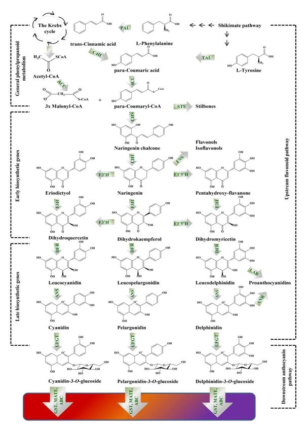
branch, including the late biosynthetic genes (LBGs), as shown on Figure 1. The anthocyanin
biosynthesis occurs in the cytoplasm, since the EBGs (chalcone synthase, chalcone isomerase, flavanone
3-hydroxylase, flavonoid 30 -hydroxylase and flavonol synthase) and LBGs (dihydroflavonol 4-reductase,
anthocyanidin synthase/leucocyanidin oxygenase and UDP-glucose:flavonoid-3-O-glycosyltransferase)
are located on the endoplasmic reticulum membranes or in the cytoplasm and further accumulated in
the vacuoles [59].
Although anthocyanin biosynthesis is well-known in many plant species, there is still no
comprehensive summary about the same in plant in vitro systems. The overall understanding of this
pathway might solve one of the biggest challenges in the commercialization of a biotechnological process
based on plant in vitro cultivation, such as slow growth, low or no productivity [13]. The anthocyanin
biosynthesis pathway has been studied in several plant in vitro model systems, in order to understand
and fine-tune its molecular regulation, e.g., V. vinifera [65], A. thaliana [61], Daucus carota [66], Hypericum
perforatum [67], Ocimum basilicum [68], and Panax sikkimensis [69] investigating the effect of elicitors [14],
precursors [13], plant growth regulators [61], nutrient supply [70,71], and light [72]. In synchronized
grape cell cultures, the CHS enzyme [73], as well as, DFR has been recognized as essential contributors
for the anthocyanins biosynthesis [57]. The significance of F3H gene was proven in purple-colored
carrot callus. Knocking out this gene by clustered regularly interspaced short palindromic repeats
(CRISPR) together with CRISPR-associated protein 9 (Cas9) resulted in the discoloration of the
calli [66]. The red/black color change in muscardine grapes (V. rotundifolia) callus was due to
induction of the enzymes UFGT and O-methyltransferase (OMT; EC 2.1.1.38, synonym anthocyanin
O-methyltransferase, AOMT), which catalyze the last steps of glycosylation and methylation in the
anthocyanins biosynthesis [74]. Elicitors (abiotic or biotic) have been recognized to stimulate defense
or stress-induced responses in plants. Some abiotic elicitors such as jasmonic acid (JA), MeJa or
salicylic acid (SA) are frequently used to enhance the SMs production in plant in vitro systems, since
they act as regulators of defense responses by initiating signals that activate multiple secondary
biosynthetic pathways and their relevant genes [75]. For example, MeJa increased the production
of total anthocyanins in V. vinifera L. cv. Gamay Fréaux cell suspension with 1.7-fold, the precursor
phenylalanine by 0.7-folds, while the combined application of MeJa (50 mg/L) and phenylalanine
(5 mg/L) had a significant synergetic effect increasing the anthocyanin content up-to 5.6-fold [13].
Similarly, the combined treatment of V. vinivera cv. Barbera cell suspension with 10 µM MeJa and
red light increased the anthocyanin production with 52% [72]. Red light stimulated mostly the
production of cyanidin and peonidin in callus cultures of O. basilicum, while during dark cultivation
the anthocyanin biosynthesis was inhibited [68]. The time-dependent biosynthesis of anthocyanins
under 100 µM SA treatment of V. vinifera “Cabernet Sauvignon” cell suspension revealed that increased
transcription of the EBGs, VvCHS and VvCHI and VvDFR began at 0.5 day after treatment and reachedMolecules 2020, 25, 2006 9 of 19
a maximum at day 1. At day 2 the expression of these genes declined, while some of the LBGs, such as
ANS revealed maximum expression, leading to 73% increase of anthocyanins accumulation [75]. Some
inorganic ions, such as Ca2+ have also important contribution as secondary signaling messengers in
elicitor-induced SMs biosynthesis. During treatment of V. vinifera L. cv. Gamay Fréaux cell suspension
with abscisic acid (ABA) or MeJa, the presence of 10 mM CaCl2 resulted in up-regulation of VvCHS,
VvDFR, and VvUFGT and down-regulation of VvFLS genes, the latest of which catalyzes the flavonols
production [76]. The addition of 20 mM MgCl2 was beneficial for the accumulation of anthocyanins in
V. vinifera cv. Gamay Red cell suspension, since Mg2+ ions prevented the degradation of anthocyanins
through the inhibition of several enzymes, e.g., peroxidases and β-glucosidases [77]. The nitrogen
(N), sulfate (S), and phosphate (P) deficiency increased the biosynthesis of anthocyanins in grape cell
suspension [65,70,71]. Under S-depletion conditions CHS, DFR, ANS, and GST were up-regulated, while
F3´5´H and UFGT showed no response [70]. During the p-deficiency, the grape cell suspension produced
dominantly cyanidin, peonidin 3-O-glucoside, and coumaroyl derivatives [71]. Auxins might also alter
gene expression, which directly or indirectly impacts anthocyanin biosynthesis. Concentrations in the
ranges from 2.2 to 27 µM 2.4-dichlorophenoxyacetic acid (2.4-D) and indole 3-acetic acid (IAA) inhibited
anthocyanin biosynthesis. During NAA treatment anthocyanin biosynthesis reached a maximum only
at 9 µM and decreased
Molecules 2020, 25, xafterwards. The same trend of expression was valid for the biosynthetic
FOR PEER REVIEW 9 of 20 genes,
among which PAL, CHS, DFR, and ANS were the most highly expressed [61].
Figure 1. Illustration, presenting the biosynthetic pathway of colored anthocyanins. PAL,
phenylalanine ammonia lyase (EC 4.3.1.24); TAL, tyrosine ammonia lyase (EC 4.3.1.23); C4H,
cinnamate 4-hydroxylase (EC 1.14.14.91); ACC, acetyl-CoA carboxylase (EC 6.4.1.2); 4CL, 4-
coumarate-CoA ligase (EC 6.2.1.12); CHS, chalcone synthase (EC 2.3.1.74); STS, stilbene synthase (EC
2.3.1.95); CHI, chalcone isomerase (EC 5.5.1.6); F3′H, flavonoid 3’-hydroxylase (EC 1.14.13.21); F3′5′H,
flavonoid 3´5´-hydroxylase (EC 1.14.13.88); FNS, flavones synthase (EC 1.14.11.22); F3H, flavanone 3-Molecules 2020, 25, 2006 10 of 19
Figure 1. Illustration, presenting the biosynthetic pathway of colored anthocyanins. PAL, phenylalanine
ammonia lyase (EC 4.3.1.24); TAL, tyrosine ammonia lyase (EC 4.3.1.23); C4H, cinnamate 4-hydroxylase
(EC 1.14.14.91); ACC, acetyl-CoA carboxylase (EC 6.4.1.2); 4CL, 4-coumarate-CoA ligase (EC 6.2.1.12);
CHS, chalcone synthase (EC 2.3.1.74); STS, stilbene synthase (EC 2.3.1.95); CHI, chalcone isomerase
(EC 5.5.1.6); F30 H, flavonoid 3’-hydroxylase (EC 1.14.13.21); F30 50 H, flavonoid 3´5´-hydroxylase
(EC 1.14.13.88); FNS, flavones synthase (EC 1.14.11.22); F3H, flavanone 3-hydroxylase (EC 1.14.11.9);
DFR, dihydroflavonol 4-reductase (EC 1.1.1.219); ANS, anthocyanidin synthase (EC 1.14.20.4); LAR,
leucoanthocyanidin reductase (EC 1.17.1.3); ANR, anthocyanidin reductase (EC 1.3.1.112); UFGT,
UDP-glucose:flavonoid-3-O-glycosyltransferase (EC 2.4.1.115); GST, glutathione S-transferase (EC EC
2.5.1.18); MATE, multidrug and toxic compound extrusion transporter; ABC, ATP-binding cassette
transporter. Phenylalanine is deaminated by PAL to form trans-cinnamic acid that is further
converted into para-coumaric acid by C4H. Para-coumaric acid is conjugated with coenzyme A
by the enzyme 4CL to obtain para-coumaroyl-CoA. The latter is condensed with three molecules
of malonyl-CoA by CHS to generated naringenin chalcone. Subsequently, CHI stereospecifically
converts the chalcone to its isomer naringenin. The B ring of naringenin is hydroxylated by F3´H or
F3´5´H to produce eriodyctiol or penthahydroxy-flavanone. The (2S)-flavanones are next subjected to
modification by the conversion of F3H into dihydroflavonols (dihydroquercetin, dihydrokaempferol
and dihydromyricetin). Dihydrokaempferol could be directly oxidized by F3´H and F3´5´H to
dihydroquercetin and dihydromyricetin. The obtained dihydroflavonols are reduced to colorless
leucoanthocyanidins by DFR, which are then oxidized to colored anthocyanidins by the activity of ANS
(synonyms leucocyanidin oxygenase: LDOX). Further, glycosylation is performed by UFGT, which
occurs in the cytoplasm and produces chemically stable water-soluble pigments.
5. Extracts (Secondary Metabolites) Activities-Gene Expression and Main Activities
The skin is an organ that has protective functions towards the external environment and its
physical integrity is essential to accomplish its functions [11]. Skin aging might happen via two
mechanisms: The first termed intrinsic or age dependent, is initiated by several factors, such as
telomere shortening, free radicals and antioxidants imbalance and hormonal changes, while the second
mechanism—photoaging is due to exposure of the skin to UV solar radiation [20]. Both mechanisms
of aging cause inhibition of tissue regeneration, growth and differentiation and mainly occur at the
level of dermal connective tissue, resulting in loss of mature collagen, elastin fibers, and hyaluronic
acid [78]. Reactive oxygen species (ROS) and heavy metals trigger skin aging via destruction of the
antioxidant system, leading to wrinkle formation, melanogenesis and severe damage of biomolecules,
such as DNA. [78,79]. Therefore, active phytochemicals that possess the ability to reduce the free
radical damage to the skin, and inhibit the activity of elastase, collagenase and hyaluronidase might
have beneficial effects in the prevention of skin aging. Several marker activities have been selected to
determine the cosmeceutical potential of the plant-derived extracts or pure molecules. For instance, the
inhibition of metalloproteinases (MMP), such as MMP1, MMP3, and MMP9 is indicative for prevention
of collagen degradation [21]. The whitening activity of extracts is determined by their ability to inhibit
the tyrosinase activity, an enzyme responsible for the synthesis of melanin, dopachrome formation
and melanogenesis, while the DNA repair mechanisms are indicated by the induction of genes, such
as gadd45α and sirtuin-1 (sirt-1) [21]. An important aspect for the wound healing process is the skin
hydration, determined by the NMF, which comprises a number of substances including ions, small
solutes and free amino acids, formed by the breakdown products of filaggrin protein. Indicators for
skin hydration are the expression of aquaporin 3 (AQP3) and filaggrin (FLG) genes. The investigations of
these diverse mechanisms require the development of different in vitro and in vivo models. The most
frequently used in vitro models comprise cell cultures of keratinocytes (preferably the immortalized
human skin cell line HaCaT), primary keratinocytes, epidermal stem cells, and fibroblasts. The in vivo
models include animal models of mice, rodents and rabbits and finally clinical trials with humans [80].
The aim of this section is to present the anti-aging, antioxidant, hydration, anti-wrinkling,
anti-inflammatory, and wound healing properties of extracts or pure compounds derived fromMolecules 2020, 25, 2006 11 of 19
plant in vitro systems. A pronounced activity, indicative for the above-mentioned properties is the
antioxidant activity of the plant-derived extracts or pure molecules. The chemical antioxidant assays,
such as oxygen radical absorbance capacity (ORAC), hydroxyl radical scavenging capacity (HOSC), and
hydroxyl radical averting capacity (HORAC) might be useful but have several limitations concerning
the prediction of the antioxidant activity in a biological environment. Therefore, this activity is
preferred to be analyzed in cellular assays based on keratinocytes (HaCaT) and fibroblast (HFF and
CCD-1112Sk) cell lines. These approaches have the advances compared to the chemical assays, since
the influence of several parameters, such as bioavailability, cellular uptake and metabolism is taken
into consideration. The ROS are induced by treatment with tert-butyl hydroperoxide (TBHP) or H2 O2
and there are two ways of treatment with the extracts: Pre-incubation of the cells with the extract before
the stressor addition and co-incubation of the extracts with the stressor. The pre-incubation treatment
gives information about the preventive activity of the extracts, while the co-incubation is closer of a
possible therapeutic approach and often seems to be the more effective one [78]. The aqueous cell
wall preparation from Nicotiana spp. suspension was investigated for its antioxidant capacity using
“in vitro” assays, such as total antioxidant capacity (TAC) and ORAC in cultured skin-derived NIH-3T3
fibroblasts. Approximately 85% reduction of ROS was observed when the cells were treated with
3.6 µg/mL extract after their induction with H2 O2 . The activity of the extract was higher than that of
ascorbate (44 µg/mL) used as a positive control. The observed activity was explained by the direct
interaction of the peptides and sugars with H2 O2 or through the activation of the defense response
signaling pathways [4].
The 80% aqueous methanolic extract of different Isodon rugosus (Wall. ex Benth.) callus cells
showed anti-aging and DNA protective activities. Two calli lines distinguished among others, the first
one was rich of caffeic and rosmarinic acids, while the second line appeared to produce triterpenoids
oleanolic, betulinic, and plectranthoic acids mostly. The pentacyclic triterpenoids were correlated
with the elastase, collagenase and tyrosinase inhibitory activity, while the phenolic acids, which were
the main contributors to the antioxidant activity correlated with the SIRT-1 (class III deacetylase)
activation, hyaluronidase and advanced glycation end-products (AGEs) inhibition [81]. Some of the
main activities and gene modulation properties of the analyzed extracts are presented in Table 2.
Table 2. Activities of plant cell culture extracts and their modulation activity on gene expression in
skin cells in vitro models.
Plant Species and Concentration of
Gene/Protein Expression Main Activity Ref.
Extract Type the Extract, µg/mL
IL-1β, IL-1α, TNFα PGE2 Anti-inflammatory activity
Calotropis
8000 inhibition Hypoxia adaptation and [82]
procera/aqueous extract
HIF1 induction wound healing activity
Tyrosinase inhibition Melanin inhibition
Citrus junos/aqueous 500 [79]
Procollagen type I induction Skin regeneration
COL I and pro-collagen I Collagen synthesis and
Hibsicus syriacus/aqueous
20 induction protection [80]
cell extract
AQP3 and FLG induction Skin hydration
Isodon rugosus (Wall. ex MMP1, hyaluronidase, Collagen synthesis and
Benth.)/80% aqueous 50 elastase inhibition protection [81]
methanol SIRT-1 activation DNA repair and protection
COL I and COL III
Lycopersicon induction, MMP1, MMP3 Collagen synthesis and
esculentum/aqueous 100 and MPP9 inhibition protection [21]
extract GADD45α and SIRT-1 DNA repair and protection
induction
Malus
Cyclin B1, cyclin E1 Retard the signs of
domestica/liposomal 100 [23]
induction senescence
extract of whole cellsMolecules 2020, 25, 2006 12 of 19
Table 2. Cont.
Plant Species and Concentration of
Gene/Protein Expression Main Activity Ref.
Extract Type the Extract, µg/mL
COL I and COL III
induction, pro-collagen I
Nicotiana tabacum BY2 (N. Collagen synthesis and
and III induction, MMP1,
sylvestris)/aqueous cell 3.6 protection [4]
MMP3 and MPP9 inhibition
wall extract DNA repair and protection
GADD45α, SIRT-1 and
SIRT-6 induction
Pyrus pyrifolia var. Tyrosinase inhibition Melanin inhibition
1000 [83]
culta/aqueous extract Pro-collagen I induction Collagen synthesis
GBA, Smpd1 induction Skin lipid production
Rubus ideaus/oil-soluble
1000 AQP3, FLG, AQP3 induction Skin hydration [22]
extract
COL I and III induction Collagen synthesis
The effect on melanogenesis of aqueous C. junos extract was evaluated on B16F10 melanoma cell
line. The tyrosinase activity was inhibited by 25.2% (at 500 µg/mL extract), while the melanin synthesis
decreased with 208% (at 50 µg/mL extract) compared to the inhibition caused by arbutin (16.3%), at the
same concentration. The skin regeneration activity was tested on fibroblasts cell model. At 500 µg/mL
extract concentration 154% fibroblast proliferation was achieved, which was higher than the effect of
the recombinant human transforming growth factor-β (TGF-β) at 500 ng/mL, utilized as a positive
control. This extract revealed also anti-wrinkle activities evaluated via the increased pro-collagen type
I C-peptide in a dose dependent manner. The highest pro-collagen synthesis (1.76-fold higher) was
achieved at 500 µg/mL extract concentration. The wound healing effect of the extract revealed 161%
increase in the recovery of the wound closure compared to the control and increased the proliferation
and migration of the fibroblasts with 15%. The main compound identified and eventually responsible
for the observed activities was p-hydroxycinnamoylmalic acid [79]. The aqueous cell walls extract
(mainly peptides and sugars) of Nicotiana tabacum BY2 suspension exhibited DNA protection and
repair functions, as well as, increased collagen synthesis and stability in human keratinocytes (HaCaT
cell line) and murine fibroblasts (NIH-3T3 cells). Indication for the DNA protective activity was the
elevated expression of GADD45α protein and SIRT-1 and SIRT-6 in the fibroblasts after treatment
with 150 µM H2 O2 . Further, this effect was confirmed by the “single cell electrophoresis assay”
(comet assay). The length of the nucleus comet, index of DNA fragmentation was significantly
reduced (with 50%), clearly indicating the genomic DNA protection. The peptide/sugar mixture
elevated the expression of collagen type I and III genes with 200% and also increased the levels of
pro-collagen I and III synthesis 2–3-folds. Beside the effect to induce new collagen production, the
same cell wall preparation inhibited the expression of MMP1, MMP3, and MMP9, responsible for
the ECM degradation [4]. The hydrosoluble extract of L. esculentum cultured stem cells protected the
NIH-3T3 fibroblasts and HaCaT keratinocytes collagen degradation and DNA integrity from heavy
metal damages [21]. The aqueous extract from H. syriacus cell culture stimulated the production of
pro-collagen type I and fibronectin with 60% and 16%, respectively and improved the wound healing
properties in HaCaT and human derma fibroblasts (HDF), isolated from 48-year-old woman with 50%
and 30% when treated with extract concentration of 20 µg/mL. The observed beneficial effects were
explained by the hydration properties of the extract, which was able to increase the expression of
AQP3 and FLG expression with 20% and 60% respectively, thus contributing to the NMF production
and maintenance of skin water balance [80]. An oil-soluble extract of R. ideaus suspension (containing
phenolic acids and wide range of fatty acids) revealed significant skin hydration and collagen protection
activities in in vitro studies, performed on HaCaT cells. The extract was found to improve the skin
lipid production through the activation of two enzymes catalyzing the induction of ceramides, such
as glucocerebrosidase (GBA) and sphingomyelin phosphodiesterase 1 (Smpd1), with 13% and 15%,
respectively. The expression of AQP1, FLG and involucrin, involved in skin hydration were increased
with 32%, 21%, and 75%. Another important enzyme, such as hyaluronan synthase 2 and 3 (HAS 2
and 3) found in the dermis and epidermis also contribute in keeping the skin in proper hydrated state.Molecules 2020, 25, 2006 13 of 19
The activity of both, HAS 2 and 3 was increased by 23% and 126% in fibroblasts and keratinocytes.
An improvement of the collagen syntheses and protection was observed by the increased production
of collagen types I and III and fibronectin by 33%, 67%, and 19% [22]. A liposomal extract of whole cell
suspension culture from Malus domestica was prepared from the Switzerland company Mibelle AG
Biochemistry by incorporation of the extract in 10% lipid fraction. The obtained liposomes resulted in
aqueous phase containing all the water-soluble ingredients and liposomes incorporating the oil soluble
ingredients. At only 0.1% of the extract was able to stimulate the human stem cells proliferation [23].
At concentration of 0.01% was able to induce with 80% its protective activities in mammalian stem
cells under exposure to UV light [56]. The extract was also able to retard the senescence in fibroblast
cells (stimulated with H2 O2 ) through the up-regulation of cyclin B1 (responsible for induction of
proliferation), cyclin E1 (controls cell cycle), p53 (tumor suppressor factor), insulin-like growth factor
(enhancer of cell proliferation) and heme oxygenase 1 (antioxidant enzyme) after incubation with 2%
of the extract for 144 h [23].
Inflammatory processes lead to skin aging, therefore many studies have investigated the protective
effects against inflammation by utilizing different in vitro or ex vivo models. The ability of C. procera
aqueous extract to protect against skin inflammation and irritation, induced by lipopolysaccharides
(LPS) and sodium dodecyl sulfate, was investigated in ex vivo human organ culture model. The extract
appeared to decrease the concentrations of inflammatory cytokines interleukin (IL)-1β, IL-1α, tumor
necrosis factor alpha (TNFα), and prostaglandin 2 (PGE2 ) with 3–4 folds compared to the control at
0.8 g/L concentration. The extract also improved the skin cells ability to adapt to hypoxia conditions
and improve their wound healing capacity by elevating the expression of hypoxia-inducible factor 1
(HIF1) with 15%. Indication for improved cellular metabolism was observed by the increased activity of
the enzyme phosphfructokinase-1 (PFK1), involved in the hydrolysis of ATP. An increase in fibronectin
production by 20% indicated the effect of the extract on regeneration and healing of the skin [82].
An interesting strategy to increase extract(s) permeability through the skin offers the encapsulation
in nanoliposomes. An encapsulated extract of C. junos displayed increased permeability of
p-hydroxycinnamoylmalic acid by 5.8-fold when tested on an artificial PAMPA membrane [79].
The incorporation of P. pyrifolia aqueous extract in nanoliposomes increased its flux across the human
epidermis by ca. 16 times and facilitated the in vitro wound recovery rates of keratinocytes and
fibroblasts [84]. Another successful approach to increase the bioavailability and permeability of
substances with low lipophilicities is to obtain their derivatives with increased lipophilic properties
or their inclusion into liposomes or lipogels. The wound healing properties of the water-soluble
verbascoside derived from the cell suspension of Buddeleia davidii were improved by the obtaining of
its lipophilic semi-synthetic derivative ES2. The ES2 is considered as a novel and potent candidate for
topical treatment of wounds, since this compound improved the wound closure more than 2-folds in
HaCaT cells and SKH1 mice, compared to that of verbascoside at 10 µM concentration [11]. Another
derivative of verbascoside, denoted as verbascoside pentapropionate, also exhibited lower hydrophilic
profile and improved antioxidant activities than verbascoside [85]. In addition, the phenylpropanoids
verbascoside and teupolioside, found in the extracts of Syringa vulgaris and Ajuga reptans, showed
remarkable anti-inflammatory and wound healing properties, which were further correlated with the
inhibition of ROS released from the whole blood leukocytes. The 56% teupoloside extract was able
to prevent the skin against excessive influx of pro-inflammatory neutrophils and against oxidative
damage by restoring GST activity. A 97% extract, containing verbascoside and teupoloside, was
the most effective in the inhibition of monocyte chemoattractant protein-1 (MCP-1), IL-8, interferon
gamma-induced protein 10 (IP-10) in the range of 1–50 µM concentration in primary cultures of human
keratinocytes [27]. Along with verbascoside, another phenylethanoid glycoside, such as forsythoside B
inhibited the inflammatory cytokines IL-8, MCP-1 and IP-10, stimulated by interferon gamma (INF-γ),
in normal human keratinocytes [86].
The final stages in plant extract testing for potential cosmeceutical application are the clinical
trials. The cream, obtained on the basis of the liposomal extract of M. domestica (PhytoCellTecTMMolecules 2020, 25, 2006 14 of 19
Malus Domestica), was tested over four weeks on 20 volunteers. The cream contained a 2% extract
and resulted in significant reduction of wrinkle depth after two and four weeks, at 8% and 15%
respectively [23]. The French company Naolys performed clinical trial of its product Refine ginger,
derived from dedifferentiated plant cell cultures, on 22 women. The product was found to improve
skin structure by 50% through pore reduction, obtained a mattifying effect by shininess reduction of
15% and sebum reduction of 19% after 28 days [18]. The Oryza sativa callus extract, containing mainly
γ-oryzanol, vitamin E homologues and phenolic acids, also showed beneficial properties when tested
on 28 volunteers, at 5% concentration, applied twice per day on facial skin for 12 weeks. The observed
anti-aging effect comprised in improvement of skin moisture (75%), elasticity (88%) and whitening
effect by 38% [6]. The oil-soluble extract of R. ideaus suspension, when applied on the skin of 20 female
volunteers (0.1%, twice per day), improved the skin hydration by 16% after 14 days and 19% after
28 days [22].
6. Conclusions and Future Perspectives
Nowadays, plant in vitro systems are superior source of SMs or complex extracts that possess
multiple beneficial activities for skin care. Due to the fast development of the cosmetic industry and
recent trends in the cosmetic market, there has been elevated demand for novel and safe natural products
(formulations). Biotechnological processes, based on cultivation of plant cells in vitro, are irreplaceable
source of bioactive ingredients, produced under controlled conditions in the absence of contaminants
and with consistent quality. A comprehensive understanding of the metabolic networks and the factors
that control the biosynthesis of SMs is necessary to optimize the (bio)molecules mass production.
These include fine tuning of the cultivation parameters (i.e., mixing and agitation) or nutrient medium
optimization, precursor feeding or elicitation, to name a few.
Due to the changing regulations in the safety assessment of cosmetics, new in vitro methods
to assess the authenticity and safety of cosmetic products have been developed, which aid our
understanding of the molecular mechanisms underlying the cosmeceutical ingredients.
Author Contributions: Conceptualization A.S.M. and M.I.G.; writing—original draft preparation A.S.M.;
writing-review M.I.G.; visualization A.S.M.; supervision M.I.G.; funding acquisition M.I.G. All authors have read
and agreed to the published version of the manuscript.
Funding: This work has been supported by a grant from the National Science Fund of Bulgaria (contract number
DFNI B02/14).
Acknowledgments: The authors are grateful to Rafe Lyall, Ramadoss Dhanushkodi and Liliya Vasileva of the
CPSBB, for proofreading and linguistic editing of the manuscript.
Conflicts of Interest: The authors declare no conflict of interest. The funders had no role in the design of the
study; in the collection, analyses, or interpretation of data; in the writing of the manuscript, or in the decision to
publish the results.
References
1. Schmitz, C.; Fritsch, L.; Fischer, R.; Schillberg, S.; Rasche, S. Statistical experimental designs for the production
of secondary metabolites in plant cell suspension cultures. Phytochem. Lett. 2016, 38, 2007–2014. [CrossRef]
2. Maia, J.; Dantas, T.; da Costa Neto, B.; Borges, K.; Lima, E.; da Mata, A.; de Medeiros, M.; Pereira, C. Extract
of spray-dried Malay apple (Syzygium malaccense L.) skin. J. Food Proc. Eng. 2019, 2019, e13275. [CrossRef]
3. Espinosa-Leal, C.; Garcia-Lara, S. Current methods for the discovery of new active ingredients from natural
products for cosmeceutical applications. Planta Med. 2019, 85, 535–551. [CrossRef]
4. Apone, F.; Tito, A.; Carola, A.; Arciello, S.; Tortora, A.; Filippini, L.; Monoli, I.; Cucchiara, M.; Gibertoni, S.;
Chrispeel, M.; et al. A mixture of peptides and sugars derived from plant cell walls increases plant defense
responses to stress and attenuates ageing-associated molecular changes in cultured skin cells. J. Biotechnol.
2010, 145, 367–376. [CrossRef]
5. Antonopoulou, I.; Varriale, S.; Topakas, E.; Rova, U.; Christakopoulous, P.; Faraco, V. Enzymatic synthesis of
bioactive compounds with high potential for cosmeceutical application. Appl. Microbiol. Biotechnol. 2016,
100, 6519–6543. [CrossRef] [PubMed]You can also read